Embed Size (px)
Citation preview

STRUCTURE OF THE
CORONARY CIRCULATION
CORONARY PHYSIOLOGY IN THE CATHLAB:
Educational Training Program ESC
European Heart House
april 24th - 26th 2014
Nico H. J. Pijls, MD, PhD
Catharina Hospital,
Eindhoven, The Netherlands

Disclosures related to this ETP course:
• Dr Pijls received institutional
research grants from St Jude Medical
and Pharma Solutions
• Dr Pijls is consultant to St Jude Medical,
and to Heartflow

• structure of the coronary circulation
• relation between vessel size and perfusion area
• endothelium and development of atherosclerosis
• the 2 or 3 compartment model of the coron circulation
• collaterals ( to be discussed tomorrow)
ISSUES TO BE DISCUSSED


03 cc/schema Braunwald
• 03 cc/schema Braunwald

Let’s have a closer look at the coronary tree…..

Fractale structure of the coronary circulation ( Gould, Finet)
X 1 X 0.75
X 4.5 X 10.5 X 90 X 1100

epicardial
compartment
( > 400 µm)
microvascular
compartment
traditionally visible by angiography
and more recently by many invasive
and non-invasive imaging methods Black box
(until recently)

Regulation of coronary blood flow by arteriolar sphincters
To be further discussed by Dirk Duncker

• structure of the coronary circulation
• relation between vessel size and perfusion area
• endothelium and development of atherosclerosis
• the 2 or 3 compartment model of the coron circulation
• collaterals
ISSUES TO BE DISCUSSED

Relationship between vessel size and myocardial mass
C. Seiler, Lance Gould, et al Circulation 1992
Regional Myocardial Mass
0
1
2
3
4
5
6
0 100 200 300 400 500
Regional Myocardial Mass (Grams)
Cross Sectional Area (~ flow) Vessel Diameter (mm)

AORTA
100 mmHg
APEX
100
100
100
100
100
100
200
150
100
75
50
25
pressure (Pd)
(mm Hg)
flow (Q)
(ml/min)
IVUS-CSA
9 mm2
7 mm2
5 mm2
3 mm2

AORTA
100 mmHg
APEX
100
100
100
100
100
100
200
150
100
75
50
25
pressure (Pd)
(mm Hg)
flow (Q)
(ml/min)
IVUS-CSA
9 mm2
7 mm2
5 mm2
3 mm2
Normal FFR
= 1.0
irrespective
where it is
measured

SIZE of the person (importance of perfusion territory)
FFR = 0.68
means exactly
the same in
both persons
CSA by IVUS
= 3.3 mm2 has
a completely
different meaning
in both persons
LAD blood flow
= 100 ml/min has
a completely
different meaning
in both persons
Suppose both of these 2 persons have a proximal LAD stenosis

Value of ANY morphologic methodology
( QCA, IVUS, OCT ) to assess functional
significance of a stenosis
is limited by definition because
there is simply no normal reference value

We cannot understand the physiologic
significance of a stenosis without taking
into account the extent of the distal
perfusion territory
……especially not under pathologic
conditions, when the “physiologic match“
between vessel size and perfusion area
has been lost
! !

With permission of
Dr Haitma Amin,
Bahrain

Normal Myocardium
Normal Myocardium
Scar
similar stenosis but different extent of perfusion area
identical CSA 4 mm2
4 mm2 is sufficient
4 mm2 is too small
identical CSA, but different significance of stenosis
QCA, IVUS

Normal Myocardium
Normal Myocardium
Scar
Anatomic stenosis severity by IVUS or QCA
is identical but physiologic severity has decreased.
FFR accounts for these changes !!!
FFR accounts for the extent of the perfusion area:
60
80
100
100
FFR=0.60
FFR=0.80

Disconnect between Anatomy and Physiology
50% Stenosis FFR=0.85
Myocardium
50% Stenosis
Collaterals Collateral-Supplied Myocardium
Vessel-Supplied
Myocardium
…During Maximal Hyperemia
FFR=0.73

FFR in the distal LAD before and After recanalization of the RCA

• structure of the coronary circulation
• relation between vessel size and perfusion area
• endothelium and development of atherosclerosis
• the 2 or 3 compartment model of the coron circulation
• collaterals
ISSUES TO BE DISCUSSED

Normal
Endothelial dysfunction
First stages of atherosclerosis:
IVUS, OCT, FFR ( abnormal pressure
decline)
Macroscopic atherosclerotic disease:
angio,
non-invasive imaging (CT, MRI)
DEVELOPMENT OF ATHEROSCLEROSIS

The earliest phase of atherosclerotic
coronary disease, is endothelial dysfunction.
This is unvisible by any imaging method,
but can be demonstrated by functional testing.

29 cc/Achol vb (6)
baseline
ACh
NTG 35-y-old male,
hypertension,
heavy smoker,
chest pain at exercise
and positive ET

Physiologic and pathologic vasomotion in 35-year old
male, heavy smoker, and chest pain at exercise

tip of infusion
catheter,
administration
of papaverin
pressure
guidewire

papaverine 10 mg
1
2
3
1 papaverine induced vasodilation
2 flow-induced vasodilation
3 flow-induced paradoxical vasoconstriction

Male, 41-year-old
early stage of atherosclerosis
22-03-2006

22-03-2006

Courtesy of Dr Pim Tonino
diffuse atherosclerosis, early stage

hyperemic pull-back LAD
diseased segment

Fibrous cap atheroma
with hemorrhage Fibrocalcific plaque
Ca2+ Hem
Ca2+
Thin fibrous cap
atheroma
NC
FC
Virmani R, et al. Arterioscler Thromb Vasc Biol 2000;20:1262
Different stages of gross coronary atherosclerosis,
easily visible on angiogram and by several
non-invasive methods

• structure of the coronary circulation
• relation between vessel size and perfusion area
• endothelium and development of atherosclerosis
• the 2 or 3 compartment model of the
coronary circulation
• collaterals
ISSUES TO BE DISCUSSED

epicardial
compartment
( > 400 µm)
microvascular
compartment
traditionally visible by angiography
and more recently by many invasive
and non-invasive imaging methods Black box
(until recently)

IMAGING OF THE EPICARDIAL COMPARTMENT • non-invasively by CT, MRI • invasively by angio, IVUS, OCT,and some newer techniques
FUNCTIONAL ASSESSMENT OF THE EPICARDIAL
COMPARTMENT
• coronary pressure & FFR

FUNCTIONAL ASSESSMENT OF THE
MICROCIRCULATION:
• IMR(Bill Fearon,Keith Oldroyd)
• absolute flow & resistance (Bernard De Bruyne)
IMAGING OF THE MICROVASCULAR COMPARTMENT • can only be done indirectly (blush, PET, MIBI. MRI)
The coronary microcirculation:
Still a black box ??
Saturday morning session

The third compartment
focal and diffuse
epicardial disease
microvascular
compartment
FFR
hard to distinguish by
traditional methods,
but easily assessed
and quantified by FFR
(hyperemic pullback recording)

How to assess the functional significance
of diffuse disease, whether or not with
super-imposed focal lesions?
Impossible by anatomic methods
CCTA,Angiography,
IVUS, or OCT

The 3rd compartment:
Diffuse epicardial coronary disease
(whether or not with superimposed focal lesions)
(Bernard De Bruyne, tomorrow morning)
easily evaluable by FFR
(pressure pull-back recording)
important consequence for treatment
(interventional or medical)

Male 58-y-old
Typical chest pain; positive MIBI-Spect inferior wall

Typical chest pain; positive MIBI-Spect inferior wall

Typical chest pain; positive MIBI-Spect inferior wall

CASE # 3

pull-back advance

Dist. stenose
Mid in-stent
restenose
Prox. stenose
Hyperemia: Pull back recording
Distal proximal Mid
FFR = 0.65

Hyperemic pull-back recordingalong the RCA

•Coronary anatomy is just one side of the coin
•There is complex interrelation between the structure
and function of the coronary circulation, not only under
physiologic circumstances in healthy persons
(vessel size/perfusion area relation, endothelium,
regulation of coronary blood flow), but also under
pathologic circumstances ( atherosclerosis, plaques,
stenosis, vulnerabilty, and ischemia).
• Understanding this relation is paramount to treat our
patients in the cathlab in the best possible way.
• Hopefully, this course will contribute both to that
understanding and to its translation into practical skills
IN SUMMARY:

EINDE

The 3rd compartment:
Diffuse epicardial coronary disease, whether
or not with super-imposed focal disease
(Bernard De Bruyne, tomorrow)

In patients with coronary artery disease,
the most important factor with respect to both
• functional class (symptoms)
• and prognosis (outcome)
Is the presence and extent of inducible ischemia
knowledge if and which lesion(s) is / are
responsible for inducible ischemia, is paramount
for adequate treatment in the cath.lab
FRACTIONAL FLOW RESERVE

Pressure pull-back curve at maximum
hyperemia:
• place sensor in distal coronary artery
• induce sustained maximum hyperemia by i.v. adenosine, or i.c. papaverine
• pull back the sensor slowly under fluoroscopy
• the individual contribution of every segment and spot to the extent of disease can be studied in this way
FFR: The Pressure Pull-back Curve
Coronary pressure is unique in this respect and such
detailed spatial information cannot be obtained by any
other invasive or non-invasive method

• structure of the coronary circulation
• relation between vessel size and perfusion area
• endothelium and development of atherosclerosis
• the 2 or 3 compartment model of the coron circulation
• collaterals
• why functional testing / FFR ?
• which lesions should be treated
ISSUES TO BE DISCUSSED

• structure of the coronary circulation
• relation between vessel size and perfusion area
• endothelium and development of atherosclerosis
• the 2 or 3 compartment model of the coron circulation
• collaterals (to be discussed tomorrow)
• which lesions should be treated ?
• why functional testing / FFR ?
ISSUES TO BE DISCUSSED
next 2 days

Qmyo = Qcor.artery + Q collateral
Quantitave assessment of the contribution of
coronary arterial and collateral flow to total
myocardial flow is possible by coronary
pressure measurements, but not trivial
Pijls & De Bruyne:
Circulation 1993
Coronary Pressure, sec edition, Kluwer 2000

26 col-schema fcf (figuur)
• 26 col-schema fcf (figuur)
Fractional collateral flow ( also called CFIp ) =
FFR coll = Pw - Pv
Pa - Pv
Venous pressure not negligible anymore !

EVIDENCE-BASED MEDICINE:
• PCI of “ischemic” lesions (associated with
reversible ischemia) makes sense and
improves symptoms and sometimes also outcome
• PCI of non-ischemic lesions has no benefit, is no
evidence-based medicine, is potentially harmful,
and unnecessary expensive
knowledge if and which lesion(s) is / are
responsible for inducible ischemia, is paramount
for adequate treatment in the cath.lab
FRACTIONAL FLOW RESERVE
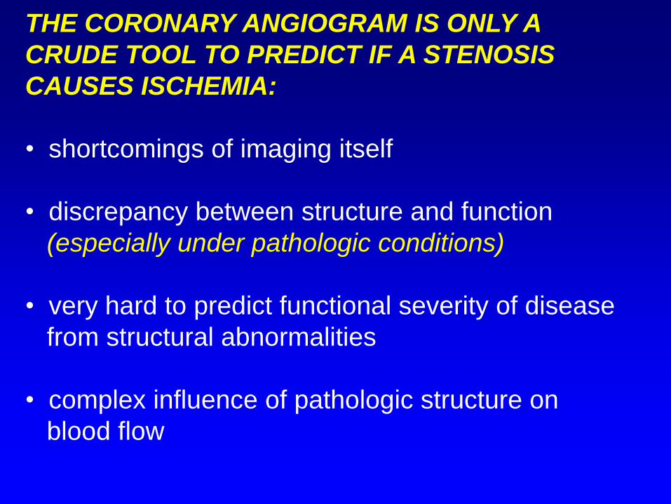
THE CORONARY ANGIOGRAM IS ONLY A
CRUDE TOOL TO PREDICT IF A STENOSIS
CAUSES ISCHEMIA:
• shortcomings of imaging itself
• discrepancy between structure and function
(especially under pathologic conditions)
• very hard to predict functional severity of disease
from structural abnormalities
• complex influence of pathologic structure on
blood flow


Normal Myocardium
Normal Myocardium
Scar
similar stenosis but different extent of perfusion area
identical CSA 4 mm2
4 mm2 is sufficient
4 mm2 is too small
identical CSA, but different significance of stenosis
QCA, IVUS
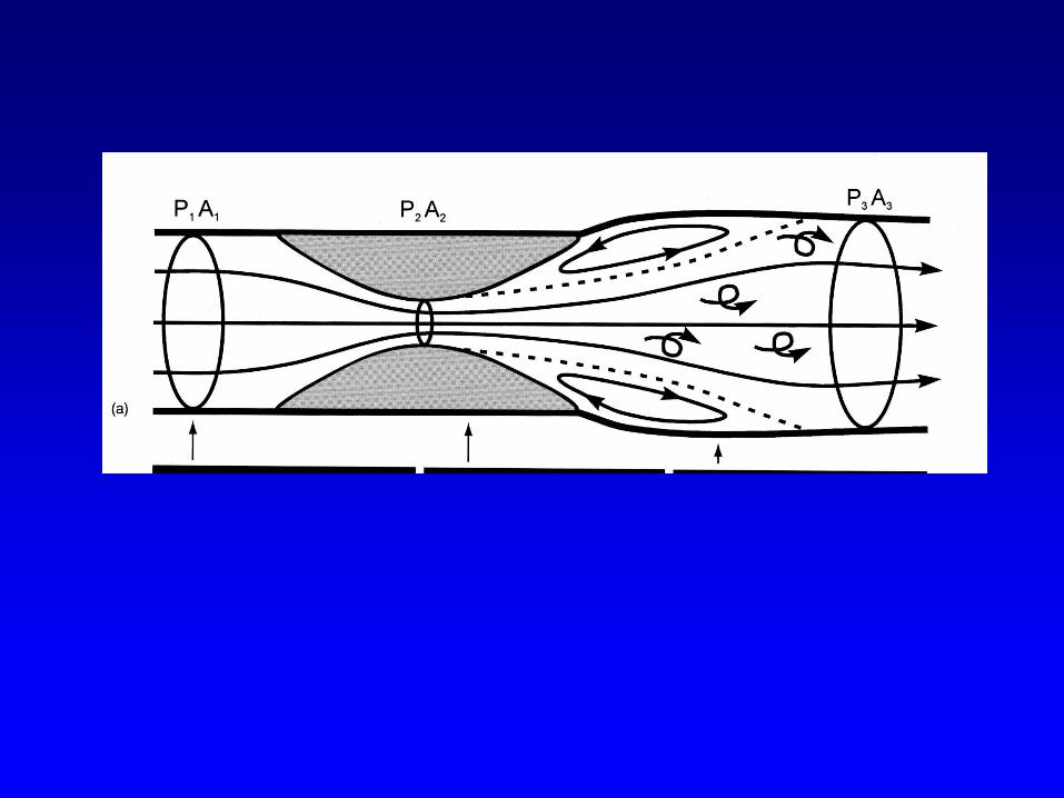
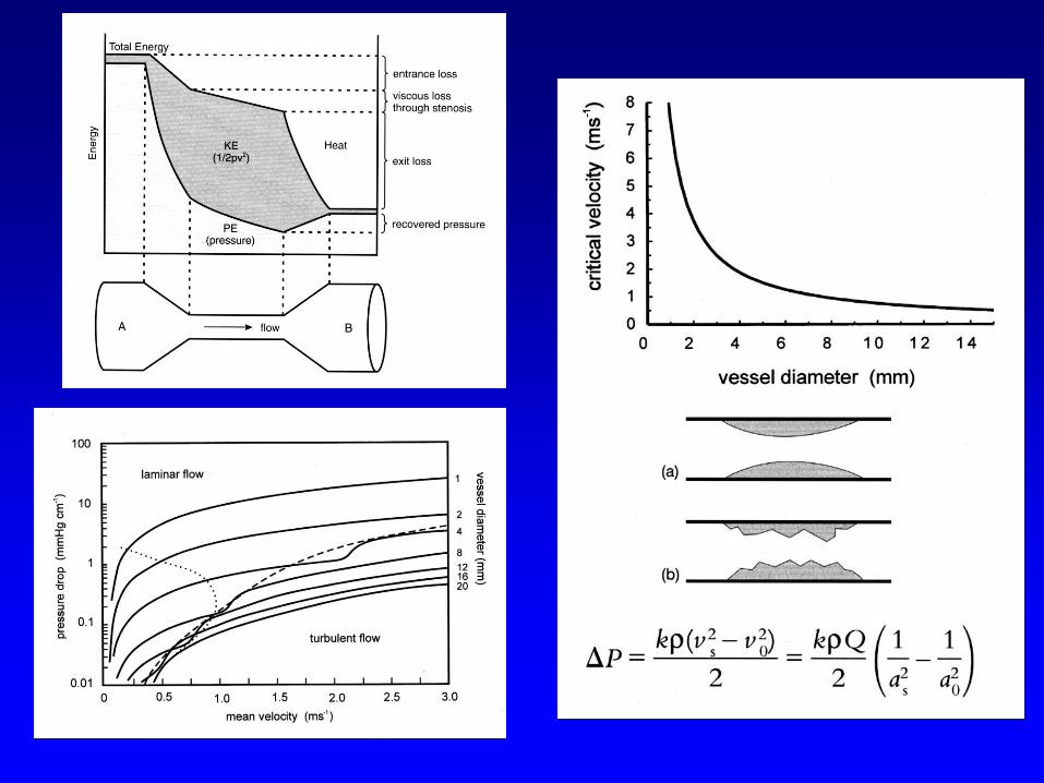

Even in the geometrically most “ideal” stenosis, it is
impossible to predict the functional severity and
influence on blood flow from hydraulic theory

In summary: EVIDENCE-BASED MEDICINE:
knowledge if and which lesion(s) is / are
responsible for inducible ischemia, is paramount
for adequate treatment in the cath.lab
The angiogram (and IVUS!) have fundamental
Shortcomings to indicate ischemia correctly
Rationale of Fractional Flow Reserve

Whatever the stenosis might look like...,
whatever the pressure/flow relations across
the stenosis might be....,
To understand the meaning of the stenosis for
the patient, the MOST important number to know is
the resulting distal perfusion pressure at
hyperemia, as a fraction of normal perfusion
pressure ( = aortic pressure)
This ratio determines completely the physiologic
significance of the stenosis
and its consequences for the patient !!
It is called FFR

Pa
100 Pd
70 Pv
0
Pa
100
Pv
0
During Maximal Vasodilatation
Q
P
FFRmyo = Pd
Pa
= 0.70

• structure of the coronary circulation
• relation between vessel size and perfusion area
• endothelium and development of atherosclerosis
• the 2 or 3 compartment model of the coron circulation
• collaterals
• why functional testing / FFR ?
• which lesions should be treated
those causing ischemia
• ischemia & vulnerability: paradox or antithesis ?
(Bernard De Bruyne, later today )
ISSUES TO BE DISCUSSED

• structure of the coronary circulation
• relation between vessel size and perfusion area
• endothelium and development of atherosclerosis
• the 2 or 3 compartment model of the coron circulation
• collaterals
• why functional testing / FFR ?
• which lesions should be treated
those causing ischemia
• ischemia & vulnerability: paradox or antithesis ?
ISSUES TO BE DISCUSSED

Excellent outcome of medical treatment in
non-ischemic stenosis
(DEFER study, many non-invasive studies)
versus
concept of vulnerable plaque
Paradox or anthithesis ?

TCFA Plaque Rupture
th ?
today tomorrow ?
Renu virmani, ETP course 2005

th
TCFA Plaque Rupture
?
today tomorrow ?
Let’s look a little bit more critical to such “plaques”….
What are the facts ?? What is the fiction ??

(Vulnerable) Plaque: Facts and Fiction
FACTS:
• plaques are very common
• majority of plaques has an excellent prognosis with
medical treatment
• only few plaques are vulnerable
• strongest indicator with respect to prognosis
is associated ischemia
FICTION:
• every plaque is vulnerable
• every vulnerable plaque leads to ACS
• most ACS occurs in mild plaques
• vulnerabilty can be assessed by imaging
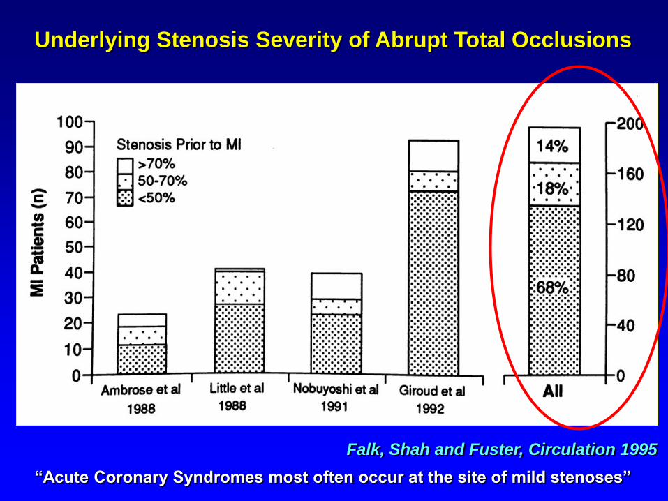
Falk, Shah and Fuster, Circulation 1995
“Acute Coronary Syndromes most often occur at the site of mild stenoses”
Underlying Stenosis Severity of Abrupt Total Occlusions

Serial Angiographic (Retrospective) Studies
in Patients with MI and a Prior Coronary Angiogram
Do Myocardial Infarctions Evolve from Mild Stenoses ?
No QCA, No IVUS but unblinded “eyebolling”
Number of Patients
Delay Angio-MI
Ambrose et al JACC 1988 23 1 month to 7 years
Little et al. Circulation 1988 42 4 days to 6.3 years
Giroud et al. AJC 1992
Moise et al. AJC 1984
Webster et al JACC 1990 abstr
Hackett et al AJC 1989
92
116
30
10
1 month to 11 years
39 months
55 months
21 months
Total 313 A few days to 11 years
(average 3.9 years !!!)

THE MYTHE OF
THE “DANGEROUS” PLAQUE
The hypothesis of the occurrence of acute MI on such
previously non-significant plaque is based upon
• 6 small retrospective studies
• with a total of 313 patients
• in whom the “index” catherization was performed
an average of 3.9 years before the acute event
All other literature (21 “meta-analyses” and
hundreds of references), refer to these 6 studies !!!

Stenosis Severity at Baseline
0
100
200
300
400
500
2161
None 5-49% 50-80% 81-95%
1% 2% 10% 24%
Occlusion at FU
Coronary Segments (n)
Adapted from Alderman et al. J Am Coll Cardiol 1993
Coronary Occlusion at 5 Years as a Function of Stenosis Severity
0
5
10
15
20
25
None 0-49% 50-80% 81-95%
% Occlusion at 5 Year
Stenosis Severity at Baseline

3.3 %
15.7 %
0
5
10
15
20 % P< 0.03
FFR > 0.75 FFR < 0.75
non-ischemic stenosis,
treated medically
ischemic stenosis,
treatment by PCI
& optimum R/x
ischemic lesion is much more dangerous than
non-ischemic lesion
JACC 2007; 49: 2105-2111
DEFER study (N=325) :
Cardiac death and Acute MI after 5 years
risk of individual non-ischemic lesion to cause death
or AMI, is very small and < 1 % per year !!

Frobert et al CCI, 2007, 70: 958-965
250 consecutive patients with ST-elevation MI
in the Catharina Hospital:
• underlying stenosis angiographically significant
in 92 % of the cases
•
At meticulous anamnesis, 80 % of patients had
recurrent chest pain in the year before the acute
myocardial infarction occurred !!

Incidence of coronary artery disease in
asymptomatic, apparently healthy persons
> 50 years old : 25%
> 60 years old : 40%
Sims et al, Am Heart J 1983
Maseri, Ischemic Heart Disease 1995
INCIDENCE OF CORONARY STENOSIS
IN A GENERAL POPULATION
What about the prognosis of these patients ?
Related to inducibility of ischemia

• structure of the coronary circulation
• relation between vessel size and perfusion area
• endothelium and development of atherosclerosis
• the 2 or 3 compartment model of the coron circulation
• collaterals
• why functional testing / FFR ?
• which lesions should be treated
• vulnerable plaques: facts & fiction
• ischemia & vulnerability: paradox or antithesis ?

Is there a link between vulnerabilty and ischemia ?
“The missing link”
Hypothesis:
• repetitive ischemia and
• high shear stress / pressure gradients
induce vulnerability
Supported by studies on the relation between
vulnerability markers and low FFR:
on-going work of Pasterkamp et.al. Heart 2007

25
50
75
Pam3Cys
5 ng/ml
Pam3Cys
50 ng/ml
Pam3Cys
500 ng/ml
0
FFR < 0.75
FFR > 0.80
p=0.008
p=0.014
p=0.014
TLR2 stimulation (Pam3Cys)
Versteeg et al, Heart 2007

th
?
Pro-inflammatory cytokines,activated monocytes, etc
Vulnerability
(“out of the blue”)
Concept of Yesterday:

th
?
Pro-inflammatory cytokines, activated monocytes, etc
Vulnerability
Concept of Tomorrow:
ischemic episodes

th
?
Pro-inflammatory cytokines etc
Vulnerability
Concept of today:
ischemic episodes by the way:
70% area
Stenosis !!

Plaque / stenosis
successful remodelling,
decrease of ischemia
overshoot,
plaque rupture
Ischemic episodes
production of remodelling-promoting substances
new paradigm:
Searching for vulnerability starts with searching for ischemia

Suppose aliens would visit us and would like to
investigate the determinants of a fire.
Living unidentified
object releasing the
substance X
Substance X, always
detected when there has
been a fire
“Substance X (also called “water”) must be dangerous substance !”

FUNCTIONAL ASSESSMENT OF BOTH
COMPARTMENTS TOGETHER:
• non-invasively
(exercise testing, stress echo, Mibi)
• invasively: intracoronary Doppler, absolute flow
FUNCTIONAL ASSESSMENT OF THE
MICROCIRCULATION:
• Index of Microcirculatory Resistance (IMR)

The coronary microcirculation:
Still a black box ??

focal and diffuse
Epicardial disease
microvascular
compartment
FFR
Specific indexes ??
Invasive indexes (saturday morning):
IMR (Bill Fearon)
absolute resistance (Nico Pijls)

We cannot understand the physiologic
significance of a stenosis without taking
into account the distal perfusion territory ! !

06 cc/Chilian (weerst)
• 06 cc/Chilian (weerst)
majority of resistance located in arterioles ( 100-400 µm)

16,2
32,4
0
5
10
15
20
25
30
35
40
Rate
of
death
or
MI
(%)
B
P=0.001
Death & MI 5 during 5 years of follow-up after
PCI vs Medical Treatment in ISCHEMIC stenosis
Shaw et al,
Circulation 2008
PCI PCI MEDICAL

Kaplan-Meier plots of Landmark Analysis of
Death or MI
FAME 2 : FFR-Guided PCI versus Medical Therapy in Stable CAD
0
5
10
15
20
25
30
Cu
mu
lati
ve
in
cid
en
ce
(%
)
0 7days 1 2 3 4 5 6 7 8 9 10 11 12
Months after randomization
p-interaction: p=0.003
> 8 days: HR 0.42 (0.17-1.04); p=0.053
≤7 days: HR 7.99 (0.99-64.6); p=0.038
0
0.5
1.0
1.5
2.0
2.5
Cu
mula
tive in
cid
ence (
%)
0 1 2 3 4 5 6 7Days after randomization
MT alone
PCI plus MT
MT alone
PCI plus MT
≤7 days
>8 days

0%
20%
40%
60%
80%
100%
baseline 1month 1 year 2 year 5 year
FFR < 0.75
freedom from angina after stenting ischemic stenosis
DEFER-study, JACC 2007; 49 : 2105-2111
Patients with proven ischemia

Death & MI 5 during 5 years of follow-up after
PCI vs Medical Treatment in NON-ischemic stenosis
MEDICAL PCI Pijls et al
JACC 2007

Mo
rtali
ty

7.4
0.6 0
1
2
3
4
5
6
7
8
12000 Patients
( 2 x 6000)
similar stenosis
severity by
coronary angio
The risk for death or acute myocardial infarction in
the next five years is 20 times higher for an ischemic
lesion compared to a non-ischemic lesion !!!
Iskander S, Iskandrian A E JACC 1998
% d
eath
or
Acu
te M
I/year
no ischemia ischemia

Risk to die or experience myocardial infarction
in the next 5 years related to a coronary stenosis:
• non-ischemic stenosis: < 1% per year *
(NUCLEAR studies, PET, MRI, DEFER, FAME)
• ischemic stenosis, if left untreated: 5-10% per year
(Many historical registries, nuclear studies, ACIP,
CCTA, MRI, FFR)
• stented stenosis: 2-3% per year
(e.g DEFER, FAME, SYNTAX,many large studies
and registries)

HIER HOREN OOK ERGENS 1 of 2 DIAs
UIT FAME 1 en FAME 2
Uit Fame 2 is er al
Uit Fame 1 de dia met het lage aantal infarcten
En dood ( 0,2%)

THE KEY ISSUE IN INTERVENTIONAL
CARDIOLOGY IS TO DISCRIMINATE
THOSE LESIONS RESPONSIBLE FOR
INDUCIBLE ISCHEMIA
Fractional Flow Reserve

THE EPICARDIAL COMPARTMENT IS RATHER EASY
TO ASSESS:
IMAGING OF THE EPICARDIAL COMPARTMENT • non-invasively by CT, MRI • invasively by angio, IVUS, OCT,and some newer techniques
FUNCTIONAL ASSESSMENT OF THE EPICARDIAL
COMPARTMENT
• coronary pressure & FFR

focal and diffuse
Epicardial disease
microvascular
compartment
FFR
Specific indexes ??
Invasive indexes:
IMR(Bill Fearon,Bernard De Bruyne)
absolute flow & resistance (Gabor Toth, Inge wijnbergen)