Embed Size (px)
Citation preview

1
Structure-guided design of a cell penetrating peptide preventing cAMP modulation of
HCN channels
Andrea Saponaro1, Francesca Cantini2, Alessandro Porro1, Annalisa Bucchi1, Dario Di
Francesco1, Vincenzo Maione2, Chiara Donadoni1, Bianca Introini1, Pietro Mesirca3, Matteo
E. Mangoni3, Gerhard Thiel4, Lucia Banci2,5, Bina Santoro6, Anna Moroni1,7
1Department of Biosciences University of Milan, Italy;
2Centro Risonanze Magnetiche (CERM) and Department of Chemistry, University of
Florence, Italy;
3Département de Physiologie, Institut de Genomique Fonctionnelle, LabEx ICST, UMR-
5203, Centre national de la recherche scientifique, F-34094, Montpellier, France.
4Department of Biology, TU-Darmstadt, Darmstadt, Germany;
5Institute of Neurosciences, Consiglio Nazionale delle Ricerche, Florence, Italy
6Department of Neuroscience, Columbia University, New York, USA;
7Institute of Biophysics, Consiglio Nazionale delle Ricerche, Milan, Italy.
* Corresponding author: Anna Moroni, Department of Biosciences, Via Celoria 26, 20133
Milano, Italy, tel. + 39 12 50314826, email: [email protected]
Running title: A synthetic regulator of HCN channels
.CC-BY-NC-ND 4.0 International licenseIt is made available under a (which was not peer-reviewed) is the author/funder, who has granted bioRxiv a license to display the preprint in perpetuity.
The copyright holder for this preprint. http://dx.doi.org/10.1101/253096doi: bioRxiv preprint first posted online Jan. 24, 2018;

2
Abstract
The auxiliary subunit TRIP8b prevents cAMP activation of HCN channels by antagonizing
its binding to their cyclic-nucleotide binding domain (CNBD). By determining an NMR-
derived structure of the complex formed by the HCN2 channel CNBD and a minimal TRIP8b
fragment, TRIPnano, we show here a bipartite interaction between the peptide and CNBD
which prevents cAMP binding in two ways: through direct competition for binding at the distal
C-helix of the CNBD; and through an allosteric reduction in cAMP affinity induced by TRIP8b
binding to the CNBD N-bundle loop. TRIPnano abolishes cAMP binding in all three isoforms,
HCN1, HCN2 and HCN4 and can be used to prevent cAMP stimulation in native f-channels.
Application of TRIP8bnano, or its delivery via a cell-penetrating sequence, in sinoatrial node
myocytes, selectively inhibits beta-adrenergic stimulation of the native If current and mimics
the physiological concentrations of acetylcholine leading to a 30% reduction in the
spontaneus rate of action potential firing.
Keywords: HCN channels/TRIP8b/cAMP/allosteric inhibition/direct competition
.CC-BY-NC-ND 4.0 International licenseIt is made available under a (which was not peer-reviewed) is the author/funder, who has granted bioRxiv a license to display the preprint in perpetuity.
The copyright holder for this preprint. http://dx.doi.org/10.1101/253096doi: bioRxiv preprint first posted online Jan. 24, 2018;

3
Introduction
Hyperpolarization-activated cyclic nucleotide-regulated (HCN1-4) channels are the
molecular correlate of the If/Ih current, which plays a key role in controlling several higher
order electrophysiological functions, including dendritic integration and intrinsic rhythmicity
both in cardiac and neuronal cells (Robinson & Siegelbaum, 2003). Unique among the
voltage-gated ion channel superfamily, HCN channels are modulated by the direct binding
of cAMP to their C-terminal CNBD. cAMP binding enhances channel opening upon
hyperpolarization via conformational changes in the CNBD that are propagated, through the
C-linker domain, to the pore (Wainger et al, 2001; Zagotta et al, 2003).
In addition to cAMP, HCN channels are regulated by TRIP8b, their brain-specific auxiliary
(β) subunit, which modulates channel trafficking and gating (Santoro et al, 2009; Zolles et
al, 2009). TRIP8b binds HCN channels in two distinct sites: the tetratricopeptide repeat
(TPR) domain, which binds the last three amino acids (SNL) of HCN channels; and the
TRIP8bcore domain, which interacts with the HCN channel CNBD domain (Santoro et al,
2011). Although TRIP8b is subject to alternative splicing giving rise to nine isoforms with
different effects on HCN trafficking (Santoro et al, 2009; Piskorowski et al, 2011; Lewis et
al, 2009), all isoforms act in the same way on the electrical activity of HCN channels: they
bind to the cAMP-free state of the CNBD and thus antagonize the effect of the ligand on the
voltage dependency of the channel (Hu et al, 2013).
In previous work, we contributed to elucidating the dual mechanism of action of cAMP and
TRIP8b on HCN channels by using a simplified system that included the HCN2 CNBD and
the TRIP8bcore protein fragments (Saponaro et al, 2014). By means of solution NMR
spectroscopy we described in atomic detail the cAMP-induced conformational changes
occurring in the HCN channel CNBD. These include a major rearrangement of the C-
terminal helix of the CNBD (C-helix), which undergoes lateral translation and folding upon
.CC-BY-NC-ND 4.0 International licenseIt is made available under a (which was not peer-reviewed) is the author/funder, who has granted bioRxiv a license to display the preprint in perpetuity.
The copyright holder for this preprint. http://dx.doi.org/10.1101/253096doi: bioRxiv preprint first posted online Jan. 24, 2018;

4
cAMP binding; and an upward movement of the N-terminal helical bundle, a helix-turn-helix
motif immediately upstream of the CNBD -roll, which presumably initiates the cAMP-
induced facilitation of pore opening. These movements are ostensibly inhibited by TRIP8b
binding to both the C-helix and N-terminal helical bundle, thus explaining the allosteric
inhibition exerted by TRIP8b on the HCN channel response to cAMP (Hu et al, 2013). The
above TRIP8b interaction sites were validated by deletion analysis, confirming that both the
N-terminal helical bundle and the C-helix are necessary but not sufficient for TRIP8bcore
binding to the CNBD (Saponaro et al, 2014). Despite these findings, several reports have
suggested a direct competition mechanism between the two ligands for a common binding
region that comprises the C-helix and the phosphate binding cassette, PBC, in the HCN
channel CNBD (Han et al, 2011; Deberg et al, 2015; Bankston et al, 2017).
A comprehensive structural model of the complex between TRIP8b and the HCN channel
CNBD, which may explain the interaction in atomic detail, is therefore crucial for solving the
apparent discrepancies between the two proposed models.
We present here a NMR-based 3D model structure of the complex formed by a minimal
TRIP8b fragment and the CNBD of the human HCN2 channel isoform, which provides
molecular support for both direct and indirect (allosteric) competition modes exerted by
TRIP8b on cAMP binding. TRIP8bnano prevented cAMP stimulation in all HCN isoform tested
(HCN1, 2 and 4) and was further employed to manipulate the native If current in cardiac
pacemaker cells. This result opens the possibility of controlling in vivo the cAMP-dependent
facilitation of HCN channel opening, which represents the basis for the autonomic regulation
of cardiac activity and spontaneous neuronal firing (Robinson & Siegelbaum, 2003).
.CC-BY-NC-ND 4.0 International licenseIt is made available under a (which was not peer-reviewed) is the author/funder, who has granted bioRxiv a license to display the preprint in perpetuity.
The copyright holder for this preprint. http://dx.doi.org/10.1101/253096doi: bioRxiv preprint first posted online Jan. 24, 2018;
5
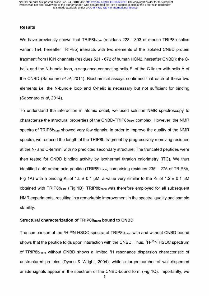
Results
We have previously shown that TRIP8bcore (residues 223 - 303 of mouse TRIP8b splice
variant 1a4, hereafter TRIP8b) interacts with two elements of the isolated CNBD protein
fragment from HCN channels (residues 521 - 672 of human HCN2, hereafter CNBD): the C-
helix and the N-bundle loop, a sequence connecting helix E’ of the C-linker with helix A of
the CNBD (Saponaro et al, 2014). Biochemical assays confirmed that each of these two
elements i.e. the N-bundle loop and C-helix is necessary but not sufficient for binding
(Saponaro et al, 2014).
To understand the interaction in atomic detail, we used solution NMR spectroscopy to
characterize the structural properties of the CNBD-TRIP8bcore complex. However, the NMR
spectra of TRIP8bcore showed very few signals. In order to improve the quality of the NMR
spectra, we reduced the length of the TRIP8b fragment by progressively removing residues
at the N- and C-termini with no predicted secondary structure. The truncated peptides were
then tested for CNBD binding activity by isothermal titration calorimetry (ITC). We thus
identified a 40 amino acid peptide (TRIP8bnano, comprising residues 235 – 275 of TRIP8b,
Fig 1A) with a binding KD of 1.5 ± 0.1 µM, a value very similar to the KD of 1.2 ± 0.1 µM
obtained with TRIP8bcore (Fig 1B). TRIP8bnano was therefore employed for all subsequent
NMR experiments, resulting in a remarkable improvement in the spectral quality and sample
stability.
Structural characterization of TRIP8bnano bound to CNBD
The comparison of the 1H-15N HSQC spectra of TRIP8bnano with and without CNBD bound
shows that the peptide folds upon interaction with the CNBD. Thus, 1H-15N HSQC spectrum
of TRIP8bnano without CNBD shows a limited 1H resonance dispersion characteristic of
unstructured proteins (Dyson & Wright, 2004), while a larger number of well-dispersed
amide signals appear in the spectrum of the CNBD-bound form (Fig 1C). Importantly, we
.CC-BY-NC-ND 4.0 International licenseIt is made available under a (which was not peer-reviewed) is the author/funder, who has granted bioRxiv a license to display the preprint in perpetuity.
The copyright holder for this preprint. http://dx.doi.org/10.1101/253096doi: bioRxiv preprint first posted online Jan. 24, 2018;
6
were now able to assign the backbone chemical shift resonances of TRIP8bnano bound to
the CNBD. The φ and ψ dihedral angles obtained from the NMR assignment indicate that
the peptide displays two α-helices (stretch L238-E250 named helix N and stretch T253-R269
named helix C) when bound to CNBD. The helices are separated by two amino acids; three
and six residues at the N- and C- termini, respectively, are unstructured (Fig 1D).
Structural characterization of CNBD bound to TRIP8bnano
NMR-analysis of the CNBD fragment bound to TRIP8bnano revealed that the interaction with
the peptide does not affect the overall fold of the protein. Thus, the CNBD adopts the typical
fold of the cAMP-free state, in line with previous evidence that this is the form bound by
TRIP8b (Deberg et al, 2015; Saponaro et al, 2014). More specifically, the secondary
structure elements of the cAMP-free CNBD are all conserved in the TRIP8bnano–bound
CNBD (Fig 2). This finding generally agrees with a recent double electron-electron
resonance (DEER) analysis of the CNBD-TRIP8b interaction, which showed that TRIP8b
binds to a conformation largely similar to the cAMP-free state (Deberg et al, 2015). Despite
the overall agreement with the DEER study, the NMR data also reveal a new and
unexpected feature of TRIP8b binding to the CNBD. Our results surprisingly show that
TRIP8bnano induces, upon binding to the CNBD, a well-defined secondary structure of the
distal region of the C-helix (Fig 2). This means that the distal region of the C-helix (residues
657-662), which is unstructured in the free form of the CNBD (Saponaro et al, 2014; Lee &
MacKinnon, 2017), extends into a helical structure upon ligand binding irrespectively of
whether the ligand is cAMP (Saponaro et al, 2014; Puljung & Zagotta, 2013; Lee &
MacKinnon, 2017) or TRIP8b (Fig 2). In contrast, and very differently from cAMP, which
directly contacts the P-helix in the Phosphate Binding cassette (PBC) and causes its folding
(Saponaro et al, 2014; Lee & MacKinnon, 2017), the NMR data show that TRIP8bnano binding
to the CNBD does not induce P-helix formation (Fig 2).
.CC-BY-NC-ND 4.0 International licenseIt is made available under a (which was not peer-reviewed) is the author/funder, who has granted bioRxiv a license to display the preprint in perpetuity.
The copyright holder for this preprint. http://dx.doi.org/10.1101/253096doi: bioRxiv preprint first posted online Jan. 24, 2018;

7
Modelling the CNBD-TRIP8bnano complex
Despite the significant improvement in sample stability and NMR spectra quality achieved
upon TRIP8bnano binding, we were still unable to assign the side chains of both proteins in
the complex and thus could not solve the solution structure of the complex by the canonical
NMR procedure. We therefore built a model of the CNBD-TRIP8bnano complex by docking
the two NMR-derived structures using the Haddock program (a detailed description of how
the respective structures were generated is provided in Materials and Methods and
Appendix Table S1).
In order to define the active residues (ambiguous interaction restraints) on the CNBD we
used the chemical shift perturbation values as described in Appendix Fig S1. For TRIP8bnano,
we defined as active a stretch of residues, E239-E243, previously identified as critical for the
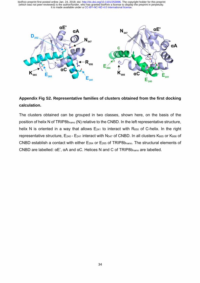
interaction (Santoro et al, 2009, 2011). Output clusters of this first molecular docking
calculation (settings can be found in Material and Methods) were further screened for
TRIP8bnano orientations in agreement with a previous DEER analysis, which placed TRIP8b
residue A248 closer to the proximal portion and TRIP8b residue A261 closer to the distal
portion of the CNBD C-helix (Deberg et al, 2015). Remarkably, in all clusters thus selected,
residues E264 or E265 in TRIP8b were found to interact with residues K665 or K666 of the CNBD
(Appendix Fig S2). This finding was notable, because we previously identified K665/K666 as
being critical for TRIP8b interaction in a biochemical binding assay (Saponaro et al, 2014).
We thus proceeded to individually mutate each of these four positions, and test their effect
on binding affinity through ITC. As expected, reverse charge mutations K665E or K666E
(CNBD) as well as E264K or E265K (TRIP8bnano) each strongly reduced the CNBD/TRIP8bnano
binding affinity (Appendix Fig S3).
Based on these observations, we performed a second molecular docking calculation,
including E264 and E265 as additional active residues for TRIP8bnano. This procedure resulted
.CC-BY-NC-ND 4.0 International licenseIt is made available under a (which was not peer-reviewed) is the author/funder, who has granted bioRxiv a license to display the preprint in perpetuity.
The copyright holder for this preprint. http://dx.doi.org/10.1101/253096doi: bioRxiv preprint first posted online Jan. 24, 2018;

8
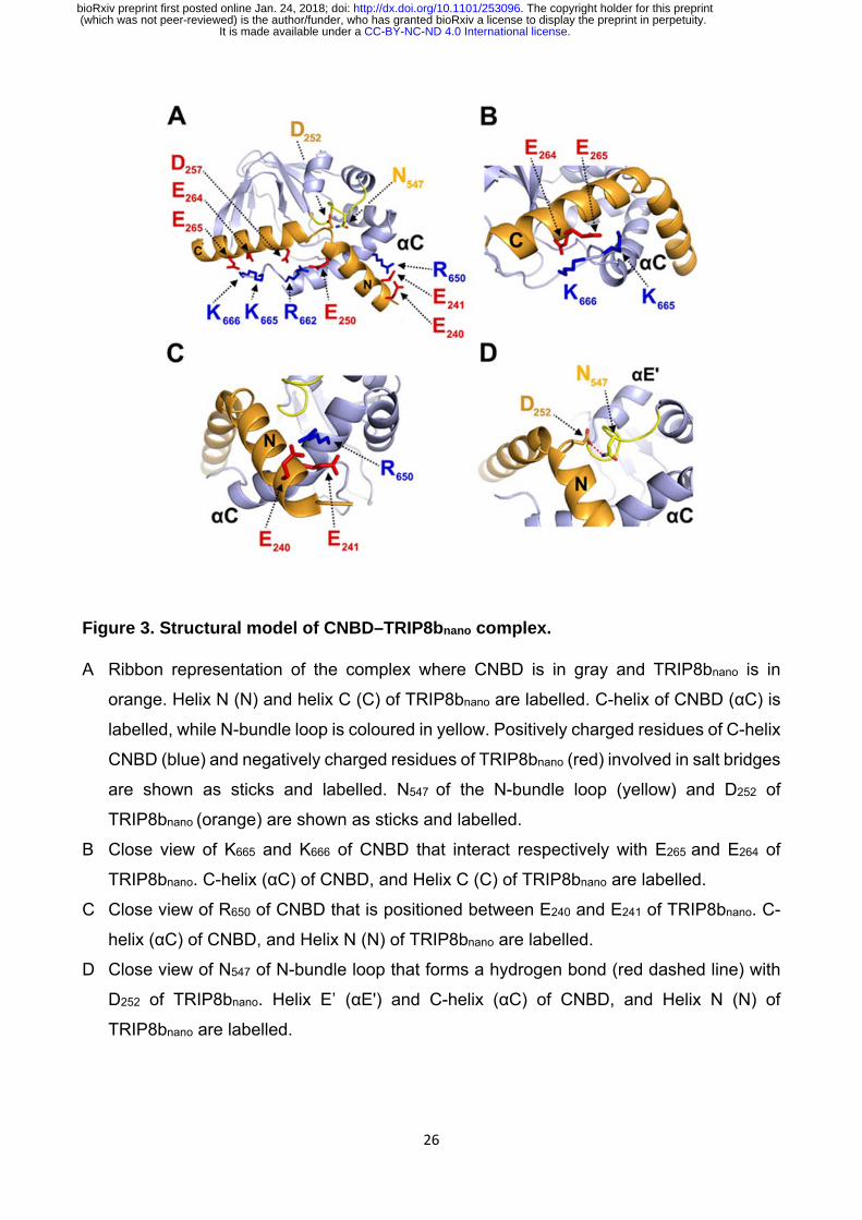
in the model shown in Fig 3, which represents the top-ranking cluster for energetic and
scoring function (Appendix Table S2) and was fully validated by mutagenesis analysis as
described below. Scrutiny of the model shows that TRIP8bnano binds to both the C-helix and
the N-bundle loop (Fig 3A). Binding to the C-helix is mainly guided by electrostatic
interactions between the negative charges on TRIP8bnano, and the positive charges on the
CNBD (Fig 3A). As shown in Fig 3B, the model highlights a double saline bridge (K665 and
K666 of CNBD with E265 and E264 of TRIP8bnano) in line with the ITC results described above
(Appendix Fig S3). Of note, the contribution of residue R662 to the binding is also consistent
with previous experiments showing residual TRIP8b interaction in a CNBD deletion mutant
ending at position 663 (Saponaro et al, 2014). Our modelling data suggest that, upon folding
of the distal portion of the C-helix, the side chains of residues R662 and R665 face to the inside
when contacting cAMP but face to the outside when binding TRIP8b (Appendix Fig S4).
In addition to clarifying the role of residues in the distal portion of the CNBD C-helix, the
model also highlights a second important cluster of electrostatic interactions with R650 in the
proximal portion of the CNBD C-helix contacting E240 and E241 in helix N of TRIP8bnano (Fig
3C). To confirm the contribution of these residues, we reversed charges and tested each
residue mutation for binding in ITC. The results in Appendix Fig S3 show that R650E caused
a more than six-fold reduction in binding affinity for TRIP8bnano, with smaller but significant
effects seen also for E240R and E241R.
A third important contact highlighted by the model is the interaction between N547 in the N-
bundle loop of the CNBD and D252 in the link between helix N and helix C of TRIP8bnano (Fig
3D). We tested this potential interaction by disrupting the expected hydrogen bond between
N547 and the carboxyl group of the negative residue (D252) in TRIP8bnano. The asparagine in
CNBD was mutated into aspartate (N547D) to generate an electrostatic repulsion for D252,
and the carboxyl group in D252 of TRIP8bnano was removed by mutation into asparagine
.CC-BY-NC-ND 4.0 International licenseIt is made available under a (which was not peer-reviewed) is the author/funder, who has granted bioRxiv a license to display the preprint in perpetuity.
The copyright holder for this preprint. http://dx.doi.org/10.1101/253096doi: bioRxiv preprint first posted online Jan. 24, 2018;

9
(D252N). As predicted, N547D greatly reduced binding to TRIP8b in ITC assays (Appendix Fig
S3 ), with a smaller but significant effect observed also for D252N (Appendix Fig S3). These
results confirm and extend our previous finding that the N-bundle loop contributes in a
substantial manner to the binding of TRIP8b (Saponaro et al, 2014).
TRIP8bnano as a tool for the direct regulation of native HCN currents
Next, we asked whether the relatively short TRIP8bnano peptide could be used to block the
response of HCN channels to cAMP by delivering the peptide to full length channels
heterologusly expressed in cells. To this end, we dialyzed TRIP8bnano into the cytosol of
HEK 293T cells transfected either with HCN1, HCN2, or HCN4 channels. The peptide was
added (10 µM) in the recording pipette together with a non-saturating concentration of cAMP
for each isoform (5 µM for HCN2, 1 µM for HCN4) expected to produce a ~10 mV rightward
shift in the mid-activation potential (V1/2) in the wildtype channels (Fig 4). No cAMP was
added in the case of HCN1, as this isoform is already fully shifted by low endogenous cAMP
levels (Fig 4 and Appendix Fig. S5). The results of the respective recordings in the presence
and absence of TRIP8bnano show that the peptide fully abolished the cAMP-induced
potentiation of channel opening in all HCN isoforms (Fig 4).
Thus, we reckoned it may be employed as a regulatory tool for native If/Ih currents. As proof
of principle, we tested whether TRIP8bnano can modulate the frequency of action potential
firing in sinoatrial node (SAN) cells. In these cells, If contributes substantially to the diastolic
depolarization phase of the action potential. Moreover, the autonomic nervous system
modulates the frequency of action potential firing by changing intracellular cAMP levels,
which in turn acts on HCN channel open probability (DiFrancesco, 1993). The native If
current from cardiomyocytes acutely isolated from rabbit sinoatrial node (SAN) was recorded
with and without 10 µM TRIP8bnano in the pipette solution (Fig 5A). Fig 5B shows that the
activation curve recorded in presence of TRIP8bnano is significantly hyperpolarized
.CC-BY-NC-ND 4.0 International licenseIt is made available under a (which was not peer-reviewed) is the author/funder, who has granted bioRxiv a license to display the preprint in perpetuity.
The copyright holder for this preprint. http://dx.doi.org/10.1101/253096doi: bioRxiv preprint first posted online Jan. 24, 2018;

10
compared to the control. This indicates that the peptide is displacing the binding of
endogenous cAMP to native HCN channels. Moreover, when the experiment was repeated
in the presence of 1 µM cAMP, TRIP8bnano prevented the typical cAMP-dependent
potentiation of the native If current (Fig 5B). In light of these results, we tested whether
TRIP8bnano is also able to modulate cardiac automaticity by antagonizing basal cAMP. The
data in Fig 5C show that TRIP8bnano indeed significantly decreased the rate of action
potential firing in single SAN cells. Strikingly, the observed 30% decrease in action potential
rate corresponds to the effect induced by physiological concentrations of acetylcholine
(DiFrancesco et al, 1989).
To conclusively prove that the inhibition of the native If current was specifically due to
TRIP8bnano rather than caused by the dilution of the cellular content followed by whole cell
configuration, we created a TAT version of TRIP8bnano (hereafter TAT-TRIP8bnano). Indeed,
TAT sequence allows the entry of biomolecules into a cell via endocytosis, thus leaving
unaltered the cytosolic content (Guidotti et al, 2017).
We thus tested whether both TRIP8bnano and TAT-TRIP8bnano were able to selectively inhibit
the beta-adrenergic stimulation of If current, while leaving unaltered the potentiation of L-
type Ca2+ current (ICa,L). To this end, we recorded either the native If or ICa,L current from
cardiomyocytes acutely isolated from mouse sinoatrial node (SAN) in the presence and in
the absence of 10 µM TRIP8bnano or TAT-TRIP8bnano, before and after stimulation with 100
nM isoproterenol, a β-adrenergic receptor agonist (Fig 6). Strikingly, TRIP8bnano prevented
the isoproterenol-induced increase of If current density, both when the peptide was added
in the recording pipette solution (Fig 6A and 6B), and when it was used in the TAT version
(Fig 6A and 6C). The specificity of TRIP8bnano for If current was confirmed by the absence
inhibition of the ICa,L current (fig 6D). Indeed, we failed to record a significant difference in
.CC-BY-NC-ND 4.0 International licenseIt is made available under a (which was not peer-reviewed) is the author/funder, who has granted bioRxiv a license to display the preprint in perpetuity.
The copyright holder for this preprint. http://dx.doi.org/10.1101/253096doi: bioRxiv preprint first posted online Jan. 24, 2018;
11
the isoproterenol-stimulated increase of the ICa,L current density between the control
condition and 10µM TRIP8bnano (fig 6E) or TAT-TRIP8bnano (fig 6F) conditions.
Discussion
TRIP8b-CNBD complex
In the present study, we describe the interaction between TRIP8b and the HCN channel
CNBD at atomic level, based on a NMR-derived structural model of their complex. The data
show that the minimal binding unit of TRIP8b, TRIP8bnano, folds in two helices upon binding,
suggesting that this region of the regulatory subunit has an intrinsically disordered behavior
when not in the complex. The model structurally validates previous indirect evidence, which
suggested that TRIP8b binds to two different elements of the CNBD, the N-bundle loop and
the C-helix. As a consequence of the interaction with TRIP8bnano, the C-helix in the CNBD
increases in length, a behavior already observed in the case of cAMP binding. The model
also identifies residues R662 and K665 in the CNBD as interaction partners with TRIP8bnano,
two cationic residues also involved in cAMP binding (Zhou & Siegelbaum, 2007). The
finding that TRIP8b and cAMP share binding sites on the C-helix provides a solid molecular
explanation for functional data which underscore a competition between the two regulators
(Han et al, 2011; Deberg et al, 2015; Bankston et al, 2017). However, it has been previously
suggested that a direct competition model does not fully explain the mutually antagonistic
effect of the two ligands (Hu et al, 2013). Specifically, the fact that the inhibitory effect of
TRIP8b on channel activity persists even at saturating cAMP concentrations suggests an
allosteric component in the regulation mechanism. Our structural model provides the
missing molecular evidence for this allosteric component. TRIP8bnano binds to the solvent-
exposed elements of CNBD (N-bundle loop and C-helix) and does not interact directly with
the buried PBC, which remains unfolded, as demonstrated by the observation that the P-
.CC-BY-NC-ND 4.0 International licenseIt is made available under a (which was not peer-reviewed) is the author/funder, who has granted bioRxiv a license to display the preprint in perpetuity.
The copyright holder for this preprint. http://dx.doi.org/10.1101/253096doi: bioRxiv preprint first posted online Jan. 24, 2018;

12
helix does not form upon TRIP8b binding. This rules out the possibility that TRIP8bnano
controls the affinity for cAMP by directly binding to the PBC. Rather, it confirms that the PBC
in the complex is indirectly kept in the low affinity state for cAMP binding (unfolded form)
through allosteric long-range interactions.
TRIP8bnano as a tool for modulating native If currents
TRIP8bnano is a minimal protein fragment, which binds the HCN channel CNBD with high
affinity and fully abolishes the cAMP effect in all tested isoforms (HCN1, 2 and 4). Given the
small size of the peptide (<5kD), TRIP8bnano may be easily adapted for in vivo delivery, and
thus constitutes a promising tool for the study or modulation of native HCN channels in
systems where the regulatory protein is not expressed, or is expressed at low levels. To this
end, we successfully fused to TRIP8bnano an internalization sequence that delivered the
peptide into sinoatrial node myocytes via the physiological pathway of the endocytosis
without affecting TRIP8bnano function. This modification may significantly expand the use of
TRIP8bnano as a tool for non-invasive and in vivo functional assays.
Our structural model explains why a previous attempt at identifying the minimal domain
required for TRIP8b activity resulted in a peptide with strongly reduced CNDB affinity, as the
fragment selected in the study by Lyman et al (Lyman et al, 2017) is lacking an important
contact residue (E240). In the present study, we successfully used TRIP8bnano to selectively
control native If currents and pacemaking in sinoatrial node cardiomyocytes. Unlike channel
blockers, which inhibit ionic currents, the peptide only interferes with the cAMP-based
regulation of HCN channels, while leaving basal HCN functions unaltered. In addition, and
in contrast to even the most selective blockers, it is entirely specific for HCN channels. The
ability to selectively control a specific molecular mechanism to modulate channel activity
represents a novel approach, which yields the promise of a more targeted therapeutic
intervention compared to pore blockers.
.CC-BY-NC-ND 4.0 International licenseIt is made available under a (which was not peer-reviewed) is the author/funder, who has granted bioRxiv a license to display the preprint in perpetuity.
The copyright holder for this preprint. http://dx.doi.org/10.1101/253096doi: bioRxiv preprint first posted online Jan. 24, 2018;

13
Materials and Methods
Constructs
The cDNA fragment encoding residues 235 – 275 (TRIP8bnano) of mouse TRIP8b (splice
variant 1a4) was cloned into pET-52b (EMD Millipore) downstream of a Strep (II) tag
sequence, while the cDNA fragment encoding residues 521–672 of human HCN2 (HCN2
CNBD) was cloned, in a previous study, into a modified pET-24b downstream of a double
His6-maltose-binding protein (MBP) (Saponaro et al, 2014). The cDNA encoding full-length
human HCN1 channel, mouse HCN2 channel, rabbit HCN4 channel and mouse TRIP8b
(1a4) were cloned into the eukaryotic expression vector pcDNA 3.1 (Clontech Laboratories).
Mutations were generated by site-directed mutagenesis (QuikChange site-directed
mutagenesis kit; Agilent Technologies) and confirmed by sequencing.
Preparation of proteins
The HCN2 CNBD WT and mutant proteins, as well as the TRIP8bcore and TRIP8bnano
proteins (WT and mutants) were produced and purified following the procedure previously
described (Saponaro et al, 2014).
Structure calculation of the cAMP-free human HCN2 CNBD in complex with
TRIP8bnano and vice versa
NMR experiments were acquired on Bruker Avance III 950, 700 and 500 MHz NMR
spectrometers equipped with a TXI-cryoprobe at 298 K. The acquired triple resonance NMR
experiments for the assignment of backbone resonances of cAMP-free HCN2 CNBD (CNBD
hereafter) in complex with TRIP8bnano and vice versa are summarized in Table S1. 15N, 13C’, 13Cα, 13Cβ, and Hα chemical shifts were used to derive ϕ and ψ dihedral angles by TALOS+
program (Cornilescu et al, 1999) for both CNBD and TRIP8bnano. For TRIP8bnano, CYANA-
2.1 structure calculation (Guntert & Buchner, 2015) was performed using 68 ϕ and ψ dihedral
angles and 40 backbone hydrogen bonds as input. For CNBD, CYANA-2.1 structure
calculation was performed using 108 ϕ and ψ dihedral angles, combined with the NOEs
obtained in our previous determination of the cAMP-free form of the CNBD (Saponaro et al,
2014) for those regions not affected by the interaction with TRIP8bnano. The 10 conformers
of TRIP8bnano and CNBD with the lowest residual target function values were subjected to
restrained energy minimization with AMBER 12.0 (Santoro et al, 2011)
.CC-BY-NC-ND 4.0 International licenseIt is made available under a (which was not peer-reviewed) is the author/funder, who has granted bioRxiv a license to display the preprint in perpetuity.
The copyright holder for this preprint. http://dx.doi.org/10.1101/253096doi: bioRxiv preprint first posted online Jan. 24, 2018;

14
(http://pyenmr.cerm.unifi.it/access/index/amps-nmr) and used as input in docking
calculations.
Docking calculations
Docking calculations were performed with HADDOCK2.2 implemented in the
WeNMR/West-Life GRID-enabled web portal (www.wenmr.eu). The docking calculations
are driven by ambiguous interaction restraints (AIRs) between all residues involved in the
intermolecular interactions (Dominguez et al, 2003). Active residues of the CNBD were
defined as the surface exposed residues (at least 50% of solvent accessibility), which show
chemical shift perturbation upon TRIP8bnano binding.
The assignment of the CNBD bound to TRIP8bnano allowed to highlight the residues of CNBD
whose backbone featured appreciable Combined Chemical Shift Perturbation (CSP) (Fig
S1). The combined CSP (∆HN) is given by the equation
∆HN=((HNfree−HNbound)2+((Nfree−Nbound)/5)2)/2½ (Garrett et al, 1997).
Passive residues of CNBD were defined as the residues close in space to active residues
and with at least 50% solvent accessibility.
In the case of TRIP8bnano, the conserved stretch E239-E243, located in helix N, was defined
as active region in a first docking calculation, while all the other solvent accessible residues
of the peptide were defined as passive. This docking calculation generated several clusters.
A post-docking filter step allowed us to select those clusters having an orientation of
TRIP8bnano bound to CNBD in agreement with a DEER study on the CNBD - TRIP8bnano
interaction (Deberg et al, 2015). The selected clusters grouped in two classes on the basis
of the orientation of helix N of TRIP8bnano (N) relative to CNBD (Fig S2). A second docking
calculation was subsequently performed introducing also residues E264-E265, located in helix
C of TRIP8bnano as active residues. The active residues for CNBD were the same used for
the first calculation. For this second HADDOCK calculation 14 clusters were obtained and
ranked according to their HADDOCK score. Among them only four clusters showed both an
orientation of TRIP8bnano bound to CNBD in agreement with the DEER study (Deberg et al,
2015) and the involvement of E239-E243 stretch of TRIP8bnano in the binding to CNBD. These
clusters were manually analyzed and subjected to a per-cluster re-analysis following the
protocol reported in http://www.bonvinlab.org/software/haddock2.2/analysis/#reanal. From
this analysis, it resulted that the top-ranking cluster, i.e. the one with the best energetic and
scoring functions, has a conformation in agreement with mutagenesis experiments (Fig S3).
.CC-BY-NC-ND 4.0 International licenseIt is made available under a (which was not peer-reviewed) is the author/funder, who has granted bioRxiv a license to display the preprint in perpetuity.
The copyright holder for this preprint. http://dx.doi.org/10.1101/253096doi: bioRxiv preprint first posted online Jan. 24, 2018;
15
Energy parameters (van der Waals energy, electrostatic energy, desolvation energy, and
the penalty energy due to violation of restraints) for this complex model are reported in Table
S2.
Both docking calculations were performed using 10 NMR conformers of both the CNBD and
the TRIP8bnano structures calculated as described above. In the TRIP8bnano structures the
unfolded N- and C-terminal regions were removed, while in the CNBD structures only the
unfolded N-terminal region was removed. This is because the C-terminal region of the CNBD
is known to comprise residues involved in TRIP8bnano binding (Saponaro et al, 2014).
Flexible regions of the proteins were defined based on the active and passive residues plus
two preceding and following residues. The residue solvent accessibility was calculated with
the program NACCESS (Hu et al, 2013). In the initial rigid body docking calculation phase,
5000 structures of the complex were generated, and the best 400 in terms of total
intermolecular energy were further submitted to the semi-flexible simulated annealing and a
final refinement in water. Random removal of the restraints was turned off. The number of
flexible refinement steps was increased from the default value of 500/500/1000/1000 to
2000/2000/2000/4000. The final 400 structures were then clustered using a cutoff of 5.0 Å
of RMSD to take into consideration the smaller size of protein-peptide interface.
Electrophysiology of HEK 293T cells
HEK 293T cells were cultured in Dulbecco’s modified Eagle’s medium (Euroclone)
supplemented with 10% fetal bovine serum (Euroclone), 1% Pen Strep (100 U/mL of
penicillin and 100 µg/ml of streptomycin), and stored in a 37°C humidified incubator with 5%
CO2. The plasmid containing cDNA of wild-type and mutant HCN1, HCN2 and HCN4
channels (1 µg) was co-transfected for transient expression into HEK 293T cells with a
plasmid containing cDNA of Green Fluorescent Protein (GFP) (1.3 µg). For co-expression
with TRIP8b (1a-4), HEK 293T cells were transiently transfected with wild-type (wt) and/or
mutant human HCN1 cDNA (1 µg), wt TRIP8b (1a-4) cDNA (1 µg) and cDNA of Green
Fluorescent Protein (GFP) (0.3 µg).
One day after transfection, GFP-expressing cells were selected for patch-clamp
experiments in whole-cell configuration. The experiments were conducted at R.T. The
pipette solution in whole cell experiments contained: 10 mM NaCl, 130 mM KCl, 1 mM
egtazic acid (EGTA), 0.5 mM MgCl2, 2 mM ATP (Mg salt) and 5 mM HEPES–KOH buffer
(pH 7.4). The extracellular bath solution contained 110 mM NaCl, 30 mM KCl, 1.8 mM CaCl2,
0.5 mM MgCl2 and 5 mM HEPES–KOH buffer (pH 7.4).
.CC-BY-NC-ND 4.0 International licenseIt is made available under a (which was not peer-reviewed) is the author/funder, who has granted bioRxiv a license to display the preprint in perpetuity.
The copyright holder for this preprint. http://dx.doi.org/10.1101/253096doi: bioRxiv preprint first posted online Jan. 24, 2018;
16
TRIP8bnano was added (10 µM) to the pipette solution. cAMP was added at different
concentration to the pipette solution depending on the HCN isoform used: 0 µM for HCN1,
5 µM for HCN2 and 1 µM for HCN4.
Whole-cell measurements of HCN channels were performed using the following voltage
clamp protocol depending on the HCN isoform measured: for HCN1, holding potential was
–30 mV (1s), with steps from –40 mV to –130 mV (10 mV interval, 3.5 s) and tail currents
recorded at –40mV (3 s); for HCN2, holding potential was –30 mV (1 s), with steps from –
40 mV to –150 mV (10 mV interval, 5 s) and tail currents recorded at -40mV (5 s); for HCN4,
holding potential was –30 mV (1s), steps from –40 mV to –160 mV (10 mV interval, 6 s) and
tail currents were recorded at -40mV (6 s).
Isolation and electrophysiology of rabbit sinoatrial node cells
Animal protocols conformed to the guidelines of the care and use of laboratory animals
established by Italian and European Directives (D. Lgs n° 2014/26, 2010/63/UE). New
Zealand white female rabbits (0.8–1.2 kg) were anesthetized (xylazine 5mg/Kg, i.m.), and
euthanized with an overdose of sodium thiopental (i.v.); hearts were quickly removed, and
the SAN region was isolated and cut in small pieces. Single SAN cardiomyocytes were
isolated following an enzymatic and mechanical procedure as previously described
(DiFrancesco et al, 1986). Following isolation, cells were maintained at 4 °C in Tyrode
solution: 140 mM NaCl, 5.4 mM KCl, 1.8 mM CaCl2, 1 mM MgCl2, 5.5 mM D-glucose, 5 mM
HEPES-NaOH (pH 7.4).
For patch clamp experiments cells were placed in a chamber on an inverted microscope
and experiments were performed in the whole-cell configuration at 35 ± 0.5 °C. The pipette
solution contained: 10 mM NaCl, 130 mM KCl, 1 mM egtazic acid (EGTA), 0.5 mM MgCl2,
and 5 mM HEPES–KOH buffer (pH 7.2). The If current was recorded from single cells
superfused with Tyrode solution with 1 mM BaCl2, and 2 mM MnCl2.
If activation curves were obtained using a two-step protocol in which test voltage steps (from
-30 to -120 mV, 15 mV interval) were applied from a holding potential of -30 mV and were
followed by a step to -125 mV. Test steps had variable durations so as to reach steady –
state activation at all voltages.
In current-clamp studies, spontaneous action potentials were recorded from single cells
superfused with Tyrode solution, and rate was measured from the interval between
.CC-BY-NC-ND 4.0 International licenseIt is made available under a (which was not peer-reviewed) is the author/funder, who has granted bioRxiv a license to display the preprint in perpetuity.
The copyright holder for this preprint. http://dx.doi.org/10.1101/253096doi: bioRxiv preprint first posted online Jan. 24, 2018;

17
successive action potential. When indicated cAMP (1 µM) and/or nanoTRIP8b (10 µM) were
added to the pipette solution.
Isolation and electrophysiology of mouse sinoatrial node cells
Mice were killed by cervical dislocation under general anesthesia consisting of 0.01 mg/g
xylazine (2% Rompun; Bayer AG), 0.1 mg/g ketamine (Imalgène; Merial) and 0.04mg/g of
Na-pentobarbital (Euthanasol VET, Laboratoire TVM, Lempdes, France), and beating hearts
were quickly removed. The SAN region was excised in warmed (35°C) Tyrode’s solution
containing: 140 mM NaCl, 5.4 mM KCl, 1.8 mM CaCl2, 1 mM MgCl2, 1 mM Hepes-NaOH
(pH = 7.4), and 5.5 mM D-glucose and cut in strips. Strips were then transferred into a “low-
Ca2+-low-Mg2+” solution containing: 140 mM NaCl; 5.4 mM KCl, 0.5 mM MgCl2, 0.2 mM
CaCl2, 1.2 mM KH2PO4, 50 mM taurine, 5.5 mM D-glucose, 1 mg/ml bovine serum albumin
(BSA), 5 mM Hepes-NaOH (pH = 6.9).
Tissue was digested by adding Liberase TH (0.15 mg/ml, Roche Diagnostics GmbH,
Mannheim, Germany), elastase (1.9 U/ml, Worthington, Lakewood, USA). Digestion was
carried out for a variable time of 15–18 minutes at 35°C. Tissue strips were then washed
and transferred into a modified “Kraftbrühe” (KB) medium containing: 70 mM L-glutamic
acid, 20 mM KCl, 80 mM KOH, (±) 10 mM D- b-OH-butyric acid; 10 mM KH2PO4, 10 mM
taurine, 1mg/ml BSA and 10 mM Hepes-KOH (pH = 7.4).
Single SAN cells were isolated by manual agitation in KB solution at 35°C for 30–50
seconds.
Cellular automaticity was recovered by re-adapting the cells to a physiological extracellular
Ca2+ concentration by addition of a solution containing: 10 mM NaCl, 1.8 mM CaCl2 and
normal Tyrode solution containing BSA (1 mg/ml). The final storage solution contained: 100
mM NaCl, 35 mM KCl, 1.3 mM CaCl2, 0.7 mM MgCl2, 14 mM L-glutamic acid, (±) 2 mM D-
b-OH-butyric acid, 2 mM KH2PO4, 2 mM taurine, 1 mg/ml BSA, (pH = 7.4). Cells were then
stored at room temperature until use. All chemicals were from SIGMA (St Quentin Fallavier,
France).
For electrophysiological recording, SAN cells in the storage solution were harvested in
special custom-made recording plexiglas chambers with glass bottoms for proper cell
attachment and mounted on the stage of an inverted microscope (Olympus IX71) and
perfused with normal Tyrode solution. The recording temperature was 36°C. We used the
whole-cell variation of the patch-clamp technique to record cellular ionic currents, by
.CC-BY-NC-ND 4.0 International licenseIt is made available under a (which was not peer-reviewed) is the author/funder, who has granted bioRxiv a license to display the preprint in perpetuity.
The copyright holder for this preprint. http://dx.doi.org/10.1101/253096doi: bioRxiv preprint first posted online Jan. 24, 2018;

18
employing a Multiclamp 700B (Axon Instruments Inc., Foster USA) patch clamp amplifier.
Recording electrodes were fabricated from borosilicate glass, by employing a WZ DMZ-
Universal microelectrode puller (Zeitz-Instruments Vertriebs GmbH, Martinsried, Germany).
If was recorded under standard whole-cell configuration during perfusion of standard
Tyrode’s containing 2 mM BaCl2 to block IK1. Patch-clamp pipettes were filled with an
intracellular solution containing: 130 mM KCl, 10 mM NaCl, 1 mM EGTA, 0.5 mM MgCl2 and
5 mM HEPES (pH 7.2).
For recording of L-type calcium currents, pipette solution contained: 125 mM CsOH, 20 mM
tetraethylammonium chloride (TEA-Cl), 1.2 mM CaCl2, 5 mM Mg-ATP, 0.1 mM Li2-GTP, 5
mM EGTA and 10 mM HEPES (pH 7.2 with aspartate). 30 µM TTX (Latoxan, Portes lès
Valence, France) to block INa was added to external solution containing: 135 mM
tetraethylammonium chloride (TEA-Cl), 4 mM CaCl2,10 mM 4-amino-pyridine, 1 mM MgCl2,
10 mM HEPES and 1 mg/ml Glucose (pH 7.4 with TEA-OH).
Electrodes had a resistance of about 3 MΩ. Seal resistances were in the range of 2–5 GΩ.
10µM TRIPb8nano was added to pipette solution. 10µM TAT-TRIPb8nano was added in cell
storage solution for at least 30 min before patch clamp recording.
Data analysis
Data were acquired at 1 kHz using an Axopatch 200B amplifier and pClamp10.5 software
(Axon Instruments). Data were analyzed off-line using Clampfit 10.5 (Molecular Devices)
and Origin 16 (OriginLab Corp., Northampton MA). Activation curves were analyzed by the
Boltzmann equation, y=1/1+exp[(V−V1/2)/s], where y is fractional activation, V is voltage,
V1/2 half-activation voltage, and s the inverse slope factor (mV) (DiFrancesco, 1999). Mean
activation curves were obtained by fitting individual curves from each cell to the Boltzmann
equation and then averaging all curves obtained.
ACKNOWLEDGEMENTS
This work has been supported by Fondazione CARIPLO grant 2014-0796 to A.M., B.S and
L.B. and partly by 2016 Schaefer Research Scholars Program of Columbia University to
A.M., by MIUR PRIN (Programmi di Ricerca di Rilevante Interesse Nazionale) 494 2015
.CC-BY-NC-ND 4.0 International licenseIt is made available under a (which was not peer-reviewed) is the author/funder, who has granted bioRxiv a license to display the preprint in perpetuity.
The copyright holder for this preprint. http://dx.doi.org/10.1101/253096doi: bioRxiv preprint first posted online Jan. 24, 2018;
19
(2015795S5W) to A.M., by European Research Council (ERC) 2015 Advanced Grant 495
(AdG) n. 695078 noMAGIC to A.M. and G.T., by National Institutes for Health Grant R01
NS036658 to B.S., by Instruct-ERIC and national member subscriptions to L.B., by
Accademia Nazionale dei Lincei (Giuseppe Levi foundation) to A.S. We specially thank the
EU ESFRI Instruct Core Centre CERM-Italy.
Author contributions
A.S. designed and prepared constructs, performed biochemical experiments and analyzed
the data, C.D. and B.I. designed constructs and purified proteins, F.C. and V.M. performed
the NMR measurements and analyzed the data, A.P. performed the patch measurements
in HEK293T cells, D.D.F. and A.B. designed the expriments and A.B: performed the
measurements in rabbit SAN myocytes, M.M. and P.M. designed the experiments and P.M.
performed the measurements in mouse SAN myocytes. L.B., B.S., G.T. and A.M. conceived
the study and wrote the manuscript. A.M. coordinated the research team.
Conflict of interest: none
References Bankston JR, DeBerg HA, Stoll S & Zagotta WN (2017) Mechanism for the inhibition of the cAMP
dependence of HCN ion channels by the auxiliary subunit TRIP8b. J. Biol. Chem.
Cornilescu G, Delaglio F & Bax A (1999) Protein backbone angle restraints from searching a database for chemical shift and sequence homology. J. Biomol. NMR 13: 289–302
Deberg HA, Bankston JR, Rosenbaum JC, Brzovic PS, Zagotta WN & Stoll S (2015) Structural mechanism for the regulation of HCN ion channels by the accessory protein TRIP8b. Structure 23: 734–744
DiFrancesco D (1993) Pacemaker mechanisms in cardiac tissue. Annu. Rev. Physiol. 55: 455–472
DiFrancesco D (1999) Dual allosteric modulation of pacemaker (f) channels by cAMP and voltage in rabbit SA node. J. Physiol. 515 ( Pt 2: 367–376
DiFrancesco D, Ducouret P & Robinson RB (1989) Muscarinic modulation of cardiac rate at low acetylcholine concentrations. Science (80‐. ). 243: 669–671 Available at: http://www.ncbi.nlm.nih.gov/pubmed/2916119
DiFrancesco D, Ferroni A, Mazzanti M & Tromba C (1986) Properties of the hyperpolarizing‐activated current (if) in cells isolated from the rabbit sino‐atrial node. J. Physiol. 377: 61–88
.CC-BY-NC-ND 4.0 International licenseIt is made available under a (which was not peer-reviewed) is the author/funder, who has granted bioRxiv a license to display the preprint in perpetuity.
The copyright holder for this preprint. http://dx.doi.org/10.1101/253096doi: bioRxiv preprint first posted online Jan. 24, 2018;
20
Dominguez C, Boelens R & Bonvin AMJJ (2003) HADDOCK: a protein‐protein docking approach based on biochemical or biophysical information. J. Am. Chem. Soc. 125: 1731–1737
Dyson HJ & Wright PE (2004) Unfolded proteins and protein folding studied by NMR. Chem. Rev. 104: 3607–3622
Garrett DS, Seok YJ, Peterkofsky A, Clore GM & Gronenborn AM (1997) Identification by NMR of the binding surface for the histidine‐containing phosphocarrier protein HPr on the N‐terminal domain of enzyme I of the Escherichia coli phosphotransferase system. Biochemistry 36: 4393–4398
Guidotti G, Brambilla L & Rossi D (2017) Cell‐Penetrating Peptides: From Basic Research to Clinics. Trends Pharmacol. Sci. 38: 406–424
Guntert P & Buchner L (2015) Combined automated NOE assignment and structure calculation with CYANA. J. Biomol. NMR 62: 453–471
Han Y, Noam Y, Lewis AS, Gallagher JJ, Wadman WJ, Baram TZ & Chetkovich DM (2011) Trafficking and gating of hyperpolarization‐activated cyclic nucleotide‐gated channels are regulated by interaction with tetratricopeptide repeat‐containing Rab8b‐interacting protein (TRIP8b) and cyclic AMP at distinct sites. J. Biol. Chem. 286: 20823–20834
Hu L, Santoro B, Saponaro A, Liu H, Moroni A & Siegelbaum S (2013) Binding of the auxiliary subunit TRIP8b to HCN channels shifts the mode of action of cAMP. J. Gen. Physiol. 142: 599–612 Available at: http://www.jgp.org/lookup/doi/10.1085/jgp.201311013
Lee C‐H & MacKinnon R (2017) Structures of the Human HCN1 Hyperpolarization‐Activated Channel. Cell 168: 111–120.e11
Lewis AS, Schwartz E, Chan CS, Noam Y, Shin M, Wadman WJ, Surmeier DJ, Baram TZ, Macdonald RL & Chetkovich DM (2009) Alternatively spliced isoforms of TRIP8b differentially control h channel trafficking and function. J. Neurosci. 29: 6250–6265
Lyman KA, Han Y, Heuermann RJ, Cheng X, Kurz JE, Lyman RE, Van Veldhoven PP & Chetkovich DM (2017) Allostery between two binding sites in the ion channel subunit TRIP8b confers binding specificity to HCN channels. J. Biol. Chem.
Piskorowski R, Santoro B & Siegelbaum SA (2011) TRIP8b Splice Forms Act in Concert to Regulate the Localization and Expression of HCN1 Channels in CA1 Pyramidal Neurons. Neuron 70: 495–509
Puljung MC & Zagotta WN (2013) A secondary structural transition in the C‐helix promotes gating of cyclic nucleotide‐regulated ion channels. J. Biol. Chem. 288: 12944–12956
Robinson RB & Siegelbaum SA (2003) Hyperpolarization‐Activated Cation Currents: From Molecules to Physiological Function. Annu. Rev. Physiol. 65: 453–480 Available at: http://www.annualreviews.org/doi/10.1146/annurev.physiol.65.092101.142734
Santoro B, Hu L, Liu H, Saponaro A, Pian P, Piskorowski RA, Moroni A & Siegelbaum SA (2011) TRIP8b Regulates HCN1 Channel Trafficking and Gating through Two Distinct C‐Terminal Interaction Sites. J. Neurosci. 31: 4074–4086 Available at: http://www.jneurosci.org/cgi/doi/10.1523/JNEUROSCI.5707‐10.2011
Santoro B, Piskorowski RA, Pian P, Hu L, Liu H & Siegelbaum SA (2009) TRIP8b Splice Variants Form a Family of Auxiliary Subunits that Regulate Gating and Trafficking of HCN Channels in the Brain. Neuron 62: 802–813
Saponaro A, Pauleta SR, Cantini F, Matzapetakis M, Hammann C, Donadoni C, Hu L, Thiel G, Banci L, Santoro B & Moroni A (2014) Structural basis for the mutual antagonism of cAMP and TRIP8b in regulating HCN channel function. Proc. Natl. Acad. Sci. U. S. A. 111: 14577–14582
.CC-BY-NC-ND 4.0 International licenseIt is made available under a (which was not peer-reviewed) is the author/funder, who has granted bioRxiv a license to display the preprint in perpetuity.
The copyright holder for this preprint. http://dx.doi.org/10.1101/253096doi: bioRxiv preprint first posted online Jan. 24, 2018;

21
Wainger BJ, DeGennaro M, Santoro B, Siegelbaum SA & Tibbs GR (2001) Molecular mechanism of cAMP modulation of HCN pacemaker channels. Nature 411: 805–810 Available at: http://www.nature.com/doifinder/10.1038/35081088
Zagotta WN, Olivier NB, Black KD, Young EC, Olson R & Gouaux E (2003) Structural basis for modulation and agonist specificity of HCN pacemaker channels. Nature 425: 200–205 Available at: http://www.nature.com/doifinder/10.1038/nature01922
Zhou L & Siegelbaum SA (2007) Gating of HCN channels by cyclic nucleotides: residue contacts that underlie ligand binding, selectivity, and efficacy. Structure 15: 655–670
Zolles G, Wenzel D, Bildl W, Schulte U, Hofmann A, Muller CS, Thumfart J‐O, Vlachos A, Deller T, Pfeifer A, Fleischmann BK, Roeper J, Fakler B & Klocker N (2009) Association with the auxiliary subunit PEX5R/Trip8b controls responsiveness of HCN channels to cAMP and adrenergic stimulation. Neuron 62: 814–825
.CC-BY-NC-ND 4.0 International licenseIt is made available under a (which was not peer-reviewed) is the author/funder, who has granted bioRxiv a license to display the preprint in perpetuity.
The copyright holder for this preprint. http://dx.doi.org/10.1101/253096doi: bioRxiv preprint first posted online Jan. 24, 2018;
22
.CC-BY-NC-ND 4.0 International licenseIt is made available under a (which was not peer-reviewed) is the author/funder, who has granted bioRxiv a license to display the preprint in perpetuity.
The copyright holder for this preprint. http://dx.doi.org/10.1101/253096doi: bioRxiv preprint first posted online Jan. 24, 2018;
23
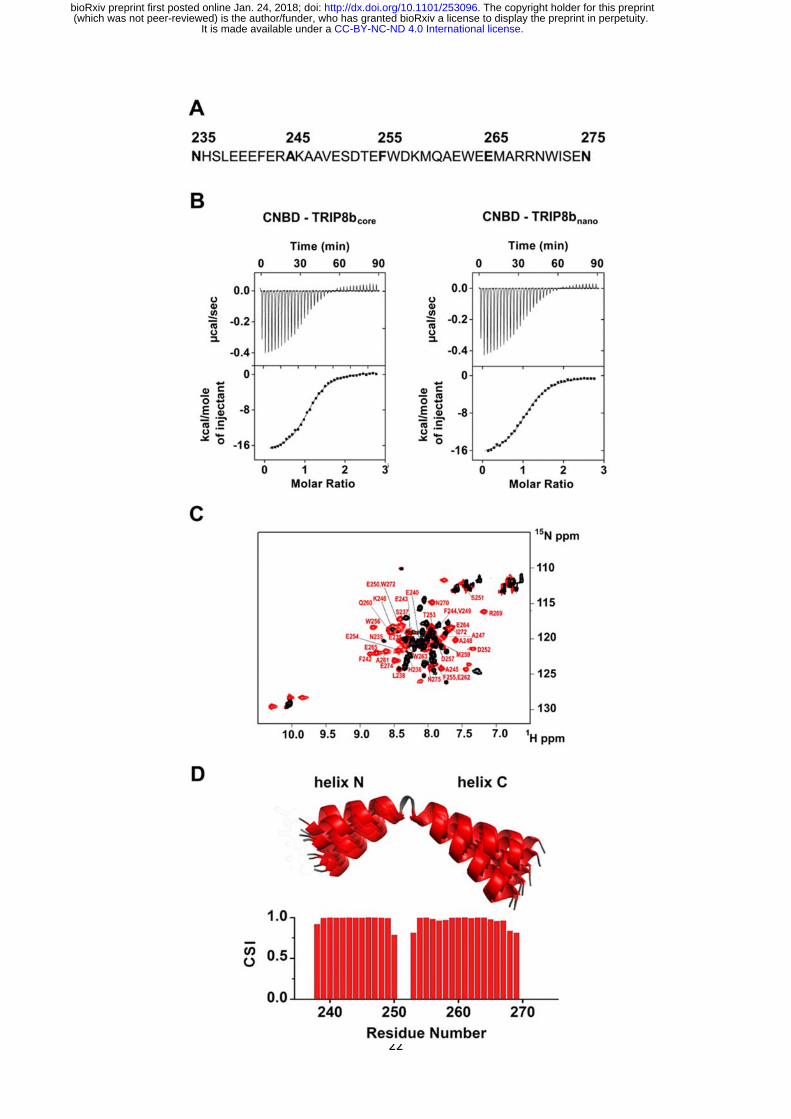
Figure 1. Functional and structural characterization of TRIP8bnano.
A Primary sequence of TRIP8bnano. Amino acid numbering refers to full length mouse
TRIP8b (1a4).
B Binding of TRIP8bcore and TRIP8bnano to purified His6-MBP-CNBD measured by
Isothermal titration calorimetry (ITC). Upper panel, heat changes (μcal/sec) during
successive injections of 8 μL of the corresponding TRIP8b peptide (200 μM) into the
chamber containing His6-MBP-CNBD (20 μM). Lower panel, binding curve obtained from
data displayed in the upper panel. The peaks were integrated, normalized to TRIP8b
peptide concentration, and plotted against the molar ratio (TRIP8b peptide / His6-MBP-
CNBD). Solid line represents a nonlinear least-squares fit to a single-site binding model,
yielding, in the present examples, a KD = 1.2 ± 0.1 μM for TRIP8bcore and KD = 1.4 ± 0.1
μM for TRIP8bnano.
C Evidence for TRIP8bnano folding upon CNBD binding based on the superimposition of the
[1H, 15N] heteronuclear single quantum coherence (HSQC) NMR spectrum of CNBD-free
TRIP8bnano (black) and CNBD-bound TRIP8bnano (red). The latter experiment was
performed at the molar ratio ([CNBD]/[ TRIP8bnano]) = 3. The backbone amide (HN)
signals of the residues of CNBD-bound TRIP8bnano are labelled in red.
D (Top) Ribbon representation of the 10 lowest energy conformers of TRIP8bnano bound to
CNBD used for in silico modelling of CNBD-TRIP8bnano complex. The unfolded regions
at the N- and C-termini of the construct (residues 235–237 and 270 – 275) are omitted
for clarity. (Bottom) Chemical Shift Index (CSI, calculated using TALOS+) plotted as a
function of the residue number of TRIP8bnano bound to CNBD. Positive values represent
helical propensity.
.CC-BY-NC-ND 4.0 International licenseIt is made available under a (which was not peer-reviewed) is the author/funder, who has granted bioRxiv a license to display the preprint in perpetuity.
The copyright holder for this preprint. http://dx.doi.org/10.1101/253096doi: bioRxiv preprint first posted online Jan. 24, 2018;

24
Figure 2. NMR structure of CNBD bound to TRIP8bnano.
A (Top) comparison of secondary structure elements of cAMP-free CNBD (Saponaro et al,
2014), cAMP-bound CNBD (Zagotta et al, 2003) and cAMP-free CNBD bound to
TRIP8bnano (this study). Secondary structure elements are indicated by arrows (-
strands) and cylinders (-helices) and labelled. The loop between β6 and β7 constitutes
the Phosphate Binding Cassette (PBC). The elements that fold upon binding of cAMP
and TRIP8bnano are shown in red. (Bottom) Chemical Shift Index (CSI, calculated using
TALOS+) plotted as a function of the residue number of CNBD bound to TRIP8bnano.
Positive values represent helical propensity, while negative values represent strands.
B Ribbon representation of the 10 lowest energy conformers of CNBD bound to TRIP8bnano
used for in silico modelling of CNBD-TRIP8bnano complex. Secondary structure elements
.CC-BY-NC-ND 4.0 International licenseIt is made available under a (which was not peer-reviewed) is the author/funder, who has granted bioRxiv a license to display the preprint in perpetuity.
The copyright holder for this preprint. http://dx.doi.org/10.1101/253096doi: bioRxiv preprint first posted online Jan. 24, 2018;
25
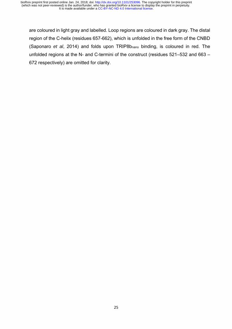
are coloured in light gray and labelled. Loop regions are coloured in dark gray. The distal
region of the C-helix (residues 657-662), which is unfolded in the free form of the CNBD
(Saponaro et al, 2014) and folds upon TRIP8bnano binding, is coloured in red. The
unfolded regions at the N- and C-termini of the construct (residues 521–532 and 663 –
672 respectively) are omitted for clarity.
.CC-BY-NC-ND 4.0 International licenseIt is made available under a (which was not peer-reviewed) is the author/funder, who has granted bioRxiv a license to display the preprint in perpetuity.
The copyright holder for this preprint. http://dx.doi.org/10.1101/253096doi: bioRxiv preprint first posted online Jan. 24, 2018;
26
Figure 3. Structural model of CNBD–TRIP8bnano complex.
A Ribbon representation of the complex where CNBD is in gray and TRIP8bnano is in
orange. Helix N (N) and helix C (C) of TRIP8bnano are labelled. C-helix of CNBD (αC) is
labelled, while N-bundle loop is coloured in yellow. Positively charged residues of C-helix
CNBD (blue) and negatively charged residues of TRIP8bnano (red) involved in salt bridges
are shown as sticks and labelled. N547 of the N-bundle loop (yellow) and D252 of
TRIP8bnano (orange) are shown as sticks and labelled.
B Close view of K665 and K666 of CNBD that interact respectively with E265 and E264 of
TRIP8bnano. C-helix (αC) of CNBD, and Helix C (C) of TRIP8bnano are labelled.
C Close view of R650 of CNBD that is positioned between E240 and E241 of TRIP8bnano. C-
helix (αC) of CNBD, and Helix N (N) of TRIP8bnano are labelled.
D Close view of N547 of N-bundle loop that forms a hydrogen bond (red dashed line) with
D252 of TRIP8bnano. Helix E’ (αE') and C-helix (αC) of CNBD, and Helix N (N) of
TRIP8bnano are labelled.
.CC-BY-NC-ND 4.0 International licenseIt is made available under a (which was not peer-reviewed) is the author/funder, who has granted bioRxiv a license to display the preprint in perpetuity.
The copyright holder for this preprint. http://dx.doi.org/10.1101/253096doi: bioRxiv preprint first posted online Jan. 24, 2018;

27
Figure 4. TRIP8bnano abolishes cAMP effect on HCN channel gating.
Effect of TRIP8bnano on human HCN1, mouse HCN2 and rabbit HCN4 half activation
potentials (V1/2). Half activation potential (V1/2) was determined as described in Material and
Methods. HCN1 (black filled circle) = -72.8 ± 0.2 mV; HCN1 + 10 µM TRIP8bnano (black open
circle) = -82 ± 0.5 mV; HCN2 (green filled circle) = -93.7± 0.3 mV; HCN2 + 5 µM cAMP
(green semi-open circle) = -83.5 ± 0.3 mV; HCN2 + 5 µM cAMP + 10 µM TRIP8bnano (green
open circle) = -94.5 ± 0.6 mV; HCN4 (blue filled circle) = -102.8 ± 0.3 mV; HCN4 + 1µM
cAMP (blue semi-open circle) = -89.2 ± 0.6 mV; HCN4 + 1µM cAMP + 10 µM TRIP8bnano
(blue open circle) = -102.1 ± 0.6 mV. Data are presented as mean ± SEM. Number of cells
(N) ≥ 11. Statistical analysis performed with ANOVA, followed by post-hoc Tukey test (***,
P < 0.001).
.CC-BY-NC-ND 4.0 International licenseIt is made available under a (which was not peer-reviewed) is the author/funder, who has granted bioRxiv a license to display the preprint in perpetuity.
The copyright holder for this preprint. http://dx.doi.org/10.1101/253096doi: bioRxiv preprint first posted online Jan. 24, 2018;

28
Figure 5. Effects of TRIP8bnano on voltage-dependent activation of If and spontaneous
rate in rabbit sinoatrial node (SAN) myocytes.
A Representative whole-cell If currents recorded, at the indicated voltages, in the control
solution and in the presence of 10 μM TRIP8bnano, without (top) and with 1 μM cAMP in
the pipette (bottom).
B Mean If activation curves measured in control (filled circles) or in the presence of: 1 µM
cAMP (open circles); 10 µM TRIP8bnano (filled squares); 1 µM cAMP + 10 µM TRIP8bnano
(open squares). Ligands were added in the patch pipette. Half activation potential (V1/2)
of If activation curves measured in control = -64.1 ± 0.4 mV or in the presence of: 1 µM
cAMP = -59.9 ± 0.4 mV; 10 µM TRIP8bnano = -67.7 ± 0.4 mV; 1 µM cAMP + 10 µM
TRIP8bnano = -67.6 ± 0.7 mV. Data are presented as mean ± SEM. Number of cells (N)
was ≥ 15. V1/2 values are significantly different between each other’s whit the exception
of V1/2 obtained in the presence TRIP8bnano and cAMP + TRIP8bnano. Statistical analysis
performed with ANOVA, followed by post-hoc Bonferroni test (*, P < 0.05)
C (Left) Representative recordings of single SAN cell spontaneous activity in control and
in the presence of 10 µM TRIP8bnano. (Right) Mean spontaneous rate (Hz) recorded in
.CC-BY-NC-ND 4.0 International licenseIt is made available under a (which was not peer-reviewed) is the author/funder, who has granted bioRxiv a license to display the preprint in perpetuity.
The copyright holder for this preprint. http://dx.doi.org/10.1101/253096doi: bioRxiv preprint first posted online Jan. 24, 2018;

29
control solution = 3.65 ± 0.29 Hz and in the presence of 10 µM TRIP8bnano added to the
pipette = 2.69 ± 0.27 Hz. Data are presented as mean ± SEM. Number of cells (N) was
≥ 7. Statistical analysis performed with t test (*, P < 0.05).
.CC-BY-NC-ND 4.0 International licenseIt is made available under a (which was not peer-reviewed) is the author/funder, who has granted bioRxiv a license to display the preprint in perpetuity.
The copyright holder for this preprint. http://dx.doi.org/10.1101/253096doi: bioRxiv preprint first posted online Jan. 24, 2018;
30
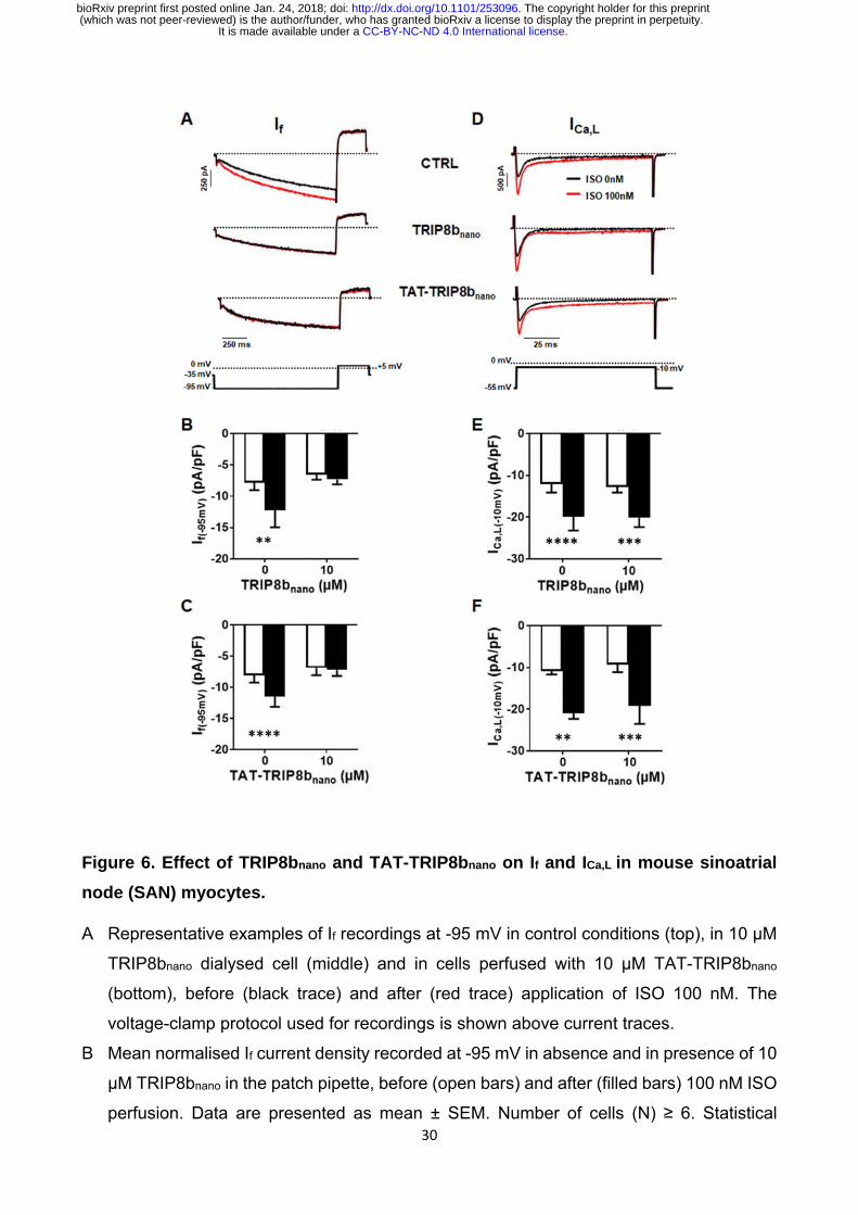
Figure 6. Effect of TRIP8bnano and TAT-TRIP8bnano on If and ICa,L in mouse sinoatrial
node (SAN) myocytes.
A Representative examples of If recordings at -95 mV in control conditions (top), in 10 µM
TRIP8bnano dialysed cell (middle) and in cells perfused with 10 µM TAT-TRIP8bnano
(bottom), before (black trace) and after (red trace) application of ISO 100 nM. The
voltage-clamp protocol used for recordings is shown above current traces.
B Mean normalised If current density recorded at -95 mV in absence and in presence of 10
µM TRIP8bnano in the patch pipette, before (open bars) and after (filled bars) 100 nM ISO
perfusion. Data are presented as mean ± SEM. Number of cells (N) ≥ 6. Statistical
.CC-BY-NC-ND 4.0 International licenseIt is made available under a (which was not peer-reviewed) is the author/funder, who has granted bioRxiv a license to display the preprint in perpetuity.
The copyright holder for this preprint. http://dx.doi.org/10.1101/253096doi: bioRxiv preprint first posted online Jan. 24, 2018;

31
analysis performed with two-way ANOVA test, followed by Sidak multiple comparisons
test (**, P < 0.01).
C Mean normalised If current density recorded at -95 mV in control solution or in the
solution containing 10 µM TAT-TRIP8bnano, in absence (open bars) and in the presence
(filled bars) of 100 nM ISO. Data are presented as mean ± SEM. Number of cells (N) ≥
8. Statistical analysis performed with two-way ANOVA test, followed by Sidak multiple
comparisons test (****, P < 0.0001).
D Representative examples of ICa,L recordings at -10 mV in control conditions (top), in 10µM
TRIP8bnano dialysed cell (middle) and in cells perfused with 10 µM TAT-TRIP8bnano
(bottom), before (black trace) and after (red trace) application of ISO 100 nM. The
voltage-clamp protocol used for recordings is shown above current traces.
E Mean normalised ICa,L current density recorded at -10 mV in absence and in presence of
10 µM TRIP8bnano in the patch pipette, before (open bars) and after (filled bars) 100 nM
ISO perfusion. Data are presented as mean ± SEM. Number of cells (N) ≥ 8. Statistical
analysis performed with two-way ANOVA test, followed by Sidak multiple comparisons
test (***, P < 0.001; ****, P < 0.0001).
F Mean normalised ICa,L current density recorded at -10 mV in control solution or in the
solution containing 10 µM TAT-TRIP8bnano, in absence (open bars) and in the presence
(filled bars) of 100 nM ISO. Data are presented as mean ± SEM. Number of cells (N) ≥
7. Statistical analysis performed with two-way ANOVA test, followed by Sidak multiple
comparisons test (**, P < 0.01; *** P < 0.001).
.CC-BY-NC-ND 4.0 International licenseIt is made available under a (which was not peer-reviewed) is the author/funder, who has granted bioRxiv a license to display the preprint in perpetuity.
The copyright holder for this preprint. http://dx.doi.org/10.1101/253096doi: bioRxiv preprint first posted online Jan. 24, 2018;

32
Supporting Information
Appendix Fig S1. CNBD residues involved in TRIP8bnano binding.
Appendix Fig S2. Representative families of clusters obtained from the first
docking calculation.
Appendix Fig S3. Biochemical validation of CNBD – TRIP8bnano complex.
Appendix Fig S4. Different orientation of R662 and K665 in the cAMP-bound and
TRIP8bnano-bound conformation of the CNBD.
Appendix Fig S5. Comparison of half activation potentials of human HCN1 WT
and of the cAMP-insensitive R549E mutant in the absence and in the presence
of cAMP.
Appendix Table S1. Acquisition parameters for NMR experiments performed on
cAMP-free human HCN2 CNBD in complex with TRIP8bnano and vice-versa.
Appendix Table S2. Docking calculation.
.CC-BY-NC-ND 4.0 International licenseIt is made available under a (which was not peer-reviewed) is the author/funder, who has granted bioRxiv a license to display the preprint in perpetuity.
The copyright holder for this preprint. http://dx.doi.org/10.1101/253096doi: bioRxiv preprint first posted online Jan. 24, 2018;

33
Appendix Fig S1. CNBD residues involved in TRIP8bnano binding.
Combined chemical shift variations of NMR signals between CNBD unbound and
TRIP8bnano-bound state. Combined chemical shift variations are calculated from the
experimental 1H and 15N chemical shift changes (∆δ(1H) and ∆δ (15N), respectively) between
the corresponding peaks in the two forms, through the following equation (Garrett et al,
1997).
Residues experiencing intermediate exchange regime (whose NMR signal becomes broad
beyond detection upon addition of TRIP8bnano) are shown in grey. The horizontal dotted line
indicates the average value plus one standard deviation. Residues above the line were set
as “active” in the docking calculation described in the text (see Materials and Methods).
Garrett DS, Seok YJ, Peterkofsky A, Clore GM & Gronenborn AM (1997) Identification by NMR of the binding surface for the histidine‐containing phosphocarrier protein HPr on the N‐terminal domain of enzyme I of the Escherichia coli phosphotransferase system. Biochemistry 36: 4393–4398
2
N25
1H
21521
combined
.CC-BY-NC-ND 4.0 International licenseIt is made available under a (which was not peer-reviewed) is the author/funder, who has granted bioRxiv a license to display the preprint in perpetuity.
The copyright holder for this preprint. http://dx.doi.org/10.1101/253096doi: bioRxiv preprint first posted online Jan. 24, 2018;
34
Appendix Fig S2. Representative families of clusters obtained from the first docking
calculation.
The clusters obtained can be grouped in two classes, shown here, on the basis of the
position of helix N of TRIP8bnano (N) relative to the CNBD. In the left representative structure,
helix N is oriented in a way that allows E241 to interact with R650 of C-helix. In the right
representative structure, E240 - E241 interact with N547 of CNBD. In all clusters K665 or K666 of
CNBD establish a contact with either E264 or E265 of TRIP8bnano. The structural elements of
CNBD are labelled: αE’, αA and αC. Helices N and C of TRIP8bnano are labelled.
.CC-BY-NC-ND 4.0 International licenseIt is made available under a (which was not peer-reviewed) is the author/funder, who has granted bioRxiv a license to display the preprint in perpetuity.
The copyright holder for this preprint. http://dx.doi.org/10.1101/253096doi: bioRxiv preprint first posted online Jan. 24, 2018;

35
Appendix Fig S3. Biochemical validation of CNBD – TRIP8bnano complex.
Dissociation constant (KD) of the interaction between the indicated CNBD and TRIP8bnano
peptides were measured by means of Isothermal Titration Calorimetry (ITC). CNBD WT -
TRIP8bnano WT (black filled circle) = 1.4 ± 0.1 µM; CNBD K665E - TRIP8bnano WT (grey filled
circle) = 6 ± 0.3 µM; CNBD K666E - TRIP8bnano WT (grey filled circle) = 9.9 ± 0.8 µM; CNBD
WT - TRIP8bnano E64K (purple filled circle) = 4.8 ± 0.2 µM; CNBD WT - TRIP8bnano E65K
(purple filled circle) = 6 ± 0.5 µM; CNBD R650E - TRIP8bnano WT (red filled circle) = 9.6 ± 0.7
µM; CNBD WT - TRIP8bnano E40R (green filled circle) = 5 ± 0.3 µM; CNBD WT - TRIP8bnano
E41R (green filled circle) = 3.8 ± 0.2 µM; CNBD N547D - TRIP8bnano WT (blue filled circle) =
11 ± 0.9 µM; CNBD WT - TRIP8bnano D52N (orange filled circle) = 3.4 ± 0.1 µM. Data are
presented as mean ± SEM. Number of experiments (N) ≥ 3. KD values of all tested
combinations are statistically different from the KD of CNBD WT for TRIP8bnano WT (*, P ≤
0.05; **, P < 0.01). Statistical analysis performed with ANOVA, followed by post-hoc Tukey
test. Dashed black vertical line indicates the KD of WT peptides.
.CC-BY-NC-ND 4.0 International licenseIt is made available under a (which was not peer-reviewed) is the author/funder, who has granted bioRxiv a license to display the preprint in perpetuity.
The copyright holder for this preprint. http://dx.doi.org/10.1101/253096doi: bioRxiv preprint first posted online Jan. 24, 2018;

36
Appendix Fig S4. Different orientation of R662 and K665 in the cAMP-bound and
TRIP8bnano-bound conformation of the CNBD.
Residues R662 and K665 of CNBD C-helix (C) which interact with cAMP (left, (Lolicato et al,
2011) and TRIP8bnano (right, this study) are represented as blue sticks and labelled.
TRIP8bnano residues E250 and D257 interacting with R662 and residue E265 interacting with K665
are shown in red sticks and labelled.
Lolicato M, Nardini M, Gazzarrini S, Moller S, Bertinetti D, Herberg FW, Bolognesi M, Martin H, Fasolini M, Bertrand JA, Arrigoni C, Thiel G & Moroni A (2011) Tetramerization dynamics of C‐terminal domain underlies isoform‐specific cAMP gating in hyperpolarization‐activated cyclic nucleotide‐gated channels. J. Biol. Chem. 286: 44811–44820
.CC-BY-NC-ND 4.0 International licenseIt is made available under a (which was not peer-reviewed) is the author/funder, who has granted bioRxiv a license to display the preprint in perpetuity.
The copyright holder for this preprint. http://dx.doi.org/10.1101/253096doi: bioRxiv preprint first posted online Jan. 24, 2018;

37
Appendix Fig S5. Comparison of half activation potentials of human HCN1 WT and of
the cAMP-insensitive R549E mutant in the absence and in the presence of cAMP.
Currents were measured by patch clamp from HEK 293T cells transfected with WT and
R549E mutant (Chen et al, 2001) in the absence and presence of 15 µM cAMP in the pipette.
Half activation potential (V1/2) was determined as described in Material and Methods. HCN1
WT (black filled circle) = -72.7 ± 0.2 mV; HCN1 WT + cAMP (black open circle) = 73.1 ± 0.3
mV; HCN1 R549E (red filled circle) = -80.4 ± 0.2 mV; HCN1 R549E + cAMP (red open circle)
= -81 ± 0.3 mV. Data are presented as mean ± SEM. Number of experiments (N) ≥ 8.
Chen S, Wang J & Siegelbaum SA (2001) Properties of Hyperpolarization‐Activated Pacemaker Current Defined by Coassembly of Hcn1 and Hcn2 Subunits and Basal Modulation by Cyclic Nucleotide. J. Gen. Physiol. 117: 491–504 Available at: http://www.jgp.org/lookup/doi/10.1085/jgp.117.5.491
.CC-BY-NC-ND 4.0 International licenseIt is made available under a (which was not peer-reviewed) is the author/funder, who has granted bioRxiv a license to display the preprint in perpetuity.
The copyright holder for this preprint. http://dx.doi.org/10.1101/253096doi: bioRxiv preprint first posted online Jan. 24, 2018;
38
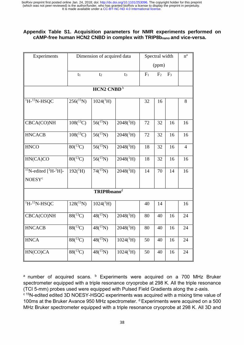
Appendix Table S1. Acquisition parameters for NMR experiments performed on cAMP-free human HCN2 CNBD in complex with TRIP8bnano and vice-versa.
Experiments Dimension of acquired data
Spectral width
(ppm)
na
t1 t2 t3 F1 F2 F3
HCN2 CNBD b
1H-15N-HSQC
256(15N) 1024(1H) 32 16 8
CBCA(CO)NH 108(13C) 56(15N) 2048(1H) 72 32 16 16
HNCACB 108(13C) 56(15N) 2048(1H) 72 32 16 16
HNCO 80(13C) 56(15N) 2048(1H) 18 32 16 4
HN(CA)CO 80(13C) 56(15N) 2048(1H) 18 32 16 16
15N-edited [1H-1H]-
NOESYc
192(1H) 74(15N) 2048(1H) 14 70 14 16
TRIP8bnanod
1H-15N-HSQC 128(15N) 1024(1H) 40 14 16
CBCA(CO)NH 88(13C) 48(15N) 2048(1H) 80 40 16 24
HNCACB 88(13C) 48(15N) 2048(1H) 80 40 16 24
HNCA 88(13C) 48(15N) 1024(1H) 50 40 16 24
HN(CO)CA 88(13C) 48(15N) 1024(1H) 50 40 16 24
a number of acquired scans. b Experiments were acquired on a 700 MHz Bruker spectrometer equipped with a triple resonance cryoprobe at 298 K. All the triple resonance (TCI 5-mm) probes used were equipped with Pulsed Field Gradients along the z-axis. c 15N-edited edited 3D NOESY-HSQC experiments was acquired with a mixing time value of 100ms at the Bruker Avance 950 MHz spectrometer. d Experiments were acquired on a 500 MHz Bruker spectrometer equipped with a triple resonance cryoprobe at 298 K. All 3D and
.CC-BY-NC-ND 4.0 International licenseIt is made available under a (which was not peer-reviewed) is the author/funder, who has granted bioRxiv a license to display the preprint in perpetuity.
The copyright holder for this preprint. http://dx.doi.org/10.1101/253096doi: bioRxiv preprint first posted online Jan. 24, 2018;

39
2D spectra were processed using the standard Bruker software TOPSPIN 2.1 and analyzed through CARA (Keller R et al, 2002; Keller RLJ. 2004). Keller R, Wüthrich KA. (2002) New Software for the Analysis of Protein NMR Spectra. Keller RLJ. (2004) The Computer Aided Resonance Assignment tutorial. Goldau: CANTINA Verlag.
Appendix Table S2. Docking calculation.
HADDOCK Score b
-130 (7)
RMSD (Å) c 1.8 (0.9)
Number of structures
71
BSA (Å2) d 1968 (106)
EAIR e 30.5 (15.2)
Einterf -398(50)
Enbg -431(46)
Statistics on the top-ranking cluster of the structural models of CNBD and TRIP8bnano complex obtained through HADDOCK2.2 calculations and in agreement with previous experimental data (Deberg et al, 2015). Averages (standard deviations are reported in parenthesis) were calculated over the best ten structures.
a Cluster rank according to the HADDOCK score. b HADDOCK score defined as a weighted sum of different energetic terms, such as: van der Waals energy, electrostatic energy, distance restraints energy, buried surface area, binding energy and desolvation energy. c Backbone RMSD from the lowest HADDOCK score structure in each cluster. Some individual energy terms are also reported: d Buried surface area, e distance restraints energy, f binding energy, g non-bonded interaction energy.
Deberg HA, Bankston JR, Rosenbaum JC, Brzovic PS, Zagotta WN & Stoll S (2015) Structural mechanism for the regulation of HCN ion channels by the accessory protein TRIP8b. Structure 23: 734–744
.CC-BY-NC-ND 4.0 International licenseIt is made available under a (which was not peer-reviewed) is the author/funder, who has granted bioRxiv a license to display the preprint in perpetuity.
The copyright holder for this preprint. http://dx.doi.org/10.1101/253096doi: bioRxiv preprint first posted online Jan. 24, 2018;