Embed Size (px)
Citation preview
Teacher Notes for Structure and Function of Cells, Organs and Organ Systems1
In this activity, students analyze multiple examples of the relationship between structure and function in diverse eukaryotic cells, in the small intestine, and in the digestive system. Students learn that cells are dynamic, with constant molecular activity. Students analyze examples that illustrate how organelles work together to accomplish cellular functions and organs and organ systems work together to accomplish functions needed by the organism.
Before students begin this activity, they should have a basic understanding of the functions of organelles in animal and plant cells. You may want to have students explore the animation, "Inside a Cell" available at https://learn.genetics.utah.edu/content/cells/insideacell/.
Table of Contents Learning Goals – pages 1-2Instructional Suggestions and Background Information
Cell Structure and Function – pages 2-6Structure and Function of Organs and Organ Systems – pages 6-8Challenge Questions – page 9
Related Activities – pages 9-10
Learning GoalsIn accord with the Next Generation Science Standards2: This activity helps students to prepare for the Performance Expectations:
o MS-LS1-2. "Develop and use a model to describe the function of a cell as a whole and ways parts of cells contribute to the function."
o MS-LS1-3. "Use argument supported by evidence for how the body is a system of interacting subsystems composed of groups of cells."
o HS-LS1-2. "Develop and use a model to illustrate the hierarchical organization of interacting systems that provide specific functions within multicellular organisms."
Students learn the following Disciplinary Core Ideas (LS1.A): o "All living things are made up of cells, which is the smallest unit that can be said to be
alive. An organism may consist of one single cell (unicellular) or many different numbers and types of cells (multicellular)."
o "Within cells, special structures are responsible for particular functions…"o "Multicellular organisms have a hierarchical structural organization, in which any one
system is made up of numerous parts and is itself a component of the next level." Students engage in recommended Scientific Practices, including "Constructing Explanations.
Apply scientific ideas, principles, and/or evidence to provide an explanation of phenomena…".
This activity focuses on the Crosscutting Concept: Structure and function. "The functions and properties of natural and designed objects and systems can be inferred from their overall structure, the way their components are shaped and used, and the molecular substructures of its various materials."
1 By Ingrid Waldron, Department of Biology, University of Pennsylvania, 2019. These Teacher Notes and the Student Handout are available at http://serendipstudio.org/exchange/bioactivities/SFCellOrgan.
2 Quotations are from http://www.nextgenscience.org/sites/default/files/HS%20LS%20topics%20combined%206.13.13.pdf
Specific Learning Goals include: To understand the function of cells, organs and organ systems, it is helpful to analyze their
components and the relationships between these components. For example, the parts of the cell work together to accomplish the activities of life.
Cells are dynamic and active. Different types of eukaryotic cells have differences in structure that correspond to their
different functions in different types of cells in the human body and in different eukaryotic organisms (animals vs. plants vs. Paramecia).
The cells in human bodies are organized in tissues, organs and organ systems. The small intestine and the digestive system illustrate how structure is related to function at the organ and organ system levels.
The digestive system and circulatory system cooperate to bring nutrients to every cell in the body.
Instructional Suggestions and Background Information To maximize student participation and learning, I suggest that you have your students work individually or in pairs to complete groups of related questions and then have a class discussion after each group of related questions. In each discussion, you can probe student thinking and help them develop a sound understanding of the concepts and information covered before moving on to the next group of related questions.
A key is available upon request to Ingrid Waldron ([email protected]). The following paragraphs provide additional instructional suggestions, links for recommended videos, and biological information – some for inclusion in your class discussions and some to provide you with relevant background that may be useful for your understanding and/or for responding to student questions.
Cell Structure and FunctionTo illustrate the dynamism of cells, you may want to show the animation available at https://www.youtube.com/watch?v=FzcTgrxMzZk. I suggest showing only part of this animation, beginning with microtubules and transport vesicles, followed by mRNA exiting the nucleus through pores in the nuclear membrane, and continuing through exocytosis of secreted proteins. This recommended section begins at about 3 minutes and 30 seconds. You will probably want to listen to the narration for your own information, but I recommend that you turn off the narration for classroom viewing because it is highly technical and likely to overwhelm your students. You will probably want to give a student-friendly narration, which you can develop, using the narration and the relevant section from http://sparkleberrysprings.com/innerlifeofcell.html.
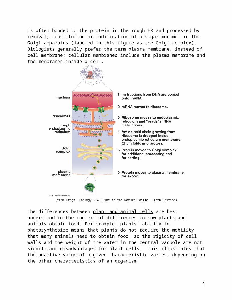
The figure below provides useful background for question 1. Many secreted proteins have carbohydrates attached; the carbohydrate is often bonded to the protein in the rough ER and processed by removal, substitution or modification of a sugar monomer in the Golgi apparatus (labeled in this figure as the Golgi complex). Biologists generally prefer the term plasma membrane, instead of cell membrane; cellular membranes include the plasma membrane and the membranes inside a cell.
2
(from Krogh, Biology - A Guide to the Natural World, Fifth Edition)
The differences between plant and animal cells are best understood in the context of differences in how plants and animals obtain food. For example, plants’ ability to photosynthesize means that plants do not require the mobility that many animals need to obtain food, so the rigidity of cell walls and the weight of the water in the central vacuole are not significant disadvantages for plant cells. This illustrates that the adaptive value of a given characteristic varies, depending on the other characteristics of an organism.
This activity focuses on eukaryotic cells in plants and animals. If your students are learning about prokaryotic cells, you may want to discuss with them that, although eukaryotic and prokaryotic cells have important differences in internal structure, both types of cells carry out the same activities of life and they share many structural similarities, including a plasma membrane, ribosomes, and many similar molecules.
Question 3 revisits protein secretion from a different point of view that includes a broader set of organelles. Be careful to avoid the common error of saying that mitochondria make energy; mitochondria transfer energy from molecules like glucose to ATP molecules (see “How do organisms use energy?”; https://serendipstudio.org/exchange/bioactivities/energy). Lysosomes contain digestive enzymes that break down damaged organelles and macromolecules into smaller molecules (e.g. amino acids, nucleotides or monosaccharides); the smaller molecules leave the lysosome and are reused by the cell (see figure on page 5).
3
In the discussion about how the parts of the cell help to carry out the various activities of life (question 4a), you may want to include other activities of life (e.g. reproduction) if your students have the relevant background knowledge. As discussed in question 4b, cells are alive, but individual molecules are not. Thus, life is an emergent property at the level of the cell. You may want to ask your students the question below in order to reinforce student understanding that life depends on the specific organization of molecules within the cell.
4c. If you ground up a cell and put all the molecules from the cell in a mini-test tube, would this mixture of molecules be alive? Explain why or why not.
When you introduce the concept that structure is related to function, you may want to: point out that structure includes shape, the component parts, and the relationships
between the component parts introduce familiar examples such as the differences between hands and feet and different
shapes of different types of teeth and the hard enamel surface of teeth explain that this relationship applies at multiple levels from cells to organ systems explain that structure includes not only overall shape, but also the component parts and
the relationships between these components explain that the relationship between structure and function is a result of natural selection.
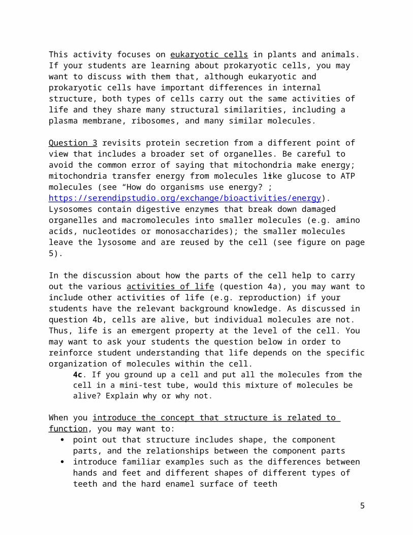
You may want to show your students a video of sperm swimming (available at http://en.wikipedia.org/wiki/Sperm ). Notice that some of these sperm move quite rapidly; natural selection favors sperm characteristics that contribute to faster swimming since the first sperm to reach an egg fertilizes the egg. The figure below shows how small human sperm are relative to a human egg. Sperm have very little cytoplasm, whereas the egg is a large cell with a lot of cytoplasm; once the egg has been fertilized, this large amount of cytoplasm is useful to supply the cytoplasm for the multiple cells that are produced by the cell divisions in early development before the developing embryo implants in the wall of the uterus.
(Figure from Krogh, Biology -- A Guide to the Natural World, Fifth Edition)
An epithelium that consists of a single layer of flattened epithelial cells (also known as a simple squamous epithelium) is observed in the walls of capillaries and also the walls of the alveoli in the lungs. In both cases, transport across the epithelium depends on diffusion, which is reasonably rapid over very short distances but very slow over any substantial distance. Thus, it is advantageous to have a minimal barrier to diffusion.3 The flattened or squamous epithelial cells
3 Polar substances diffuse through the interstitial fluid between the cells of the capillary wall, whereas lipid soluble substances like oxygen and carbon dioxide diffuse across the cells; the latter drastically increases the area available
4
also reflect the fact that the cells of capillaries and alveoli have minimal metabolic activity, so there is minimal need for cytoplasm.
You may want to show your students a time lapse video of a phagocytic cell (neutrophil) chasing a bacterium (available at https://www.youtube.com/watch?v=5yimbhkTqJo or https://www.youtube.com/watch?v=I_xh-bkiv_c). In this video, the phagocytic cell uses chemical information to pursue a bacterium and then eat it. This video shows the dynamic changes in shape as the phagocytic cell moves.
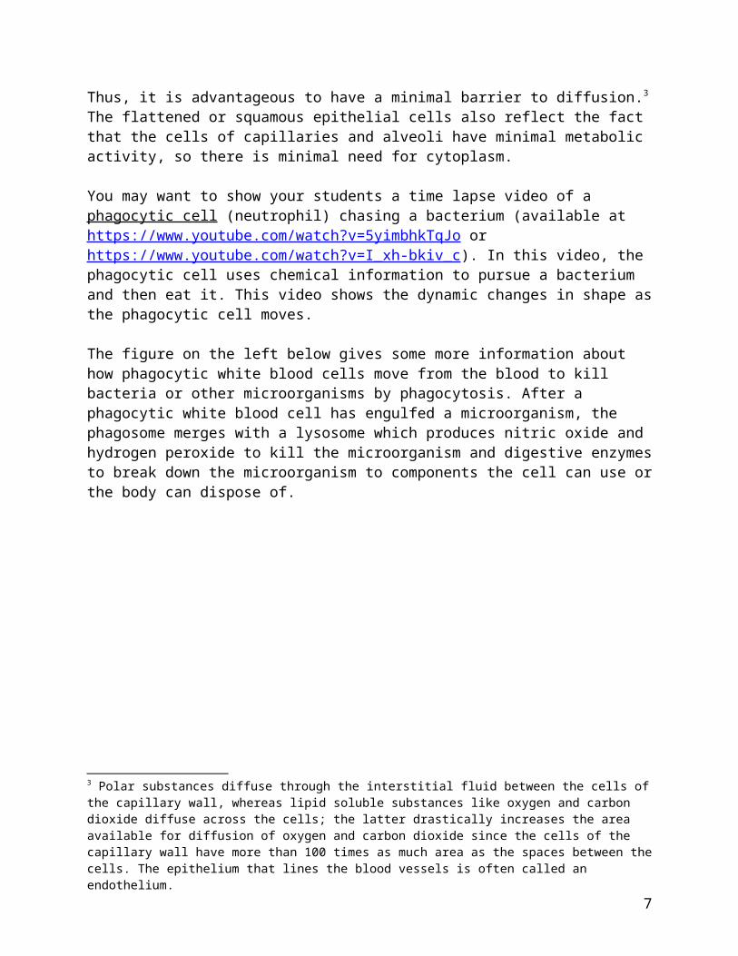
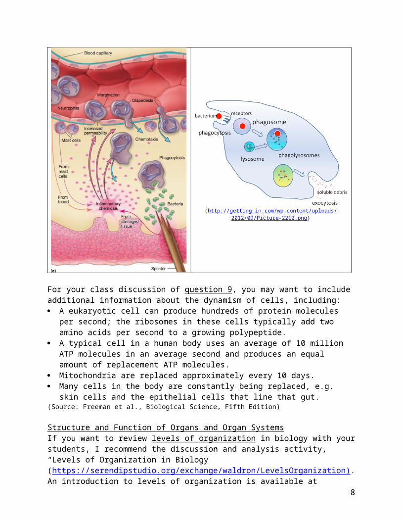
The figure on the left below gives some more information about how phagocytic white blood cells move from the blood to kill bacteria or other microorganisms by phagocytosis. After a phagocytic white blood cell has engulfed a microorganism, the phagosome merges with a lysosome which produces nitric oxide and hydrogen peroxide to kill the microorganism and digestive enzymes to break down the microorganism to components the cell can use or the body can dispose of.
(http://getting-in.com/wp-content/uploads/2012/09/Picture-2212.png)
For your class discussion of question 9, you may want to include additional information about the dynamism of cells, including: A eukaryotic cell can produce hundreds of protein molecules per second; the ribosomes in
these cells typically add two amino acids per second to a growing polypeptide.
for diffusion of oxygen and carbon dioxide since the cells of the capillary wall have more than 100 times as much area as the spaces between the cells. The epithelium that lines the blood vessels is often called an endothelium.
5
A typical cell in a human body uses an average of 10 million ATP molecules in an average second and produces an equal amount of replacement ATP molecules.
Mitochondria are replaced approximately every 10 days. Many cells in the body are constantly being replaced, e.g. skin cells and the epithelial cells
that line that gut.(Source: Freeman et al., Biological Science, Fifth Edition)
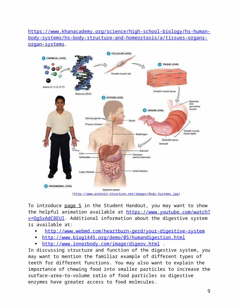
Structure and Function of Organs and Organ SystemsIf you want to review levels of organization in biology with your students, I recommend the discussion and analysis activity, “Levels of Organization in Biology” (https://serendipstudio.org/exchange/waldron/LevelsOrganization ) . An introduction to levels of organization is available at https://www.khanacademy.org/science/high-school-biology/hs-human-body-systems/hs-body-structure-and-homeostasis/a/tissues-organs-organ-systems.
(http://www.protein-structure.net/images/Body-Systems.jpg)
To introduce page 5 in the Student Handout, you may want to show the helpful animation available at https://www.youtube.com/watch?v=Og5xAdC8EUI. Additional information about the digestive system is available at:
http://www.webmd.com/heartburn-gerd/your-digestive-system http://www.biog1445.org/demo/05/humandigestion.html http://www.innerbody.com/image/digeov.html .
6
In discussing structure and function of the digestive system, you may want to mention the familiar example of different types of teeth for different functions. You may also want to explain the importance of chewing food into smaller particles to increase the surface-area-to-volume ratio of food particles so digestive enzymes have greater access to food molecules.
The two diagrams of the digestive system on page 5 of the Student Handout illustrate how different diagrams of the same structure convey somewhat different information and are useful for different purposes. The first diagram clearly shows the sequence of the organs in the digestive system, whereas the second diagram more accurately shows the relative sizes and arrangement of the organs of the digestive system.
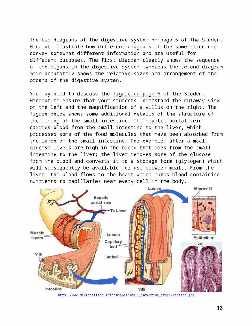
You may need to discuss the figure on page 6 of the Student Handout to ensure that your students understand the cutaway view on the left and the magnification of a villus on the right. The figure below shows some additional details of the structure of the lining of the small intestine. The hepatic portal vein carries blood from the small intestine to the liver, which processes some of the food molecules that have been absorbed from the lumen of the small intestine. For example, after a meal, glucose levels are high in the blood that goes from the small intestine to the liver; the liver removes some of the glucose from the blood and converts it to a storage form (glycogen) which will subsequently be available for use between meals. From the liver, the blood flows to the heart which pumps blood containing nutrients to capillaries near every cell in the body.
http://www.daviddarling.info/images/small_intestine_cross-section.jpg
The length of the small intestine, the folds, the villi and the microvilli all increase the surface area for absorption. (The narrow diameter of the small intestines ensures that digested food molecules in the lumen are relatively near to the wall of the small intestine where they can be absorbed.)
The cells in the epithelium that lines the inner surface of the small intestine synthesize protein enzymes that help to complete the digestion of food molecules. These cells also pump in sugars
7
and amino acids, which increases absorption of these useful molecules from the lumen of the small intestine. To accomplish these functions, these cells need lots of cytoplasm with lots of mitochondria, ribosomes, rough endoplasmic reticulum and Golgi apparatus (the latter two are needed since the digestive enzymes and pump proteins are inserted in the plasma membrane (in the region that faces the lumen of the small intestine)). These metabolically active epithelial cells in the lining of the small intestine are tall and have much more cytoplasm per epithelial area than the relatively inactive flattened cells in the capillary wall. (The capillary wall cells do not synthesize enzymes or pump useful molecules; instead the flattened shape provides a minimum barrier to diffusion, as discussed above.) This is another example of how structure is related to function.
In discussing the role of the circulatory system (questions 12c and 13), you may want to explain that nutrients diffuse into the blood in the capillaries in the villi of the small intestine and then later diffuse out of the blood in capillaries in other parts of the body (as shown in question 6).
Question 13 illustrates the important general principle that our bodies consist of multiple body systems that cooperate to accomplish important functions. For example, the digestive and circulatory systems cooperate to provide cells all over the body with digested food molecules that can be used for cellular respiration and as building blocks to synthesize needed molecules.
You may want to include additional examples such as the cooperation between the respiratory system and the circulatory system to provide oxygen to every cell in the body and also to remove the waste product, carbon dioxide. This diagram illustrates how the circulatory system is crucial for allowing the specialized digestive, respiratory, and excretory systems to serve needed functions for all the cells in the body.
You also may want to point out the role of the nervous system in controlling the activity of the tongue and jaw muscles, regulating other aspects of digestion, controlling breathing, and regulating the circulatory system (e.g. heart rate). A rap video that reviews body systems is available at http://mr.powner.org/b/lessons/humans/So%20Many%20Systems%20-%20Human%20Body%20Systems%20Rap.mp4.Challenge QuestionsQuestion 14 provides an additional example of the diversity of eukaryotic cell structure. This question challenges students to develop their skills in obtaining and evaluating information. A useful resource for students to learn about Paramecia is available at
8
http://labs.7bscience.com/lab-2---paramecium.html. This webpage includes an excellent video that shows the motion of cilia, contraction of the contractile vacuoles, and food being swept into the oral groove; seeing this video will help students understand how dynamic cells are, with constant activity of their organelles.
Discussion of Challenge Question 15 introduces the concept that the digestive systems of different types of animals have different structures, which correspond to differences in their diets (as well as their evolutionary history and ecological niche). A useful introduction to ruminant digestive systems is available at http://sci.waikato.ac.nz/farm/content/animalstructure.html. This source also describes "hindgut fermenters" like horses and rabbits which have digestive systems that extract a lot of the energy in high-fiber diets by bacterial breakdown of cellulose after food has passed through the small intestine. You may want to mention that the human gut is home to many bacteria which help to provide nutrients, protect against invasion by disease germs, and stimulate the development of the intestine and immune system (http://www.karger.com/Article/FullText/89775). You might want to mention an extreme example of diversity – tapeworms that live in the small intestine where they are surrounded by a solution of digested nutrient molecules do not have a digestive system, but instead absorb nutrients through their body surface.
You may want to add the following optional question:16. Write an argument to support the claim that "The structure of a cell is related to its function." Give examples and explain how your examples support this claim.
If your students are not familiar with scientific argumentation, you may want to introduce them to the idea that, in a scientific argument, a claim is supported by observable evidence with justification to explain how the evidence is relevant to the claim. Criteria for evaluating a scientific argument include:
how well the claim fits with all the available evidence the relevance and sufficiency of the evidence (as explained in the justification) the quality of the evidence.4
Additional information for teaching about scientific argumentation is available at http://www.indiana.edu/~ensiweb/Sci.Argumentation.html http://undsci.berkeley.edu/article/0_0_0/howscienceworks_07 . (This website takes a
somewhat different approach to the components of a scientific argument.)
Related Activities Cell Membrane Structure and Functionhttp://serendipstudio.org/sci_edu/waldron/#diffusionThis activity includes two hands-on experiments and numerous analysis and discussion questions to help students understand how the molecular composition and organization of a cell membrane result in its selective permeability. In the hands-on experiments, students test whether a synthetic membrane is selectively permeable and students observe how a layer of oil can be a barrier to diffusion of an aqueous solution. Then, students learn how the phospholipid bilayer and membrane proteins play key roles in the cell membrane function of regulating what gets into and
4 These criteria are paraphrased from Argument-Driven Inquiry in Biology (by Sampson et al., NSTA Press). This book presents a very useful, more extensive format for developing students’ ability to engage in scientific argument as well as multiple argument-driven inquiry laboratory investigations designed for high school students.
9
out of the cell. Topics covered include ions, polar and nonpolar molecules; simple diffusion through the phospholipid bilayer; facilitated diffusion through ion channels or carrier proteins; and active transport. An optional final page introduces exocytosis and endocytosis. (This activity supports the Next Generation Science Standards = NGSS.)
Cell Structure and Function – Major Concepts and Learning Activities http://serendipstudio.org/exchange/bioactivities/cellsThis overview presents key concepts that students often do not learn from standard textbook presentations and suggests learning activities to help students understand how the parts of a cell work together to accomplish the multiple functions of a dynamic living cell. Suggested activities also reinforce student understanding of the relationships between molecules, organelles and cells and the importance and limitations of diffusion. This overview provides links to web resources, analysis and discussion activities, and hands-on activities.
10