Embed Size (px)
Citation preview
i
STRUCTURE FUNCTION ANALYSIS OF THE
DEUBIQUITYLATING ENZYME FAM
Poon-Yu Khut, B.Sc
School of Molecular & Biomedical Science (Biochemistry)
University of Adelaide
Adelaide, South Australia 5005
October 2006
1
CHAPTER 1
INTRODUCTION
Ubiquitin: a Multipurpose Tag
Ubiquitin is a highly conserved 76 amino acid protein that serves in a complex, post-
translational modification system that is largely used by the cell to either target a protein
for destruction at the proteasome or affect its intracellular trafficking. Although exclusively
found in eukaryotes, prokaryotes (that do not have any form of signalling system based on
covalent protein-protein attachments) appear to possess clear ancestors of the ubiquitin
fold (reviewed in Hochstrasser 2000). Ubiquitylation is the process by which ubiquitin is
covalently attached to a specific target protein via an isopeptide bond between the C-
terminal glycine of ubiquitin to a -amino group of a substrate’s lysine residue. The protein
can remain with only one attached ubiquityl moiety (monoubiquitylation), have multiple
single ubiquityl moieties attached to several substrate lysine residues (multiubiquitylation),
or through successive ubiquitylation reactions form a chain of ubiquitins
(polyubiquitylation). As ubiquitin itself contains several lysine residues, it is possible to
form a variety of polyubiquityl chains based on which lysine residue the subsequent
ubiquitin is attached to. It has become apparent that the number and nature of these
ubiquitin attachments is paramount in affecting the appropriate, and very different cellular
outcomes.
UBIQUITIN AND PROTEIN DEGRADATION
Ubiquitin’s cellular role has been best characterised in protein turnover where addition of a
polyubiquityl chain serves as a destruction tag, targeting the protein to the proteasome
where it is rapidly and irreversibly degraded (figure 1.1). To form an effective substrate for
proteasome recognition, proteins must have multiple ubiquitin moieties attached to it
(polyubiquitylation) via gly76-lys48 linkages (reviewed in Coux et al. 1996). In vitro
binding studies with Rpn10, a polyubiquityl receptor subunit of the proteasome, showed
that only ubiquitin chains of at least four moieties were efficiently recognised, and binding
affinity increased with length. Indeed, binding affinity to proteasomes increases more than
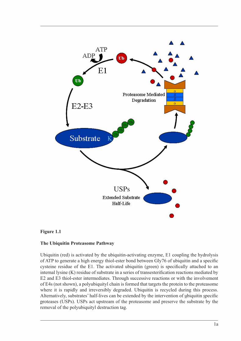
Figure 1.1
The Ubiquitin Proteasome Pathway
Ubiquitin (red) is activated by the ubiquitin-activating enzyme, E1 coupling the hydrolysis
E2 and E3 thiol-ester intermediates. Through successive reactions or with the involvement of E4s (not shown), a polyubiquityl chain is formed that targets the protein to the proteasome where it is rapidly and irreversibly degraded. Ubiquitin is recycled during this process. Alternatively, substrates’ half-lives can be extended by the intervention of ubiquitin sproteases (USPs). USPs act upstream of the proteasome and preserve the substrate by the removal of the polyubiquityl destruction tag.
USPs
1a

2
100-fold when the chain is lengthened from two to four ubiquitins, but only 10-fold more
when the chain is increased to eight (Deveraux et al. 1994).
It is estimated that over 30% of newly synthesised cellular proteins are disposed of without
being properly folded i.e., misfolded and/or unassembled, despite lacking mutational or
translational error (Schubert et al. 2000). Additionally, properly folded proteins often are
damaged through various external stresses such as heat, oxidation and ultraviolet damage.
Where the chaperone system has either not rescued or acted upon these proteins, the
ubiquitin system is largely responsible for their removal and its role in “cellular garbage
disposal” has been long established. Indeed failure to remove such proteins can lead to
protein aggregation and formation of toxic inclusion bodies. Within the nervous system,
defects in the ubiquitin-dependent removal of certain proteins is closely associated with the
pathogenesis of several major human neurodegenerative diseases such as Alzheimer’s and
Parkinson’s disease (reviewed in Layfield et al. 2001, Tanaka et al. 2004)
Extending from initial views that the ubiquitin pathway served only a housekeeping
function in the destruction of unwanted proteins, it has since been revealed through various
molecular, biochemical, cellular, genetic and clinical studies, that it plays a significant role
in a broad array of cellular processes (reviewed in Ciechanover et al. 2000). Examples
include the regulation of cell cycle, differentiation and development, the cellular response
to extracellular effectors and stress, modulation of cell surface receptors and ion channels,
DNA repair, regulation of the immune and inflammatory responses and biogenesis of
organelles. Due to its speed, permanency and capacity for specificity, ubiquitin mediated
protein degradation is almost always featured in regulatory mechanisms involving timing
control (reviewed in Hochstrasser 1995). Substrates thus include cell cycle regulators,
tumour suppressors and growth modulators, transcriptional activators and inhibitors, cell
surface receptors and endoplasmic reticulum proteins (reviewed in Ciechanover et al.
2000).
UBIQUITIN AND TRAFFICKING
In recent years, the significance of the ubiquitin system in cellular functions other than that
of pure protein half-life regulation has become better understood. Ubiquitin K63-linked
chains are used to regulate processes such as, DNA repair (Spence et al. 1995), activation
3
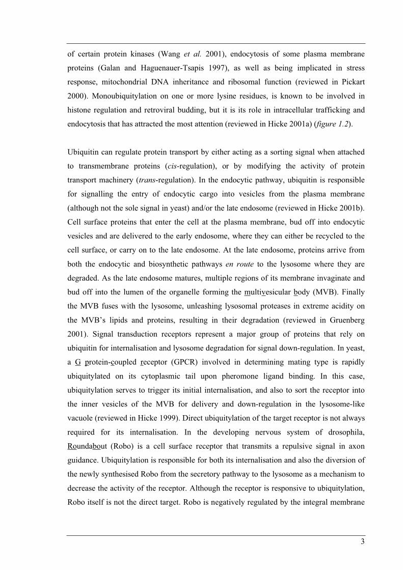
of certain protein kinases (Wang et al. 2001), endocytosis of some plasma membrane
proteins (Galan and Haguenauer-Tsapis 1997), as well as being implicated in stress
response, mitochondrial DNA inheritance and ribosomal function (reviewed in Pickart
2000). Monoubiquitylation on one or more lysine residues, is known to be involved in
histone regulation and retroviral budding, but it is its role in intracellular trafficking and
endocytosis that has attracted the most attention (reviewed in Hicke 2001a) (figure 1.2).
Ubiquitin can regulate protein transport by either acting as a sorting signal when attached
to transmembrane proteins (cis-regulation), or by modifying the activity of protein
transport machinery (trans-regulation). In the endocytic pathway, ubiquitin is responsible
for signalling the entry of endocytic cargo into vesicles from the plasma membrane
(although not the sole signal in yeast) and/or the late endosome (reviewed in Hicke 2001b).
Cell surface proteins that enter the cell at the plasma membrane, bud off into endocytic
vesicles and are delivered to the early endosome, where they can either be recycled to the
cell surface, or carry on to the late endosome. At the late endosome, proteins arrive from
both the endocytic and biosynthetic pathways en route to the lysosome where they are
degraded. As the late endosome matures, multiple regions of its membrane invaginate and
bud off into the lumen of the organelle forming the multivesicular body (MVB). Finally
the MVB fuses with the lysosome, unleashing lysosomal proteases in extreme acidity on
the MVB’s lipids and proteins, resulting in their degradation (reviewed in Gruenberg
2001). Signal transduction receptors represent a major group of proteins that rely on
ubiquitin for internalisation and lysosome degradation for signal down-regulation. In yeast,
a G protein-coupled receptor (GPCR) involved in determining mating type is rapidly
ubiquitylated on its cytoplasmic tail upon pheromone ligand binding. In this case,
ubiquitylation serves to trigger its initial internalisation, and also to sort the receptor into
the inner vesicles of the MVB for delivery and down-regulation in the lysosome-like
vacuole (reviewed in Hicke 1999). Direct ubiquitylation of the target receptor is not always
required for its internalisation. In the developing nervous system of drosophila,
Roundabout (Robo) is a cell surface receptor that transmits a repulsive signal in axon
guidance. Ubiquitylation is responsible for both its internalisation and also the diversion of
the newly synthesised Robo from the secretory pathway to the lysosome as a mechanism to
decrease the activity of the receptor. Although the receptor is responsive to ubiquitylation,
Robo itself is not the direct target. Robo is negatively regulated by the integral membrane
3a
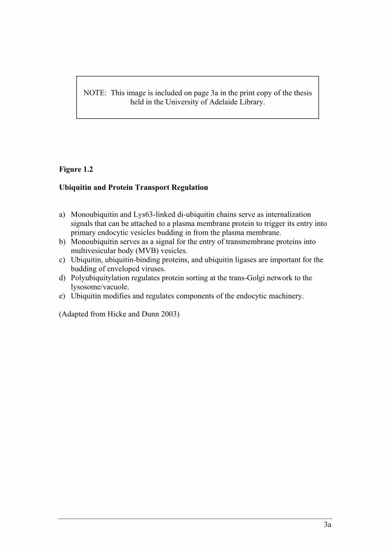
Figure 1.2
Ubiquitin and Protein Transport Regulation a) Monoubiquitin and Lys63-linked di-ubiquitin chains serve as internalization
signals that can be attached to a plasma membrane protein to trigger its entry into primary endocytic vesicles budding in from the plasma membrane.
b) Monoubiquitin serves as a signal for the entry of transmembrane proteins into multivesicular body (MVB) vesicles.
c) Ubiquitin, ubiquitin-binding proteins, and ubiquitin ligases are important for the budding of enveloped viruses.
d) Polyubiquitylation regulates protein sorting at the trans-Golgi network to the lysosome/vacuole.
e) Ubiquitin modifies and regulates components of the endocytic machinery. (Adapted from Hicke and Dunn 2003)
NOTE: This image is included on page 3a in the print copy of the thesis
held in the University of Adelaide Library.
4
protein Commissureless (Comm), which when ubiquitylated, supplies the trafficking
signals to the Robo/Comm complex in trans (reviewed in Hicke and Dunn 2003).
Clearly the ubiquitin signal has important and diverse roles within the cell and so must be
tightly regulated. As with virtually all biological systems, ubiquitylation can be reversed,
this process is known as deubiquitylation. Regulation of certain proteins at the fundamental
level of ubiquitylation and deubiquitylation can either directly or indirectly, affect cellular
processes.
Ubiquitylation
Conjugation of ubiquitin to a substrate involves a hierarchal three-step process requiring
the activity of at least three different classes of enzymes. The first step involves activation
of ubiquitin by a single dedicated ubiquitin-activating (E1) enzyme. In eukaryotes,
activation comprises two steps: the initial coupling of ATP to form an ubiquitin-adenylate
intermediate, followed by the formation of a high energy thiol-ester bond between the
carboxyl-terminal glycine (Gly76) of the intermediate with a specific cysteine residue of
the E1. One of several members of the ubiquitin-conjugating enzyme family (E2) then
moves the activated ubiquitin in a trans-esterification reaction via an E2 ubiquitin thiol-
ester intermediate, to the substrate which has been specifically bound to an ubiquitin-
protein ligase (E3). The E3-assisted transfer of ubiquitin to substrate occurs either directly
or via an additional E3 ubiquitin thiol-ester intermediate (reviewed in Varshavsky, 1997)
(figure 1.3 A). Attachment of the activated carboxyl-terminal of ubiquitin is directed to the
-amino groups of internal lysine residues of the substrate to form an isopeptide bond,
which in successive reactions or with the involvement of the recently described E4 class of
enzymes (Keog et al. 1999) generates a polyubiquityl chain (figure 1.1).
REGULATION AND SPECIFICITY OF UBIQUITYLATION
There exists no single conserved peptide motif that targets substrates for ubiquitylation.
Specificity is imparted by protein-protein interactions with the ubiquitylation machinery,
specifically the E2s and E3s that recognise their substrates via specific motifs. The human
4a
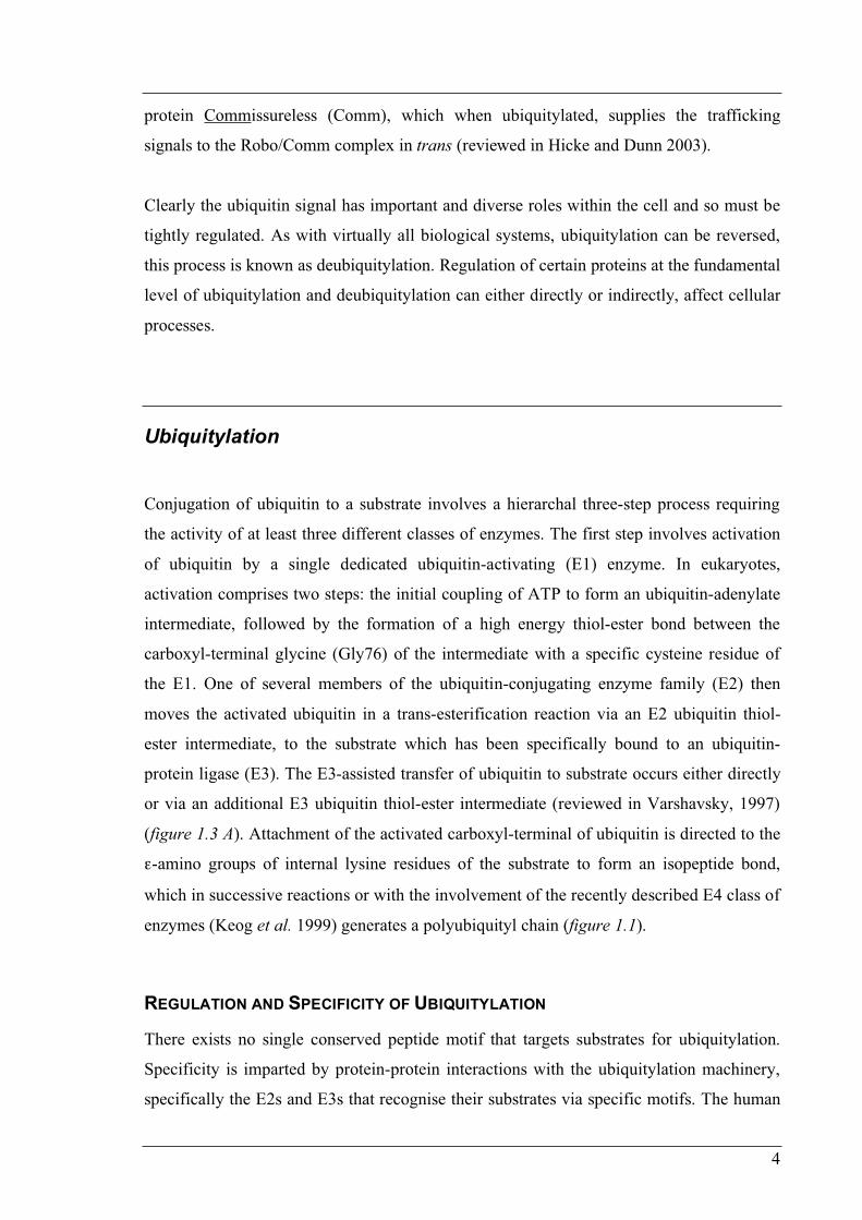
Figure 1.3
Ubiquitin Conjugation a) Basic steps in substrate modification by the ubiquitylation machinery consisting of
ubiquitin-activating (E1), ubiquitin-conjugating (E2) and ubiquitin-protein ligase (E3) classes of enzymes
b) The ubiquitin conjugation cascade. Humans are predicted to contain genes for one E1, over 40 E2s and more than 500 different E3s
c) Modes of E3 Recognition of Protein Substrates: 1) Constitutive recognition by the E3 via a primary motif, 2) Recognition of the substrate following its post-translational modification (eg. phosphorylation), 3) Recognition of the substrate following the post-translational modification of the E3, 4) Recognition of the substrate following its association with an ancillary protein.
(A, B adapted from Pickart and Eddins 2004, and C adapted from Ciechanover et al. 2000)
NOTE: This image is included on page 4a in the print copy of the thesis
held in the University of Adelaide Library.
5
genome project has revealed the presence of 1 E1 (although alternative translation
initiation sites gives rise to nuclear and cytoplasmic isoforms), >40 different E2s and >500
different E3s (Wong et al. 2003). This large repertoire of enzymes undoubtedly reflects the
requirement for regulation and specificity of the ubiquitin system. The hierarchical layout
of these enzymes is such that one E1 interacts with all the E2s, and each different E2
species may be able to form an E2-E3 ligase complex with one or more E3s (figure 1.3 B).
This type of combinatorial expansion is a way to potentially increase the ubiquitin
systems’ substrate specificity repertoire. Furthermore, each class of enzyme in this cascade
affords an extra level of control over the ubiquitylation process and are regulated both
temporally and spatially. For example, the intracellular localisation of some E2 enzymes
has recently been shown to be regulated. A group of E2’s, murine UbcM2, and human
UbcH6 and UBE2E2 are imported into the nucleus upon transfer of the activated ubiquitin
intermediate to the E2’s active site cysteine (Plafker et al. 2004). Although these enzymes
are theoretically small enough to freely diffuse into the nucleus, their importation relies on
their recognition by importin-11, a member of the karyopherin family of nuclear transport
receptors that mediates translocation through nuclear pore complexes. Importin-11
however only has affinity for ubiquitin charged forms of these E2s and selectively
transports them into the nucleus to perform their function. Previous observations that
UbcM2 shuttles continuously between the nucleus and cytoplasm has led to the proposition
that these E2s are first activated by cytoplasmic E1s, imported into the nucleus and then
returned to the cytosol once they have completed their function. It has been further
proposed that the E2 delivery maybe directed to a particular sub-nuclear compartment as
another member of the karyopherin family, Kap104 in yeast has been shown to release
their cargo only when delivered to RNA. Importin-11 may deliver their activated E2 cargo
to specific E3’s and thus regulate the formation of specific E2-E3 complexes within the
nucleus (reviewed in Zhang and Matunis 2005).
The ubiquitin system gains another level of exquisite specificity from the E3s, which act as
molecular scaffolds, binding substrates for presentation to the various E2s. E3s employ a
number of strategies to recognise and bind substrates. In most cases, an E3 recognises a
subset of protein substrates that share a particular structural motif. Some substrates contain
more than one distinct recognition motif, allowing for recognition by different E3s. In
other cases, the mode of recognition can be dependent on loss of DNA binding (as in the
case of transcription factors which require dissociation from DNA binding sites in order to
6
be recognised), post-translational modification (such as phosphorylation of either substrate
or E3) or association with ancillary proteins (eg. molecular chaperones acting as
recognition elements in trans, reviewed in Ciechanover et al. 2000) (figure 1.3 C). Several
classes of ubiquitin ligases have been described based on the presence of certain domains:
HECT (homologous to the E6-AP C-terminus) domain, RING (really interesting new gene)
finger, the related PHD (pleckstrin homology domain) finger and U-box (UFD2 homology)
domain (reviewed in Pickart and Eddins 2004). Additionally, certain E3s are regulated by
tissue specific expression. For example, expression of MAFbx, a muscle-specific E3
ubiquitin ligase is dramatically up-regulated in atrophying muscle. Through the ubiquitin-
proteasome pathway, MAFbx ubiquitylates and regulates the protein levels of MyoD, a
helix-loop-helix transcription factor that directs myogenic differentiation. In this way,
selective expression of MAFbx and its affect on MyoD in muscle is thought to regulate the
overall balance of differentiated cells and undifferentiated quiescent cells. Thus
upregulation of MAFbx leads to MyoD degradation resulting in muscle atrophy while
downregulation increases MyoD half-life, leading to muscle fibre regeneration (Tintignac
et al. 2005)
Use of a polyubiquitylating class of enzymes could theoretically provide the ubiquitin
system an extra level of regulatory control once a protein has been monoubiquitylated. The
cell would then have the options of either to degrade the protein by creating the K48
polyubiquityl tag (a step E4s have been implicated in), to direct it to another fate by
creation of different polyubiquityl tag (such as a K63-linked di-ubiquitin tag implicated in
DNA repair) or have its intracellular localisation regulated by leaving it
monoubiquitylated.
The ubiquitin system is regulated on yet another level by the localisation of its enzymes. It
has been suggested that the more enzymatically promiscuous deubiquitylating enzymes are
prevented from indiscriminate activity if tethered to the proteasome (Wilkinson 1997).
Additionally, certain ubiquitin conjugases involved in the polyubiquitylation of misfolded
proteins may localise tightly to the endoplasmic reticulum, where quality control of
membrane and secreted proteins occur (Lord et al. 2000).
7
Deubiquitylation
Deubiquitylation is mediated by deubiquitylating enzymes (Dubs), a large family of
proteins that the human genome project has estimated more than 90 members (reviewed in
Baek 2003). Dubs function to specifically cleave ubiquitin-linked moieties after the last C-
terminal residue of ubiquitin (Gly76) and their cellular roles vary from housekeeping to
modification of protein fate/localisation (figure 1.4).
Dubs can be classified into several distinct sub-families. The first family termed ubiquitin
carboxyl-terminal hydrolases (UCHs) consist of relatively small sized proteins of around
20-30 kDa. Their catalytic core domain spans around 230 amino acids and contains a
catalytic triad of spatially conserved Cys, His and Asp residues, a geometry similar to that
of cysteine proteases (figure 1.5 A). UCHs primarily serve housekeeping functions by
preferentially cleaving small adducts from ubiquitin, such as peptides degraded by the
proteasome. Beyond ubiquitin recycling, UCHs are also required in processing newly
synthesised ubiquitin molecules that are translated as linear “head” to “tail” polyubiquityl
precursors, and also releasing some ribosomal proteins that are translated with an ubiquityl
group at their N-terminus that targets them to the ribosome (reviewed in Ciechanover,
2000).
A second group of Dubs belongs to the ubiquitin specific peptidase (USP) family
(otherwise known as the ubiquitin-specific proteases (UBP) family). They exhibit substrate
specificity, display various tissue-specific expression patterns (Baker et al. 1999, reviewed
in Hochstrasser 1996) and are involved in a broad number of cellular processes such as
control of growth, differentiation, oncogenesis and genome integrity (reviewed in
Wilkinson, 1997). Of the 90 odd deubiquitylating enzymes identified by the human
genome project, around 80% of them are USPs. The molecular weights of USPs are quite
large compared to UCHs ranging from 50 to 300 kDa, which may be attributed to their
requirement to recognise multiple substrates. The USP catalytic core is roughly delimited
by two highly conserved motifs, the Cys and His boxes however, unlike UCHs the USP
catalytic core displays considerable sequence and size variance, ranging from 300 to 600
amino acids (figure 1.5 B). Within the catalytic core there are several other highly
conserved motifs including the Asp box and KRF box (figure 1.5 C). The remaining
7a
Figure 1.4 Roles of Deubiquitylating Enzymes
• Proprotein processing: Ubiquitin and several ubiquitin like proteins are synthesised as fusion proteins, requiring cleavage of the ubiquityl group to be functionally active
• Salvage: recycling of ubiquitin from degraded proteins • Editing of ubiquitylated proteins: Either as a proofreading function or to
reverse ubiquitylation to alter protein fate • Disassembly of degradation intermediates: disassembly of polyubiquitin
chains to regenerate free ubiquitin. If not removed, these chains may act as competitive inhibitors to the proteasome
(Adapted from Wilkinson 2000)
NOTE: This image is included on page 7a in the print copy of the thesis
held in the University of Adelaide Library.
7b
Figure 1.5 Conservation of Deubiquitylating Enzyme Catalytic Cores. Alignment of the conserved Cys and His box motifs surrounding catalytically active amino acid residues (asterisks) for (A) UCHs and (B) USPs. (C) Schematic representation of various USPs highlighting the relative positions of several conserved motifs. Conserved motifs are labelled C (Cys box that contains the catalytic C), D (Asp box), Zn (Zinc binding region), K (KRF box), and H (His box that contains the catalytic H and D). Proteins are to scale, except for the N- and C-terminal regions of FAM, which are given in amino acids. (A, B Adapted from Amerik and Hochstrasser 2004)
NOTE: This image is included on page 7b in the print copy of the thesis
held in the University of Adelaide Library.
8
regions of these proteins consist of a variety of N-terminal extensions, occasional C-
terminal extensions and insertions within the catalytic core. The sequences of these
extensions are not conserved amongst the family and it has been proposed that these
regions of diversity may function in substrate recognition, subcellular localisation and
protein-protein interactions (reviewed in Kim et al. 2003). Indeed substrate specificity has
been shown for the USP, herpesvirus-associated ubiquitin-specific protease (HAUSP).
Through mammalian cell culture and biochemical assays, HAUSP has been demonstrated
to stabilise p53 but not p27 (both of which are polyubiquitylated), while the unrelated
human USP11 neither binds or deubiquitylates p53 (Li et al. 2002).
Recently, three other subfamilies of Dubs have been identified. They include the OTU
(ovarian tumour)-related proteases, ataxin-3 proteases and the JAMM/MPN+ proteases. It
is known that these families catalyse the disassembly of ubiquitin conjugates, but much of
their biological function and significance as a separate subfamily from UCHs and USPs
remains unclear. Few members have been characterised for these subfamilies but one
OTU-related protease, A20 has been shown to inhibit nuclear factor- B (NF- B)
activation by its surprising ability to both deubiquitylate and ubiquitylate (reviewed in
Heyninck and Beyaert 2005). NF- B is a transcriptional regulator involved in innate and
adaptive immunity, inflammation, development, cell proliferation and survival. NF- B is
activated by the tumour necrosis factor (TNF) signal transduction pathway, which requires
the recruitment of several proteins to the signalling complex to mediate signal
transduction. Receptor-interacting protein (RIP) is a key signalling protein that is recruited
to the activated TNF-receptor complex when polyubiquitylated via Lys63 linkages. Signal
termination is mediated by A20, which acts to remove RIP from the complex.
Interestingly, A20 directs the removal of RIP by first removing the Lys63 polyubiquityl tag
via its OTU domain (deubiquitylation), and then finally conjugating a new Lys48 linked
polyubiquityl tag (ubiquitylation) that targets the protein for destruction at the proteasome.
A20 expression is induced by NF- B activation thus completing the negative feedback
loop. The manner by which the two opposing activities of A20 are regulated still remain
unclear but does represent a new paradigm in the ubiquitin field.
9
CATALYTIC CORE STRUCTURE OF DUBS
Structural data for representative members of the UCH, USP, MPN+/JAMM and OTU
classes of Dubs have been determined through X-ray crystallography. Of particular
significance, crystal structures of UCH and USP enzymes in both the free enzyme form,
and also covalently complexed to an ubiquitin derivative, have revealed the mechanisms
employed in deubiquitylation. This was made possible by use of ubiquitin aldehyde (Ubal),
a form of ubiquitin that has had its C-terminal carboxylate reduced. When the cysteine of a
Dubs catalytic triad attacks Ubal, a relatively stable hemi-thioacetal intermediate is
trapped, locking the active site into a functional conformation. Mimicking this reaction
intermediate allows sufficient quantities of Dub-Ubal to be crystallised. Studies into the
structures of UCHs UCH-L3 (Johnston et al. 1997) and Yuh1 (Johnston et al. 1999), and
the USP catalytic core of HAUSP (Hu et al. 2002) revealed remarkable similarities to the
active site geometry of the classical papain family of cysteine proteases such as cathepsin
B, particularly in the region including the catalytic triad (reviewed in Amerik and
Hochstrasser 2004) (figure 1.6 A, B). In these regions, the UCH and USP proteins have
nearly indistinguishable three-dimensional folds despite their sequence divergence, and the
conformations of the catalytic triad residues are superimposable (figure 1.6 C). In their
respective substrate free forms, the active sites are not in a catalytically competent
conformation. Only upon an apparent ubiquitin-induced conformational rearrangement, do
these enzymes become catalytically active by elimination of steric obstructions in the
active cleft (UCH-L3, Yuh1) or by the bringing together of the catalytic residues into their
proper relative positions (HAUSP).
In order to cleave ubiquitin from conjugates, the catalytic triad residues of cysteine,
histidine and aspartic acid must be properly aligned. The catalytic cysteine undergoes
deprotonation, unleashing a nucleophilic attack on the carbonyl carbon atom of the
ubiquitin gly76 at the scissile peptide bond, forming an initial tetrahedral intermediate,
followed by a more stable acyl intermediate through the expulsion of the C-terminal
leaving group. Attack by a water molecule generates a carboxylate on the ubiquitin product
(its negative potential dissipated by the proteases’ oxyanion hole) and this simultaneously
regenerates a free thiol on the enzyme. The charged groups on the histidine and aspartic
acid side chains interact to assist and stabilise the reaction, rendering the thiol group on the
9a
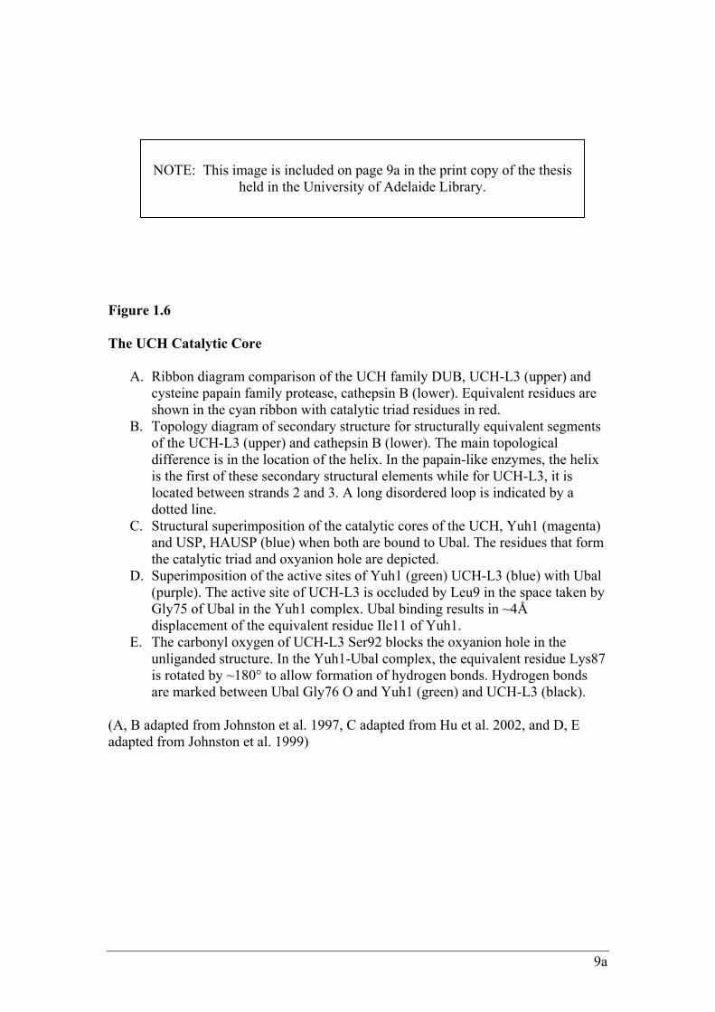
Figure 1.6 The UCH Catalytic Core
A. Ribbon diagram comparison of the UCH family DUB, UCH-L3 (upper) and cysteine papain family protease, cathepsin B (lower). Equivalent residues are shown in the cyan ribbon with catalytic triad residues in red.
B. Topology diagram of secondary structure for structurally equivalent segments of the UCH-L3 (upper) and cathepsin B (lower). The main topological difference is in the location of the helix. In the papain-like enzymes, the helix is the first of these secondary structural elements while for UCH-L3, it is located between strands 2 and 3. A long disordered loop is indicated by a dotted line.
C. Structural superimposition of the catalytic cores of the UCH, Yuh1 (magenta) and USP, HAUSP (blue) when both are bound to Ubal. The residues that form the catalytic triad and oxyanion hole are depicted.
D. Superimposition of the active sites of Yuh1 (green) UCH-L3 (blue) with Ubal (purple). The active site of UCH-L3 is occluded by Leu9 in the space taken by Gly75 of Ubal in the Yuh1 complex. Ubal binding results in ~4Å displacement of the equivalent residue Ile11 of Yuh1.
E. The carbonyl oxygen of UCH-L3 Ser92 blocks the oxyanion hole in the unliganded structure. In the Yuh1-Ubal complex, the equivalent residue Lys87 is rotated by ~180° to allow formation of hydrogen bonds. Hydrogen bonds are marked between Ubal Gly76 O and Yuh1 (green) and UCH-L3 (black).
(A, B adapted from Johnston et al. 1997, C adapted from Hu et al. 2002, and D, E adapted from Johnston et al. 1999)
NOTE: This image is included on page 9a in the print copy of the thesis
held in the University of Adelaide Library.
10
cysteine protease an effective nucleophile for attack on the peptide bond (reviewed in
Wing 2003).
In an inactive conformation, the UCH active site and oxyanion hole is inaccessible due to
the presence of a disordered active site-crossover loop (not present in classical cysteine
proteases), which becomes ordered upon ubiquitin binding (figure 1.6 D, E). However,
even at its most open state, the loop diameter is no greater than 15 Å, much smaller than
the majority of folded proteins and thus limits UCHs to the cleavage of small adducts or
unfolded polypeptides from the C-terminus of ubiquitin. Additionally, the active site
cysteine is located at the bottom of a narrow groove in the surface of the enzyme that
restricts access to large side-chain residues. Ubiquitin, which terminates with a pair of
glycines, is small enough to be accommodated in this groove. These factors (along with the
extensive binding interactions between ubiquitin and the UCH) determine the specificity of
UCH enzymes.
Crystal structures of the catalytic core of HAUSP in both its free and Ubal-complexed
form reveal a three globular domain configuration for USPs that resembles an extended
right hand comprised of Fingers, Palm and Thumb (Hu et al. 2002) (figure 1.7 A). The
general conservation of the residues that constitute the secondary structural elements
within the three domains appears conserved among other USP enzymes, and more
specifically, residues that contribute to the structural integrity of the Fingers-Palm-Thumb
architecture are invariant. Work into predicting the 3D-structure of putative domains for
human USP9Y (Ginalski et al. 2004) found that four cysteine residues in the Fingers
domain of the catalytic core that may coordinate a zinc ion. These cysteines form a
putative zinc ribbon-like structure, which was recently confirmed in HAUSP by
comparative sequence and structural analysis, as a zinc ribbon that has lost its zinc-binding
ability (Krishna and Grishin, 2004).
Using this hand analogy, the highly conserved Cys and His boxes are positioned on
opposite sides of a deep, inter-domain catalytic cleft created by the Palm and Thumb.
Correct positioning of the ubiquitin moiety is coordinated by the Fingers, with the C-
terminal glycines of ubiquitin placed in the active site between the Palm and the Thumb
(figure 1.7 B). In the free form of HAUSP, the catalytic triad is misaligned with the
catalytic Cys and His residues too far apart to allow any meaningful interactions. Binding
10a
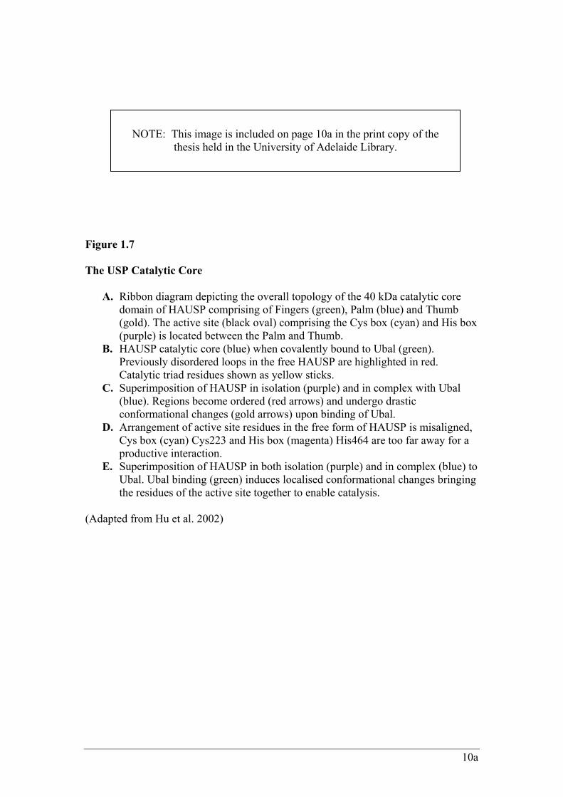
Figure 1.7 The USP Catalytic Core
A. Ribbon diagram depicting the overall topology of the 40 kDa catalytic core domain of HAUSP comprising of Fingers (green), Palm (blue) and Thumb (gold). The active site (black oval) comprising the Cys box (cyan) and His box (purple) is located between the Palm and Thumb.
B. HAUSP catalytic core (blue) when covalently bound to Ubal (green). Previously disordered loops in the free HAUSP are highlighted in red. Catalytic triad residues shown as yellow sticks.
C. Superimposition of HAUSP in isolation (purple) and in complex with Ubal (blue). Regions become ordered (red arrows) and undergo drastic conformational changes (gold arrows) upon binding of Ubal.
D. Arrangement of active site residues in the free form of HAUSP is misaligned, Cys box (cyan) Cys223 and His box (magenta) His464 are too far away for a productive interaction.
E. Superimposition of HAUSP in both isolation (purple) and in complex (blue) to Ubal. Ubal binding (green) induces localised conformational changes bringing the residues of the active site together to enable catalysis.
(Adapted from Hu et al. 2002)
NOTE: This image is included on page 10a in the print copy of the
thesis held in the University of Adelaide Library.
11
of Ubal caused a dramatic, highly localised conformational change in the catalytic cleft,
bringing together the relevant residues of the catalytic triad and causing the ordering of two
previously flexible surface loops, switching the enzyme into an active conformation (figure
1.7 C-E). HAUSP makes extensive contacts with ubiquitin involving interactions with both
the Fingers and the Palm-Thumb scaffold resulting in the burial of ~3600 Å2 of solvent
accessible surface area. Binding of Ubal by HAUSP can be visualised as grabbing Ubal
with the tip of the Fingers and the catalytic cleft between the Palm and Thumb, while a
cushion of water mediates the contact between Ubal and the middle portion of the Fingers.
While the three domain structure of USP allows for recognition of larger substrates, such
as polyubiquitylated proteins and free polyubiquityl chains, the HAUSP catalytic core by
itself is not sufficient for recognition of ubiquitylated substrates in vivo as gauged by high
dissociation constants. HAUSP instead recognises its substrate p53, via a domain N-
terminal to the catalytic core that contains the elements required for its specific interaction
with p53 (Hu et al. 2002). Interestingly, HAUSP only requires a short 26 amino acid C-
terminal segment of p53 for this interaction, and this p53 element includes several lysine
residues thought to be involved in p53 ubiquitylation. This data supports a model whereby
an N-terminal extension of HAUSP directly binds p53 proximal to the ubiquitin linkage
sites to enable the catalytic core, which normally has weak affinity for ubiquitin, to bind
the conjugate and become activated for proteolysis (reviewed in Lima 2003). Such a model
has implications for other USPs and lends strength to the argument that the sequence
divergent extensions of the various USPs comprise in part, substrate recognition sites.
Although UCHs and USPs share nearly identical active site geometries, the overall
structure and topology are vastly different. UCHs lack the Fingers domain and have a
shortened thumb. Although the Palm domain is present with the same overall fold, large
deviations are apparent throughout the structure. UCHs make contact with ubiquitin largely
from one surface due to a lack of Fingers, and the catalytic triad within the active site does
not undergo any significant conformational change during catalysis. Crystal structures of
both human otubain2 (OTU family of Dubs) and an even more distantly related cysteine
protease that acts on the ubiquitin-like molecule SUMO, are also found to have the same
active site configuration as UCHs and USPs demonstrating a conserved catalytic
mechanism for deubiquitylation (reviewed in Amerik and Hochstrasser 2004).
12
Drosophila Fat Facets (faf), a Developmentally Regulated USP
Fat facets (faf) is a developmentally regulated, 2747 amino acid USP that was identified in
a mutation screen for genes that affect drosophila eye development. It is essential in at least
two key developmental events: regulation of photoreceptor number in compound-eye
formation, and nuclear migration during syncytial stage oocyte development (Fischer-Vize
et al. 1992). Using a -galactosidase fusion to the first 392 amino acids of FAF, it was also
shown that FAF protein is spatially regulated. A complex localisation pattern was observed
in the imaginal eye disc and localisation within the syncytial oocyte was detected at the
posterior pole, indicating a requirement of specifically localised FAF activity for proper
eye and oocyte development in drosophila (Fischer-Vize et al. 1992).
FAF AND COMPOUND-EYE DEVELOPMENT
In drosophila, the compound eye consists of a hexagonal array of around 800 ommatidia
eye units, or facets that develop from a monolayer of cells termed the eye imaginal disc. A
depression called the morphogenetic furrow, moves across the disc of undifferentiated cells
as a wave, posterior to anterior and in the process of this movement, facet preclusters
emerge, mature and recruit additional cells in a precise series of inductions. Contained
within these initial facet preclusters are seven cells, five of which will become
photoreceptors that have a key role in directing the differentiation of the rest of the facet,
while the other two, termed mystery cells, will detach from the precluster and re-enter the
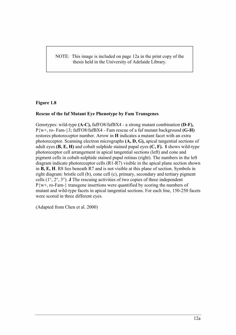
surrounding pool of undifferentiated cells (reviewed in Ready 1989) (figure 1.8 A-C, I).
Although faf null mutant flies were viable, they showed abnormal “rough” eye morphology
characterised by the appearance of extra photoreceptors in addition to the normal
complement of eight in each facet (figure 1.8 D-F). Ectopic photoreceptor formation is the
result of inappropriate differentiation of mystery cells that would otherwise have retreated
into the imaginal disc. Analysis of eye cells mosaic for faf+/faf
-, showed that the cell
communication pathway that negatively regulates the neural cell fate in the developing eye
requires FAF in cells near to, but outside the eight photoreceptors in wild-type facets.
Additionally, mutant alleles of a 20S proteasome subunit strongly suppress the faf mutant
phenotype, suggesting that FAF acts to limit the ubiquitylation and therefore degradation
of one or more regulators of eye development (Huang et al. 1995, Wu et al. 1999).
12a
Figure 1.8 Rescue of the faf Mutant Eye Phenotype by Fam Transgenes Genotypes: wild-type (A-C), fafFO8/fafBX4 - a strong mutant combination (D-F), P{w+, ro- Fam-}3; fafFO8/fafBX4 - Fam rescue of a faf mutant background (G-H) restores photoreceptor number. Arrow in H indicates a mutant facet with an extra photoreceptor. Scanning electron micrographs (A, D, G), apical tangential sections of adult eyes (B, E, H) and cobalt sulphide stained pupal eyes (C, F). I shows wild-type photoreceptor cell arrangement in apical tangential sections (left) and cone and pigment cells in cobalt-sulphide stained pupal retinas (right). The numbers in the left diagram indicate photoreceptor cells (R1-R7) visible in the apical plane section shown in B, E, H. R8 lies beneath R7 and is not visible at this plane of section. Symbols in right diagram: bristle cell (b), cone cell (c), primary, secondary and tertiary pigment cells (1°, 2°, 3°). J The rescuing activities of two copies of three independent P{w+, ro-Fam-} transgene insertions were quantified by scoring the numbers of mutant and wild-type facets in apical tangential sections. For each line, 150-250 facets were scored in three different eyes. (Adapted from Chen et al. 2000)
NOTE: This image is included on page 12a in the print copy of the
thesis held in the University of Adelaide Library.
13
A genetic screen to uncover candidates for the critical substrate of FAF in the eye
identified liquid facets (lqf), an endocytic protein orthologous to vertebrate Epsin1
(Cadavid et al. 2000). Four genetic observations were made that suggested a role for FAF
in preventing Lqf degradation: (1) lqf loss-of-function mutants are strong dominant
enhancers of the faf mutant eye phenotype, (2) faf and lqf loss-of–function mutations have
similar mutant eye phenotypes, (3) the faf+ and lqf
+ genes are required in the same group
of cells in the eye, and (4) one extra copy of the lqf+ gene overcomes the need for the faf
+
gene in the eye. These genetic observations were confirmed with biochemical data that
showed: (1) there is less Lqf protein in the developing drosophila eye in the absence of
functional FAF protein, (2) Lqf is ubiquitylated in the developing eye and is
deubiquitylated by FAF, and (3) Lqf and FAF interact physically (Chen et al. 2002).
Therefore it was concluded that FAF regulates the levels of Lqf by deubiquitylating it, thus
preventing its degradation at the proteasome. The fact that Lqf is involved in endocytosis
carries added significance given the recent understanding that the ubiquitin pathway and its
Dubs can influence endocytosis and intracellular trafficking. Indeed, a role for Lqf in
endocytosis is supported by genetic and co-localisation evidence that shows that lqf
interacts with several critical endocytosis genes (Cadavid et al. 2000, Chen et al. 2002).
Recent genetic experiments have provided a molecular model for how FAF, Lqf, and
endocytosis relate to cell signalling to inhibit photoreceptor development by the
surrounding cells. The Notch pathway participates in a wide range of cell communication
events, either inhibiting or promoting a variety of cell fates, and has been implicated in the
FAF/Lqf control of photoreceptor development (Overstreet et al. 2003). Notch activation
by Delta/Serrate/Lag2 (DSL) ligands requires endocytosis in both signalling and signal-
receiving cells and it has been proposed that the extracellular domain of the Notch receptor
(on the signal-receiving cell) when bound to Delta, is trans-endocytosed into the Delta-
expressing (signalling) cell. Processing of the intracellular domain of the cleaved Notch
receptor in the signal-receiving cell leads to signal transduction (and in this case, inhibition
of photoreceptor development). It has been shown that Lqf is required by signalling cells to
transmit DSL signals (Wang and Struhl 2004). Through immunoctyochemistry and genetic
experiments, it was deduced that FAF through its substrate Lqf, promotes Delta
internalisation (a process dependent on its ubiquitylation by the E3, Neuralised) and
subsequent Delta signalling by signalling cells. The signalling cells then activate Notch in
surrounding undifferentiated cells, preventing ectopic photoreceptor differentiation
14
(Overstreet et al. 2003, 2004). It has also been proposed that endocytosis of
monoubiquitylated DSL signalling ligands by Lqf, may target them to an endocytic
recycling compartment, converting them from inactive ‘pro-ligands’ into active ligands
(Wang and Struhl 2004).
FAF has also been shown to genetically interact with Rap1 and Ras1, members of the
mitogen-activated protein kinase (MAPK) pathway. Analysis of these interactions has
revealed that faf has an additional function later in eye development involving these
proteins, in the continued influence of facet assembly from the adjacent signalling cells (Li
et al. 1997).
FAF AND OOCYTE DEVELOPMENT
In addition to abnormal eye development, faf mutations have a maternal effect phenotype.
Homozygous mutant females are sterile despite possessing seemingly normal ovaries, and
their eggs are unable to reach the syncytial blastoderm stage (Fischer-Vize et al. 1992).
Normally in drosophila development, a fertilised egg will undergo a series of 14
synchronous divisions to form a multi-nucleated cell termed the syncytium. By division 10,
the nuclei migrate to the periphery of the cell forming the “syncytial blastoderm”, and the
primordial germ cells (pole cells) then form at the posterior end of the embryo. The nuclei
continue to the 14th synchronous division when cellularisation occurs around each nucleus
to form the cellular blastoderm. In faf mutant embryos, it was observed through nuclei and
cell membrane staining, that syncytial blastoderm development had been disrupted in all
cases. Embryos were able to reach at least the 10th division as determined by the presence
of the pole cells, however most nuclei (except patches of asynchronously divided nuclei)
did not migrate to the periphery. With the exception of the pole cells (which were fewer in
number and more spread out) cellularisation failed to occur (Fischer-Vize et al. 1992).
Although no direct critical substrate of FAF has been identified for oogenesis, FAF has
been shown to interact with Vasa, an RNA helicase that is a component of polar granules
and maternally essential for posterior patterning and germ cell specification (Liu et al.
2003). FAF has been shown to reverse Vasa ubiquitylation and stabilise it in the pole
plasm by inference, but has not been biochemically proven to be a bona fide, nor critical
substrate of FAF in oogenesis.
15
Fat Facets in Mouse (Fam)
The mouse orthologue of faf, Fat Facets in Mouse (Fam) was identified in a gene trap
screen in embryonic stem cells for genes expressed during gastrulation and neurulation
(Wood et al. 1997). One particular clone identified by this screen displayed a
developmentally restricted -galactosidase expression pattern, beginning only at the late
primitive streak stage. After isolating the complete cDNA, sequence analysis revealed an
open reading frame encoding a 2554 amino acid USP bearing strong sequence similarity to
faf. FAM and FAF are collinear over nearly the entire length and show approximately 50%
amino acid identity and 70% similarity, which increase considerably over the conserved
Cys and His boxes. Furthermore, over-expression of Fam in faf mutants is able to rescue
the phenotype which suggests that Fam and faf are true orthologues (Chen et al. 2000)
(figure 1.8 G, H, J).
FAM is critical for early mouse development as anti-sense oligodeoxynucleotides
knockdowns of FAM protein levels in pre-implantation mouse embryos inhibits their
progression from the two-cell to morulae or blastocyst stages. These embryos exhibit an
apparent diminution in cell-cell adhesion (figure 1.9). Depletion of FAM also
corresponded with decreased protein levels of two of its substrates, -catenin and AF-6.
However, following an initial decrease, after 48 hours AF-6 levels returned to normal but
the nascent protein was mislocalised to the apical surface of blastomeres (Pantaleon et al.
2001).
There are several other known isoforms, homologues and orthologues of FAM. Northern
analysis of Fam transcripts performed on post-implantation embryos revealed the existence
of three different length transcripts: 8.5 kb, 10.0 kb and 11.5 kb of which the 10.0 kb
transcript was the most abundant. All the transcripts encode the same protein and the
variance in size is due to differences in the length of the 3' untranslated region however,
their functional significance remains unclear (Wood et al. 1997). Fam also exists as two
isoforms caused by alternate splicing of exons. This splice variation results in the frame
shift insertion or exclusion of 15 bps, which encode the amino acids SSSRF (at position
589-590 aa). Thus the isoforms are named SSSRF+ (2559aa) and SSSRF- (2554aa)
however again, the functional significance is unknown.
15a
Figure 1.9 FAM is Essential for Pre-Implantation Mouse Development Immuno-localisation of FAM following treatment with Fam sense (A, C) and anti-sense (B, D) oligodeoxynucleotides (ODN). Two-cell stage embryos were cultured in the respective ODNs for 24 h (A, B) or for 72 h to the late blastocyst stage (C, D) and then probed with anti-FAM antibodies. The perinuclear and cytoplasmic positive FAM immuno-reactivity present in the sense treated embryos (A, C) is reduced in the 24 h anti-sense ODN treated embryos (B) and nearly abolished by 72 h (D). Colour wedge indicates highest intensity of immunofluorescence as white. Bar = 25µm. (Adapted from Pantaleon et al. 2001)
NOTE: This image is included on page 15a in the print copy of the
thesis held in the University of Adelaide Library.
16
Fam is located on the sex chromosomes and so two homologues exist, an X and Y copy.
The Y homologue is exclusively expressed in the testes and maps to the Sxrb deletion,
which is associated with an early post-natal blockage of spermatogonial proliferation and
differentiation and results in the adult testis being almost totally devoid of germ cells
(Brown et al. 1998). The X homologue which will be referred to as Fam, displays
temporally and spatially restricted expression throughout embryogenesis. The remainder of
this thesis describes research carried out on the SSSRF+, X-chromosome homologue
unless otherwise indicated.
The only described orthologue of Fam other than drosophila faf, are the human DFFRX
and DFFRY (drosophila fat-facets related X or Y) genes, otherwise known as USP9X or
USP9Y according to new nomenclature conventions. USP9X escapes X-inactivation and
both the X and Y copies are expressed in a wide and similar range of tissues (Brown et al.
1998) however, USP9Y is additionally expressed in the testes and has been shown to be a
functional USP (Lee et al. 2003). USP9X shares 97% identity and 99% similarity to FAM
while USP9X and USP9Y share 89 % identity and 98% similarity (Brown et al. 1998).
TEMPORAL AND SPATIAL REGULATION OF FAM DURING DEVELOPMENT
Whole-mount in situ analysis of post-implantation mouse embryos revealed that Fam is
expressed in a complex temporal and spatial pattern throughout development (Wood et al.
1997). Transcripts were first detected at the mid-streak stage (E7.5) but switches from
relatively ubiquitous expression to more tissue specific after E10.5, where expression
became progressively restricted to the developing central nervous system (CNS), limb buds
and branchial arches. By E13.5 Fam transcripts were undetectable by whole-mount in situ
analysis but were still expressed strongly as shown by in situ analysis (figure 1.10 A, B, C).
Within the CNS, Fam is expressed in a complex fashion. Fam expression in the nervous
system is strongest in the third, fourth and lateral ventricles, and in the developing spinal
cord. At E14.5, Fam expression is obvious in the olfactory lobe and in certain ganglia:
trigeminal, inferior glossopharyngeal and dorsal root ganglia. Within the developing
cortex, Fam is expressed by the neuroblasts of the ventricular zone, as well as in migrating
and differentiating neurons of the intermediate zone and cortical plate (Friocourt et al.
2005).
16a
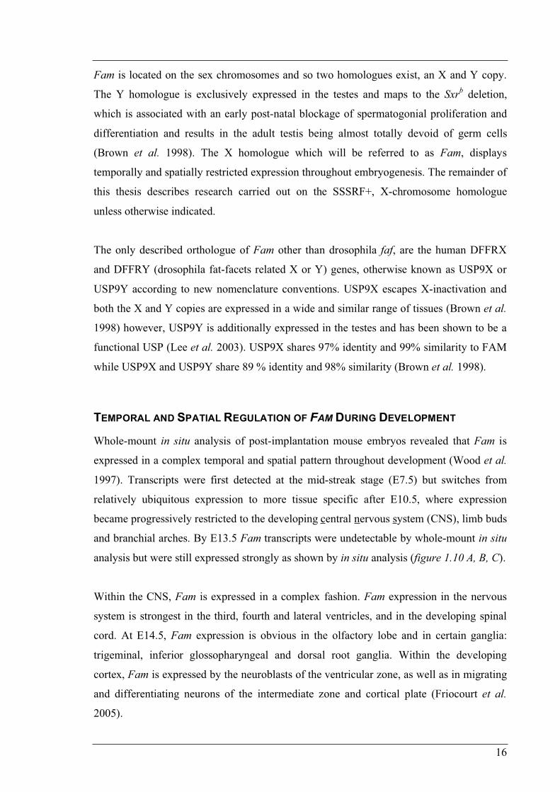
Figure 1.10 Fam Expression in Wholemount Mouse Embryos Wholemount in situ hybridisation analysis of E9.5 (A) E11.5 (B) and E12.5 (C) with a Fam anti-sense probe. Ubiquitous expression at E9.5 is increasingly restricted to the E12.5 stage. Expression is lost from the body wall while being maintained longest in the distal halves of the limb buds, branchial arches as well as the eye and central nervous system. At E12.5 only the eye, mesencephalon, telencephalon and the apoptotic regions between the digits remain positive for Fam. Bar represents 500µm in each panel. Sections of E12.5 midbrain (D) reveal strong Fam expression in the diencephalon (Di) and metencephalon (Met) which is progressively lost laterally. Sagittal sections of an E14.5 eye (E) show strong expression in the outer nuclear layer (on) but expression is lost from the inner nuclear layer (in) of the retina. Sections of the outer root sheath of the vibrissae (F) show strong expression around the hair follicle. (Adapted from Wood et al. 1997)
NOTE: This image is included on page 16a in the print copy of the
thesis held in the University of Adelaide Library.
17
Strong expression was detected in the eyes, ubiquitously at first, but later becoming more
neurally confined until retinal differentiation when expression weakened (figure 1.10 E).
Fam expression throughout eye development has been well characterised and found to co-
localise extensively with one of its substrates AF-6 (discussed later), an epithelial tight-
junction protein (Kanai-Azuma et al. 2000). At E10.5, FAM and AF-6 are both expressed
in the outer layer of the optic cup and a single layer of cells immediately adjacent in the
inner layer (figure 1.11 A, B). The outer layer goes on to form the retinal pigment
epithelium (RPE) and continues to maintain FAM and AF-6 expression. During lens
development, FAM and AF-6 were initially expressed at the apical surface of cells lining
the cavity of the lens vesicle and later (E13.5), present at the contact zone between the
differentiated lens fibres and sub-capsular epithelium (figure 1.11 C). FAM and AF-6 are
also co-expressed throughout corneal epithelium development. In late embryonic
development, both FAM and AF-6 are expressed in the inner layer of the neural retina and
in the adult, localise to the outer plexiform layers. Fam is expressed in the outer nuclear
layer by E18.5 (figure 1.11 D, E).
Interestingly, expression within the neural tube and brain was seen to become increasingly
polarised (Wood et al. 1997). Expression within the diencephalon and metencephalon at
E12.5 is strong in the ependymal layer, but is laterally lost by some cells through the
mantle and even fewer in the marginal layer giving the appearance of a gradient of FAM
expression (figure 1.10 D). Polarised expression was also observed during limb bud
development where it was initially ubiquitous, but gradually was confined to only the distal
halves at E11.5, and only present in the apoptotic regions between the digits twenty four
hours later.
Fam transcripts were additionally detected in many internal organs, including the liver and
other epithelial tissues such as the bronchi of the lungs, gut and nasal vibrissae (figure 1.10
F). Such diverse and specific expression patterns during early mouse development imply
that Fam is associated with multiple developmental or cellular events (Wood et al. 1997).
Fam has also been characterised during gonadal development and gametogenesis where it
was found to be stage-dependently expressed in the germ cells and supporting cells (Noma
et al. 2002). FAM further colocalises with AF-6 in Sertoli and granulosa cells of the testis
and ovary in a stage-dependent, synchronous manner going from a diffuse cytoplasmic
17a
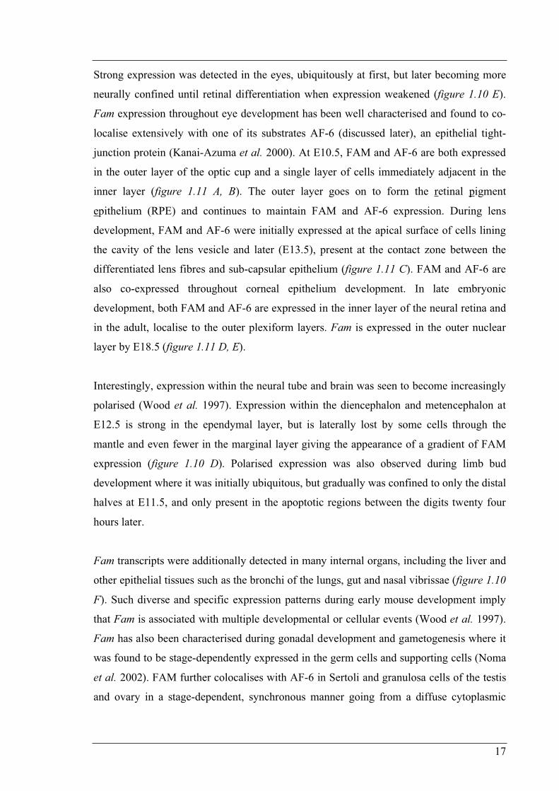
Figure 1.11 Fam Expression in the Developing Mouse Eye Immunofluorescence micrographs showing FAM localisation during eye development in E10.5 (A, B), E13.5 (C) and E18.5 (D, E) mouse embryos. (B) is an enlargement of (A) while (E) is an enlargement of (D). At E10.5, the eye primordium is composed of lens (l) and inner and outer layer of optic cup, which are precursors of the nervous layer of the retina (nr) and retinal pigmented epithelium (rpe) respectively. FAM is restricted to the outer layer and cells of the inner layer immediately adjacent (Arrows in A and B). FAM is strongly expressed in the retinal pigment epithelial cells (arrows in C-E) and the lens subcapsular epithelia cells (arrowheads in C). In the retina, the inner portion of the ventricular layer (in) increases expression from E13.5 to E18.5 while there also appears to be some FAM expression in the outer nuclear layer (on) at 18.5. (Adapted from Kanai-Azuma et al. 2000)
NOTE: This image is included on page 17a in the print copy of the
thesis held in the University of Adelaide Library.
18
distribution, to the Sertoli-Sertoli and Sertoli-spermatid junctions at later stages (Sato et al.
2004).
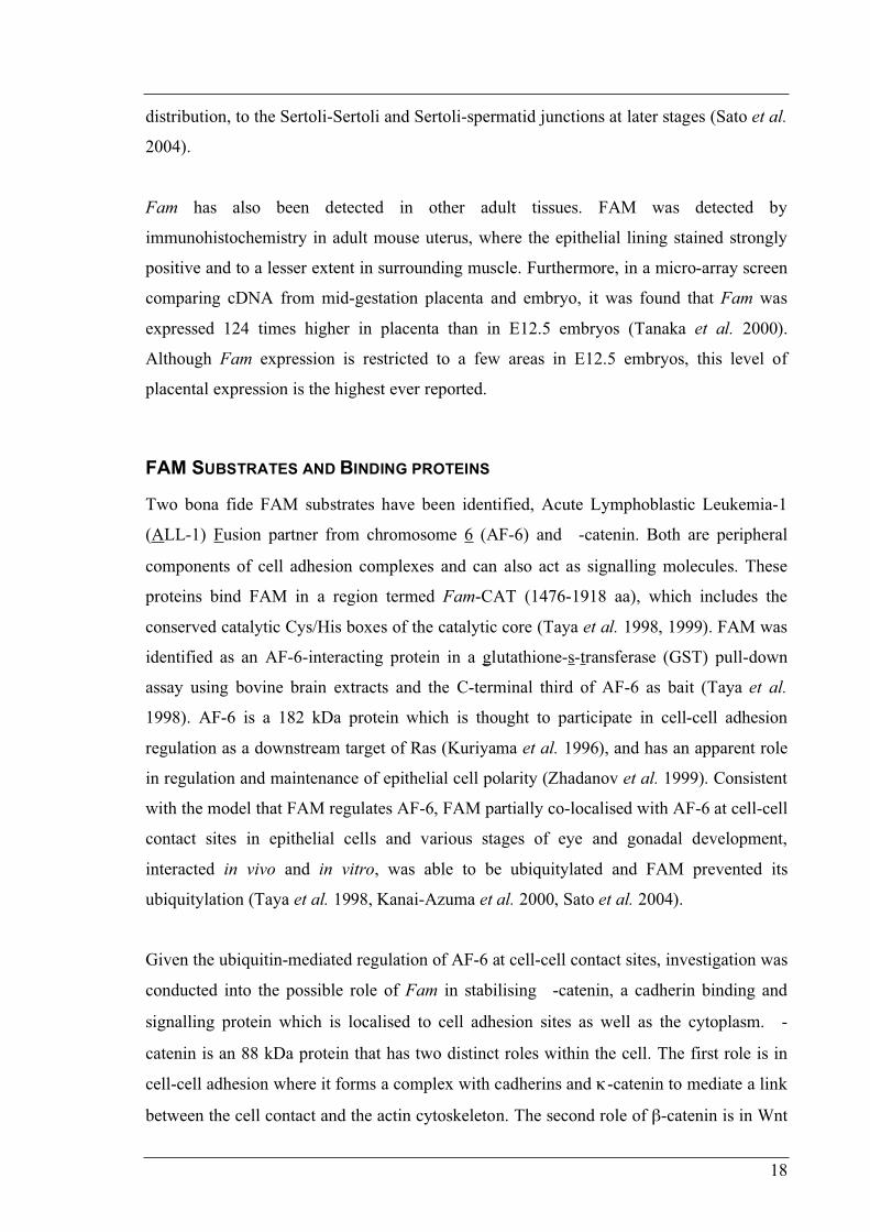
Fam has also been detected in other adult tissues. FAM was detected by
immunohistochemistry in adult mouse uterus, where the epithelial lining stained strongly
positive and to a lesser extent in surrounding muscle. Furthermore, in a micro-array screen
comparing cDNA from mid-gestation placenta and embryo, it was found that Fam was
expressed 124 times higher in placenta than in E12.5 embryos (Tanaka et al. 2000).
Although Fam expression is restricted to a few areas in E12.5 embryos, this level of
placental expression is the highest ever reported.
FAM SUBSTRATES AND BINDING PROTEINS
Two bona fide FAM substrates have been identified, Acute Lymphoblastic Leukemia-1
(ALL-1) Fusion partner from chromosome 6 (AF-6) and -catenin. Both are peripheral
components of cell adhesion complexes and can also act as signalling molecules. These
proteins bind FAM in a region termed Fam-CAT (1476-1918 aa), which includes the
conserved catalytic Cys/His boxes of the catalytic core (Taya et al. 1998, 1999). FAM was
identified as an AF-6-interacting protein in a glutathione-s-transferase (GST) pull-down
assay using bovine brain extracts and the C-terminal third of AF-6 as bait (Taya et al.
1998). AF-6 is a 182 kDa protein which is thought to participate in cell-cell adhesion
regulation as a downstream target of Ras (Kuriyama et al. 1996), and has an apparent role
in regulation and maintenance of epithelial cell polarity (Zhadanov et al. 1999). Consistent
with the model that FAM regulates AF-6, FAM partially co-localised with AF-6 at cell-cell
contact sites in epithelial cells and various stages of eye and gonadal development,
interacted in vivo and in vitro, was able to be ubiquitylated and FAM prevented its
ubiquitylation (Taya et al. 1998, Kanai-Azuma et al. 2000, Sato et al. 2004).
Given the ubiquitin-mediated regulation of AF-6 at cell-cell contact sites, investigation was
conducted into the possible role of Fam in stabilising -catenin, a cadherin binding and
signalling protein which is localised to cell adhesion sites as well as the cytoplasm. -
catenin is an 88 kDa protein that has two distinct roles within the cell. The first role is in
cell-cell adhesion where it forms a complex with cadherins and -catenin to mediate a link
between the cell contact and the actin cytoskeleton. The second role of -catenin is in Wnt
19
signalling where activation of the pathway leads to the accumulation of -catenin in the
cytoplasm followed by translocation to the nucleus where it activates Wnt target genes
(cell cycle regulators) after heterodimerisation. FAM was shown to interact with -catenin
in vivo and in vitro. In mouse L cells, over-expression of the carboxyl-terminal half of Fam
including the catalytic domain, leads to elevation of -catenin levels and extension of its
half-life. Immunofluorescence studies in these mouse L cells also show partial co-
localisation of Fam-CAT with -catenin at dot-like structures in the cytoplasm (Taya et al.
1999). Interestingly, -catenin binds FAM via a series of armadillo repeats, a region that
bears homology to the ENTH (Epsin N-Terminal Homology) domain of drosophila Lqf
(Chen et al. 2000). This raises the possibility that FAM may recognise a conserved binding
motif contained on a subset of its substrates.
Fam has also been shown to regulate Epsin1 (mammalian orthologue of Lqf). FAM and
Epsin1 display overlapping immunostaining in synapses of rat brain sections, FAM
coprecipitates with anti-Epsin1 immunoprecipitates, and Epsin1 is immobilised in a GST
pull-down from rat-brain cytosol with a C-terminal portion of FAM (1554-1953aa) as bait
(Chen et al. 2003). Further, when Fam expression in HeLa cells is suppressed by short
interfering RNA (siRNA), deubiquitylation of Epsin1 was specifically inhibited. These
results are consistent with the biochemical results obtained in drosophila that show that Lqf
is a direct substrate of FAF (Chen et al. 2002).
Fam was also recently identified in a yeast two-hybrid screen, searching for interactors
with Doublecortin (DCX), a microtubule-associated protein involved in neuronal migration
(Friocourt et al. 2005). Although this interaction was confirmed by targeted mutagenesis,
colocalisation, and immunoprecipitation studies, DCX does not appear to be ubiquitylated
and is unlikely to be a FAM substrate. DCX was found to bind to human FAM (USP9X) in
a region outside of the catalytic core, a novel C-terminal recognition domain comprising
the last 256 amino acids (USP9X 2292-2547 aa). Interestingly, DCX also interacts with the
subunits of the clathrin adaptor complexes AP-1 and AP-2 (Friocourt et al. 2001), known
to be involved in vesicle trafficking, and associated with the trans-golgi network and
plasma membrane respectively. Given that DCX is a microtubule-associated protein, it has
been suggested that it may act to localise and/or regulate FAM in specialised
compartments of neuronal cells (Friocourt et al. 2005). Interestingly these investigators
20
also report that DCLK1C, a protein similar to DCX which is expressed in astroglial and
neuronal cells also interacts with FAM.
SUBCELLULAR LOCALISATION OF FAM
Through immunohistochemistry and confocal microscopy, analysis of Fam expression in
pre-implantation mouse embryos revealed complex subcellular localisation patterns
(Pantaleon et al. 2001). In the unfertilised ovulated egg, FAM was strongly associated with
the spindle and chromosomes, as well as displaying low level punctate staining in the
cytoplasm. However upon fertilisation, FAM was observed to vacate to the cytoplasm
where it displayed very strong punctate staining while being completely absent from the
female and male pronuclei. In the following cleavage stages, FAM was still observed as
puncta in the cytoplasm but also highly localised in the perinuclear region, but not to the
chromosomes and spindle during mitosis. This pattern of localisation was maintained
following the initiation of differentiation at compaction and also in the blastocyst (figure
1.12). It is noteworthy that a cytoplasmic punctate distribution has also been observed for
FAF in drosophila S2 cells (Wu et al. 1999).
Investigation of FAM’s subcellular localisation in non-differentiated proliferating PC12
cells (derived from rat adrenal gland) has revealed different localisations depending of the
stage of cell cycle (Friocourt et al. 2005). FAM is located at microtubule-containing
structures such as the midbody during cytokinesis, the microtubule-organising centre, and
mitotic spindle in dividing cells. In confluent glial cells and MDCKII cells, FAM
colocalises at cell-cell contacts with -catenin (Friocourt et al. 2005, Taya et al. 1999) and
AF-6 in MDCKII cells (Taya et al. 1998).
Recent immunofluorescence microscopy has further analysed FAM’s subcellular
localisation in polarised epithelial cells. FAM was found to localise to various puncta
throughout the basolateral but not the apical cytoplasm (Murray et al. 2004). These puncta
represented sites of protein sorting and trafficking as FAM partially colocalised with the
Golgi apparatus, late endosomes and the lysosome. Through immunoprecipitation and gel
filtration assays, FAM also was found to associate with nascent -catenin and E-cadherin
complexes in the cytoplasm, but not at the plasma membrane in sub-confluent cells. These
observations suggest a role for FAM in facilitating the vesicular transport of nascent -

20a
Figure 1.12 Subcellular Localisation of FAM During Mouse Pre-Implantation Development Confocal immunofluorescent optical sections of mouse embryos. (A) Fertilised egg and (B) blastocyst incubated with preimmune rabbit serum IgG. In unfertilised oocytes (C), FAM is strongly associated with the spindle and chromosomes, and also detected at lower levels in the cytoplasm. Fertilised oocytes (D) displayed positive FAM immunoreactivity in the cytoplasm, while both the male and female pronuclei are devoid of FAM. Two-cell (E), four-cell (F), morula (G), and blastocyst (H) embryos all show positive staining for FAM, mainly associated with the perinuclear membrane but is also present in cytoplasmic puncta. Colour wedge indicates highest intensity of immunofluorescence as white. Bar = 25µm. (Adapted from Pantaleon et al. 2001)
NOTE: This image is included on page 20a in the print copy of the
thesis held in the University of Adelaide Library.
21
catenin E-cadherin complexes from the trans-golgi network to the basolateral plasma
membrane of sub-confluent epithelial cells that are establishing cell-cell contacts (Murray
et al. 2004).
It is possible that these observations made in epithelial cells may also explain the punctate
staining pattern observed during pre-implantation development, and may also provide a
mechanistic reason as to why a lack of FAM leads to a reduction in cellular adhesion.
FAM-depleted embryos may not be able to establish strong cell-cell contacts because
adhesion complexes are not able to be trafficked to the plasma membrane in the absence of
FAM. FAM depletion correlates with an initial reduction in AF-6 protein levels which later
return to normal; however the nascent protein is mislocalised to the apical surface of
blastomeres. Assuming that FAM normally localises and sequesters AF-6 to the baso-
lateral compartment (as observed in polarised epithelia), it is conceivable then that a lack
of FAM results in the mislocalisation of AF-6 to the opposite compartment.
The findings that FAM is localised to multiple points of protein trafficking, coupled with
the role of FAF in regulating the endocytic factor Lqf, implies that FAM and its
orthologues may have a role beyond that of mere half-life extension of specific substrates.
Their additional roles in modifying specific proteins for intracellular transportation,
imparts to them a far more active role in regulating cellular events, the consequences of
which have dramatic effects on development.
Structure and Function of USPs
It is well established that the E2-E3 ubiquitin ligase family of proteins supply the ubiquitin
machinery a high level of specificity. By deduction, the reverse process, deubiquitylation
must also have the same level of specificity in order to avoid futile cycles of ubiquitylation
and deubiquitylation. However, little is known of the structure-function relationship of
USPs such as FAM. Given that USPs display significant sequence diversity (excluding the
conserved catalytic boxes) in the form of insertions within the catalytic core and a variety
of different N and/or C-terminal extensions, it has been proposed that these unique regions
22
may serve (amongst other things) as substrate-binding sites, conferring substrate specificity
to USPs (Baker et al. 1992). This proposition makes two predictions. The first is that these
unique regions specifically bind substrates and secondly, these regions preclude
inappropriate enzymatic activity. Indeed, a USP has been identified where these extensions
have been shown to specifically bind proteins. The mouse deubiquitylating enzyme
mUBPy binds the SH3 domain of Hrs-binding protein (Hbp) via two novel SH3-binding
motifs located in its N-terminal extension (Kato et al. 2000). A clear example where the
second prediction of the proposition is demonstrated comes from testis USP. Testis USP
exists as two isoforms, USP-t1 and USP-t2, each containing the same catalytic core
regions, but with distinct N-termini. Not only do these N-termini target the two isoforms to
different subcellular locations (Lin et al. 2000) but they also inhibit the ability of the
enzyme to indiscriminately cleave ubiquitin from an artificial substrate as compared to the
catalytic core alone, without significantly affecting catalytic function (Lin et al. 2001).
This modular model of function also has precedence in a member of the E3 HECT ligase
family, RSP5. Like USPs, Hect E3 ligases are diverse in size (from 92 to over 500 kDa)
and generally only have the HECT domain in common and so the same model, whereby
the highly variable regions define substrate specificity, has been proposed. In the case of
RSP5, it has been shown that the HECT domain by itself is enzymatically active, while its
interaction with the binding site of its substrate Rpb1 is independent of this (Wang et al.
1999).
However, the modular model of USP structure/function may prove to be an
oversimplification. In a study to determine the regions of FAF that are essential in its role
in drosophila eye development, a two interesting pieces of genetic evidence were raised
that do not support this model. Firstly, all protein domains along the entire length of the
FAF protein are seemingly required for full activity as a series of deletion constructs fail to
rescue the mutant eye phenotype (Chen and Fischer, 2000). Furthermore, analysis of 14
point mutant faf alleles with a maternal affect lethal phenotype show a distribution over the
full length of FAF. Secondly the functionally active catalytic core is also insufficient to
rescue the eye phenotype (Chen and Fischer, 2000) whereas the expression of Fam and the
yeast USPs, USP2 and USP3 can substitute for endogenous FAF more effectively than
most of the prementioned deletions (Chen et al. 2000, Wu et al. 1999).
23
It should also be noted that although protein binding can occur in these extension regions
outside the catalytic core of USPs as in the case of mUBPy, several examples are
documented where protein binding regions exist within the catalytic core, presumably
binding to the unique sequences interspersed between the Cys, His, Asp and KRF boxes. A
region of FAM containing the catalytic core (Fam-CAT 1476-1918 aa) binds both AF-6
and -catenin and its over-expression leads to their stabilisation (Taya et al. 1998, 1999).
The catalytic core of yet another USP, Unp, binds Retinoblastoma tumour suppressor
protein (Rb) and Rb family members p107 and p130 (Blanchette et al. 2001, Desalle et al.
2001). These examples raise the possibility that these catalytic cores and other regions may
consist of multi-substrate binding domains, as is the organization of proteins such as
CBP/p300 (Reviewed in Goodman and Smolik 2000).
Aims
Of the 2554 amino acids of FAM, only a region that spans the catalytic core has been
directly characterised. This region has been shown to bind its substrates -catenin and AF-
6 (Fam-CAT: 1476-1918 aa) (Taya et al. 1998, 1999), and also Epsin (1554-1953aa)
(Chen et al. 2003). By extrapolation from human USP9X (97% identity and 99% similarity
to FAM), Doublecortin binds FAM in the last 256 amino acids of its C-terminal extension
(2299-2554 aa) (Friocourt et al. 2005). The identity and function of the remainder of the
FAM, some 1800 odd amino acids remain unclear largely due to the lack of significant
homology to any other protein. Some recent work has predicted the 3D structure of some
putative human USP9Y domains (Ginalski et al. 2004). They found that four cysteine
residues (Cys-1726, Cys-1729, Cys-1773, and Cys-1776) in the Fingers domain of the
catalytic core (1553-1996 aa) may coordinate a zinc ion. These cysteines form a putative
zinc ribbon-like structure, absent from the crystal structure of HAUSP. Three previously
uncharacterised long -helical regions were also predicted in the N- and C-terminal
extensions (71-868, 1008-1532, 2004-2476 aa), the most C-terminal presumably the
Doublecortin interacting domain. Lastly a domain located in the N-terminus between two
presumptive -helical regions, appears to have a -grasp fold (884-971 aa) characteristic
of ubiquitin-like proteins (figure 1.13, includes the corresponding residue positions of
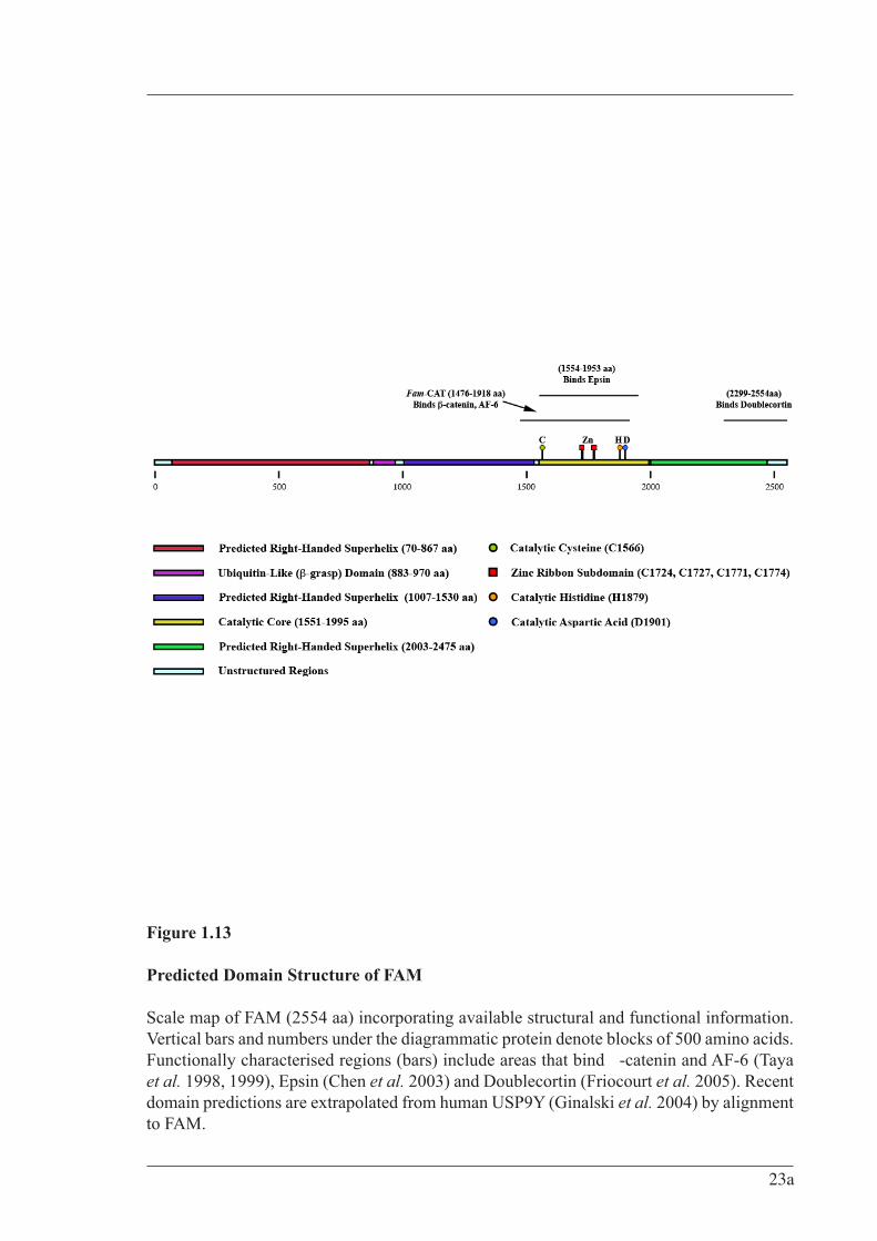
Figure 1.13
Predicted Domain Structure of FAM
Scale map of FAM (2554 aa) incorporating available structural and functional information. Vertical bars and numbers under the diagrammatic protein denote blocks of 500 amino acids. Functionally characterised regions (bars) include areas that bind -catenin and AF-6 (Taya et al. 1998, 1999), Epsin (Chen et al. 2003) and Doublecortin (Friocourt et al. 2005). Recent domain predictions are extrapolated from human USP9Y (Ginalski et al. 2004) by alignment to FAM.
23a
24
these putative domains for FAM). It has been postulated that this ubiquitin-like domain
corresponds to a distant homologue of other ubiquitin-like proteins and that it functions to
target USP9Y to its specific cellular localisation (Ginalski et al. 2004).
Without any experimental structural data, the boundaries of these putative domains remain
purely theoretical. Several attempts at expressing regions of FAM in various bacterial and
mammalian systems have either failed to express or have been found in the insoluble
fraction, presumably due to the disruption of a folding domain. Precise knowledge of
FAM’s domain structure would not only provide insight into regions of amino acids that
hold no homology to any other known domains, but would also provide a valuable
experimental tool for the design of FAM domain constructs to dissect FAM’s functions.
Use of individual domains would aid in the discovery of novel binding proteins, be it
substrate or non-substrate proteins such as regulatory, localisation or ancillary factors. To
this effect, expression of full-length FAM and subsequent partial proteolysis were
undertaken to identify protease resistant regions of FAM that would theoretically
correspond to folding domains.
Given the complex temporal and spatial expression of Fam and its orthologues throughout
development, it has been proposed that FAM maybe involved in several developmental
events. One way to investigate FAM’s role in development is to search for
developmentally relevant binding partners. Identification of such proteins would not only
gain insight into the developmental events that FAM may regulate, but also shed light on
the molecular mechanism. This thesis will also detail the establishment of a developmental
screen that sought to identify dominant-negative developmental defects from the ectopic
expression of regions of FAM in Danio rerio (zebrafish).
25
CHAPTER 2
MATERIALS AND METHODS
Abbreviations
Aa Amino Acid AF-6 ALL-1 Fusion partner from chromosome 6 ALL-1 Acute Lymphoblastic Leukemia-1 Amp Ampicillin APC Adenomatous Polyposis Coli ARM Armadillo BES N,N-Bis-(2-hydroxyethyl)-2-aminoethanesulfonic acid BCIP 5-bromo-4-chloro-3-indolyl-phosphate Bp base pair °C Degrees Celsius CAM Calmodulin homology CBC Cap Binding Complex cDNA Complementary Deoxyribonucleic Acid CNS Central Nervous System Comm Commissureless Cul1 Cullin homolog 1 DCX Doublecortin Dub Deubiquitylating enzyme DFFRX Drosophila Fat-Facets Related X DFFRY Drosophila Fat-Facets Related Y DMEM Dulbecco’s Modified Eagles MediumDMSO Dimethyl Sulfoxide Dpf Days Post-fertilisation DSL Delta/Serrate/Lag2 DTT Dithiothreitol Dub Deubiquitylating enzymes eGFP Enhanced Green Fluorescent Protein E. coli Escherichia coliECL Enhanced Chemiluminescence EDTA Ethylene Diamine Tetra Acetic acid EGTA Ethyleneglycol-bis-(2-aminoethyl)-N,N,N,N'-tetraacetic acid ENTH Epsin N-Terminal Homology EPL cells Early Primitive Ectoderm-Like Cells ER Endoplasmic Reticulum ES cells Embryonic Stem Cells Faf Drosophila Fat Facets
Fam Fat Facets in Mouse (chromosome X) FamY Fat Facets in Mouse (chromosome Y) Fam-CAT Fam catalytic core (1476 – 1918aa)
26
FCS Fetal Calf Serum G Gravitational forces GPCR G Protein-Coupled Receptor GST Glutathione-S-Transferase HAUSP Herpesvirus-Associated Ubiquitin-Specific Protease HBP Hrs-Binding Protein HCL Hydrochloric Acid HCRs Highly Conserved Regions HEAT Huntingtin-Elongation-A subunit - TOR HECT Homologous to the E6-AP C-Terminus HEK293T Human Embryonic Kidney 293T fibroblasts Hepes N-2-hydroxyethylpiperazine-N’-2-ethane sulphonic acid His tag Histidine tag Hpf Hours Post-Fertilisation Hr Hour HRP Horseradish Peroxidase I B Inhibitor of nuclear factor BIBB Importin- Binding IPTG Isopropyl- -D-Thiogalactopyranoside kDa Kilodalton LOOPP Learning, Observing and Outputting Protein Patterns Lqf Liquid facets (drosophila epsin1)MALDI Matrix-Assisted Laser Desorption/Ionisation MAPK Mitogen-Activated Protein Kinase MDCKII Madin-Darby Canine Kidney II Min / ‘ Minutes mL Millilitres mM Millimoles M Molar concentration MQ Milli-Q mRNA messenger RNA MVB Multivesicular Body NBT 4-nitro blue tetrazolium chloride NF B Nuclear Factor- BNMR Nuclear Magnetic Resonance NP-40 NonidetP-40 NPC Nuclear Pore Complex NTMT buffer NaCl, Tris, MgCl2, Tween-20 buffer OD Optical Density ODN Oligodeoxynucleotides OUT Ovarian Tumour PAGE Polyacrylamide Gel Electorphoresis PBS Phosphate Buffered Saline PBT PBS + 0.1% Tween 20 PCR Polymerase Chain Reaction PHD Pleckstrin Homology Domain PIP2 Phosphatidylinositol 4,5-bisphosphate PIP3 Phosphatidylinositol 3,4,5-trisphosphate PMSF Phenyl Methyl Sulfonyl Fluoride Ppm Parts per million
27
PTC Phenylthiocarbamide QTOF2 Quadrupole/time-of-flight Rb Retinoblastoma RING Really Interesting New Gene RIP Receptor-Interacting Protein Robo Roundabout RPE Retinal Pigment Epithelium Rpm Revolutions per minute SCF Skp1-Cullin-F-box protein SDS Sodium Dodecyl Sulphate Sec Seconds siRNA Short Interfering RNA SNARE Soluble NSF attachment protein receptors snRNA small nuclear RNA TBS Tris-buffered saline TCA Trichloroacetic Acid TNF Tumour Necrosis Factor TTBS Tris-buffered saline with 1% Tween-20 Ubl Ubiquitin-like U-box UFD2 homology UBP Ubiquitin specific proteases UCH Ubiquitin Carboxyl-terminal Hydrolases
l microliter m micrometer
USP Ubiquitin Specific Peptidase Usp9X Ubiquitin Specific Protease 9 on Chromosome X (Human)Usp9Y Ubiquitin Specific Protease 9 on Chromosome Y (Human)usp9 Zebrafish Orthologue of Fam(X)UTP Uracil triphosphate UV Ultraviolet
28
Materials
Platinum Pfx DNA polymerase was obtained from Invitrogen (Mount Waverly, VIC),
Expand High Fidelity PCR System and dNTPs from Roche Applied Sciences (Castle Hill,
NSW), and custom primers from Geneworks (Adelaide SA). All Gateway related materials
including enzymes and vectors were obtained through Invitrogen. All Fam cDNA
templates were obtained from Dr. Stephen Wood (Child Health Research Institute,
Adelaide SA). All restriction enzymes were obtained from New England Biolabs Inc.
(Ipswich, MA, USA) and used according to the manufacturer’s specifications. Benchmark
Molecular Weight Markers and RPN800 Rainbow Molecular Weight Markers were
obtained through Invitrogen and Amersham Biosciences (Castle Hill, NSW) respectively.
Foetal Calf Serum (FCS), Dulbecco’s Modified Eagle Medium (DMEM), Phosphate
Buffered Saline (PBS) and antibiotics for tissue culture were obtained from CSL
Biosciences (Parkville, NSW) and Gibco BRL (Invitrogen). All Zebrafish related materials
were kindly and generously provided by Dr. Michael Lardelli (Department of Molecular
Bioscience, University of Adelaide). All other materials were obtained from Sigma-
Aldrich (Sydney, NSW) unless otherwise stated.
ANTIBODIES
Monoclonal anti-V5 antibody was obtained from Invitrogen. Monoclonal anti-Myc
antibody was a kind gift from Dr. Michael Lardelli and originally sourced from Sigma-
Aldrich. Polyclonal anti-GST serum raised in rabbit was kindly donated by Dr. Stephen
Rodda, (Department of Molecular Bioscience, University of Adelaide). Rabbit polyclonal
anti-N1 FAM antibodies raised against a synthetic peptide corresponding to the first 20aa
of murine FAM (TATTRGSPVGGNDNQGQAPC), denoted N1, which was produced by
Susan Millard (Department of Molecular Bioscience, University of Adelaide). Goat anti-
rabbit and rabbit anti-mouse secondary antibodies conjugated with HRP were obtained
from DAKO (Carpinteria, CA, USA).
29
BACTERIAL STRAINS
DH5 : supE44 lac U169 ( 80 lacZ M15) hsdR17 recA 1 endA 1 gyrA96 thi 1
relA1
BL21 (DE3): F- ompT hsdSB (rB-mB-) gal dcm lon- (DE3)
DB3.1: F- gyrA462 endA1 (sr1-recA) mcrB mrr hsdS20(rB-, mB-) supE44 ara14
galK2 lacY1 proA2 rpsL20(Smr) xyl5 leu mtl1
DH10Bac: F–mcrA (mrr-hsdRMS-mcrBC) 80lacZ M15 lacX74 recA1 endA1
araD139 (ara, leu)7697 galU galK –rpsL nupG /pMON14272 /
pMON7124
Plasmids
Much of the cloning presented in this thesis was performed using gateway technology
(Invitrogen). Gateway technology allows the transfer of genes of interest in the context of
an “entry clone” into a variety of “destination vectors” using site-specific recombination
borrowed from phage. Destination vectors are available commercially and can be
constructed by insertion of a cassette (in frame with any fusion tags) that contains the attR
recombination sites, along with a ccdB gene for negative selection against non-
recombinants in standard bacterial lines. The entry clones used in this research were
constructed by PCR of the gene of interest with flanking attB sites to facilitate the “BP”
reaction into the attP sites of the entry vector pDONR201 (figure 2.1 A, B). The resultant
“entry clone” now contains attL sites for “LR” reaction transfer of the gene of interest into
the attR sites of the destination vector. All gateway reactions were performed as directed
by the manufacturer’s protocols. In all instances, constructed entry clones used for
experiments were verified by restriction digests and then sequenced with the pDONR
sequencing primers (table 2.1) uncovering no errors.
Two different PCR kits were used according to the manufacturer’s instructions. The
Expand High Fidelity PCR System (Roche) and the Platinum Pfx DNA polymerase
(Invitrogen) kit, which polymerise at 72°C and 68°C respectively. A general PCR program
29a
Figure 2.1 Entry Vector and the BP Reaction A. Vector map of entry vector pDONR201. B. Diagram detailing the BP reaction to transfer a PCR product with attB sites into
the attP sites of pDONR201 to form an entry clone. (Adapted from the Invitrogen website (http://invitrogen.com))
NOTE: This image is included on page 29a in the print copy of the
thesis held in the University of Adelaide Library.
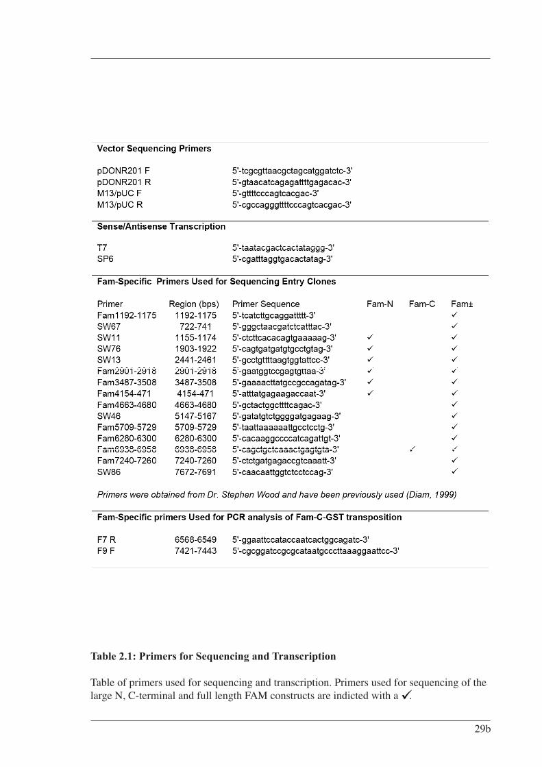
Table 2.1: Primers for Sequencing and Transcription
Table of primers used for sequencing and transcription. Primers used for sequencing of the large N, C-terminal and full length FAM constructs are indicted with a .
29b
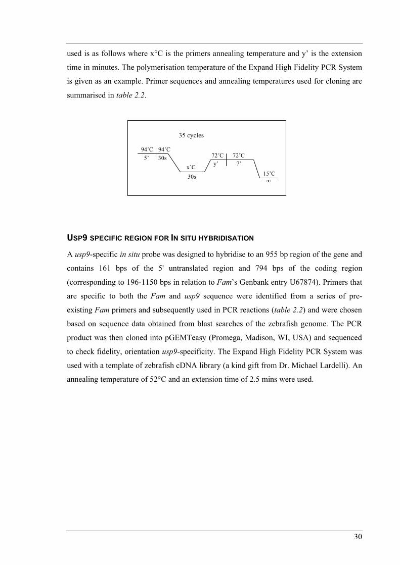
30
used is as follows where x°C is the primers annealing temperature and y’ is the extension
time in minutes. The polymerisation temperature of the Expand High Fidelity PCR System
is given as an example. Primer sequences and annealing temperatures used for cloning are
summarised in table 2.2.
USP9 SPECIFIC REGION FOR IN SITU HYBRIDISATION
A usp9-specific in situ probe was designed to hybridise to an 955 bp region of the gene and
contains 161 bps of the 5' untranslated region and 794 bps of the coding region
(corresponding to 196-1150 bps in relation to Fam’s Genbank entry U67874). Primers that
are specific to both the Fam and usp9 sequence were identified from a series of pre-
existing Fam primers and subsequently used in PCR reactions (table 2.2) and were chosen
based on sequence data obtained from blast searches of the zebrafish genome. The PCR
product was then cloned into pGEMTeasy (Promega, Madison, WI, USA) and sequenced
to check fidelity, orientation usp9-specificity. The Expand High Fidelity PCR System was
used with a template of zebrafish cDNA library (a kind gift from Dr. Michael Lardelli). An
annealing temperature of 52°C and an extension time of 2.5 mins were used.
94˚C 94˚C72˚C 72˚C
x˚C15˚C
5’ 30s
30s
y’ 7’
35 cycles
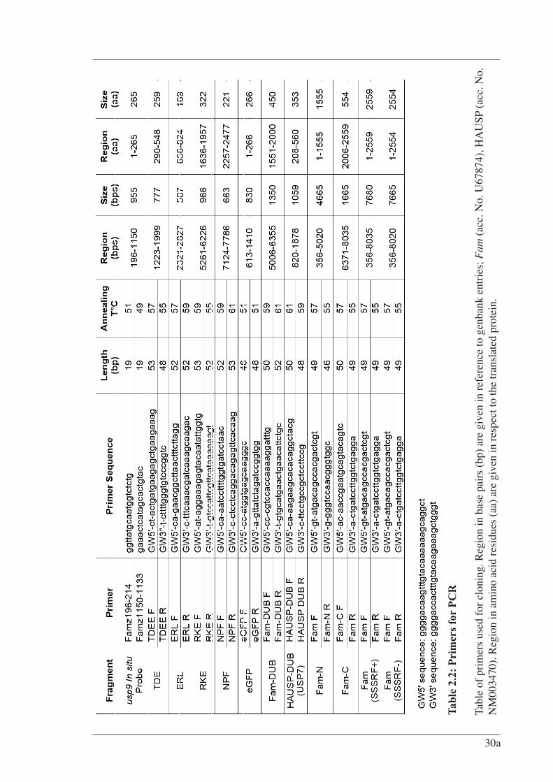
Tab
le 2
.2:
Pri
mer
s fo
r P
CR
Tabl
e of
pri
mer
s us
ed f
or c
loni
ng. R
egio
n in
bas
e pa
irs
(bp)
are
giv
en in
ref
eren
ce to
gen
bank
ent
ries
; Fam
(ac
c. N
o. U
6787
4), H
AU
SP (
acc.
No.
N
M00
3470
). R
egio
n in
am
ino
acid
res
idue
s (a
a) a
re g
iven
in r
espe
ct to
the
tran
slat
ed p
rote
in.
30a
31
Entry Clones
HIGHLY CONSERVED REGIONS (HCRS)
Entry clone construction of FAM’s four highly conserved regions; TDEE, ERL, RKE and
NPF (see pg. 56 for a description) was achieved by amplifying regions of full-length Fam
SSSRF+ cDNA (290-548, 356-824, 1636-1957, 2257-2477 respectively) by PCR with the
Expand High Fidelity PCR System according to the manufactures instructions. An
annealing temperature of 55°C and an extension time of 1.5 mins were used.
EGFP
A plasmid containing eGFP was kindly donated from Dr. Gavin Chapman (Karolinska
Institute, Sweden). This plasmid, pEGFP-C3FC is a destination vector, a modified version
of pEGFP (Clontech Laboratories, Inc., Palo Alto, CA, USA) that has had Gateway
cassette frame C inserted into the SmaI site of the multiple cloning site. The gateway
cassette was removed by digestion with SacII and BamHI, blunted and religated. This
strategy allowed retention of the 3’ multiple cloning site with three frames of stop codons.
Not only was the removal of the cassette necessary to provide a stop codon for eGFP, but it
also allowed flexibility for any future cloning into the multiple cloning sites. As such, this
plasmid can only be used as a C terminal fusion when combined with destination vectors
containing N terminal tags, such as GW-pCS2+MT. A Gateway compatible eGFP entry
clone was constructed by amplification of the eGFP gene from the pre-mentioned pEGFP
derived plasmid with similar PCR reaction conditions used for the pre-mentioned HCRs
and then inserted into pDONR201 via a BP reaction and verified by restriction digests and
sequenced.
USP CATALYTIC CORES
Entry clone construction of Fam-DUB was achieved by PCR off full-length Fam SSSRF-
cDNA template with the Platinum Pfx DNA polymerase PCR system according to the
manufactures instructions. An annealing temperature of 59°C and an extension time of 2
mins were used.

32
Entry clone construction of HAUSP-DUB was achieved by PCR off HAUSP (USP7)
cDNA template (accession number Z72499, kindly donated by Dr. RD Everett, Medical
Research Council Virology Unit, Scotland) with the Expand High Fidelity PCR system
according to the manufactures instructions. An annealing temperature of 55°C and an
extension time of 3 mins were used.
FAM N AND C-TERMINAL EXTENSIONS
Entry clone construction of Fam N and C terminal extensions was achieved by PCR off
full-length Fam SSSRF+ cDNA template using the Platinum Pfx DNA polymerase PCR
system according to the manufactures instructions. For the Fam N-terminal extension, an
annealing temperature of 55°C and an extension time of 5.5 mins was used. For the Fam
C-terminal extension, an annealing temperature of 55°C and an extension time of 2.5 mins
was used. In addition to verification by restriction digests and sequencing with pDONR
sequencing primers, several other Fam-specific primers were used for full sequence
coverage (table 2.1). No errors were detected.
FULL-LENGTH FAM (SSSRF±)
Entry clone construction of both isoforms of full-length Fam was achieved by PCR off
Fam SSSRF+ or Fam SSSRF- cDNA template with the Expand High Fidelity PCR system
according to the manufactures instructions. An annealing temperature of 57°C and an
extension time of 8.5 mins were used. In addition to verification by restriction digests and
sequencing with pDONR sequencing primers, several other Fam-specific primers were
used for full sequence coverage (table 2.1). Conservative errors were detected (figure 4.7).
FAM-CYS MUTANT (C1566S)
An entry clone containing full-length Fam (SSSRF+) with the critical cysteine of the
catalytic triad mutated to a serine was obtained from Susan Millard (Department of
Molecular Bioscience, University of Adelaide). Its construction involved site directed
mutagenesis on the Fam (SSSRF+) entry clone described above.
33
Destination Vectors
GWPCS2+MT
A vector for in vitro transcription pCS2+MT was kindly obtained by Dr. Michael Lardelli
and allows mRNA transcription and in vivo translation with an N terminal 6-myc tag. This
vector was made Gateway compatible by insertion of a Gateway cassette (reading frame
A) into blunted StuI and XbaI sites within the multiple cloning region (figure 2.2).
COMMERCIALLY AVAILABLE DESTINATION VECTORS
The pDEST15 vector (which generates a bacterially expressed N-terminal GST fusion), the
pEF-DEST51 vector (which adds a C-terminal V5 6x His tag fusion for mammalian
expression) and the pDEST20 vector (which adds an N-terminal GST fusion for
baculoviral expression) were obtained through Invitrogen (figure 2.3)

Figure 2.2
Constructed Destination Vector GWpCS2+MT
Vector map of GWpCS2+MT constructed by ligation of a gateway cassette (reading frame A) into blunted Stu1 and Xba1 sites.
33a
33b
Figure 2.3 Commercially Available Destination Vectors A. Vector map of pDEST15 for the bacterial expression of N-terminal GST fusion
proteins B. Vector map of pEF-DEST51 for the mammalian expression of C-terminal V5
6xHis tagged fusion proteins C. Vector map of pDEST20 for the transposition of the cloned gene of interest into a
DH10Bac (Invitrogen) bacmid. Recombinant bacmid is expressed in insect cells and produces N-terminal GST fusion proteins.
(Figures adapted from the Invitrogen website (http://invitrogen.com))
NOTE: This image is included on page 33b in the print copy of the
thesis held in the University of Adelaide Library.
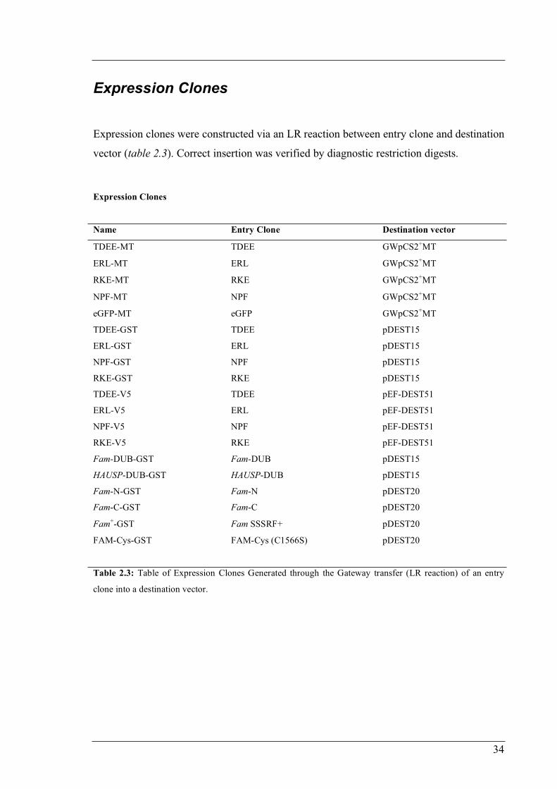
34
Expression Clones
Expression clones were constructed via an LR reaction between entry clone and destination
vector (table 2.3). Correct insertion was verified by diagnostic restriction digests.
Table 2.3: Table of Expression Clones Generated through the Gateway transfer (LR reaction) of an entry
clone into a destination vector.
Expression Clones
Name Entry Clone Destination vector
TDEE-MT TDEE GWpCS2+MT
ERL-MT ERL GWpCS2+MT
RKE-MT RKE GWpCS2+MT
NPF-MT NPF GWpCS2+MT
eGFP-MT eGFP GWpCS2+MT
TDEE-GST TDEE pDEST15
ERL-GST ERL pDEST15
NPF-GST NPF pDEST15
RKE-GST RKE pDEST15
TDEE-V5 TDEE pEF-DEST51
ERL-V5 ERL pEF-DEST51
NPF-V5 NPF pEF-DEST51
RKE-V5 RKE pEF-DEST51
Fam-DUB-GST Fam-DUB pDEST15
HAUSP-DUB-GST HAUSP-DUB pDEST15
Fam-N-GST Fam-N pDEST20
Fam-C-GST Fam-C pDEST20
Fam+-GST Fam SSSRF+ pDEST20
FAM-Cys-GST FAM-Cys (C1566S) pDEST20
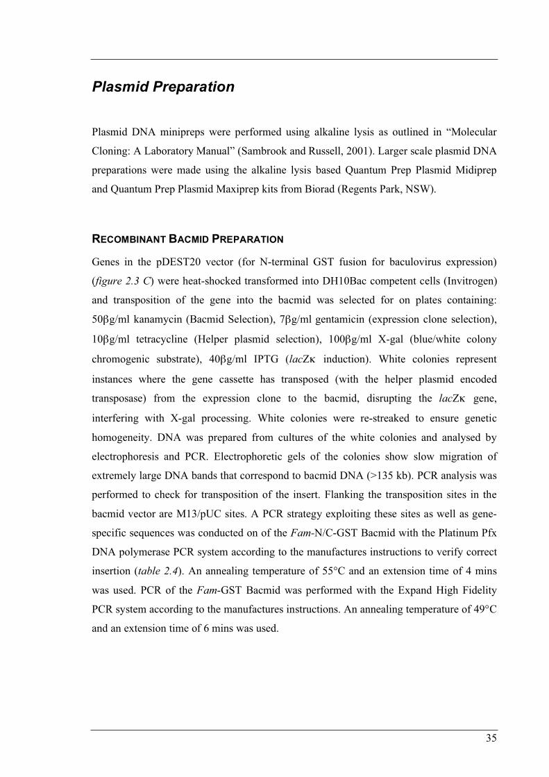
35
Plasmid Preparation
Plasmid DNA minipreps were performed using alkaline lysis as outlined in “Molecular
Cloning: A Laboratory Manual” (Sambrook and Russell, 2001). Larger scale plasmid DNA
preparations were made using the alkaline lysis based Quantum Prep Plasmid Midiprep
and Quantum Prep Plasmid Maxiprep kits from Biorad (Regents Park, NSW).
RECOMBINANT BACMID PREPARATION
Genes in the pDEST20 vector (for N-terminal GST fusion for baculovirus expression)
(figure 2.3 C) were heat-shocked transformed into DH10Bac competent cells (Invitrogen)
and transposition of the gene into the bacmid was selected for on plates containing:
50 g/ml kanamycin (Bacmid Selection), 7 g/ml gentamicin (expression clone selection),
10 g/ml tetracycline (Helper plasmid selection), 100 g/ml X-gal (blue/white colony
chromogenic substrate), 40 g/ml IPTG (lacZ induction). White colonies represent
instances where the gene cassette has transposed (with the helper plasmid encoded
transposase) from the expression clone to the bacmid, disrupting the lacZ gene,
interfering with X-gal processing. White colonies were re-streaked to ensure genetic
homogeneity. DNA was prepared from cultures of the white colonies and analysed by
electrophoresis and PCR. Electrophoretic gels of the colonies show slow migration of
extremely large DNA bands that correspond to bacmid DNA (>135 kb). PCR analysis was
performed to check for transposition of the insert. Flanking the transposition sites in the
bacmid vector are M13/pUC sites. A PCR strategy exploiting these sites as well as gene-
specific sequences was conducted on of the Fam-N/C-GST Bacmid with the Platinum Pfx
DNA polymerase PCR system according to the manufactures instructions to verify correct
insertion (table 2.4). An annealing temperature of 55°C and an extension time of 4 mins
was used. PCR of the Fam-GST Bacmid was performed with the Expand High Fidelity
PCR system according to the manufactures instructions. An annealing temperature of 49°C
and an extension time of 6 mins was used.
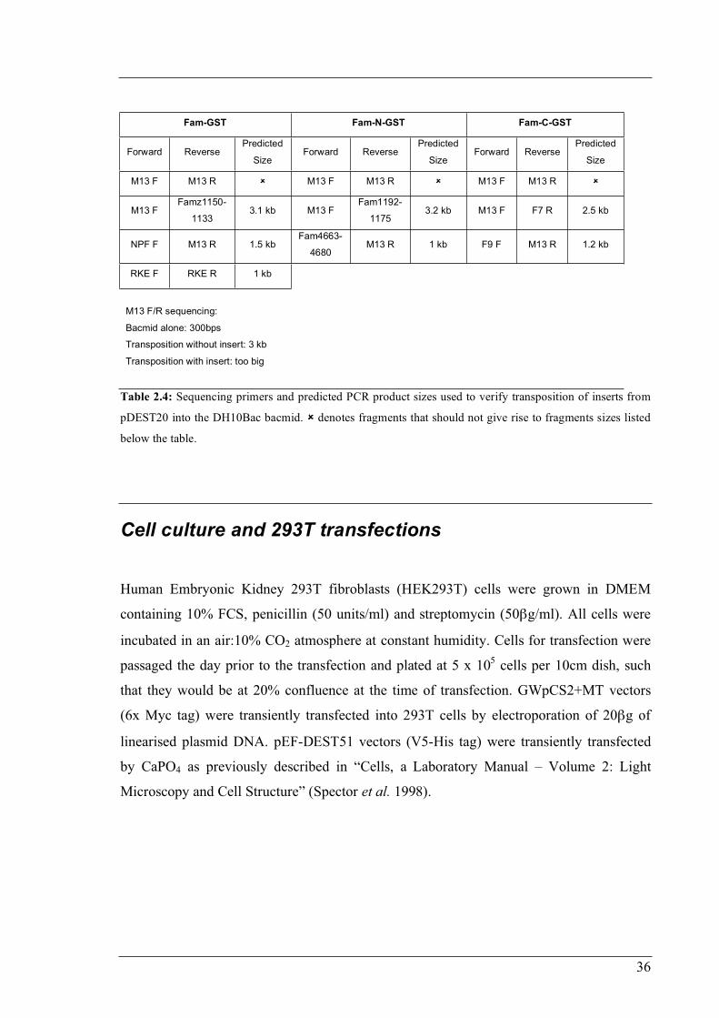
36
Fam-GST Fam-N-GST Fam-C-GST
Forward Reverse Predicted
Size Forward Reverse
Predicted
Size Forward Reverse
Predicted
Size
M13 F M13 R M13 F M13 R M13 F M13 R
M13 F Famz1150-
1133 3.1 kb M13 F
Fam1192-
1175 3.2 kb M13 F F7 R 2.5 kb
NPF F M13 R 1.5 kb Fam4663-
4680 M13 R 1 kb F9 F M13 R 1.2 kb
RKE F RKE R 1 kb
M13 F/R sequencing:
Bacmid alone: 300bps
Transposition without insert: 3 kb
Transposition with insert: too big
Table 2.4: Sequencing primers and predicted PCR product sizes used to verify transposition of inserts from
pDEST20 into the DH10Bac bacmid. denotes fragments that should not give rise to fragments sizes listed
below the table.
Cell culture and 293T transfections
Human Embryonic Kidney 293T fibroblasts (HEK293T) cells were grown in DMEM
containing 10% FCS, penicillin (50 units/ml) and streptomycin (50 g/ml). All cells were
incubated in an air:10% CO2 atmosphere at constant humidity. Cells for transfection were
passaged the day prior to the transfection and plated at 5 x 10⁵ cells per 10cm dish, such
that they would be at 20% confluence at the time of transfection. GWpCS2+MT vectors
(6x Myc tag) were transiently transfected into 293T cells by electroporation of 20 g of
linearised plasmid DNA. pEF-DEST51 vectors (V5-His tag) were transiently transfected
by CaPO4 as previously described in “Cells, a Laboratory Manual – Volume 2: Light
Microscopy and Cell Structure” (Spector et al. 1998).
37
Whole-Mount In Situ Hybridisation
A usp9-specific in situ probe was designed and cloned into the pGEMTeasy vector
(Promega, Madison, WI, USA) as previously described. To produce linear, RNAase-free
cDNA template for in vitro transcription, another PCR reaction was performed using M13
primers to amplify the usp9 sequence as well as flanking vector sequence, which contains
SP6 and T7 transcriptional start sites in opposing orientations. Due to the orientation of the
insert, Antisense RNA was produced with SP6 primers while sense RNA from T7 (table
2.1).
For whole-mount in situ transcript hybridisation, zebrafish embryos were raised at 28ºC
and staged as previously described (Kimmel et al. 1995). 24 hours post-fertilisation (hpf)
up to 5 days post-fertilisation (dpf) embryos were treated with Phenylthiocarbamide (PTC)
to inhibit their pigmentation. A 20mM stock solution in 70% DMSO (1:200 in embryo
media) was applied to embryos whose chorions were punctured with hypodermic needles.
Media and PTC was changed daily. In situ transcript hybridisation was performed as
described by Jowett (1997) using single-stranded RNA probes labelled with digoxigenin-
UTP (Roche, Basel, Switzerland). Photos were taken under Normaski optics under a light
microscope.
mRNA Injections Into Zebrafish Embryos
mRNA for injection into zebrafish embryos was in vitro transcribed using the mRNA
mMessage mMachine kit (Ambion Inc., Austin, TX, USA) as per manufacturer’s
instructions. 1 g of template linearised plasmid (HCRs were linearised with NotI) was
used in each reaction. After precipitation, mRNA was resuspended in 20 l H2O. In all
instances, yields of mRNA were estimated at 500ng/ l. The RNA was then aliquoted into
5 l volumes (to minimise freeze thaw cycles) at dilutions of 1:10 (50ng/ l) and 1:5
(100ng/ l).
38
Pre-prepared injection needles were used to draw up aliquots of mRNA by capillary action
and then loaded on to a gas pump injection apparatus positioned under a light microscope.
With the use of fine forceps, individual 2-cell stage embryos were penetrated first through
the chorion, then the yolk sac, then finally into one of the cells. A small amount of mRNA
was then deposited within the cell and the needle removed. Embryos were then placed into
a Petri dish of embryo media and allowed to develop at 28.5°C for the required amount of
time. After observation, embryos were fixed in PBS 4% formaldehyde.
Protein Processing
BACTERIAL INDUCTION
GST fusions were expressed in the E. coli BL21 (DE3) strain. Pilot studies of induction
and solubility of fusion proteins at 37°C and 25°C were conducted from starter cultures,
diluted 1:100 into 50 ml cultures (luria broth, 50 g/ml amp). Although aiming to induce at
OD 0.6-0.8, due to the unusually slow growth of the HCR-GST cultures, induction was
performed after 2-4hr of growth with [0.2mM] IPTG for 2.5-4hrs. Cells were lysed by
sonication 3x 30 sec in 2mls TTBS with addition of PMSF before, between and after each
interval to a final [1mM]. Lysed cells were then spun at 3000xg for 10 min at 4°C. The
supernatant was removed and the pellet resuspended in 2mls TBS. Uninduced, induced,
soluble and insoluble samples were analysed by SDS-PAGE. Large scale inductions were
conducted at 37°C and 25°C using essentially the same protocol as for the pilot studies
except; 400ml cultures were used, lysis was performed in 20ml TTBS by French Press and
lysed cells were spun 3000xg for 20 min at 4°C.
Uninduced and Induced samples for gel loading were prepared by removing 2mls and 1ml
respectively from the appropriate bacterial culture followed by spinning down a pellet and
removing the supernatant. The pellet was then resuspended in 100 l of 2x protein load
buffer, of which 10 l was loaded. The Soluble and Insoluble samples for gel loading were
prepared from 20 l each of the prepared protein solutions, mixed with 100 l of 2x protein
39
load buffer, of which 10 l was loaded. All samples were boiled at 100°C for 5 minutes
prior to loading.
The pDEST15 vector expresses GST fusions under the T7 promoter. The original bacterial
strain designed for its induced expression is the BL21-SI (Gibco BRL, Invitrogen), which
transcribes a chromosomal copy of T7 RNA polymerase (DE3 lysogen) in response to 0.3
M NaCl instead of IPTG. BL21-SI is an expression host designed for high-level, tightly
regulated expression from the proU, salt-inducible promoter. However, the BL21-SI strain
was discontinued and not available for use at the time of experimentation. The vector can
be expressed in a new strain called BL21-AI (Invitrogen) which is induced with L-
arabinose and repressed by glucose (Invitrogen, 2004), however others have continued to
use this vector with traditional IPTG induction (Evans et al. 2003).
PREPARATION OF MAMMALIAN CELL LYSATES
Cells were harvested and lysed by resuspension in 1-2 pellet volumes of mammalian lysis
buffer (0.5% triton X, 60mM KCl, 5mM MgCl2, 2mM EDTA, 2mM EGTA, 30mM Hepes
pH 7.4, 1mM Na3Vo4, 50mM NaF, 2mM PMSF, 10 g/ml aprotinin), incubation with
gentle rocking at 4°C for 30 mins, spin at 14,000 rpm in a microfuge, and removal of
supernatant lysate. Protein concentration was assayed with Bradford reagent (Biorad).
PREPARATION OF INSECT CELL LYSATES
Frozen pellets of cultures transfected with recombinant bacmid were obtained from Dr.
Sassan Asgari. Cell pellets were resuspended in five volumes of lysis buffer per gram of
cells with one “Complete Mini” protease inhibitor cocktail tablet per 10ml (Roche Applied
Sciences). Insect cell lysis buffer (50mM Tris-HCL (pH 8.5), 5mM 2-mercaptoethaol,
100mM KCL, 1mM PMSF, 1% NP-40 at 4°C). Tubes were inverted for 1 minute to lyse
the cells and then cellular debris removed by centrifugation at 10,000 x g for 10 minutes.
The supernatant containing soluble protein was then transferred to a new tube. The pellet
was resuspended in TTBS to constitute the insoluble fraction.
PROTEIN PURIFICATION
Soluble fractions filtered with a 0.45 m filter and bound to glutathione agarose. Bound
proteins were eluted off the column with reduced glutathione (10 mM pH8.0) in TBS.
40
Samples were then concentrated to 2 mls and buffer exchanged in to binding buffer (50mM
Tris/HCL at pH 8.0, 1 mM EDTA, 0.1% Triton X-100, 0.1% CHAPS). The protein sample
was then concentrated with a Macrosep centrifugal device (Pall Life Sciences, Lane Cove,
NSW) to around 6 mg/ml.
PARTIAL PROTEOLYSIS
Partial proteolysis of FAM-Cys-GST was carried out with Chymotrypsin sequencing
grade, Endoproteinase Glu-C (Protease V8) Trypsin, modified, sequencing grade (Roche
Diagnostics, Mannheim, Germany) in their respective protease specific buffers and
temperatures (See below) with the addition of DTT [1mM]. Reactions were stopped by the
addition of PMSF [5mM] for 15 minutes on ice before addition of load buffer and boiling
prior to gel loading.
Trypsin digestion buffer: TBS pH 8 @ 37°C
V8 protease digestion buffer: 100mM NH4HCO3 pH 7.8 @ 25°C
Chymotrypsin digestion buffer: 100mM Tris-HCl, 10mM CaCl2 pH 7.8 @ 25°C
PROTEIN PRECIPITATION
Scaled-up partial proteolysis reactions required the proteins to be precipitated in order to
achieve a suitable volume for loading onto a gel. This was achieved by the addition of
TCA (trichloroacetic acid) to a final concentration of 10%, followed by incubation of the
sample on ice for 30 minutes to allow the protein to precipitate. Samples were then
centrifuged at 14,000 rpm in a microfuge for 15 minutes, washed in an equal volume of
100% EtOH centrifuged for a further 15 minutes and air-dried. Samples then had load
buffer added and were boiled at 100% for 5 min.
PROTEIN ELECTROPHORESIS
Proteins samples and prestained RPN800 molecular weight size markers (Amersham
Biosciences) or Benchmark molecular weight size markers (Invitrogen) were subjected to
SDS-PAGE at 40mA on either 7.5% or 12.5% polyacrylamide resolving gels poured and
41
electrophoresed using the Miniprotean III system (Biorad) or large gels at 20mA on the SE
600 series electrophoresis unit (Hoefer).
Protein Detection
WESTERN BLOTTING
Proteins were electrophoretically transferred to nitrocellulose membrane (Biotrace NT;
Pall Corporation, NY, USA) using a semi-dry transfer apparatus (Biorad) for 1 hour at
80mA. Membranes were immunoblotted with monoclonal or polyclonal antibodies as
indicated in figure legends. Primary antibodies were detected with HRP conjugated
secondary antibodies and visualised by ECL (Enhanced Chemi-Luminescence).
Chemiluminescent bands were detected with SuperRX Fuji Medical X-ray Film (Fuji
Photo Film, Tokyo, Japan). Chemiflorescent bands were detected using a Typhoon 8600
variable mode imager (Amersham Biosciences).
SILVER STAIN
Polyacrylamide gels (0.75mm) were first fixed in fixative solution (10% Acetic Acid, 40%
EtOH) for 30 minutes to overnight. Gels were incubated for 30 minutes to overnight in
incubation solution (0.5M Sodium acetate (anhydrous), 8mM Sodium thiosulphate, 0.13%
Glutaraldehyde, 30% EtOH (added after the other reagents were dissolved)). Gels were
then washed three times for 5 minutes in MQ water. Gels then were incubated in silver
solution (0.1% silver nitrate, 0.004% formaldehyde) for 40 minutes. Gels were then briefly
rinsed in water and then developed in developing solution (2.5% sodium carbonate,
0.004% formaldehyde) for up to 15 minutes. Staining was stopped in stop solution (1.46%
EDTA.Na2.2H2O) for 5-10 minutes. Incubation, silver and developing solutions were made
just prior to use.
42
COLLOIDAL COOMASSIE STAINING
After electrophoresis, gels were rinsed in water and then washed in water for a further 10
minutes. The gels were then fixed with 20% (w/v) Trichloroacetic Acid (TCA) overnight at
room temperature to fix the proteins. After removal of the fixing solution, gels were
covered with the colloidal Coomassie stain (0.1% (w/v) Coomassie blue in 20% (w/v)
TCA) for 3 to 4 hours with gentle agitation. After aspiration of the stain, the acid was
washed out of the gel with several changes of water over ~2 hours to enhance contrast.
Gels stored in 5% acetic acid.
Mass Spectrometry
The scaled-up digest gels containing the partial proteolysis products were submitted to the
Hanson Institute’s Protein Laboratory for mass spectrometry analysis at the Institute of
Medical and Veterinary Science. The researchers there were responsible for the selection
and excision of the correct bands from the gels guided by a provided scanned printout of
the bands, as well as making decisions on whether particular bands were suitable for
analysis. For instance, when excising bands from the chymotrypsin 1:500 lane (figure 4.13
C), care was taken to avoid cutting across a smear that runs vertically down the lane. Bands
of interest were excised from the gel and the protein was treated with iodoacetamide (S-
carbamidomethylation) prior to its full digestion with trypsin. The resultant peptides from
bands were resolved through a short C18 reverse phase silica column into the Micromass
quadrupole/time-of-flight (QTOF2) mass spectrometer for matrix-assisted laser
desorption/ionisation (MALDI) peptide mapping. The eluted peaks were analysed and the
monoisotopic (C12, H1, N14, O16, and S32) forms of the component ions and their charge
states were measured. Spectra were recorded as abundance versus mass/charge ratio
(Thomson, Th), and were calibrated against a standard solution including glutathione,
apomyoglobin and [Glu1]-Fibrinopeptide B. Further calibration was achieved through
identifying a trypsin autolysis product, which was used as a lock mass. The raw data from
each band was then transformed with MAXENT-3 which resolves the observed masses
back to a single charge state corresponding to the mass of the peptide plus one proton for
ease of analysis.
43
Computer Analyses
ASSEMBLY OF USP9 SEQUENCE
Predicted zebrafish Fam sequence (usp9) was obtained by a BLAST search of the Zv4
assembly of zebrafish genomic sequence at the Sanger Institute
(http://www.ensembl.org/Multi/blastview?species=Danio_rerio). Where amino acid
residues differed from the pufferfish and other vertebrate orthologues, the sequence was
verified by examining several electrophoretograms available from the Ensembl Trace
Server (http://trace.ensembl.org/perl/ssahaview?server=danio_rerio).
To define the putative coding sequence of zebrafish Fam (usp9, Acc. no. DQ086492), a
predicted mRNA sequence from the Zv4 assembly of zebrafish genomic sequence by the
Sanger Institute (http://www.ensembl.org/Multi/blastview?species=Danio_rer-io) was
aligned with the known Fam cDNA sequences from: Mouse Famx (DQ086491) and Famy
(Acc. no. AJ307017) and Humans Usp9x (Acc. no. X98296) and Usp9y (Acc. no.
Y13618), and with predicted mRNA sequences from Fugu rubripes (Sanger centre
SINFRUG00000128324), Gallus gallus (Sanger centre ENSGALG00000016236),
Xenopus tropicalis (Sanger centre ENSXETG000000154-89), Canis familiaris (Sanger
centre ENSCAFG00000014207) and Rattus norvegicus (Sanger centre
ENSRNOG00000003261). When positions of variation in the predicted zebrafish sequence
relative to both the Fugu rubripes sequence and the other sequences were identified, or
where, (in one instance), an intron-exon boundary was not conserved relative to mouse
Fam, the variant sequence was checked by examination of electrophoretograms from the
zebrafish genome project (Ensembl Trace Server;
http://trace.ensembl.org/perl/ssahaview?server=danio_rerio). In this way, all uncertainties
in the usp9 open reading frame were resolved.
PHYLOGENETIC ANALYSIS
Phylogenetic analysis was performed using the BioManager facility provided by the
Australian National Genomic Information Service (ANGIS, http://www.angis.org.au/). The
protein multiple sequence alignment shown in figure 3.4 was constructed using Gendoc
44
Pairwise alignment (Nicholas, 1997) with default parameters, and was prepared for
publication using the GenDoc program (Nicholas, 1997). The phylogenetic tree was
constructed from known and predicted FAM orthologue amino acid sequences using
Protdist. The Protdist distance matrix was generated under the Dayhoff PAM matrix
method of amino acid substitution, and a phylogenetic tree subsequently constructed using
the ‘Neighbour Joining Method’ (figure 3.5). Bootstrap analysis was conducted using
Seqboot (Felsenstein, 1989) to produce 1,000 resampled datasets that were then analysed
with Protdist (Felsenstein, 1989). Bootstrap values were finally generated using the
Consense program (Felsenstein, 1989), and are shown in figure 3.5.
STATISTICAL ANALYSIS OF INJECTION PHENOTYPES
Embryos injected with FAM HCR mRNA were scored according to the presence or
absence of several observed phenotypes. The data was then processed by cross-tabulating
embryo phenotype against the type of injected HCR mRNA. Included in the cross-
tabulation were the negative controls of eGFP and H2O. The data was then assessed for
significant differences with a Pearson Chi-Square test to obtain a p-value. All statistical
analyses were performed with SPSS version 10.
FAM PEPTIDE MASS ANALYSIS
The raw data from the mass spectrometry analysis of each band was transformed with
MAXENT-3 which resolves observed masses back to a single charge state (monoisotpic
masses [M+H]+) corresponding to the mass of the peptide plus one proton for ease of
analysis. This data was used to query the FAM-Cys-GST sequence in Findmod
(http://ca.expasy.org/cgi-bin/findmod_form.pl) to find peptides that matched the FAM
sequence. Search parameters included the likely carbamidomethyl modification to cysteine
(from reaction of the band with iodoacetamide prior to full digestion with trypsin) and
oxidation of methionine. Matching peptides were found with an error of ±50 ppm (parts
per million) and allowed for up to 2 missed cleavages (Appendix A).
The occurrence of the matched peptides was manually plotted against a FAM-Cys-GST
sequence that had been theoretically digested with trypsin (http://ca.expasy.org/cgi-
bin/peptide-mass.pl). This table (Appendix B) shows the potential carbamidomethyl
45
modification to cysteine and oxidation of methionine modifications but does not include
peptides < 500 Da.
SECONDARY STRUCTURE PREDICTION
The primary sequence of both FAM (SSSRF+) and FAM-GST (SSSRF+) (including the
additional residues in the linker between FAM and GST, and vector encoded C-terminal
residues before the vector stop sequence) were analysed by the JUFO server (Meiler et al.
2002) found at www.jens-meiler.de. This server offers a protein secondary structure
prediction from its primary sequence only. A neural network was trained with an amino
acid property profile and the position based scoring matrix of a blast run (as used by Jones
DT, 1999). It achieves a three state prediction accuracy of about 75%.
INTRINSIC PROTEIN DISORDER, DOMAIN & GLOBULARITY PREDICTION
The FAM-GST sequence was analysed with GlobPlot version 2.2 (http://globplot.embl.de/)
using default settings and the Russell/Linding definition (Linding et al. 2003).
STATISTICAL ANALYSIS OF PROTEIN SEQUENCES
The primary sequence of FAM (SSSRF+) was analysed by the ISREC-Server (Version of
April 11, 1996, http://www.isrec.isb-sib.ch/software/SAPS_form.html) to give a SAPS
(Statistical Analysis of Protein Sequences) report. SAPS evaluates by various statistical
criteria, a wide variety of protein sequence properties including composition, charge
distribution, distribution of other amino acid types, repetitive structures, mulitplets,
periodicity and spacing (Brendel et al. 1992).
FOLD PREDICTION
Predicted domain sequences were analysed for potential structural similarities to other
known folds by the LOOPP (Learning, Observing and Outputting Protein Patterns) server
(version 3.0, http://cbsuapps.tc.cornell.edu/loopp.aspx). LOOPP is a fold recognition
program that collates straightforward sequence alignments, sequence profiles, threading,
secondary structure and exposed surface area predictions to merge them into a single score,

46
and generate atomic coordinates based on an alignment into a homologue template
structure (Teodorescu et al. 2004).