Embed Size (px)
Citation preview
Learning physics confers pose-sensitivity instructure-based virtual screening
Pawel GniewekAtomwise Inc
San Francisco, CA, [email protected]
Bradley WorleyAtomwise Inc
San Francisco, CA, [email protected]
Kate StaffordAtomwise Inc
San Francisco, CA, [email protected]
Henry van den BedemAtomwise Inc
University of California, San FranciscoSan Francisco, CA, [email protected]
Brandon AndersonAtomwise Inc
San Francisco, CA, [email protected]
Abstract
In drug discovery, structure-based virtual high-throughput screening (vHTS) cam-paigns aim to identify bioactive ligands or “hits” for therapeutic protein targetsfrom docked poses at specific binding sites. However, while generally successful atthis task, many deep learning methods are known to be insensitive to protein-ligandinteractions, decreasing the reliability of hit detection and hindering discovery atnovel binding sites. Here, we overcome this limitation by introducing a class ofmodels with two key features: 1) we condition bioactivity on pose quality score,and 2) we present poor poses of true binders to the model as negative examples.The conditioning forces the model to learn details of physical interactions. Weevaluate these models on a new benchmark designed to detect pose-sensitivity.
1 Introduction
Vast, make-on-demand chemical libraries like ENAMINE or Mcule have transformed the scaleof pharmaceutical, structure-based virtual high-throughput screening (vHTS) campaigns [1]. Toidentify a “hit” from a library of candidate molecules, structure-based vHTS methods predict bindingaffinity between a protein and a ligand from their docked, bound complex, thereby assuming thatexperimentally observed affinities correlate with protein-ligand interactions. Conventional methodsuse empirical, physics-based approaches, which attempt to calculate the binding free energy ofcomplex formation. By contrast newer deep learning (DL) approaches are trained on large data setsusing implicit features to predict activity. These statistical models can outperform physics-basedapproaches in retrospective tests for predicting activity.
Early structure-based DL methods in vHTS centered on Convolutional Neural Networks (CNNs),representing protein-ligand structures by a 3D voxel grid to predict activity [2–5]. Although generallyeffective [6], a drawback of CNNs is that they are not rotationally invariant, and require moreparameters than alternative representations. Consequently, Graph Convolutional Networks (GCNs) [7,8], and their position-based generalizations [9–16], have gained popularity by capitalizing on physicalsymmetries and using data more efficiently.
Recent studies have suggested that the performance of structure-based machine learning methodsis partly driven by proteochemometric (PCM)-like features [5, 17, 18]. Rather than responding tospecific interactions between the ligand and the binding site, the model learns independent ligand and
Machine Learning for Structural Biology Workshop, NeurIPS 2021.
arX
iv:2
110.
1545
9v3
[q-
bio.
QM
] 2
Dec
202
1
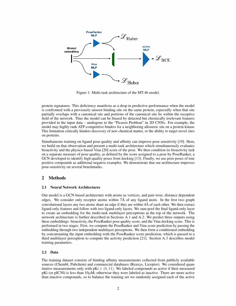
Figure 1: Multi-task architecture of the MT-4b model.
protein signatures. This deficiency manifests as a drop in predictive performance when the modelis confronted with a previously unseen binding site on the same protein, especially when that sitepartially overlaps with a canonical site and portions of the canonical site lie within the receptivefield of the network. Thus the model can be biased by detected but chemically irrelevant featuresprovided in the input data – analogous to the “Picasso Problem” in 2D CNNs. For example, themodel may highly rank ATP-competitive binders for a neighboring allosteric site on a protein kinase.This limitation critically hinders discovery of new chemical matter, or the ability to target novel siteson proteins.
Simultaneous training on ligand pose quality and affinity can improve pose sensitivity [19]. Here,we build on that observation and present a multi-task architecture which simultaneously evaluatesbioactivity and the physics-based Vina [20] score of the pose. We then condition its bioactivity taskon a separate measure of pose quality, as defined by the score assigned to a pose by PoseRanker, aGCN developed to identify high-quality poses from docking [13]. Finally, we use poor poses of truepositive compounds as additional negative examples. We demonstrate that our architecture improvespose-sensitivity on several benchmarks.
2 Methods
2.1 Neural Network Architectures
Our model is a GCN-based architecture with atoms as vertices, and pair-wise, distance dependentedges. We consider only receptor atoms within 7Å of any ligand atom. In the first two graphconvolutional layers any two atoms share an edge if they are within 4Å of each other. We then extractligand-only features and follow with two ligand-only layers. We sum-pool the final ligand-only layerto create an embedding for the multi-task multilayer perceptrons at the top of the network. Thenetwork architecture is further described in Sections A.1 and A.2. We predict three outputs usingthese embeddings: bioactivity, the PoseRanker pose quality score, and the Vina docking score. This isperformed in two stages. First, we compute the PoseRanker and Vina score prediction by passing theembedding through two independent multilayer perceptrons. We then form a conditioned embeddingby concatenating the input embedding with the PoseRanker score prediction, which is passed to athird multilayer perceptron to compute the activity prediction [21]. Section A.3 describes modeltraining parameters.
2.2 Data
The training dataset consists of binding affinity measurements collected from publicly availablesources (Chembl, Pubchem) and commercial databases (Reaxys, Liceptor). We considered quan-titative measurements only with pKi ∈ (0, 11). We labeled compounds as active if their measuredpKi (or pIC50) is less than 10µM; otherwise they were labeled as inactive. There are more activethan inactive compounds, so to balance the training set we randomly assigned each of the active
2
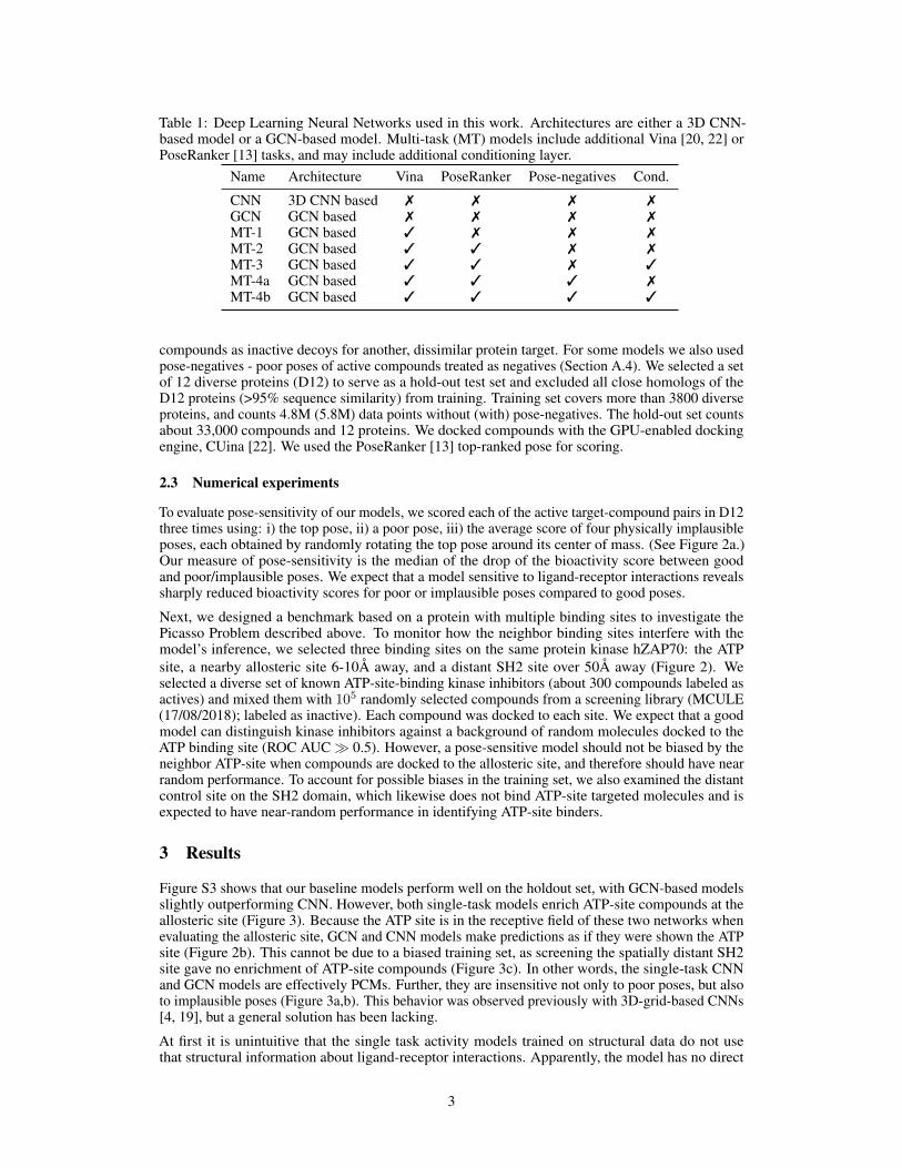
Table 1: Deep Learning Neural Networks used in this work. Architectures are either a 3D CNN-based model or a GCN-based model. Multi-task (MT) models include additional Vina [20, 22] orPoseRanker [13] tasks, and may include additional conditioning layer.
Name Architecture Vina PoseRanker Pose-negatives Cond.
CNN 3D CNN based 7 7 7 7GCN GCN based 7 7 7 7MT-1 GCN based 3 7 7 7MT-2 GCN based 3 3 7 7MT-3 GCN based 3 3 7 3MT-4a GCN based 3 3 3 7MT-4b GCN based 3 3 3 3
compounds as inactive decoys for another, dissimilar protein target. For some models we also usedpose-negatives - poor poses of active compounds treated as negatives (Section A.4). We selected a setof 12 diverse proteins (D12) to serve as a hold-out test set and excluded all close homologs of theD12 proteins (>95% sequence similarity) from training. Training set covers more than 3800 diverseproteins, and counts 4.8M (5.8M) data points without (with) pose-negatives. The hold-out set countsabout 33,000 compounds and 12 proteins. We docked compounds with the GPU-enabled dockingengine, CUina [22]. We used the PoseRanker [13] top-ranked pose for scoring.
2.3 Numerical experiments
To evaluate pose-sensitivity of our models, we scored each of the active target-compound pairs in D12three times using: i) the top pose, ii) a poor pose, iii) the average score of four physically implausibleposes, each obtained by randomly rotating the top pose around its center of mass. (See Figure 2a.)Our measure of pose-sensitivity is the median of the drop of the bioactivity score between goodand poor/implausible poses. We expect that a model sensitive to ligand-receptor interactions revealssharply reduced bioactivity scores for poor or implausible poses compared to good poses.
Next, we designed a benchmark based on a protein with multiple binding sites to investigate thePicasso Problem described above. To monitor how the neighbor binding sites interfere with themodel’s inference, we selected three binding sites on the same protein kinase hZAP70: the ATPsite, a nearby allosteric site 6-10Å away, and a distant SH2 site over 50Å away (Figure 2). Weselected a diverse set of known ATP-site-binding kinase inhibitors (about 300 compounds labeled asactives) and mixed them with 105 randomly selected compounds from a screening library (MCULE(17/08/2018); labeled as inactive). Each compound was docked to each site. We expect that a goodmodel can distinguish kinase inhibitors against a background of random molecules docked to theATP binding site (ROC AUC� 0.5). However, a pose-sensitive model should not be biased by theneighbor ATP-site when compounds are docked to the allosteric site, and therefore should have nearrandom performance. To account for possible biases in the training set, we also examined the distantcontrol site on the SH2 domain, which likewise does not bind ATP-site targeted molecules and isexpected to have near-random performance in identifying ATP-site binders.
3 Results
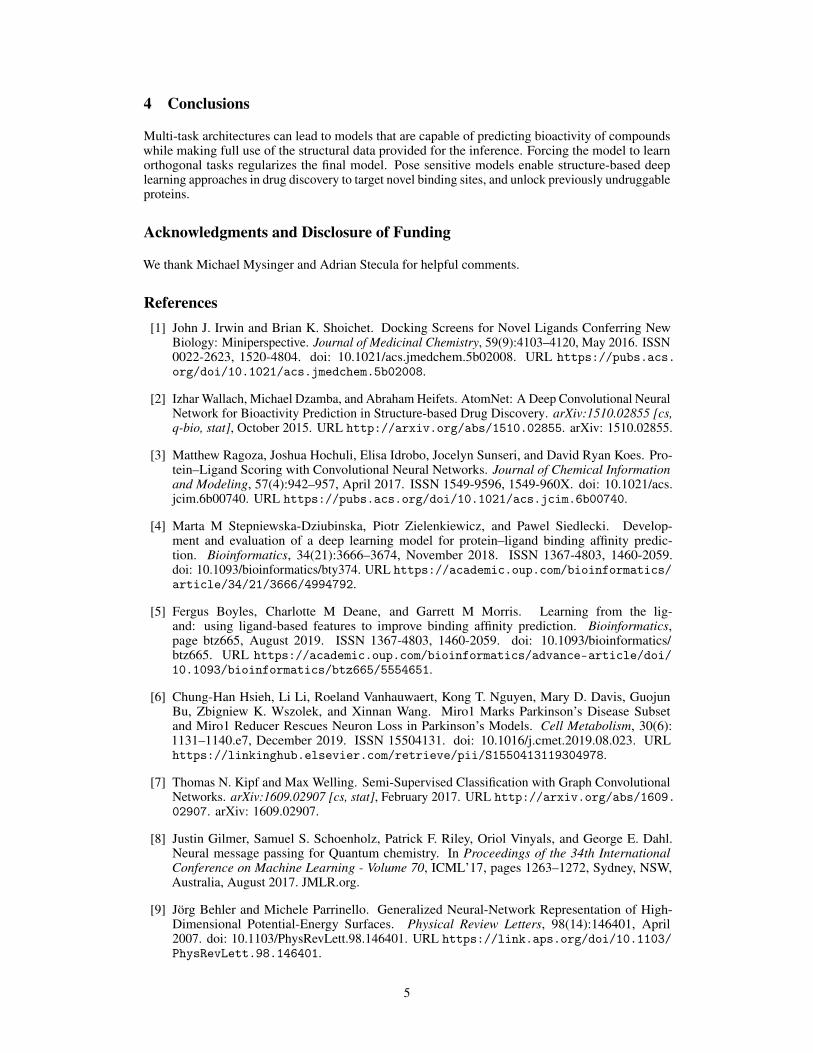
Figure S3 shows that our baseline models perform well on the holdout set, with GCN-based modelsslightly outperforming CNN. However, both single-task models enrich ATP-site compounds at theallosteric site (Figure 3). Because the ATP site is in the receptive field of these two networks whenevaluating the allosteric site, GCN and CNN models make predictions as if they were shown the ATPsite (Figure 2b). This cannot be due to a biased training set, as screening the spatially distant SH2site gave no enrichment of ATP-site compounds (Figure 3c). In other words, the single-task CNNand GCN models are effectively PCMs. Further, they are insensitive not only to poor poses, but alsoto implausible poses (Figure 3a,b). This behavior was observed previously with 3D-grid-based CNNs[4, 19], but a general solution has been lacking.
At first it is unintuitive that the single task activity models trained on structural data do not usethat structural information about ligand-receptor interactions. Apparently, the model has no direct
3
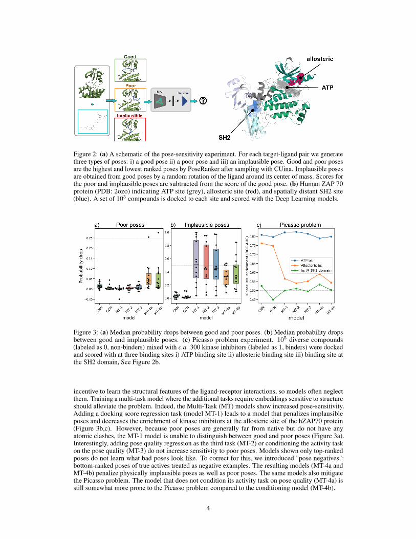
Figure 2: (a) A schematic of the pose-sensitivity experiment. For each target-ligand pair we generatethree types of poses: i) a good pose ii) a poor pose and iii) an implausible pose. Good and poor posesare the highest and lowest ranked poses by PoseRanker after sampling with CUina. Implausible posesare obtained from good poses by a random rotation of the ligand around its center of mass. Scores forthe poor and implausible poses are subtracted from the score of the good pose. (b) Human ZAP 70protein (PDB: 2ozo) indicating ATP site (grey), allosteric site (red), and spatially distant SH2 site(blue). A set of 105 compounds is docked to each site and scored with the Deep Learning models.
Figure 3: (a) Median probability drops between good and poor poses. (b) Median probability dropsbetween good and implausible poses. (c) Picasso problem experiment. 105 diverse compounds(labeled as 0, non-binders) mixed with c.a. 300 kinase inhibitors (labeled as 1, binders) were dockedand scored with at three binding sites i) ATP binding site ii) allosteric binding site iii) binding site atthe SH2 domain, See Figure 2b.
incentive to learn the structural features of the ligand-receptor interactions, so models often neglectthem. Training a multi-task model where the additional tasks require embeddings sensitive to structureshould alleviate the problem. Indeed, the Multi-Task (MT) models show increased pose-sensitivity.Adding a docking score regression task (model MT-1) leads to a model that penalizes implausibleposes and decreases the enrichment of kinase inhibitors at the allosteric site of the hZAP70 protein(Figure 3b,c). However, because poor poses are generally far from native but do not have anyatomic clashes, the MT-1 model is unable to distinguish between good and poor poses (Figure 3a).Interestingly, adding pose quality regression as the third task (MT-2) or conditioning the activity taskon the pose quality (MT-3) do not increase sensitivity to poor poses. Models shown only top-rankedposes do not learn what bad poses look like. To correct for this, we introduced "pose negatives":bottom-ranked poses of true actives treated as negative examples. The resulting models (MT-4a andMT-4b) penalize physically implausible poses as well as poor poses. The same models also mitigatethe Picasso problem. The model that does not condition its activity task on pose quality (MT-4a) isstill somewhat more prone to the Picasso problem compared to the conditioning model (MT-4b).
4
4 Conclusions
Multi-task architectures can lead to models that are capable of predicting bioactivity of compoundswhile making full use of the structural data provided for the inference. Forcing the model to learnorthogonal tasks regularizes the final model. Pose sensitive models enable structure-based deeplearning approaches in drug discovery to target novel binding sites, and unlock previously undruggableproteins.
Acknowledgments and Disclosure of Funding
We thank Michael Mysinger and Adrian Stecula for helpful comments.
References[1] John J. Irwin and Brian K. Shoichet. Docking Screens for Novel Ligands Conferring New
Biology: Miniperspective. Journal of Medicinal Chemistry, 59(9):4103–4120, May 2016. ISSN0022-2623, 1520-4804. doi: 10.1021/acs.jmedchem.5b02008. URL https://pubs.acs.org/doi/10.1021/acs.jmedchem.5b02008.
[2] Izhar Wallach, Michael Dzamba, and Abraham Heifets. AtomNet: A Deep Convolutional NeuralNetwork for Bioactivity Prediction in Structure-based Drug Discovery. arXiv:1510.02855 [cs,q-bio, stat], October 2015. URL http://arxiv.org/abs/1510.02855. arXiv: 1510.02855.
[3] Matthew Ragoza, Joshua Hochuli, Elisa Idrobo, Jocelyn Sunseri, and David Ryan Koes. Pro-tein–Ligand Scoring with Convolutional Neural Networks. Journal of Chemical Informationand Modeling, 57(4):942–957, April 2017. ISSN 1549-9596, 1549-960X. doi: 10.1021/acs.jcim.6b00740. URL https://pubs.acs.org/doi/10.1021/acs.jcim.6b00740.
[4] Marta M Stepniewska-Dziubinska, Piotr Zielenkiewicz, and Pawel Siedlecki. Develop-ment and evaluation of a deep learning model for protein–ligand binding affinity predic-tion. Bioinformatics, 34(21):3666–3674, November 2018. ISSN 1367-4803, 1460-2059.doi: 10.1093/bioinformatics/bty374. URL https://academic.oup.com/bioinformatics/article/34/21/3666/4994792.
[5] Fergus Boyles, Charlotte M Deane, and Garrett M Morris. Learning from the lig-and: using ligand-based features to improve binding affinity prediction. Bioinformatics,page btz665, August 2019. ISSN 1367-4803, 1460-2059. doi: 10.1093/bioinformatics/btz665. URL https://academic.oup.com/bioinformatics/advance-article/doi/10.1093/bioinformatics/btz665/5554651.
[6] Chung-Han Hsieh, Li Li, Roeland Vanhauwaert, Kong T. Nguyen, Mary D. Davis, GuojunBu, Zbigniew K. Wszolek, and Xinnan Wang. Miro1 Marks Parkinson’s Disease Subsetand Miro1 Reducer Rescues Neuron Loss in Parkinson’s Models. Cell Metabolism, 30(6):1131–1140.e7, December 2019. ISSN 15504131. doi: 10.1016/j.cmet.2019.08.023. URLhttps://linkinghub.elsevier.com/retrieve/pii/S1550413119304978.
[7] Thomas N. Kipf and Max Welling. Semi-Supervised Classification with Graph ConvolutionalNetworks. arXiv:1609.02907 [cs, stat], February 2017. URL http://arxiv.org/abs/1609.02907. arXiv: 1609.02907.
[8] Justin Gilmer, Samuel S. Schoenholz, Patrick F. Riley, Oriol Vinyals, and George E. Dahl.Neural message passing for Quantum chemistry. In Proceedings of the 34th InternationalConference on Machine Learning - Volume 70, ICML’17, pages 1263–1272, Sydney, NSW,Australia, August 2017. JMLR.org.
[9] Jörg Behler and Michele Parrinello. Generalized Neural-Network Representation of High-Dimensional Potential-Energy Surfaces. Physical Review Letters, 98(14):146401, April2007. doi: 10.1103/PhysRevLett.98.146401. URL https://link.aps.org/doi/10.1103/PhysRevLett.98.146401.
5
[10] Kristof T. Schütt, Pieter-Jan Kindermans, Huziel E. Sauceda, Stefan Chmiela, AlexandreTkatchenko, and Klaus-Robert Müller. SchNet: A continuous-filter convolutional neuralnetwork for modeling quantum interactions. arXiv:1706.08566 [physics, stat], December 2017.URL http://arxiv.org/abs/1706.08566. arXiv: 1706.08566.
[11] Evan N. Feinberg, Debnil Sur, Zhenqin Wu, Brooke E. Husic, Huanghao Mai, Yang Li, SaisaiSun, Jianyi Yang, Bharath Ramsundar, and Vijay S. Pande. PotentialNet for Molecular PropertyPrediction. ACS Central Science, 4(11):1520–1530, November 2018. ISSN 2374-7943, 2374-7951. doi: 10.1021/acscentsci.8b00507. URL https://pubs.acs.org/doi/10.1021/acscentsci.8b00507.
[12] Jaechang Lim, Seongok Ryu, Kyubyong Park, Yo Joong Choe, Jiyeon Ham, and Woo YounKim. Predicting Drug–Target Interaction Using a Novel Graph Neural Network with 3DStructure-Embedded Graph Representation. Journal of Chemical Information and Modeling, 59(9):3981–3988, September 2019. ISSN 1549-9596, 1549-960X. doi: 10.1021/acs.jcim.9b00387.URL https://pubs.acs.org/doi/10.1021/acs.jcim.9b00387.
[13] Kate Stafford, Brandon M. Anderson, Jon Sorenson, and Henry van den Bedem. AtomNetPoseRanker: Enriching Ligand Pose Quality for Dynamic Proteins in Virtual High ThroughputScreens. September 2021. doi: 10.33774/chemrxiv-2021-t6xkj. URL https://chemrxiv.org/engage/chemrxiv/article-details/614b905e39ef6a1c36268003.
[14] Nathaniel Thomas, Tess Smidt, Steven Kearnes, Lusann Yang, Li Li, Kai Kohlhoff, and PatrickRiley. Tensor field networks: Rotation- and translation-equivariant neural networks for 3D pointclouds. arXiv:1802.08219 [cs], May 2018. URL http://arxiv.org/abs/1802.08219.arXiv: 1802.08219.
[15] Brandon Anderson, Truong Son Hy, and Risi Kondor. Cormorant: Covariant Molecu-lar Neural Networks. In Advances in Neural Information Processing Systems, volume 32.Curran Associates, Inc., 2019. URL https://papers.nips.cc/paper/2019/hash/03573b32b2746e6e8ca98b9123f2249b-Abstract.html.
[16] Raphael J. L. Townshend, Martin Vögele, Patricia Suriana, Alexander Derry, Alexander Powers,Yianni Laloudakis, Sidhika Balachandar, Bowen Jing, Brandon Anderson, Stephan Eismann,Risi Kondor, Russ B. Altman, and Ron O. Dror. ATOM3D: Tasks On Molecules in ThreeDimensions. arXiv:2012.04035 [physics, q-bio], June 2021. URL http://arxiv.org/abs/2012.04035. arXiv: 2012.04035.
[17] Jochen Sieg, Florian Flachsenberg, and Matthias Rarey. In Need of Bias Control: EvaluatingChemical Data for Machine Learning in Structure-Based Virtual Screening. Journal of Chem-ical Information and Modeling, 59(3):947–961, March 2019. ISSN 1549-9596, 1549-960X.doi: 10.1021/acs.jcim.8b00712. URL https://pubs.acs.org/doi/10.1021/acs.jcim.8b00712.
[18] Lieyang Chen, Anthony Cruz, Steven Ramsey, Callum J. Dickson, Jose S. Duca, ViktorHornak, David R. Koes, and Tom Kurtzman. Hidden bias in the DUD-E dataset leads tomisleading performance of deep learning in structure-based virtual screening. PLOS ONE,14(8):e0220113, August 2019. ISSN 1932-6203. doi: 10.1371/journal.pone.0220113. URLhttps://dx.plos.org/10.1371/journal.pone.0220113.
[19] Paul G. Francoeur, Tomohide Masuda, Jocelyn Sunseri, Andrew Jia, Richard B. Iovanisci,Ian Snyder, and David R. Koes. Three-Dimensional Convolutional Neural Networks and aCross-Docked Data Set for Structure-Based Drug Design. Journal of Chemical Information andModeling, 60(9):4200–4215, September 2020. ISSN 1549-9596, 1549-960X. doi: 10.1021/acs.jcim.0c00411. URL https://pubs.acs.org/doi/10.1021/acs.jcim.0c00411.
[20] Oleg Trott and Arthur J. Olson. AutoDock Vina: Improving the speed and accuracy ofdocking with a new scoring function, efficient optimization, and multithreading. Journalof Computational Chemistry, 31(2):455–461, 2010. ISSN 1096-987X. doi: https://doi.org/10.1002/jcc.21334. URL https://onlinelibrary.wiley.com/doi/abs/10.1002/jcc.21334. _eprint: https://onlinelibrary.wiley.com/doi/pdf/10.1002/jcc.21334.
6
[21] Mingsheng Long, Zhangjie Cao, Jianmin Wang, and Michael I. Jordan. Conditional AdversarialDomain Adaptation. arXiv:1705.10667 [cs], December 2018. URL http://arxiv.org/abs/1705.10667. arXiv: 1705.10667.
[22] Adrian Morrison, Greg Friedland, and Izhar Wallach. CUina: An Efficient GPU Im-plementation of AutoDock Vina. August 2020. URL https://blog.atomwise.com/efficient-gpu-implementation-of-autodock-vina.
A Appendix
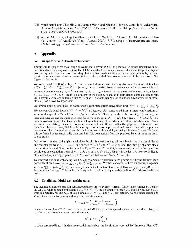
A.1 Graph Neural Network architecture
Throughout the paper we use a graph convolutional network (GCN) to generate the embeddings used in ourconditional multi-tasking architecture. Our GCN takes the three dimensional coordinates of the protein-ligandpose, along with a one-hot atom encoding that simultaneously identifies element type, protein/ligand, andhybridization state. We define our connectivity purely by radial functions without use of chemical bonds. SeeFigure S1 for details.
We use a radial cutoff Rlc at layer l to define a radial graph, with the neighborhood for atom i defined as
N (i) = {j : dij < Rc}, where dij = ‖ri − rj‖ is the pairwise distance between atoms i and j. At each layer lwe have a feature vector f l
i ∈ RNlf at atom i ∈ SP/L/P+L, where N l
f is the number of features at layer l, andSP , SL, SP+L = SP ∪ SL are the set of atoms in the protein, ligand, or protein-ligand complex respectively.Our network can be configured so that P , L, or P + L atoms can be used as either source atoms (i) or targetatoms (j) on a layer-by-layer basis.
Our graph convolutional block is based upon a continuous filter convolution [10], fconvi ←
∑j W
l(dij)flj .
We use convolutional kernels W l(r) =∑Nl
bn wl
nj0(rz0n/Rlc) constructed from a linear combinations of
zeroth-order spherical Bessel functions j0(x) = sinx/x. Here z0n is the n-th zero of j0(x), and wln are
learnable weights, and the number of basis functions is chosen as N lb = dRl
c/δle, where δl = 0.15625Å. Thisparametrization ensures that the convolutional kernels vanish at the edge of an internal neighborhood. Sincewe are not calculating forces, we do not need a smooth cutoff here. After the graph convolution step, weinclude a Linear − LeakyReLU − Linear layer. We do not apply a residual connection at the output of aconvolution block. Instead, each convolutional layer takes as input all layers using a bottleneck layer. We foundthis performed better empirically than standard skip connections from the previous layer of the same set ofsource atoms.
Our network has five total graph convolutional blocks. In the first two graph conv blocks, we consider all ligandand receptor atoms so i, j ∈ SP+L, and choose Rc = 5Å and N l
F = 64 filters. The third graph conv block,the cutoff radius and filters are increased to Rc = 7Å and N l
F = 128, however only atoms in the ligand areconsidered as destination atoms (i. e., i ∈ SP+L, but j ∈ SL only). Finally, in the last two layers only ligandatom embeddings are aggregated (i, j ∈ SL) with a cutoff Rc = 7Å and N l
F = 128.
To construct our final embedding, we first apply a readout operation to the protein and ligand features inde-pendently at each layer: zlP =
∑i∈SP
f li , zlL =
∑i∈SL
f li . We then concatenate these embeddings together,
zread =⊕1
l=0 zlP ⊕
⊕5l=0 z
lL, and finally construct z from two iterations of Dropout0.2 − LeakyReLU −
Linear applied to zread. This final embedding is then used as the input to the conditional multi-task predictionlayer.
A.2 Conditional Multi-task architectures
The techniques used to condition network outputs on others (Figure 1) largely follow those outlined by Long etal. [21]. Given the shared embedding zread = g(f0...L), the PoseRanker score ypose and the Vina score yvinawere computed by passing zread through separate MLPs ypose and yvina, respectively. A conditioned embeddingz′ was then formed by passing z through the conditional map:
h : zread 7→(
zread σ(ypose)zread (1− σ(ypose))
)where σ : x 7→ (1 + e−x)−1, and passed to a final MLP pactive to compute the activity score. Alternatively, z′
may be passed through a second conditional map,
h′ : z′ 7→(
z′
yvina
)to obtain an embedding z′′ that has been conditioned on both the PoseRanker score and the Vina score (Figure S2).
7
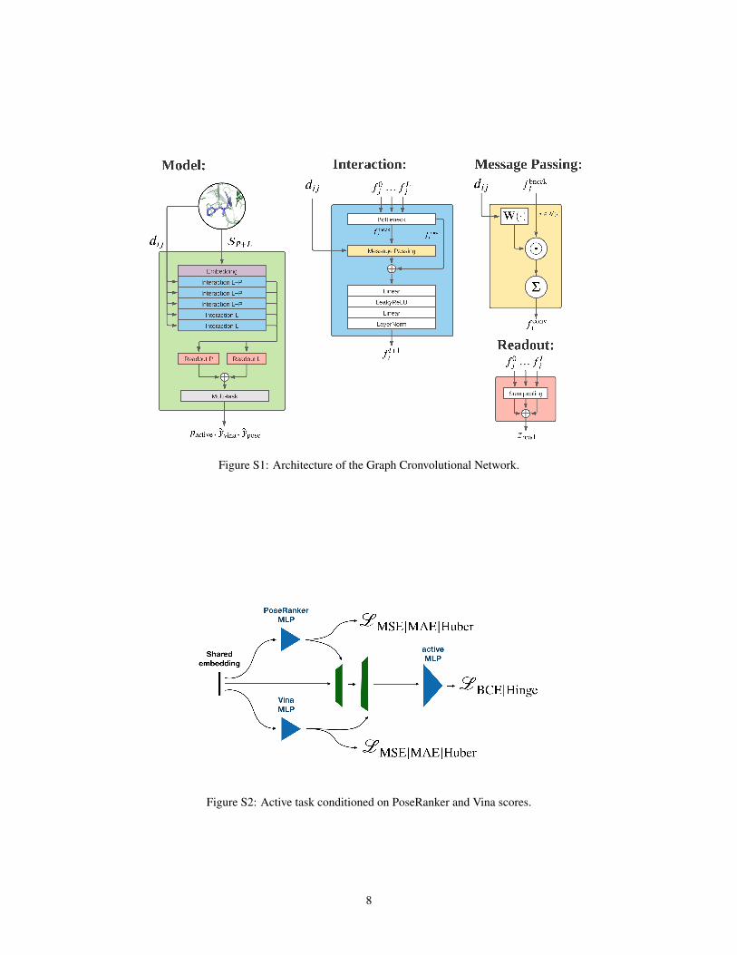
Figure S1: Architecture of the Graph Cronvolutional Network.
Figure S2: Active task conditioned on PoseRanker and Vina scores.
8
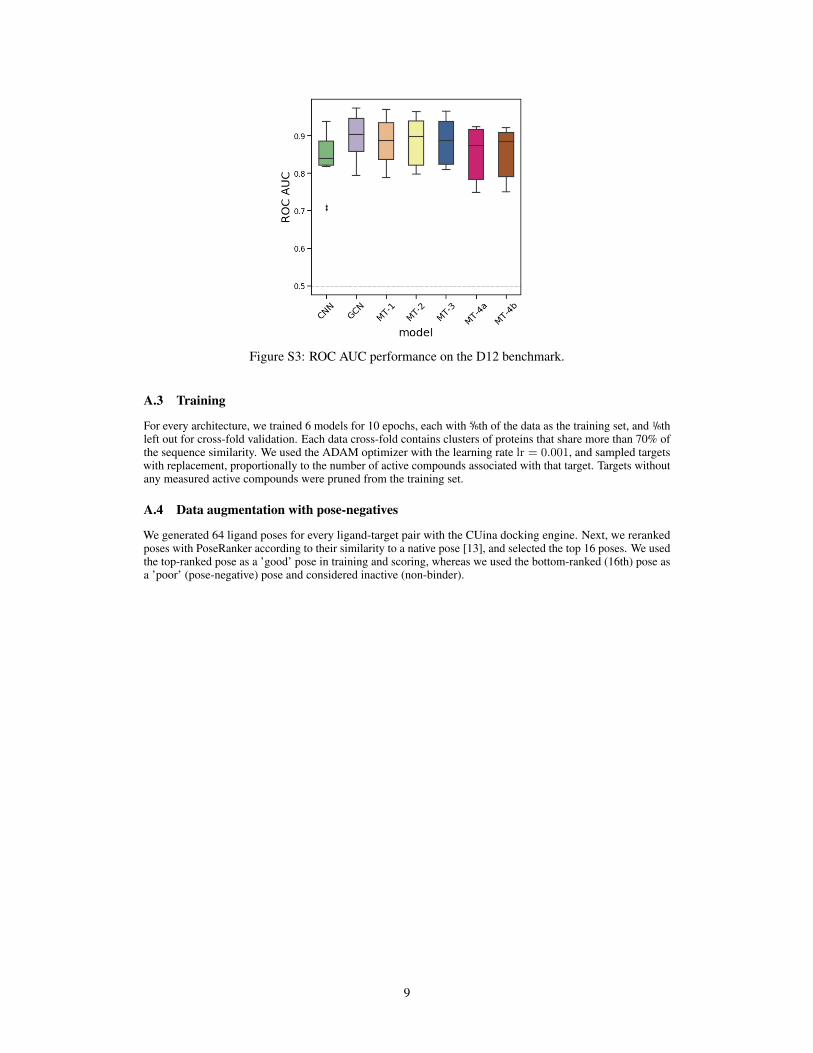
Figure S3: ROC AUC performance on the D12 benchmark.
A.3 Training
For every architecture, we trained 6 models for 10 epochs, each with 5/6th of the data as the training set, and 1/6thleft out for cross-fold validation. Each data cross-fold contains clusters of proteins that share more than 70% ofthe sequence similarity. We used the ADAM optimizer with the learning rate lr = 0.001, and sampled targetswith replacement, proportionally to the number of active compounds associated with that target. Targets withoutany measured active compounds were pruned from the training set.
A.4 Data augmentation with pose-negatives
We generated 64 ligand poses for every ligand-target pair with the CUina docking engine. Next, we rerankedposes with PoseRanker according to their similarity to a native pose [13], and selected the top 16 poses. We usedthe top-ranked pose as a ’good’ pose in training and scoring, whereas we used the bottom-ranked (16th) pose asa ’poor’ (pose-negative) pose and considered inactive (non-binder).
9