Embed Size (px)
Citation preview
doi:10.1006/jmbi.2000.3629 available online at http://www.idealibrary.com on J. Mol. Biol. (2000) 297, 1145±1158
Structure and Thermodynamics of RNA-proteinBinding: Using Molecular Dynamics and Free EnergyAnalyses to Calculate the Free Energies of Bindingand Conformational Change
Carolina M. Reyes and Peter A. Kollman*
Department of PharmaceuticalChemistry, University ofCalifornia San FranciscoSan Francisco, CA94122-0446, USA
E-mail address of the [email protected]
Abbreviations used: BADH, bondelec, electrostatic; MD, molecular dymolecular mechanics; PB, Poisson-BRNA-binding domain; RNP, ribonuRNA recognition motif; vdW, van d
0022-2836/00/051145±14 $35.00/0
An adaptive binding mechanism, requiring large conformationalrearrangements, occurs commonly with many RNA-protein associations.To explore this process of reorganization, we have investigated the con-formational change upon spliceosomal U1A-RNA binding with moleculardynamics (MD) simulations and free energy analyses. We computed theenergetic cost of conformational change in U1A-hairpin and U1A-internalloop binding using a hybrid of molecular mechanics and continuum sol-vent methods. Encouragingly, in all four free energy comparisons (twoslightly different proteins, two different RNAs), the free macromoleculewas more stable than the bound form by the physically reasonable valueof �10 kcal/mol. We calculated the absolute binding free energies forboth complexes to be in the same range as that found experimentally.
# 2000 Academic Press
Keywords: human U1A protein; RNA; molecular dynamics; continuumsolvent models; MM/PBSA
*Corresponding authorIntroduction
Structural and biochemical studies of RNA-pro-tein interactions examine how a protein recognizesa speci®c RNA site, what effect it has on RNAstructure, and how their interactions promote aspeci®c function (Draper, 1995). For ribonucleopro-tein (RNP) complexes, these functions include tran-scription, splicing, and translation, which arecrucial in regulating gene expression. In manycases, RNA-protein complexes are formed by an``adaptive binding'' mechanism, wherein both mol-ecules undergo signi®cant conformational changesupon binding (De Guzman et al., 1998). What is thecontribution of structural reorganization in RNA-protein binding? The free energy cost of structuraladaptation is experimentally dif®cult, if notimpossible, to obtain, but amenable to theoreticalstudies. Theoretical investigations of RNA-proteinbinding can complement biochemical and structur-
ing author:
, angle, dihedral;namics; MM,oltzmann; RBD,cleoprotein; RRM,er Waals.
al studies and formulate insights into theirdynamics and energetics. So, to better understandRNA-protein recognition, we computed the absol-ute binding free energies and the adaptation freeenergies of one of the best-characterized RNP com-plexes, U1A-RNA (Allain et al., 1996; Avis et al.,1996; Gubser & Varani, 1996; Nagai et al., 1990;Oubridge et al., 1994).
U1A-RNA is a component of the U1 small nucle-ar ribonucleoprotein that binds to the 50 splice siteof a primary transcript. The U1A protein containstwo RNA-recognition motifs (RRM) or RNA-bind-ing domains (RBD), which fold into bab-bab struc-ture, found in many RNA-binding proteins (Lu &Hall, 1997; Nagai et al., 1995). The N-terminal RBDof U1A (U1A RBD1) has been studied extensively,both structurally and biochemically, which makesit an ideal system for computational studies (Aviset al., 1996; Hall & Kranz, 1995; Jessen et al., 1991;Kranz & Hall, 1998; Kranz et al., 1996; Nagai et al.,1990; Zeng & Hall, 1997).
U1A RBD1 binds a hairpin RNA and an internalloop RNA with subnanomolar af®nities (Hall,1994). Both the crystal structure of hairpin complex(Oubridge et al., 1994) and the NMR structure ofthe internal loop complex (Allain et al., 1996) showhydrophobic stacking of RNA loop bases with resi-dues on the b-sheet. In both structures, illustrated
# 2000 Academic Press
1146 Estimating the Free Energy Cost of Preorganization
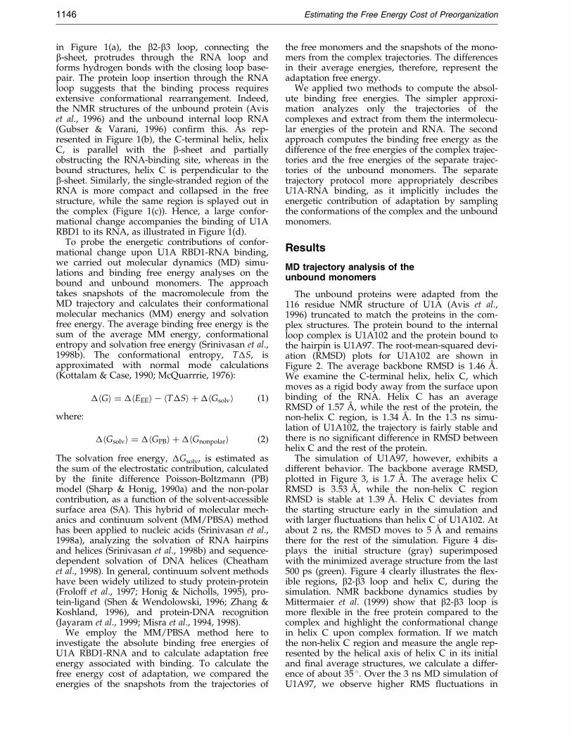
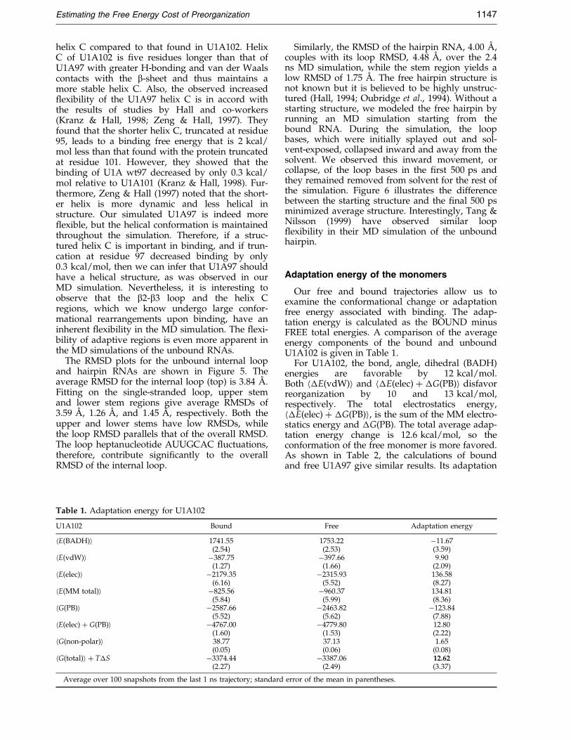
in Figure 1(a), the b2-b3 loop, connecting theb-sheet, protrudes through the RNA loop andforms hydrogen bonds with the closing loop base-pair. The protein loop insertion through the RNAloop suggests that the binding process requiresextensive conformational rearrangement. Indeed,the NMR structures of the unbound protein (Aviset al., 1996) and the unbound internal loop RNA(Gubser & Varani, 1996) con®rm this. As rep-resented in Figure 1(b), the C-terminal helix, helixC, is parallel with the b-sheet and partiallyobstructing the RNA-binding site, whereas in thebound structures, helix C is perpendicular to theb-sheet. Similarly, the single-stranded region of theRNA is more compact and collapsed in the freestructure, while the same region is splayed out inthe complex (Figure 1(c)). Hence, a large confor-mational change accompanies the binding of U1ARBD1 to its RNA, as illustrated in Figure 1(d).
To probe the energetic contributions of confor-mational change upon U1A RBD1-RNA binding,we carried out molecular dynamics (MD) simu-lations and binding free energy analyses on thebound and unbound monomers. The approachtakes snapshots of the macromolecule from theMD trajectory and calculates their conformationalmolecular mechanics (MM) energy and solvationfree energy. The average binding free energy is thesum of the average MM energy, conformationalentropy and solvation free energy (Srinivasan et al.,1998b). The conformational entropy, T�S, isapproximated with normal mode calculations(Kottalam & Case, 1990; McQuarrrie, 1976):
�hGi � �hEEEi ÿ hT�Si ��hGsolvi �1�where:
�hGsolvi � �hGPBi ��hGnonpolari �2�The solvation free energy, �Gsolv, is estimated asthe sum of the electrostatic contribution, calculatedby the ®nite difference Poisson-Boltzmann (PB)model (Sharp & Honig, 1990a) and the non-polarcontribution, as a function of the solvent-accessiblesurface area (SA). This hybrid of molecular mech-anics and continuum solvent (MM/PBSA) methodhas been applied to nucleic acids (Srinivasan et al.,1998a), analyzing the solvation of RNA hairpinsand helices (Srinivasan et al., 1998b) and sequence-dependent solvation of DNA helices (Cheathamet al., 1998). In general, continuum solvent methodshave been widely utilized to study protein-protein(Froloff et al., 1997; Honig & Nicholls, 1995), pro-tein-ligand (Shen & Wendolowski, 1996; Zhang &Koshland, 1996), and protein-DNA recognition(Jayaram et al., 1999; Misra et al., 1994, 1998).
We employ the MM/PBSA method here toinvestigate the absolute binding free energies ofU1A RBD1-RNA and to calculate adaptation freeenergy associated with binding. To calculate thefree energy cost of adaptation, we compared theenergies of the snapshots from the trajectories of
the free monomers and the snapshots of the mono-mers from the complex trajectories. The differencesin their average energies, therefore, represent theadaptation free energy.
We applied two methods to compute the absol-ute binding free energies. The simpler approxi-mation analyzes only the trajectories of thecomplexes and extract from them the intermolecu-lar energies of the protein and RNA. The secondapproach computes the binding free energy as thedifference of the free energies of the complex trajec-tories and the free energies of the separate trajec-tories of the unbound monomers. The separatetrajectory protocol more appropriately describesU1A-RNA binding, as it implicitly includes theenergetic contribution of adaptation by samplingthe conformations of the complex and the unboundmonomers.
Results
MD trajectory analysis of theunbound monomers
The unbound proteins were adapted from the116 residue NMR structure of U1A (Avis et al.,1996) truncated to match the proteins in the com-plex structures. The protein bound to the internalloop complex is U1A102 and the protein bound tothe hairpin is U1A97. The root-mean-squared devi-ation (RMSD) plots for U1A102 are shown inFigure 2. The average backbone RMSD is 1.46 AÊ .We examine the C-terminal helix, helix C, whichmoves as a rigid body away from the surface uponbinding of the RNA. Helix C has an averageRMSD of 1.57 AÊ , while the rest of the protein, thenon-helix C region, is 1.34 AÊ . In the 1.3 ns simu-lation of U1A102, the trajectory is fairly stable andthere is no signi®cant difference in RMSD betweenhelix C and the rest of the protein.
The simulation of U1A97, however, exhibits adifferent behavior. The backbone average RMSD,plotted in Figure 3, is 1.7 AÊ . The average helix CRMSD is 3.53 AÊ , while the non-helix C regionRMSD is stable at 1.39 AÊ . Helix C deviates fromthe starting structure early in the simulation andwith larger ¯uctuations than helix C of U1A102. Atabout 2 ns, the RMSD moves to 5 AÊ and remainsthere for the rest of the simulation. Figure 4 dis-plays the initial structure (gray) superimposedwith the minimized average structure from the last500 ps (green). Figure 4 clearly illustrates the ¯ex-ible regions, b2-b3 loop and helix C, during thesimulation. NMR backbone dynamics studies byMittermaier et al. (1999) show that b2-b3 loop ismore ¯exible in the free protein compared to thecomplex and highlight the conformational changein helix C upon complex formation. If we matchthe non-helix C region and measure the angle rep-resented by the helical axis of helix C in its initialand ®nal average structures, we calculate a differ-ence of about 35 �. Over the 3 ns MD simulation ofU1A97, we observe higher RMS ¯uctuations in
Estimating the Free Energy Cost of Preorganization 1147
helix C compared to that found in U1A102. HelixC of U1A102 is ®ve residues longer than that ofU1A97 with greater H-bonding and van der Waalscontacts with the b-sheet and thus maintains amore stable helix C. Also, the observed increased¯exibility of the U1A97 helix C is in accord withthe results of studies by Hall and co-workers(Kranz & Hall, 1998; Zeng & Hall, 1997). Theyfound that the shorter helix C, truncated at residue95, leads to a binding free energy that is 2 kcal/mol less than that found with the protein truncatedat residue 101. However, they showed that thebinding of U1A wt97 decreased by only 0.3 kcal/mol relative to U1A101 (Kranz & Hall, 1998). Fur-thermore, Zeng & Hall (1997) noted that the short-er helix is more dynamic and less helical instructure. Our simulated U1A97 is indeed more¯exible, but the helical conformation is maintainedthroughout the simulation. Therefore, if a struc-tured helix C is important in binding, and if trun-cation at residue 97 decreased binding by only0.3 kcal/mol, then we can infer that U1A97 shouldhave a helical structure, as was observed in ourMD simulation. Nevertheless, it is interesting toobserve that the b2-b3 loop and the helix Cregions, which we know undergo large confor-mational rearrangements upon binding, have aninherent ¯exibility in the MD simulation. The ¯exi-bility of adaptive regions is even more apparent inthe MD simulations of the unbound RNAs.
The RMSD plots for the unbound internal loopand hairpin RNAs are shown in Figure 5. Theaverage RMSD for the internal loop (top) is 3.84 AÊ .Fitting on the single-stranded loop, upper stemand lower stem regions give average RMSDs of3.59 AÊ , 1.26 AÊ , and 1.45 AÊ , respectively. Both theupper and lower stems have low RMSDs, whilethe loop RMSD parallels that of the overall RMSD.The loop heptanucleotide AUUGCAC ¯uctuations,therefore, contribute signi®cantly to the overallRMSD of the internal loop.
Table 1. Adaptation energy for U1A102
U1A102 Bound
hE(BADH)i 1741.55(2.54)
hE(vdW)i ÿ387.75(1.27)
hE(elec)i ÿ2179.35(6.16)
hE(MM total)i ÿ825.56(5.84)
hG(PB)i ÿ2587.66(5.52)
hE(elec) � G(PB)i ÿ4767.00(1.60)
hG(non-polar)i 38.77(0.05)
hG(total)i � T�S ÿ3374.44(2.27)
Average over 100 snapshots from the last 1 ns trajectory; standard
Similarly, the RMSD of the hairpin RNA, 4.00 AÊ ,couples with its loop RMSD, 4.48 AÊ , over the 2.4ns MD simulation, while the stem region yields alow RMSD of 1.75 AÊ . The free hairpin structure isnot known but it is believed to be highly unstruc-tured (Hall, 1994; Oubridge et al., 1994). Without astarting structure, we modeled the free hairpin byrunning an MD simulation starting from thebound RNA. During the simulation, the loopbases, which were initially splayed out and sol-vent-exposed, collapsed inward and away from thesolvent. We observed this inward movement, orcollapse, of the loop bases in the ®rst 500 ps andthey remained removed from solvent for the rest ofthe simulation. Figure 6 illustrates the differencebetween the starting structure and the ®nal 500 psminimized average structure. Interestingly, Tang &Nilsson (1999) have observed similar loop¯exibility in their MD simulation of the unboundhairpin.
Adaptation energy of the monomers
Our free and bound trajectories allow us toexamine the conformational change or adaptationfree energy associated with binding. The adap-tation energy is calculated as the BOUND minusFREE total energies. A comparison of the averageenergy components of the bound and unboundU1A102 is given in Table 1.
For U1A102, the bond, angle, dihedral (BADH)energies are favorable by 12 kcal/mol.Both h�E(vdW)i and h�E(elec) � �G(PB)i disfavorreorganization by 10 and 13 kcal/mol,respectively. The total electrostatics energy,h�E(elec) � �G(PB)i, is the sum of the MM electro-statics energy and �G(PB). The total average adap-tation energy change is 12.6 kcal/mol, so theconformation of the free monomer is more favored.As shown in Table 2, the calculations of boundand free U1A97 give similar results. Its adaptation
Free Adaptation energy
1753.22 ÿ11.67(2.53) (3.59)ÿ397.66 9.90
(1.66) (2.09)ÿ2315.93 136.58
(5.52) (8.27)ÿ960.37 134.81
(5.99) (8.36)ÿ2463.82 ÿ123.84
(5.62) (7.88)ÿ4779.80 12.80
(1.53) (2.22)37.13 1.65(0.06) (0.08)ÿ3387.06 12.62
(2.49) (3.37)
error of the mean in parentheses.
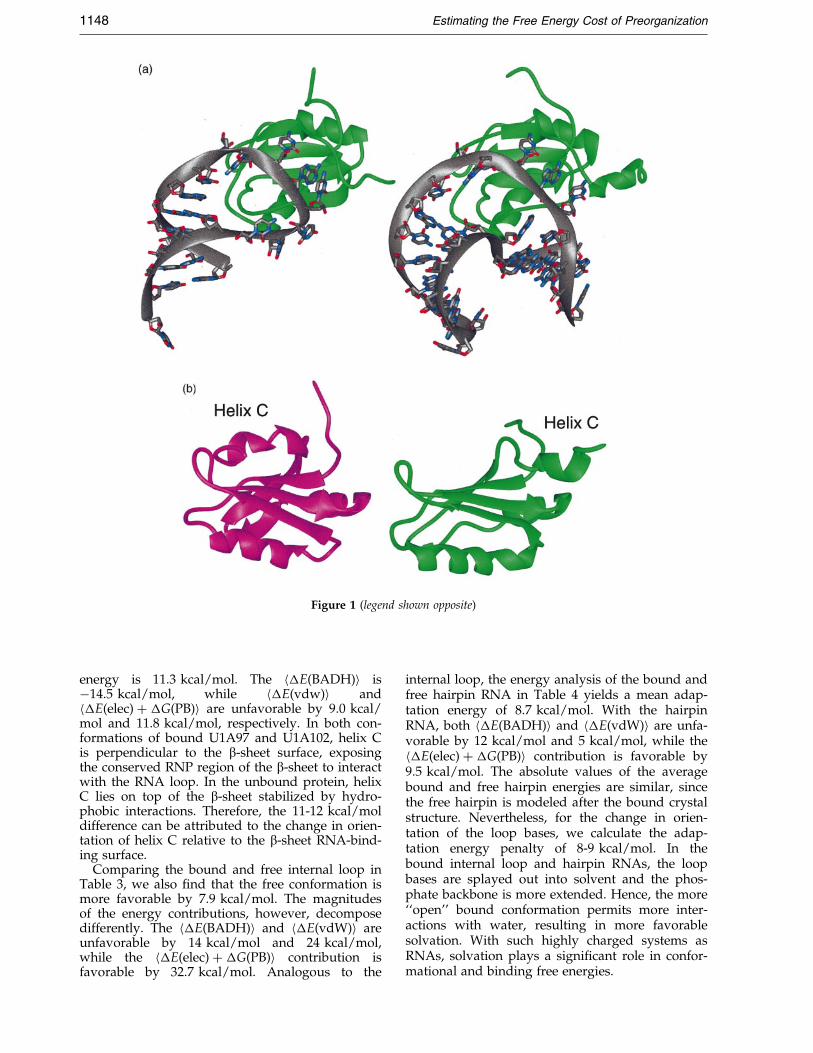
Figure 1 (legend shown opposite)
1148 Estimating the Free Energy Cost of Preorganization
energy is 11.3 kcal/mol. The h�E(BADH)i isÿ14.5 kcal/mol, while h�E(vdw)i andh�E(elec) � �G(PB)i are unfavorable by 9.0 kcal/mol and 11.8 kcal/mol, respectively. In both con-formations of bound U1A97 and U1A102, helix Cis perpendicular to the b-sheet surface, exposingthe conserved RNP region of the b-sheet to interactwith the RNA loop. In the unbound protein, helixC lies on top of the b-sheet stabilized by hydro-phobic interactions. Therefore, the 11-12 kcal/moldifference can be attributed to the change in orien-tation of helix C relative to the b-sheet RNA-bind-ing surface.
Comparing the bound and free internal loop inTable 3, we also ®nd that the free conformation ismore favorable by 7.9 kcal/mol. The magnitudesof the energy contributions, however, decomposedifferently. The h�E(BADH)i and h�E(vdW)i areunfavorable by 14 kcal/mol and 24 kcal/mol,while the h�E(elec) � �G(PB)i contribution isfavorable by 32.7 kcal/mol. Analogous to the
internal loop, the energy analysis of the bound andfree hairpin RNA in Table 4 yields a mean adap-tation energy of 8.7 kcal/mol. With the hairpinRNA, both h�E(BADH)i and h�E(vdW)i are unfa-vorable by 12 kcal/mol and 5 kcal/mol, while theh�E(elec) � �G(PB)i contribution is favorable by9.5 kcal/mol. The absolute values of the averagebound and free hairpin energies are similar, sincethe free hairpin is modeled after the bound crystalstructure. Nevertheless, for the change in orien-tation of the loop bases, we calculate the adap-tation energy penalty of 8-9 kcal/mol. In thebound internal loop and hairpin RNAs, the loopbases are splayed out into solvent and the phos-phate backbone is more extended. Hence, the more``open'' bound conformation permits more inter-actions with water, resulting in more favorablesolvation. With such highly charged systems asRNAs, solvation plays a signi®cant role in confor-mational and binding free energies.
Figure 1. (a) The U1A protein (green) bound to the hairpin RNA (left) and to the internal loop RNA (right).(b) The conformational change of U1A102 upon binding; in the free form (magenta), helix C sits on top of theb-sheet, while in the bound form (green), helix C is oriented away from the b-sheet. (c) The internal loop RNA under-goes signi®cant structural change upon binding. The free internal loop (blue) is more collapsed, while the boundinternal loop has its loop bases extruding out to interact with hydrophobic residues on the protein's b-sheet.(d) Adaptive binding of U1A-RNA.
Estimating the Free Energy Cost of Preorganization 1149
Binding free energies
The calculated average binding free energies ofU1A RBD1-RNA are reported in Table 5. In gener-al, the MM energies and the electrostatic solvationenergies compensate each other; both are largenumbers, but opposite in sign. For example,extended conformations expose lots of surfacecharges to solvent, lowering the solvation energy.However, the distance between the charges is nowincreased, increasing E(elec) and E(vdW). Thebinding of U1A RBD1 to its RNA is an energeticbalance of the vdW energies and the total electro-
static energies (sum of MM electrostatic and PBterms). In all four calculations, the vdW contri-butions to binding are favorable by 82-116 kcal/mol, but the total electrostatic contributions areunfavorable by 40-60 kcal/mol. The non-polar sol-vation term, solvent-accessible surface area-depen-dent term, is always favorable by 10-14 kcal/mol.The sum of the MM energies, h�E(MM total)i, andthe solvation free energies, h�G(PB)i � h�G(non-polar)i, yield favorable binding for both hairpincomplex (ÿ69 kcal/mol for single trajectory andÿ48.4 kcal/mol for separate trajectory) and
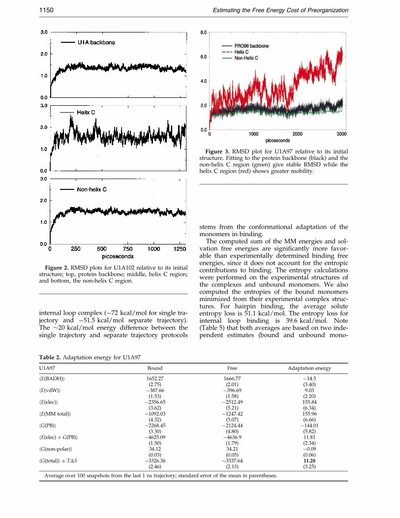
Figure 2. RMSD plots for U1A102 relative to its initialstructure; top, protein backbone; middle, helix C region;and bottom, the non-helix C region.
Figure 3. RMSD plot for U1A97 relative to its initialstructure. Fitting to the protein backbone (black) and thenon-helix C region (green) give stable RMSD while thehelix C region (red) shows greater mobility.
1150 Estimating the Free Energy Cost of Preorganization
internal loop complex (ÿ72 kcal/mol for single tra-jectory and ÿ51.5 kcal/mol separate trajectory).The �20 kcal/mol energy difference between thesingle trajectory and separate trajectory protocols
Table 2. Adaptation energy for U1A97
U1A97 Bound
hE(BADH)i 1652.27(2.75)
hE(vdW)i ÿ387.66(1.53)
hE(elec)i ÿ2356.65(3.62)
hE(MM total)i ÿ1092.03(4.32)
hG(PB)i ÿ2268.45(3.30)
hE(elec) � G(PB)i ÿ4625.09(1.50)
hG(non-polar)i 34.12(0.03)
hG(total)i � T�S ÿ3326.36(2.46)
Average over 100 snapshots from the last 1 ns trajectory; standard
stems from the conformational adaptation of themonomers in binding.
The computed sum of the MM energies and sol-vation free energies are signi®cantly more favor-able than experimentally determined binding freeenergies, since it does not account for the entropiccontributions to binding. The entropy calculationswere performed on the experimental structures ofthe complexes and unbound monomers. We alsocomputed the entropies of the bound monomersminimized from their experimental complex struc-tures. For hairpin binding, the average soluteentropy loss is 51.1 kcal/mol. The entropy loss forinternal loop binding is 39.6 kcal/mol. Note(Table 5) that both averages are based on two inde-pendent estimates (bound and unbound mono-
Free Adaptation energy
1666.77 ÿ14.5(2.01) (3.40)ÿ396.69 9.03
(1.58) (2.20)ÿ2512.49 155.84
(5.21) (6.34)ÿ1247.42 155.96
(5.07) (6.66)ÿ2124.44 ÿ144.01
(4.80) (5.82)ÿ4636.9 11.81
(1.79) (2.34)34.21 ÿ0.09(0.05) (0.06)ÿ3337.64 11.28
(2.13) (3.25)
error of the mean in parentheses.
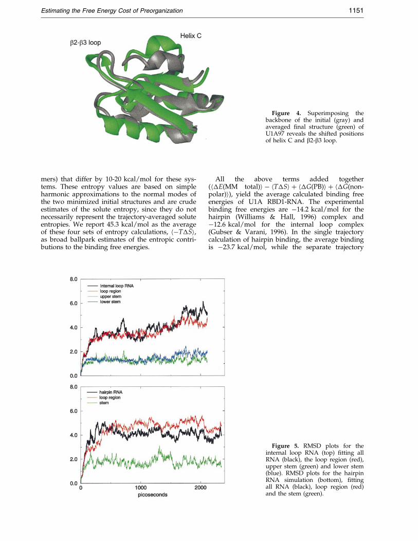
Figure 4. Superimposing thebackbone of the initial (gray) andaveraged ®nal structure (green) ofU1A97 reveals the shifted positionsof helix C and b2-b3 loop.
Estimating the Free Energy Cost of Preorganization 1151
mers) that differ by 10-20 kcal/mol for these sys-tems. These entropy values are based on simpleharmonic approximations to the normal modes ofthe two minimized initial structures and are crudeestimates of the solute entropy, since they do notnecessarily represent the trajectory-averaged soluteentropies. We report 45.3 kcal/mol as the averageof these four sets of entropy calculations, hÿT�Si,as broad ballpark estimates of the entropic contri-butions to the binding free energies.
All the above terms added together(h�E(MM total)i ÿ hT�Si � h�G(PB)i � h�G(non-polar)i), yield the average calculated binding freeenergies of U1A RBD1-RNA. The experimentalbinding free energies are ÿ14.2 kcal/mol for thehairpin (Williams & Hall, 1996) complex andÿ12.6 kcal/mol for the internal loop complex(Gubser & Varani, 1996). In the single trajectorycalculation of hairpin binding, the average bindingis ÿ23.7 kcal/mol, while the separate trajectory
Figure 5. RMSD plots for theinternal loop RNA (top) ®tting allRNA (black), the loop region (red),upper stem (green) and lower stem(blue). RMSD plots for the hairpinRNA simulation (bottom), ®ttingall RNA (black), loop region (red)and the stem (green).
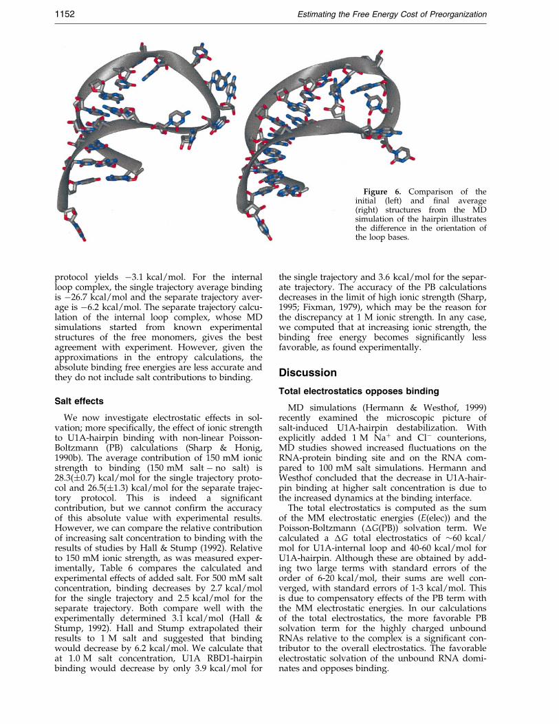
Figure 6. Comparison of theinitial (left) and ®nal average(right) structures from the MDsimulation of the hairpin illustratesthe difference in the orientation ofthe loop bases.
1152 Estimating the Free Energy Cost of Preorganization
protocol yields ÿ3.1 kcal/mol. For the internalloop complex, the single trajectory average bindingis ÿ26.7 kcal/mol and the separate trajectory aver-age is ÿ6.2 kcal/mol. The separate trajectory calcu-lation of the internal loop complex, whose MDsimulations started from known experimentalstructures of the free monomers, gives the bestagreement with experiment. However, given theapproximations in the entropy calculations, theabsolute binding free energies are less accurate andthey do not include salt contributions to binding.
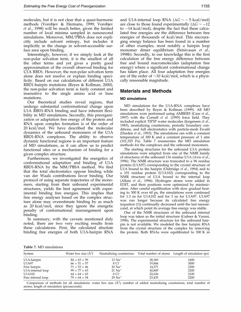
Salt effects
We now investigate electrostatic effects in sol-vation; more speci®cally, the effect of ionic strengthto U1A-hairpin binding with non-linear Poisson-Boltzmann (PB) calculations (Sharp & Honig,1990b). The average contribution of 150 mM ionicstrength to binding (150 mM salt ÿ no salt) is28.3(�0.7) kcal/mol for the single trajectory proto-col and 26.5(�1.3) kcal/mol for the separate trajec-tory protocol. This is indeed a signi®cantcontribution, but we cannot con®rm the accuracyof this absolute value with experimental results.However, we can compare the relative contributionof increasing salt concentration to binding with theresults of studies by Hall & Stump (1992). Relativeto 150 mM ionic strength, as was measured exper-imentally, Table 6 compares the calculated andexperimental effects of added salt. For 500 mM saltconcentration, binding decreases by 2.7 kcal/molfor the single trajectory and 2.5 kcal/mol for theseparate trajectory. Both compare well with theexperimentally determined 3.1 kcal/mol (Hall &Stump, 1992). Hall and Stump extrapolated theirresults to 1 M salt and suggested that bindingwould decrease by 6.2 kcal/mol. We calculate thatat 1.0 M salt concentration, U1A RBD1-hairpinbinding would decrease by only 3.9 kcal/mol for
the single trajectory and 3.6 kcal/mol for the separ-ate trajectory. The accuracy of the PB calculationsdecreases in the limit of high ionic strength (Sharp,1995; Fixman, 1979), which may be the reason forthe discrepancy at 1 M ionic strength. In any case,we computed that at increasing ionic strength, thebinding free energy becomes signi®cantly lessfavorable, as found experimentally.
Discussion
Total electrostatics opposes binding
MD simulations (Hermann & Westhof, 1999)recently examined the microscopic picture ofsalt-induced U1A-hairpin destabilization. Withexplicitly added 1 M Na� and Clÿ counterions,MD studies showed increased ¯uctuations on theRNA-protein binding site and on the RNA com-pared to 100 mM salt simulations. Hermann andWesthof concluded that the decrease in U1A-hair-pin binding at higher salt concentration is due tothe increased dynamics at the binding interface.
The total electrostatics is computed as the sumof the MM electrostatic energies (E(elec)) and thePoisson-Boltzmann (�G(PB)) solvation term. Wecalculated a �G total electrostatics of �60 kcal/mol for U1A-internal loop and 40-60 kcal/mol forU1A-hairpin. Although these are obtained by add-ing two large terms with standard errors of theorder of 6-20 kcal/mol, their sums are well con-verged, with standard errors of 1-3 kcal/mol. Thisis due to compensatory effects of the PB term withthe MM electrostatic energies. In our calculationsof the total electrostatics, the more favorable PBsolvation term for the highly charged unboundRNAs relative to the complex is a signi®cant con-tributor to the overall electrostatics. The favorableelectrostatic solvation of the unbound RNA domi-nates and opposes binding.
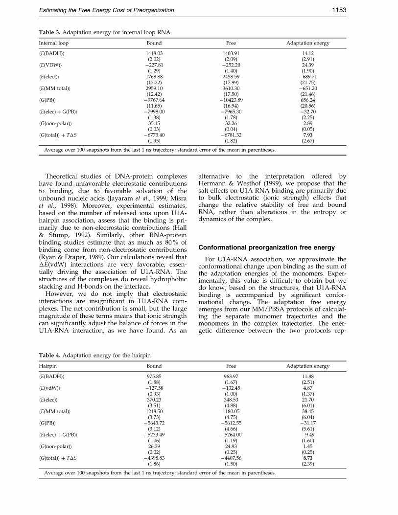
Table 3. Adaptation energy for internal loop RNA
Internal loop Bound Free Adaptation energy
hE(BADH)i 1418.03 1403.91 14.12(2.02) (2.09) (2.91)
hE(VDW)i ÿ227.81 ÿ252.20 24.39(1.29) (1.40) (1.90)
hE(elect)i 1768.88 2458.59 ÿ689.71(12.22) (17.99) (21.75)
hE(MM total)i 2959.10 3610.30 ÿ651.20(12.42) (17.50) (21.46)
hG(PB)i ÿ9767.64 ÿ10423.89 656.24(11.65) (16.94) (20.56)
hE(elec) � G(PB)i ÿ7998.00 ÿ7965.30 ÿ32.70(1.38) (1.78) (2.25)
hG(non-polar)i 35.15 32.26 2.89(0.03) (0.04) (0.05)
hG(total)i � T�S ÿ6773.40 ÿ6781.32 7.93(1.95) (1.82) (2.67)
Average over 100 snapshots from the last 1 ns trajectory; standard error of the mean in parentheses.
Estimating the Free Energy Cost of Preorganization 1153
Theoretical studies of DNA-protein complexeshave found unfavorable electrostatic contributionsto binding, due to favorable solvation of theunbound nucleic acids (Jayaram et al., 1999; Misraet al., 1998). Moreover, experimental estimates,based on the number of released ions upon U1A-hairpin association, assess that the binding is pri-marily due to non-electrostatic contributions (Hall& Stump, 1992). Similarly, other RNA-proteinbinding studies estimate that as much as 80 % ofbinding come from non-electrostatic contributions(Ryan & Draper, 1989). Our calculations reveal that�E(vdW) interactions are very favorable, essen-tially driving the association of U1A-RNA. Thestructures of the complexes do reveal hydrophobicstacking and H-bonds on the interface.
However, we do not imply that electrostaticinteractions are insigni®cant in U1A-RNA com-plexes. The net contribution is small, but the largemagnitude of these terms means that ionic strengthcan signi®cantly adjust the balance of forces in theU1A-RNA interaction, as we have found. As an
Table 4. Adaptation energy for the hairpin
Hairpin Bound
hE(BADH)i 975.85(1.88)
hE(vdW)i ÿ127.58(0.93)
hE(elec)i 370.23(3.51)
hE(MM total)i 1218.50(3.73)
hG(PB)i ÿ5643.72(3.12)
hE(elec) � G(PB)i ÿ5273.49(1.06)
hG(non-polar)i 26.39(0.02)
hG(total)i � T�S ÿ4398.83(1.86)
Average over 100 snapshots from the last 1 ns trajectory; standard
alternative to the interpretation offered byHermann & Westhof (1999), we propose that thesalt effects on U1A-RNA binding are primarily dueto bulk electrostatic (ionic strength) effects thatchange the relative stability of free and boundRNA, rather than alterations in the entropy ordynamics of the complex.
Conformational preorganization free energy
For U1A-RNA association, we approximate theconformational change upon binding as the sum ofthe adaptation energies of the monomers. Exper-imentally, this value is dif®cult to obtain but wedo know, based on the structures, that U1A-RNAbinding is accompanied by signi®cant confor-mational change. The adaptation free energyemerges from our MM/PBSA protocols of calculat-ing the separate monomer trajectories and themonomers in the complex trajectories. The ener-getic difference between the two protocols rep-
Free Adaptation energy
963.97 11.88(1.67) (2.51)ÿ132.45 4.87
(1.00) (1.37)348.53 21.70(4.88) (6.01)
1180.05 38.45(4.75) (6.04)ÿ5612.55 ÿ31.17
(4.66) (5.61)ÿ5264.00 ÿ9.49
(1.19) (1.60)24.93 1.45(0.25) (0.25)ÿ4407.56 8.73
(1.50) (2.39)
error of the mean in parentheses.
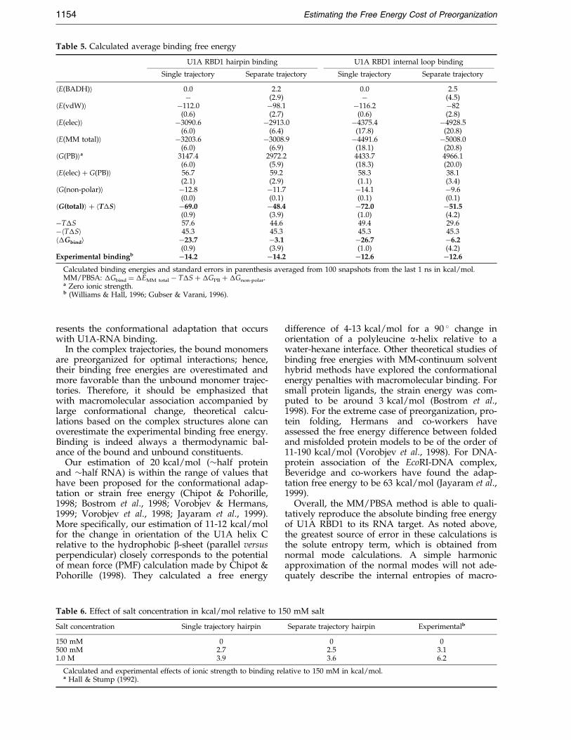
Table 5. Calculated average binding free energy
U1A RBD1 hairpin binding U1A RBD1 internal loop binding
Single trajectory Separate trajectory Single trajectory Separate trajectory
hE(BADH)i 0.0 2.2 0.0 2.5ÿ (2.9) ÿ (4.5)
hE(vdW)i ÿ112.0 ÿ98.1 ÿ116.2 ÿ82(0.6) (2.7) (0.6) (2.8)
hE(elec)i ÿ3090.6 ÿ2913.0 ÿ4375.4 ÿ4928.5(6.0) (6.4) (17.8) (20.8)
hE(MM total)i ÿ3203.6 ÿ3008.9 ÿ4491.6 ÿ5008.0(6.0) (6.9) (18.1) (20.8)
hG(PB)ia 3147.4 2972.2 4433.7 4966.1(6.0) (5.9) (18.3) (20.0)
hE(elec) � G(PB)i 56.7 59.2 58.3 38.1(2.1) (2.9) (1.1) (3.4)
hG(non-polar)i ÿ12.8 ÿ11.7 ÿ14.1 ÿ9.6(0.0) (0.1) (0.1) (0.1)
hG(total)i � hT�Si ÿ69.0 ÿ48.4 ÿ72.0 ÿ51.5(0.9) (3.9) (1.0) (4.2)
ÿT�S 57.6 44.6 49.4 29.6ÿhT�Si 45.3 45.3 45.3 45.3h�Gbindi ÿ23.7 ÿ3.1 ÿ26.7 ÿ6.2
(0.9) (3.9) (1.0) (4.2)Experimental bindingb ÿ14.2 ÿ14.2 ÿ12.6 ÿ12.6
Calculated binding energies and standard errors in parenthesis averaged from 100 snapshots from the last 1 ns in kcal/mol.MM/PBSA: �Gbind � �EMM total ÿ T�S � �GPB � �Gnon-polar.a Zero ionic strength.b (Williams & Hall, 1996; Gubser & Varani, 1996).
1154 Estimating the Free Energy Cost of Preorganization
resents the conformational adaptation that occurswith U1A-RNA binding.
In the complex trajectories, the bound monomersare preorganized for optimal interactions; hence,their binding free energies are overestimated andmore favorable than the unbound monomer trajec-tories. Therefore, it should be emphasized thatwith macromolecular association accompanied bylarge conformational change, theoretical calcu-lations based on the complex structures alone canoverestimate the experimental binding free energy.Binding is indeed always a thermodynamic bal-ance of the bound and unbound constituents.
Our estimation of 20 kcal/mol (�half proteinand �half RNA) is within the range of values thathave been proposed for the conformational adap-tation or strain free energy (Chipot & Pohorille,1998; Bostrom et al., 1998; Vorobjev & Hermans,1999; Vorobjev et al., 1998; Jayaram et al., 1999).More speci®cally, our estimation of 11-12 kcal/molfor the change in orientation of the U1A helix Crelative to the hydrophobic b-sheet (parallel versusperpendicular) closely corresponds to the potentialof mean force (PMF) calculation made by Chipot &Pohorille (1998). They calculated a free energy
Table 6. Effect of salt concentration in kcal/mol relative to 1
Salt concentration Single trajectory hairpin
150 mM 0500 mM 2.71.0 M 3.9
Calculated and experimental effects of ionic strength to binding rea Hall & Stump (1992).
difference of 4-13 kcal/mol for a 90 � change inorientation of a polyleucine a-helix relative to awater-hexane interface. Other theoretical studies ofbinding free energies with MM-continuum solventhybrid methods have explored the conformationalenergy penalties with macromolecular binding. Forsmall protein ligands, the strain energy was com-puted to be around 3 kcal/mol (Bostrom et al.,1998). For the extreme case of preorganization, pro-tein folding, Hermans and co-workers haveassessed the free energy difference between foldedand misfolded protein models to be of the order of11-190 kcal/mol (Vorobjev et al., 1998). For DNA-protein association of the EcoRI-DNA complex,Beveridge and co-workers have found the adap-tation free energy to be 63 kcal/mol (Jayaram et al.,1999).
Overall, the MM/PBSA method is able to quali-tatively reproduce the absolute binding free energyof U1A RBD1 to its RNA target. As noted above,the greatest source of error in these calculations isthe solute entropy term, which is obtained fromnormal mode calculations. A simple harmonicapproximation of the normal modes will not ade-quately describe the internal entropies of macro-
50 mM salt
Separate trajectory hairpin Experimentalb
0 02.5 3.13.6 6.2
lative to 150 mM in kcal/mol.
Estimating the Free Energy Cost of Preorganization 1155
molecules, but it is not clear that a quasi-harmonicmethods (Vorobjev & Hermans, 1999; Vorobjevet al., 1998) will be much better, given the limitednumber of local minima sampled in nanosecondsimulations. Moreover, MM/PBSA does not expli-citly include solvent entropy, but includes itimplicitly in the change in solvent-accessible sur-face area upon binding.
Interestingly, however, if we simply look at thenon-polar solvation term, it is the smallest of allthe other terms and yet gives a pretty goodapproximation of the overall observed binding forU1A RBD1. However, the non-polar solvation termalone does not resolve or explain binding speci-®city. Based on our calculations of different U1ARBD1-hairpin mutations (Reyes & Kollman, 2000),the non-polar solvation term is fairly constant andinsensitive to the single amino acid or basemutations.
Our theoretical studies reveal regions, thatundergo substantial conformational change uponU1A RBD1-RNA binding and have inherent ¯exi-bility in MD simulations. Secondly, this preorgani-zation or adaptation free energy of the protein andRNA upon complex formation is of the order of20 kcal/mol. We have described the moleculardynamics of the unbound monomers of the U1ARBD1-RNA complexes. The ability to observedynamic functional regions is an important featureof MD simulations, as it can allow us to predictfunctional sites or a mechanism of binding for agiven complex structure.
Furthermore, we investigated the energetics ofconformational adaptation and binding of U1ARBD1-RNA by the MM/PBSA method. We ®ndthat the total electrostatics oppose binding whilevan der Waals contributions favor binding. Ourprotocol of using separate trajectories of the mono-mers, starting from their unbound experimentalstructures, yields the best agreement with exper-imental binding free energies. This implies thatfree energy analyses based on the complex struc-ture alone may overestimate binding by as muchas 20 kcal/mol, since they ignore the energeticpenalty of conformational rearrangement uponbinding.
In summary, with the caveats mentioned dulynoted, there are two very exciting results fromthese calculations. First, the calculated absolutebinding free energies of both U1A-hairpin RNA
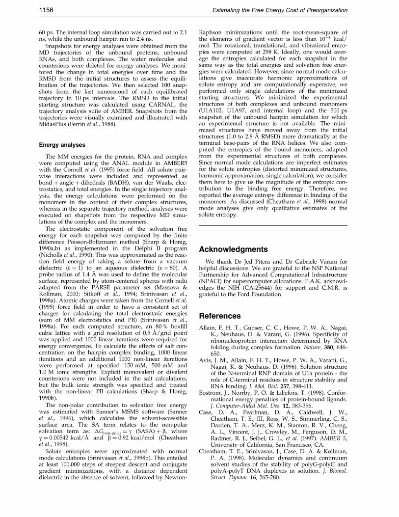
Table 7. MD simulations
System Water box size (AÊ 3) Neutralizing c
U1A-hairpin 84 � 63 � 59 13 NaU1A97 66 � 51 � 57 8 Clÿ
Free hairpin 71 � 53 � 46 20 NaU1A-internal loop 99 � 77 � 63 21 NaU1A102 64 � 64 � 63 8 Clÿ
Free internal loop 79 � 64 � 54 29 Na
Comparison of methods for all simulations: water box size (AÊ 3
atoms, length of simulation (picoseconds).
and U1A-internal loop RNA (�G � ÿ 5 kcal/mol)are close to those found experimentally (�G � ÿ12to ÿ14 kcal/mol), despite the fact that these calcu-lated free energies are the difference between freeenergies of thousands of kcal/mol. This encoura-ging energy balance has been found in a numberof other examples, most notably a hairpin loopmonomer dimer equilibrium (Srinivasan et al.,1998b). Secondly, to our knowledge this is the ®rstcalculation of the free energy difference betweenfree and bound macromolecules (adaptation freeenergy) where a signi®cant conformational changehas taken place. All four adaptation free energiesare of the order of �10 kcal/mol, which is a physi-cally reasonable magnitude.
Materials and Methods
MD simulations
MD simulations for the U1A-RNA complexes havebeen described by Reyes & Kollman (1999). All MDsimulations were performed using AMBER5 (Case et al.,1997) with the Cornell et al. (1995) force ®eld. Theyincluded explicit TIP3P water molecules (Jorgensen et al.,1983), neutralizing counterions, periodic boundary con-ditions, and full electrostatics with particle-mesh Ewald(Darden et al., 1993). The simulations ran with a constanttemperature of 300 K and a constant pressure of 1 atm(101,325 Pa). Table 7 enumerates the MD simulationmethods for the complexes and the unbound monomers.
The starting structures for the unbound U1A proteinsimulations were adapted from one of the NMR familyof structures of the unbound 116 residue U1A (Avis et al.,1996). The NMR structure was truncated to a 96 residueprotein (U1A97) corresponding to the crystal structure ofU1A bound to the hairpin (Oubridge et al., 1994) and toa 101 residue protein (U1A102) corresponding to theNMR structure of U1A bound to the internal loop(Allain et al., 1996). Hydrogen atoms were added inEDIT, and their positions were optimized by minimiz-ation. After careful equilibration with slow gradual heat-ing to 300 K over 60 ps, the simulations were continuedfor 1.3 ns for U1A102 and for 3 ns for U1A97. U1A97was run longer because its calculated free energy(equation (1)) continually decreased until the last nanose-cond, at which point its average free energy was stable.
One of the NMR structures of the unbound internalloop was taken as the initial structure (Gubser & Varani,1996). The experimental structure for the unbound hair-pin is not available. We modeled the free hairpin RNAfrom the crystal structure of the complex by removingthe protein. Both RNAs were equilibrated to 300 K in
ounterions Total number of atoms Length of simulation (ps)
� 28,369 160019,844 3000
� 14,371 2300� 42,845 2200
22,636 2100� 23,435 2200
), number of added neutralizing counterions, total number of
1156 Estimating the Free Energy Cost of Preorganization
60 ps. The internal loop simulation was carried out to 2.1ns, while the unbound hairpin ran to 2.4 ns.
Snapshots for energy analyses were obtained from theMD trajectories of the unbound proteins, unboundRNAs, and both complexes. The water molecules andcounterions were deleted for energy analyses. We moni-tored the change in total energies over time and theRMSD from the initial structures to assess the equili-bration of the trajectories. We then selected 100 snap-shots from the last nanosecond of each equilibratedtrajectory in 10 ps intervals. The RMSD to the initialstarting structure was calculated using CARNAL, thetrajectory analysis suite of AMBER. Snapshots from thetrajectories were visually examined and illustrated withMidasPlus (Ferrin et al., 1988).
Energy analyses
The MM energies for the protein, RNA and complexwere computed using the ANAL module in AMBER5with the Cornell et al. (1995) force ®eld. All solute pair-wise interactions were included and represented asbond � angle � dihedrals (BADH), van der Waals, elec-trostatics, and total energies. In the single trajectory anal-ysis, the energy calculations were performed on themonomers in the context of their complex structures,whereas in the separate trajectory method, analyses wereexecuted on shapshots from the respective MD simu-lations of the complex and the monomers.
The electrostatic component of the solvation freeenergy for each snapshot was computed by the ®nitedifference Poisson-Boltzmann method (Sharp & Honig,1990a,b) as implemented in the Delphi II program(Nicholls et al., 1990). This was approximated as the reac-tion ®eld energy of taking a solute from a vacuumdielectric (e � 1) to an aqueous dielectric (e � 80). Aprobe radius of 1.4 AÊ was used to de®ne the molecularsurface, represented by atom-centered spheres with radiiadapted from the PARSE parameter set (Massova &Kollman, 2000; Sitkoff et al., 1994; Srinivasan et al.,1998a). Atomic charges were taken from the Cornell et al.(1995) force ®eld in order to have a consistent set ofcharges for calculating the total electrostatic energies(sum of MM electrostatics and PB) (Srinivasan et al.,1998a). For each computed structure, an 80 % box®llcubic lattice with a grid resolution of 0.5 AÊ /grid pointwas applied and 1000 linear iterations were required forenergy convergence. To calculate the effects of salt con-centration on the hairpin complex binding, 1000 lineariterations and an additional 1000 non-linear iterationswere performed at speci®ed 150 mM, 500 mM and1.0 M ionic strengths. Explicit monovalent or divalentcounterions were not included in the salt calculations,but the bulk ionic strength was speci®ed and treatedwith the non-linear PB calculations (Sharp & Honig,1990b).
The non-polar contribution to solvation free energywas estimated with Sanner's MSMS software (Sanneret al., 1996), which calculates the solvent-accessiblesurface area. The SA term relates to the non-polarsolvation term as: �Gnon-polar � g (SASA) � b, whereg � 0.00542 kcal/AÊ and b � 0.92 kcal/mol (Cheathamet al., 1998).
Solute entropies were approximated with normalmode calculations (Srinivasan et al., 1998b). This entailedat least 100,000 steps of steepest descent and conjugategradient minimizations, with a distance dependentdielectric in the absence of solvent, followed by Newton-
Raphson minimizations until the root-mean-square ofthe elements of gradient vector is less than 10ÿ4 kcal/mol. The rotational, translational, and vibrational entro-pies were computed at 298 K. Ideally, one would aver-age the entropies calculated for each snapshot in thesame way as the total energies and solvation free ener-gies were calculated. However, since normal mode calcu-lations give inaccurate harmonic approximations ofsolute entropy and are computationally expensive, weperformed only single calculations of the minimizedstarting structures. We minimized the experimentalstructures of both complexes and unbound monomers(U1A102, U1A97, and internal loop) and the 500 pssnapshot of the unbound hairpin simulation for whichan experimental structure is not available. The mini-mized structures have moved away from the initialstructures (1.0 to 2.8 AÊ RMSD) more dramatically at theterminal base-pairs of the RNA helices. We also com-puted the entropies of the bound monomers, adaptedfrom the experimental structures of both complexes.Since normal mode calculations are imperfect estimatesfor the solute entropies (distorted minimized structures,harmonic approximation, single calculation), we considerthem here to give us the magnitude of the entropic con-tribution to the binding free energy. Therefore, wereported the average entropy difference in binding of themonomers. As discussed (Cheatham et al., 1998) normalmode analyses give only qualitative estimates of thesolute entropy.
Acknowledgments
We thank Dr Jed Pitera and Dr Gabriele Varani forhelpful discussions. We are grateful to the NSF NationalPartnership for Advanced Computational Infrastructure(NPACI) for supercomputer allocations. P.A.K. acknowl-edges the NIH (CA-25644) for support and C.M.R. isgrateful to the Ford Foundation
References
Allain, F. H. T., Gubser, C. C., Howe, P. W. A., Nagai,K., Neuhaus, D. & Varani, G. (1996). Speci®city ofribonucleoprotein interaction determined by RNAfolding during complex formation. Nature, 380, 646-650.
Avis, J. M., Allain, F. H. T., Howe, P. W. A., Varani, G.,Nagai, K. & Neuhaus, D. (1996). Solution structureof the N-terminal RNP domain of U1a protein - therole of C-terminal residues in structure stability andRNA binding. J. Mol. Biol. 257, 398-411.
Bostrom, J., Norrby, P. O. & Liljefors, T. (1998). Confor-mational energy penalties of protein-bound ligands.J. Computer-Aided Mol. Des. 12, 383-396.
Case, D. A., Pearlman, D. A., Caldwell, J. W.,Cheatham, T. E., III, Ross, W. S., Simmerling, C. S.,Darden, T. A., Merz, K. M., Stanton, R. V., Cheng,A. L., Vincent, J. J., Crowley, M., Ferguson, D. M.,Radmer, R. J., Seibel, G. L., et al. (1997). AMBER 5,University of California, San Francisco, CA.
Cheatham, T. E., Srinivasan, J., Case, D. A. & Kollman,P. A. (1998). Molecular dynamics and continuumsolvent studies of the stability of polyG-polyC andpolyA-polyT DNA duplexes in solution. J. Biomol.Struct. Dynam. 16, 265-280.
Estimating the Free Energy Cost of Preorganization 1157
Chipot, C. & Pohorille, A. (1998). Folding and transloca-tion of the undecamer of poly-L-leucine across thewater-hexane interface. A molecular dynamicsstudy. J. Am. Chem. Soc. 120, 11912-11924.
Cornell, W. D., Cieplak, P., Bayly, C. I., Gould, I. R.,Merz, K. M., Ferguson, D. M., Spellmeyer, D. C.,Fox, T., Caldwell, J. W. & Kollman, P. A. (1995). Asecond generation force ®eld for the simulation ofproteins, nucleic acids, and organic molecules. J. Am.Chem. Soc. 117, 5179-5197.
Darden, T., York, D. & Pedersen, L. (1993). Particlemesh Ewald - an N. log(N) method for ewald sumsin large systems. J. Chem. Phys. 98, 10089-10092.
De Guzman, R. N., Turner, R. B. & Summers, M. F.(1998). Protein-RNA recognition. Biopolymers, 48,181-195.
Draper, D. E. (1995). Protein-RNA recognition. Annu.Rev. Biochem. 64, 593-620.
Ferrin, T. E., Huang, C. C., Jarvis, L. E. & Langridge, R.(1988). The MIDAS display system. J. Mol. Graph. 6,13-27.
Fixman, M. (1979). The Poisson-Boltzmann equation andits application to polyelectrolytes. J. Chem. Phys. 70,4995-5005.
Froloff, N., Windemuth, A. & Honig, B. (1997). On thecalculation of binding free energies using conti-nuum methods: application to MHC class I protein-peptide interactions. Protein Sci. 6, 1293-1301.
Gubser, C. C. & Varani, G. (1996). Structure of the poly-adenylation regulatory element of the human U1apre-mRNA 30-untranslated region and interactionwith the U1a protein. Biochemistry, 35, 2253-2267.
Hall, K. B. (1994). Interaction of RNA hairpins with thehuman U1a N-terminal RNA binding domain. Bio-chemistry, 33, 10076-10088.
Hall, K. B. & Kranz, J. K. (1995). Thermodynamics andmutations in RNA-protein interactions. MethodsEnzymol. 259, 261-281.
Hall, K. B. & Stump, W. T. (1992). Interaction of N-term-inal domain of U1a protein with an RNA stemloop. Nucl. Acids Res. 20, 4283-4290.
Hermann, T. & Westhof, E. (1999). Simulations of thedynamics at an RNA-protein interface. NatureStruct. Biol. 6, 540-544.
Honig, B. & Nicholls, A. (1995). Classical electrostaticsin biology and chemistry. Science, 268, 1144-1149.
Jayaram, B., McConnell, K. J., Dixit, S. B. & Beveridge,D. L. (1999). Free energy analysis of protein-DNAbinding: the EcoRI endonuclease-DNA complex.J. Comput. Phys. 151, 333-357.
Jorgensen, W. L., Chandreskhar, J., Madura, J. D.,Imprey, R. W. & Klein, M. L. (1983). Comparison ofsimple potential functions for simulating liquidwater. J. Chem. Phys. 79, 926-935.
Jessen, T. H., Oubridge, C., Teo, C. H., Pritchard, C. &Nagai, K. (1991). Identi®cation of molecular contactsbetween the U1-a small nuclear ribonucleoproteinand U1 RNA. EMBO J. 10, 3447-3456.
Kottalam, J. & Case, D. A. (1990). Langevin modes ofmacromolecules: applications to crambin and DNAhexamers. Biopolymers, 29, 1409-1421.
Kranz, J. K. & Hall, K. B. (1998). RNA binding mediatesthe local cooperativity between the beta-sheet andthe C-terminal tail of the human U1A RBD1 pro-tein. J. Mol. Biol. 275, 465-481.
Kranz, J. K., Lu, J. R. & Hall, K. B. (1996). Contributionof the tyrosines to the structure and function of thehuman U1a N-terminal RNA binding domain. Pro-tein Sci. 5, 1567-1583.
Lu, J. & Hall, K. B. (1997). Tertiary structure of RBD2and backbone dynamics of RBD1 and RBD2 of thehuman U1A protein determined by NMR spec-troscopy. Biochemistry, 36, 10393-10406.
Massova, I. & Kollman, P. A. (2000). Combined molecu-lar mechanical and continuum solvent approach(MM-PBSA/GBSA) to predict ligand binding. Per-spect. Drug Discovery Des. 18, 1-23.
McQuarrie, D. A. (1976). Statistical Mechanics, Harperand Rowe, New York.
Misra, V. K., Hecht, J. L., Sharp, K. A., Friedman, R. A.& Honig, B. (1994). Salt effects on protein-DNAinteractions - the lambda-CI repressor and EcoRIendonuclease. J. Mol. Biol. 238, 264-280.
Misra, V. K., Hecht, J. L., Yang, A. S. & Honig, B.(1998). Electrostatic contributions to the binding freeenergy of the lambda cl repressor to DNA. Biophys.J. 75, 2262-2273.
Mittermaier, A., Varani, L., Muhandiram, D. R., Kay, L.& Varani, G. (1999). Changes in side-chain andbackbone dynamics identify determinants of speci-®city in RNA recognition by human U1A protein.J. Mol. Biol. 294, 967-979.
Nagai, K., Oubridge, C., Jessen, T. H., Li, J. & Evans,P. R. (1990). Crystal structure of the RNA-bindingdomain of the U1 small nuclear ribonucleoprotein-A. Nature, 348, 515-520.
Nagai, K., Oubridge, C., Ito, N., Avis, J. & Evans, P.(1995). The RNP domain - a sequence-speci®c RNA-binding domain involved in processing and trans-port of RNA. Trends Biochem. Sci. 20, 235-240.
Nicholls, A., Sharp, K. A. & Honig, B. (1990). DelPhi,Department of Biochemistry and Molecular Bio-physics, Columbia University, NY.
Oubridge, C., Ito, H., Evans, P. R., Teo, C. H. & Nagai,K. (1994). Crystal structure At 1.92 AÊ resolution ofthe RNA-binding domain of the U1a spliceosomalprotein complexed with an RNA hairpin. Nature,372, 432-438.
Reyes, C. M. & Kollman, P. A. (1999). Moleculardynamics studies of U1A-RNA complexes. RNA, 5,235-244.
Reyes, C. M. & Kollman, P. A. (2000). Investigating thebinding speci®city of U1A-RNA by computationalmutagenesis. J. Mol. Biol. 295, 1-6.
Ryan, P. C. & Draper, D. E. (1989). Thermodynamics ofprotein RNA recognition in a highly conservedregion of the large-subunit ribosomal RNA. Bio-chemistry, 28, 9949-9956.
Sanner, M. F., Olson, A. J. & Spehner, J. C. (1996).Reduced surface - an ef®cient way to compute mol-ecular surfaces. Biopolymers, 38, 305-320.
Sharp, K. A. (1995). Polyelectrolyte electrostatics: saltdependence, entropic, and enthalpic contributionsto free energy in the nonlinear Poisson-Boltzmannmodel. Biopolymers, 36, 227-243.
Sharp, K. A. & Honig, B. (1990a). Electrostatic inter-actions in macromolecules - theory and appli-cations. Annu. Rev. Biophys. Biophys. Chem. 19,301-332.
Sharp, K. A. & Honig, B. (1990b). Calculating total elec-trostatic energies with the nonlinear Poisson-Boltz-mann equation. J. Phys. Chem. 94, 7684-7692.
Shen, J. & Wendoloski, J. (1996). Electrostatic bindingenergy calculation using the ®nite difference sol-ution to the linearized Poisson-Boltzmann equation:assessment of its accuracy. J. Comput. Chem. 17, 350-357.
1158 Estimating the Free Energy Cost of Preorganization
Sitkoff, D., Sharp, K. A. & Honig, B. (1994). Accuratecalculation of hydration free energies using macro-scopic solvent models. J. Phys. Chem. 98, 1978-1988.
Srinivasan, J., Cheatham, T. E., Cieplak, P., Kollman,P. A. & Case, D. A. (1998a). Continuum solventstudies of the stability of DNA, RNA, and phos-phoramidate - DNA helices. J. Am. Chem. Soc. 120,9401-9409.
Srinivasan, J., Miller, J., Kollman, P. A. & Case, D. A.(1998b). Continuum solvent studies of the stabilityof RNA hairpin loops and helices. J. Biomol. Struct.Dynam. 16, 671-682.
Tang, Y. & Nilsson, L. (1999). Molecular dynamics simu-lations of the complex between human U1A proteinand hairpin II of small nuclear RNA and of freeRNA in solution. Biophys. J. 77, 1284-1305.
Vorobjev, Y. N. & Hermans, J. (1999). ES/IS: estimationof conformational free energy by combiningdynamics simulations with explicit solvent with an
implicit solvent continuum model. Biophys. Chem.78, 195-205.
Vorobjev, Y. N., Almagro, J. C. & Hermans, J. (1998).Discrimination between native and intentionallymisfolded conformations of proteins: ES/IS, a newmethod for calculating conformational free energythat uses both dynamics simulations with an expli-cit solvent and an implicit solvent continuummodel. Proteins: Struct. Funct. Genet. 32, 399-413.
Williams, D. J. & Hall, K. B. (1996). RNA hairpins withnon-nucleotide spacers bind ef®ciently to thehuman U1a protein. J. Mol. Biol. 257, 265-275.
Zeng, Q. Y. & Hall, K. B. (1997). Contribution of theC-terminal tail of U1A RBD1 to RNA recognitionand protein stability. RNA, 3, 303-314.
Zhang, T. & Koshland, D. E. (1996). Computationalmethod for relative binding energies of enzyme-substrate complexes. Protein Sci. 5, 348-356.
Edited by B. Honig
(Received 18 October 1999; received in revised form 14 February 2000; accepted 18 February 2000)