Embed Size (px)
Citation preview
step. Notably, the free energy required to de-stabilize binding at S2 ultimately stems from thebinding energy of the incoming ion, best seenduring the transition of the central ion (Fig. 3E,red arrows).The proposed mechanism predicts an impor-
tant experimental characteristic of K+ channels.Because the rate of K+ ions leaving the SF at theextracellular side is determined by the rate withwhich incoming intracellular ions arrive at thefilter (movie S1), our model inherently impliesthat K+ channels are diffusion-limited as long asan ion pair occupies the inner SF binding sites.We therefore recorded the occupancy of the in-ner SF sites under varying K+ concentrations. In-deed,we found that ions occupy these positionsover a broad range of concentrations from 10mMto 400 mM (fig. S3). Taken together, our modelthus not only accounts for the diffusion controlof K+ channels, but also explains the wide lin-ear regime of K+ channel conductance above~10 mM K+ (9), which is a prerequisite for ro-bust K+ channel function under variable externalconditions.Our permeation model for ion transfer in K+
channels at physiological voltages relies on re-pulsive Coulomb interactions between adjacentions in the SF as the main driver for conductionnear the diffusion limit (Fig. 3). In re-investigatingseveral K+ channel structures, we found directionic contacts to be compatible with the avail-able crystallographic data. The results presentedabove demonstrate that these direct contacts arenot energetically prohibitive. Rather, they serveto enhance ion flux to the maximum attainablespeed over a broad range of concentrations.
REFERENCES AND NOTES
1. B. Hille, Ion Channels of Excitable Membranes (Sinauer,Sunderland, MA, ed. 3, 2001).
2. D. A. Doyle et al., Science 280, 69–77 (1998).3. Y. Zhou, J. H. Morais-Cabral, A. Kaufman, R. MacKinnon,
Nature 414, 43–48 (2001).4. Y. Zhou, R. MacKinnon, J. Mol. Biol. 333, 965–975 (2003).5. L. G. Cuello et al., Nature 466, 272–275 (2010).6. C. Domene, M. S. P. Sansom, Biophys. J. 85, 2787–2800
(2003).7. A. L. Hodgkin, R. D. Keynes, J. Physiol. 128, 61–88 (1955).8. C. Miller, Nature 414, 23–24 (2001).9. J. H. Morais-Cabral, Y. Zhou, R. MacKinnon, Nature 414, 37–42
(2001).10. J. Åqvist, V. Luzhkov, Nature 404, 881–884 (2000).11. L. Ceccarini, M. Masetti, A. Cavalli, M. Recanatini, PLOS ONE 7,
e49017 (2012).12. F. Khalili-Araghi, E. Tajkhorshid, K. Schulten, Biophys. J. 91,
L72–L74 (2006).13. G. Yellen, Nature 419, 35–42 (2002).14. T. Lu et al., Nat. Neurosci. 4, 239–246 (2001).15. S. Bernèche, B. Roux, Biophys. J. 78, 2900–2917 (2000).16. I. H. Shrivastava, M. S. P. Sansom, Biophys. J. 78, 557–570
(2000).17. T. Sumikama, S. Saito, I. Ohmine, J. Phys. Chem. B 110,
20671–20677 (2006).18. S. Bernèche, B. Roux, Nature 414, 73–77 (2001).19. M. Ø. Jensen et al., Proc. Natl. Acad. Sci. U.S.A. 107,
5833–5838 (2010).20. M. Ø. Jensen, V. Jogini, M. P. Eastwood, D. E. Shaw, J. Gen.
Physiol. 141, 619–632 (2013).21. K. Kasahara, M. Shirota, K. Kinoshita, PLOS ONE 8, e56342
(2013).22. S. Furini, C. Domene, Proc. Natl. Acad. Sci. U.S.A. 106,
16074–16077 (2009).23. P. W. Fowler, E. Abad, O. Beckstein, M. S. P. Sansom, J. Chem.
Theory Comput. 9, 5176–5189 (2013).
24. L. G. Cuello, V. Jogini, D. M. Cortes, E. Perozo, Nature 466,203–208 (2010).
25. C. Kutzner, H. Grubmüller, B. L. de Groot, U. Zachariae,Biophys. J. 101, 809–817 (2011).
26. G. Hummer, D. M. Soumpasis, M. Neumann, Mol. Phys. 81,1155–1163 (1994).
27. C. B. Hübschle, G. M. Sheldrick, B. Dittrich, J. Appl. Crystallogr.44, 1281–1284 (2011).
28. G. M. Sheldrick, Acta Crystallogr. A 64, 112–122 (2008).29. S. Ye, Y. Li, Y. Jiang, Nat. Struct. Mol. Biol. 17, 1019–1023
(2010).30. M. Nishida, M. Cadene, B. T. Chait, R. MacKinnon, EMBO J. 26,
4005–4015 (2007).31. O. B. Clarke et al., Cell 141, 1018–1029 (2010).32. M. Iwamoto, S. Oiki, J. Neurosci. 31, 12180–12188 (2011).33. T. Hoomann, N. Jahnke, A. Horner, S. Keller, P. Pohl, Proc. Natl.
Acad. Sci. U.S.A. 110, 10842–10847 (2013).34. S. M. Saparov, P. Pohl, Proc. Natl. Acad. Sci. U.S.A. 101,
4805–4809 (2004).35. S. Imai, M. Osawa, K. Takeuchi, I. Shimada, Proc. Natl. Acad.
Sci. U.S.A. 107, 6216–6221 (2010).36. M. LeMasurier, L. Heginbotham, C. Miller, J. Gen. Physiol. 118,
303–314 (2001).
ACKNOWLEDGMENTS
We thank H. Sun and S. Wacker for simulations of Kv1.2,R. Sknepnek for assistance with HOOMD-blue, and H. Grubmüller,M.S.P. Sansom, P. Pohl, T. Graen, and K. Zeth for helpful discussions.Supported by the International Max Planck Research Schoolfor Biology and Complex Systems (D.A.K.), a Marie Curie IntraEuropean Fellowship within the 7th European CommunityFramework Programme (C.S.), the Volkswagen Stiftung (viathe Niedersachsenprofessur awarded to G.M.S.) (T.G.), theScottish Universities’ Physics Alliance (U.Z.), and the Germanresearch foundation DFG through SFB803 (B.L.d.G.).
SUPPLEMENTARY MATERIALS
www.sciencemag.org/content/346/6207/352/suppl/DC1Materials and MethodsFigs. S1 to S6Tables S1 to S14Movie S1References (37–63)
15 April 2014; accepted 27 August 201410.1126/science.1254840
ION CHANNELS
Structure and selectivity inbestrophin ion channelsTingting Yang,1 Qun Liu,2 Brian Kloss,3 Renato Bruni,3 Ravi C. Kalathur,3 Youzhong Guo,1
Edda Kloppmann,3,4 Burkhard Rost,3,4 Henry M. Colecraft,5 Wayne A. Hendrickson1,2,3,5*
Human bestrophin-1 (hBest1) is a calcium-activated chloride channel from the retinalpigment epithelium, where mutations are associated with vitelliform macular degeneration,or Best disease. We describe the structure of a bacterial homolog (KpBest) of hBest1and functional characterizations of both channels. KpBest is a pentamer that forms afive-helix transmembrane pore, closed by three rings of conserved hydrophobic residues,and has a cytoplasmic cavern with a restricted exit. From electrophysiological analysisof structure-inspired mutations in KpBest and hBest1, we find a sensitive control of ionselectivity in the bestrophins, including reversal of anion/cation selectivity, and dramaticactivation by mutations at the cytoplasmic exit. A homology model of hBest1 shows thelocations of disease-causing mutations and suggests possible roles in regulation.
The human BEST1 gene encodes a protein[human bestrophin-1 (hBest1)] that is high-ly expressed in retinal pigment epithelium(1–4). More than 120 distinct mutations inhBest1 have been identified that result in
multiple retinal degeneration disorders (5–11),notably vitelliformmacular degeneration or Bestdisease. Functionally, hBest1 was identified as aCl− channel that canbe activated byCa2+ (8, 12, 13),and most of the disease-causing mutations inhBest1 are point mutations that cause channeldysfunction (8, 12, 14–16). Thus, understanding
the structure of the hBest1 channel holds valuefrom both biological and biomedical perspectives.The bestrophin family identified by hBest1 is
distributed widely, with representatives in mostmetazoan animals, including four in humans,and also in other eukaryotes and in prokaryotes(7, 8). The animal bestrophins are characterizedby a highly conserved N-terminal domain thatincludes four predicted transmembrane helices(TMs) and diverse C-terminal domains thatmay be involved in protein-protein interactions(8, 12, 15, 17). Bacterial bestrophins lack the var-iable C-terminal domain and are more divergentin the transmembrane portion. Using a structuralgenomics approach,we identified a homolog fromKlebsiella pneumoniae (KpBest) that could beproduced by recombinant expression for struc-tural and functional characterization. The structure-based sequence alignment implies 14% identitybetween KpBest and hBest1 (fig. S1).Initial crystals of detergent-solubilized KpBest
diffracted poorly; however, constructs from atruncation series did yield suitable crystals. Theinitial structure was solved from one of these,
SCIENCE sciencemag.org 17 OCTOBER 2014 • VOL 346 ISSUE 6207 355
RESEARCH | REPORTS
1Department of Biochemistry and Molecular Biophysics,Columbia University, New York, NY 10032, USA. 2New YorkStructural Biology Center, Synchrotron Beamlines,Brookhaven National Laboratory, Upton, NY 11973, USA.3New York Consortium on Membrane Protein Structure,New York Structural Biology Center, 89 Convent Avenue,New York, NY 10027, USA. 4Department of Informatics,Bioinformatics and Computational Biology, TUM (TechnischeUniversität München), Garching 85748, Germany.5Department of Physiology and Cellular Biophysics, ColumbiaUniversity, New York, NY 10032, USA.*Corresponding author. E-mail: [email protected]
on A
pril
3, 2
016
Dow
nloa
ded
from
on
Apr
il 3,
201
6D
ownl
oade
d fr
om o
n A
pril
3, 2
016
Dow
nloa
ded
from
on
Apr
il 3,
201
6D
ownl
oade
d fr
om o
n A
pril
3, 2
016
Dow
nloa
ded
from
grown from a solution containing zinc acetate, at2.9 Å resolution by single-wavelength anomalousdiffraction (SAD) at the zinc K-edge resonance. Im-proved diffraction was obtained after further trun-cation, removing a total of 11 residues from the Cterminus (fig. S1), and the structure was furtherrefined to 2.3 Å resolution (tables S1 and S2). Thebuilding and refinement of the structural modelwere facilitated by five-fold noncrystallographic sym-metry.Therefinedmodel comprisesorderedresiduesfrom 22, 23, or 24 through 285 or 289 in differentprotomers, plus Zn2+ ions and water molecules.Bestrophins have been predicted by different
groups to form dimers, tetramers, or pentamers(12, 18). Here, we found that KpBest forms astable pentamer (Fig. 1, A and B, and fig. S2) withlarge intersubunit contacts (26,880 Å2 total buriedsurface area). The electrostatic potential surfaceis largely negative on the extracellular surface,
neutral in the transmembrane region, and posi-tive at the cytoplasmic membrane surface (Fig. 1,C and D). Consistent with the experimentallydetermined topology of hBest1 (19), each proto-mer has four transmembrane helices and the Nand C termini both reside on the cytoplasmicside (Fig. 1, E and F). Extracellular interhelixloops TM1-TM2 (12 residues) and TM3-TM4 (3residues) are short, whereas the intracellularconnection between TM2 and TM3 is long (105residues), comprising five helices (a3 to a7) thatform a separate cytoplasmic domain together withtheC-terminal helixa10 (red inFig. 1, E andF). Boththe transmembrane and the cytoplasmic helicalbundles appear to have novel folds: Dali searchesfindmatches only to fragments of these structures.TheTM2helices line the putative ion-conducting
pore through the membrane, and they continueintracellularly as long and curved, but uninter-
rupted, helices a2 (light blue in Fig. 1, E and F).In contrast, TM3/a8 and TM4/a9 are connectedto the cytoplasmic domain by extended segments(4 and 12 residues, respectively). The a9-a10 con-nection is a loop structure that corresponds to aconserved carboxylate-rich segment (EDDDDFE)in eukaryotes, possibly playing a role in Ca2+ regu-lation. This segmenthas fewer carboxylate residuesin prokaryotic homologs, but it presents an elec-tronegative surface patch in KpBest nevertheless(Fig. 1D). The a7 helices (light yellow in Fig. 1F)and cytoplasmic portions of a2 line a cytoplasmiccavern beneath the transmembrane pore.An apparent ion conduction pathway is at the
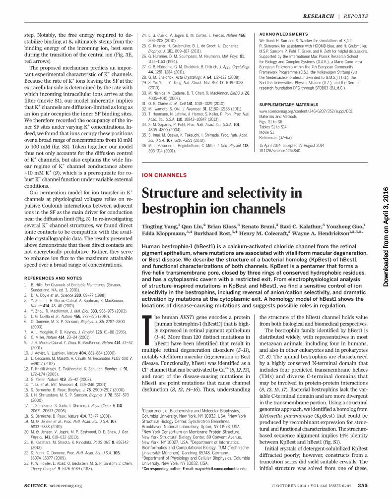
center of the KpBest pentamer. A funnel-shapedelectronegative vestibule, penetratingmidway intothe membrane, precedes a hydrophobic five-helixtransmembrane pore (Fig. 2A). The pore is fol-lowed by a cytoplasmic cavern with a restricted,
356 17 OCTOBER 2014 • VOL 346 ISSUE 6207 sciencemag.org SCIENCE
Fig. 1. Crystal structure of KpBest. (A and B) Ribbon diagram of the KpBest pentamer with each protomer colored differently: (A) as viewed from outside themembrane and (B) as viewed from the side (rotated 90° through the x axis). (C and D) Electrostatic potential at the molecular surface viewed as in (A) and (B),respectively. The contour level is at T5 kT/e; red for negative potential and blue for positive potential. Membrane boundaries in (B) and (D) were calculated byOPM (Orientations of Proteins in Membranes) server. (E) 2D topology of a protomer, colored spectrally from dark blue at its N-terminal segment to red atits C-terminal segment. (F) Ribbon diagram of a protomer. Colored as in (E).
RESEARCH | REPORTS
on-axis exit ~46 Å below themembrane. The poreand upper parts of the cavern are highly con-served, whereas outer surfaces and lower parts ofthe cytoplasmic domain are not (Fig. 2B). Over-all, the ion permeation pathway has a flower-vase shape, with one restriction (radius < 2.0 Å)from three rings of TM2 residues (I62, I66, andF70) at the pore and another (I180, radius = 1.2 Å)at the start of cytoplasmic helix a7 (Fig. 2C).Therefore, the structure of KpBest predicts twodistinct permeation restrictions in the ion passage-way, providing a vital clue for the functionalmechanism of bestrophin channels. Notably, allfour residues located at the predicted restrictionsare highly conserved and/or disease related inhBest1: I76, F84, and I205 (KpBest I62, F70, andI180, respectively) are identical (fig. S1), whereaspoint mutation of either F80 or I205 (KpBest I66and I180, respectively) causes retinal disorders(6, 20, 21).Although eukaryotic bestrophins are known
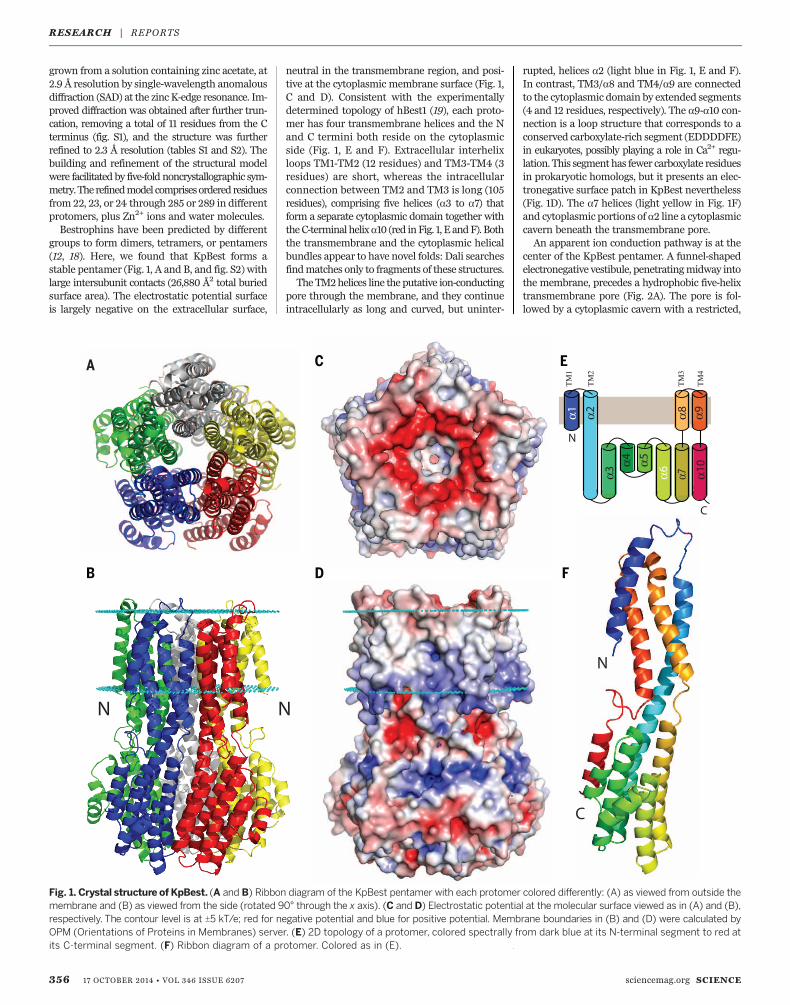
as Ca2+-activated Cl– channels, the function ofKpBest had not been previously examined. Totest its function, purified KpBest was fused into aplanar lipid bilayer with 150 mM of NaCl in boththe trans (internal) and cis (external) solutions.Applying a range of transmembrane potentialsresulted in well-resolved unitary currents with alinear single-channel I-V relationship (Fig. 3, Aand B), confirming that KpBest is indeed an ionchannel. Ca2+ was not required for KpBest acti-vation, as might have been expected given thatKpBest lacks the C-terminal domain that con-tains putative Ca2+ binding sites in eukaryoticbestrophins (7, 13, 22). Strikingly, with 150mMofNaCl on the cis side andnoNaCl on the trans side,inward single-channel currents were recorded(Fig. 3C) with mean amplitude of –5.3 pA (fig.S3A, left), demonstrating that KpBest is a cation
channel that conducts Na+, unlike Cl–-conductingeukaryotic bestrophins. To fully assess KpBestion selectivity, reversal potentials under variousbionic conditions were measured. KpBest is per-meable to monovalent cations with rank orderNa+ > K+ ≈ Cs+ but not to bivalent cations Mg2+,Ca2+, or Ba2+ (Fig. 3D). It is noteworthy that ionchannels in the same family can have reversedcharge selectivity, as exemplified by TMEM16Ca2+-activated channels: TMEM16A and 16B areanion channels, whereas TMEM16Fmay conductcations (23).Despite extensive studies on the ion-conducting
pores of eukaryotic bestrophins, including hBest1and mouse bestrophin-2 (mBest2) (15, 24–26),the molecular basis for ion selection in thesechannels is not clear. Our KpBest model predictsthree critical residues (I62, I66, and F70) at thefirst permeation restriction (Fig. 2C) that likelycontrol ion selectivity. To test this hypothesis, wefirst examined I66, because it is the only residueamong the three that is different in KpBest com-pared with anion-conducting bestrophin chan-nels (where this residue is F) (Fig. 3F and fig. S1)(12, 15, 24–27). In bilayer experiments, KpBestI66F showed outward current with 150 mM ofNaCl on the cis side and no NaCl on the transside, indicating that this mutant channel con-ducts Cl– rather than Na+ (Figs. 3E, top, and fig.S4). To further test this premise in the eukaryoticcounterpart, wild-type (WT) and F80I mutanthBest1 (corresponding to KpBest I66) were trans-fected into human embryonic kidney (HEK) 293cells, and their reversal potentials were deter-mined in whole-cell voltage clamp experiments.Consistent with the KpBest1 I66F results, hBest1F80I was much less permeable to Cl– comparedwithWT (PNa/PCl = 0.39 for F80I, compared withPNa/PCl = 0.03 for the WT) (Fig. 3G and fig. S5).
Inspired in part by selectivity-flipping changesmade at F81 (equivalent toKpBest I66) inDrosophilamelanogaster bestrophin-1 (dBest1) (28), we nextexamined whether KpBest ion selectivity couldbe altered by individually substituting each ofthe hydrophobic pore-lining residues with pos-itively charged arginine (R), which in principlemight favor negatively charged Cl–. When puri-fied KpBest I62R, I66R, and F70R mutants weretested in bilayer experiments, I62R (but not I66Ror F70R) conducted Cl– rather than Na+ (Fig. 3Eand fig. S4). Thesemutants showed perturbed con-ductance amplitudes (fig. S4), indicating effectson permeation at all of these sites. Following thesame logic as for KpBest, the equivalent residueson hBest1 (I76, F80, and F84, respectively) wereindividually mutated to negatively charged glu-tamic acid (E). Consistent with the KpBestresults, only I76E flipped the ion selectivity to Na+
(PNa/PCl = 1.54) (Fig. 3Gand fig. S5B), althoughF80Eand F84E were also less permeable to Cl– than WT(PNa/PCl = 0.33 and 0.46, respectively) (Fig. 3G).None of the hBest1mutations significantly affectedcurrent density (fig. S6). Notably, our results arein accord with previous reports: The rectificationof mBest2 could be altered in opposite directionsby replacing F80 with either R or E (26), and thecorresponding F81E mutation of dBest1 flippedthe cation/anion selectivity (28). Taken together,using our KpBest model as a guide, we have iden-tified three residues that sensitively affect ionselectivity in bestrophins.The dramatic change in ion selectivity from
substitutions at the first hydrophobic residue ofthe pore (I62R in KpBest and I76E in hBest1)suggests a particularly critical role for this posi-tion. Interestingly, the expression level of I62R inE. coli was much lower compared with that ofWT KpBest (about 1/14) (fig. S7), suggesting that
SCIENCE sciencemag.org 17 OCTOBER 2014 • VOL 346 ISSUE 6207 357
RESEARCH | REPORTS
Fig. 2. Structure of the ion-conducting pathway through KpBest. (A) Cross section through the pore center. The model is viewed as in Fig. 1D, with theelectrostatic potential shown on exposed surfaces of the molecular envelope. (B) Cross section as in (A), but colored by Consurf sequence conservation.Turquoise marks the most variable positions, and maroon marks those most conserved.The calculation used 150 prokaryotic homologs with 95%maximal and35%minimal sequence identities comparedwith KpBest. (C) Ribbon diagramof two oppositely facing (144°) protomers of a KpBest pentamer are shownwith theextracellular side on the top.The side chains of critical residues are red.
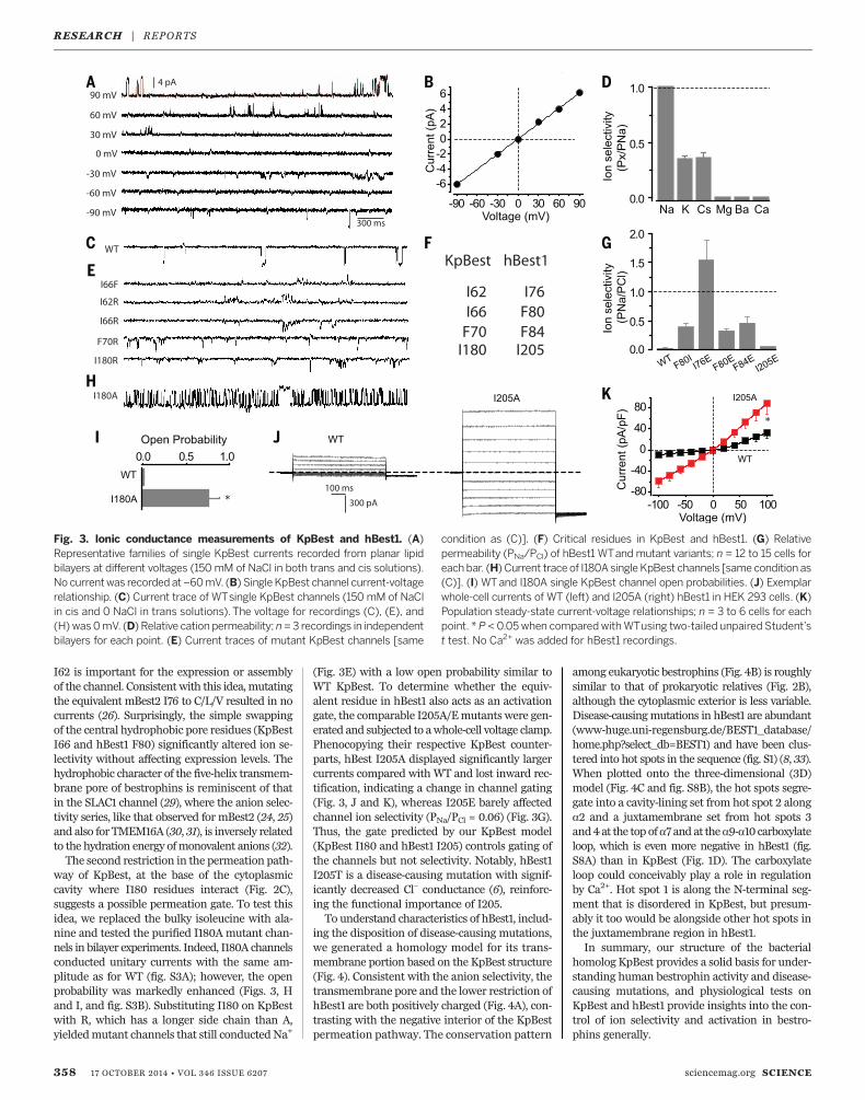
I62 is important for the expression or assemblyof the channel. Consistent with this idea,mutatingthe equivalent mBest2 I76 to C/L/V resulted in nocurrents (26). Surprisingly, the simple swappingof the central hydrophobic pore residues (KpBestI66 and hBest1 F80) significantly altered ion se-lectivity without affecting expression levels. Thehydrophobic character of the five-helix transmem-brane pore of bestrophins is reminiscent of thatin the SLAC1 channel (29), where the anion selec-tivity series, like that observed for mBest2 (24, 25)and also for TMEM16A (30, 31), is inversely relatedto the hydration energy ofmonovalent anions (32).The second restriction in the permeation path-
way of KpBest, at the base of the cytoplasmiccavity where I180 residues interact (Fig. 2C),suggests a possible permeation gate. To test thisidea, we replaced the bulky isoleucine with ala-nine and tested the purified I180A mutant chan-nels in bilayer experiments. Indeed, I180A channelsconducted unitary currents with the same am-plitude as for WT (fig. S3A); however, the openprobability was markedly enhanced (Figs. 3, Hand I, and fig. S3B). Substituting I180 on KpBestwith R, which has a longer side chain than A,yieldedmutant channels that still conductedNa+
(Fig. 3E) with a low open probability similar toWT KpBest. To determine whether the equiv-alent residue in hBest1 also acts as an activationgate, the comparable I205A/Emutants were gen-erated and subjected to awhole-cell voltage clamp.Phenocopying their respective KpBest counter-parts, hBest I205A displayed significantly largercurrents compared with WT and lost inward rec-tification, indicating a change in channel gating(Fig. 3, J and K), whereas I205E barely affectedchannel ion selectivity (PNa/PCl = 0.06) (Fig. 3G).Thus, the gate predicted by our KpBest model(KpBest I180 and hBest1 I205) controls gating ofthe channels but not selectivity. Notably, hBest1I205T is a disease-causing mutation with signif-icantly decreased Cl– conductance (6), reinforc-ing the functional importance of I205.To understand characteristics of hBest1, includ-
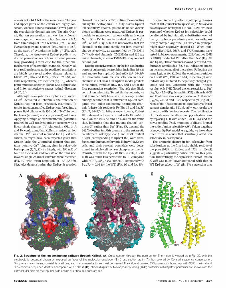
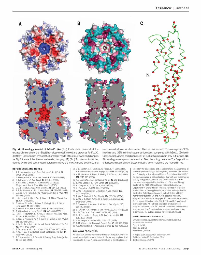
ing the disposition of disease-causing mutations,we generated a homology model for its trans-membrane portion based on the KpBest structure(Fig. 4). Consistent with the anion selectivity, thetransmembrane pore and the lower restriction ofhBest1 are both positively charged (Fig. 4A), con-trasting with the negative interior of the KpBestpermeation pathway. The conservation pattern
among eukaryotic bestrophins (Fig. 4B) is roughlysimilar to that of prokaryotic relatives (Fig. 2B),although the cytoplasmic exterior is less variable.Disease-causingmutations in hBest1 are abundant(www-huge.uni-regensburg.de/BEST1_database/home.php?select_db=BEST1) and have been clus-tered into hot spots in the sequence (fig. S1) (8, 33).When plotted onto the three-dimensional (3D)model (Fig. 4C and fig. S8B), the hot spots segre-gate into a cavity-lining set from hot spot 2 alonga2 and a juxtamembrane set from hot spots 3and4 at the top ofa7 and at thea9-a10 carboxylateloop, which is even more negative in hBest1 (fig.S8A) than in KpBest (Fig. 1D). The carboxylateloop could conceivably play a role in regulationby Ca2+. Hot spot 1 is along the N-terminal seg-ment that is disordered in KpBest, but presum-ably it too would be alongside other hot spots inthe juxtamembrane region in hBest1.In summary, our structure of the bacterial
homolog KpBest provides a solid basis for under-standing human bestrophin activity and disease-causing mutations, and physiological tests onKpBest and hBest1 provide insights into the con-trol of ion selectivity and activation in bestro-phins generally.
358 17 OCTOBER 2014 • VOL 346 ISSUE 6207 sciencemag.org SCIENCE
Fig. 3. Ionic conductance measurements of KpBest and hBest1. (A)Representative families of single KpBest currents recorded from planar lipidbilayers at different voltages (150 mM of NaCl in both trans and cis solutions).No currentwas recorded at–60mV. (B) Single KpBest channel current-voltagerelationship. (C) Current trace of WTsingle KpBest channels (150 mM of NaClin cis and 0 NaCl in trans solutions). The voltage for recordings (C), (E), and(H) was0mV. (D) Relative cation permeability; n=3 recordings in independentbilayers for each point. (E) Current traces of mutant KpBest channels [same
condition as (C)]. (F) Critical residues in KpBest and hBest1. (G) Relativepermeability (PNa/PCl) of hBest1 WTand mutant variants; n = 12 to 15 cells foreach bar. (H) Current trace of I180A single KpBest channels [same condition as(C)]. (I) WTand I180A single KpBest channel open probabilities. (J) Exemplarwhole-cell currents of WT (left) and I205A (right) hBest1 in HEK 293 cells. (K)Population steady-state current-voltage relationships; n = 3 to 6 cells for eachpoint. * P < 0.05when comparedwithWTusing two-tailed unpaired Student’st test. No Ca2+ was added for hBest1 recordings.
RESEARCH | REPORTS
REFERENCES AND NOTES
1. A. D. Marmorstein et al., Proc. Natl. Acad. Sci. U.S.A. 97,12758–12763 (2000).
2. A. Marquardt et al., Hum. Mol. Genet. 7, 1517–1525 (1998).3. K. Petrukhin et al., Nat. Genet. 19, 241–247 (1998).4. R. Neussert, C. Müller, V. M. Milenkovic, O. Strauss,
Pflugers Arch: Eur. J. Phys. 460, 163–175 (2010).5. C. J. Boon et al., Prog. Retin. Eye Res. 28, 187–205 (2009).6. A. E. Davidson et al., Am. J. Hum. Genet. 85, 581–592 (2009).7. Q. Xiao, H. C. Hartzell, K. Yu, Pflugers Arch: Eur. J. Phys. 460,
559–569 (2010).8. H. C. Hartzell, Z. Qu, K. Yu, Q. Xiao, L. T. Chien, Physiol. Rev.
88, 639–672 (2008).9. F. Krämer, N. Mohr, U. Kellner, G. Rudolph, B. H. F. Weber,
Hum. Mutat. 22, 418 (2003).10. F. Krämer et al., Eur. J. Hum. Genet. 8, 286–292 (2000).11. R. Allikmets et al., Hum. Genet. 104, 449–453 (1999).12. H. Sun, T. Tsunenari, K. W. Yau, J. Nathans, Proc. Natl. Acad.
Sci. U.S.A. 99, 4008–4013 (2002).13. Q. Xiao, A. Prussia, K. Yu, Y. Y. Cui, H. C. Hartzell, J. Gen. Physiol.
132, 681–692 (2008).14. K. Yu, Z. Qu, Y. Cui, H. C. Hartzell, Invest. Ophthalmol. Vis. Sci.
48, 4694–4705 (2007).15. T. Tsunenari et al., J. Biol. Chem. 278, 41114–41125 (2003).16. K. Yu, Y. Cui, H. C. Hartzell, Invest. Ophthalmol. Vis. Sci. 47,
4956–4961 (2006).17. A. D. Marmorstein, H. E. Cross, N. S. Peachey, Prog. Retin. Eye Res.
28, 206–226 (2009).
18. J. B. Stanton, A. F. Goldberg, G. Hoppe, L. Y. Marmorstein,A. D. Marmorstein, Biochim. Biophys. Acta 1758, 241–247 (2006).
19. V. M. Milenkovic, A. Rivera, F. Horling, B. H. Weber, J. Biol. Chem.282, 1313–1321 (2007).
20. A. J. Lotery et al., Invest. Ophthalmol. Vis. Sci. 41, 1291–1296 (2000).21. S. Maia-Lopes et al., Hum. Genet. 123, 112 (2008).22. A. Kranjc et al., PLOS ONE 4, e4672 (2009).23. H. Yang et al., Cell 151, 111–122 (2012).24. Z. Qu, R. Fischmeister, C. Hartzell, J. Gen. Physiol. 123,
327–340 (2004).25. Z. Qu, C. Hartzell, J. Gen. Physiol. 124, 371–382 (2004).26. Z. Qu, L. T. Chien, Y. Cui, H. C. Hartzell, J. Neurosci. 26,
5411–5419 (2006).27. T. Tsunenari, J. Nathans, K. W. Yau, J. Gen. Physiol. 127,
749–754 (2006).28. L. T. Chien, H. C. Hartzell, J. Gen. Physiol. 132, 537–546 (2008).29. Y. H. Chen et al., Nature 467, 1074–1080 (2010).30. B. C. Schroeder, T. Cheng, Y. N. Jan, L. Y. Jan, Cell 134,
1019–1029 (2008).31. Y. D. Yang et al., Nature 455, 1210–1215 (2008).32. E. M. Wright, J. M. Diamond, Physiol. Rev. 57, 109–156 (1977).33. A. D. Marmorstein, T. R. Kinnick, Exp. Eye Res. 85, 423–424 (2007).
ACKNOWLEDGMENTS
We thank O. Clarke for help in the diffraction analysis; A. Marks foruse of planar lipid instrumentation; W. Xie for help in planar lipidexperiments; Q. Fan, Y. Geng, and members of the Hendrickson
laboratory for discussions; and J. Schwanof and R. Abramowitz atNational Synchrotron Light Source (NSLS) beamlines X4A and X4Cand F. Murphy at the Advanced Photon Source beamline 24-ID-Efor their assistance in data collection. This work was supported inpart by NIH grants GM095315 and GM107462 to W.A.H. X4beamlines are supported by the New York Structural BiologyCenter at the NSLS of Brookhaven National Laboratory, aDepartment of Energy facility. The data reported in this paperare tabulated in the supplementary materials and deposited tothe Protein Data Bank with access codes listed in table S2.Author contributions: T.Y. and W.A.H. designed research,analyzed data, and wrote the paper; T.Y. performed experiments;Q.L. analyzed diffraction data; B.K., R.C.K., and R.B. performedexpression tests; Y.G. advised on protein production andanalyzed diffraction data; E.K. and B.R. performed bioinformaticsanalyses; and H.M.C. designed and analyzed electrophysiologyexperiments. The authors declare no conflicts of interest.
SUPPLEMENTARY MATERIALS
www.sciencemag.org/content/346/6207/355/suppl/DC1Materials and MethodsFigs. S1 to S8Table S1 and S2References (34–44)
7 August 2014; accepted 17 September 2014Published online 25 September 2014;10.1126/science.1259723
SCIENCE sciencemag.org 17 OCTOBER 2014 • VOL 346 ISSUE 6207 359
RESEARCH | REPORTS
Fig. 4. Homology model of hBest1. (A) (Top) Electrostatic potential at theextracellular surface of the hBest1 homologymodel.Viewed and drawn as for Fig. 1C.(Bottom)Crosssection through thehomologymodelofhBest1.Viewedanddrawnasfor Fig. 2A, except that the cut surface is plain gray. (B) (Top) Top view as in (A), butcolored by surface conservation. Turquoise marks the most variable positions, and
maroonmarks thosemost conserved.This calculation used 150 homologswith 95%maximal and 35% minimal sequence identities compared with hBest1. (Bottom)Cross section viewed and drawn as in Fig. 2B but having a plain gray cut surface. (C)Ribbondiagramofaprotomer fromthehBest1homologypentamer.TheCa positionsof residues that are sites of disease-causing point mutations aremarked in red.
DOI: 10.1126/science.1259723, 355 (2014);346 Science
et al.Tingting YangStructure and selectivity in bestrophin ion channels
This copy is for your personal, non-commercial use only.
clicking here.colleagues, clients, or customers by , you can order high-quality copies for yourIf you wish to distribute this article to others
here.following the guidelines
can be obtained byPermission to republish or repurpose articles or portions of articles
): April 3, 2016 www.sciencemag.org (this information is current as of
The following resources related to this article are available online at
/content/346/6207/355.full.htmlversion of this article at:
including high-resolution figures, can be found in the onlineUpdated information and services,
/content/suppl/2014/09/24/science.1259723.DC1.html can be found at: Supporting Online Material
/content/346/6207/355.full.html#relatedfound at:
can berelated to this article A list of selected additional articles on the Science Web sites
/content/346/6207/355.full.html#ref-list-1, 18 of which can be accessed free:cites 44 articlesThis article
/content/346/6207/355.full.html#related-urls1 articles hosted by HighWire Press; see:cited by This article has been
/cgi/collection/biochemBiochemistry
subject collections:This article appears in the following
registered trademark of AAAS. is aScience2014 by the American Association for the Advancement of Science; all rights reserved. The title
CopyrightAmerican Association for the Advancement of Science, 1200 New York Avenue NW, Washington, DC 20005. (print ISSN 0036-8075; online ISSN 1095-9203) is published weekly, except the last week in December, by theScience
on A
pril
3, 2
016
Dow
nloa
ded
from