Embed Size (px)
Citation preview
Seediscussions,stats,andauthorprofilesforthispublicationat:http://www.researchgate.net/publication/283450752
Structureandmechanicalpropertiesofselectedprotectivesystemsinmarineorganisms
ARTICLE·FEBRUARY2016
DOI:10.1016/j.msec.2015.10.033
READS
69
5AUTHORS,INCLUDING:
StevenE.Naleway
UniversityofCalifornia,SanDiego
15PUBLICATIONS24CITATIONS
SEEPROFILE
JenniferR.A.Taylor
UniversityofCalifornia,SanDiego
9PUBLICATIONS71CITATIONS
SEEPROFILE
MichaelMPorter
ClemsonUniversity
44PUBLICATIONS72CITATIONS
SEEPROFILE
MarcAMeyers
UniversityofCalifornia,SanDiego
484PUBLICATIONS11,198CITATIONS
SEEPROFILE
Availablefrom:MarcAMeyers
Retrievedon:15December2015
Materials Science and Engineering C xxx (2015) xxx–xxx
MSC-05849; No of Pages 25
Contents lists available at ScienceDirect
Materials Science and Engineering C
j ourna l homepage: www.e lsev ie r .com/ locate /msec
Review
Structure and mechanical properties of selected protective systems inmarine organisms
Steven E. Naleway a,⁎, Jennifer R.A. Taylor b, Michael M. Porter e, Marc A. Meyers a,c,d, Joanna McKittrick a,c
a Materials Science and Engineering Program, University of California, San Diego, La Jolla, CA 92093, USAb Scripps Institution of Oceanography, University of California, San Diego, La Jolla, CA 92037, USAc Department of Mechanical and Aerospace Engineering, University of California, San Diego, La Jolla, CA 92093, USAd Department of NanoEngineering, University of California, San Diego, La Jolla, CA 92093, USAe Department of Mechanical Engineering, Clemson University, Clemson, SC 29634, USA
⁎ Corresponding author.E-mail addresses: [email protected] (S.E. Nalew
[email protected] (J. McKittrick).
http://dx.doi.org/10.1016/j.msec.2015.10.0330928-4931/© 2015 Elsevier B.V. All rights reserved.
Please cite this article as: S.E. Naleway, et al.,Eng., C (2015), http://dx.doi.org/10.1016/j.m
a b s t r a c t
a r t i c l e i n f oArticle history:Received 21 January 2015Received in revised form 29 September 2015Accepted 12 October 2015Available online xxxx
Keywords:Marine organismsProtective mechanismsStructural biological materialsBioinspired design
Marine organisms have developed a wide variety of protective strategies to thrive in their native environments.These biologicalmaterials, although formed from simple biopolymer and biomineral constituents, take onmany in-tricate and effective designs. The specific environmental conditions that shape all marine organisms have helpedmodify thesematerials into their current forms: complete hydration, and variation in hydrostatic pressure, temper-ature, salinity, as well asmotion from currents and swells. These conditions vary throughout the ocean, beingmoreconsistent in the pelagic and deep benthic zoneswhile experiencingmore variability in the nearshore and shallows(e.g. intertidal zones, shallow bays and lagoons, salt marshes andmangrove forests). Of note, many marine organ-isms are capable of migrating between these zones. In this review, the basic building blocks of these structural bi-ological materials and a variety of protective strategies in marine organisms are discussed with a focus on theirstructure and mechanical properties. Finally, the bioinspired potential of these biological materials is discussed.
© 2015 Elsevier B.V. All rights reserved.
Contents
1. Introduction . . . . . . . . . . . . . . . . . . . . . . . . . . . . . . . . . . . . . . . . . . . . . . . . . . . . . . . . . . . . . . . 02. Basic building blocks of marine organisms . . . . . . . . . . . . . . . . . . . . . . . . . . . . . . . . . . . . . . . . . . . . . . . . . 0
2.1. Biopolymers . . . . . . . . . . . . . . . . . . . . . . . . . . . . . . . . . . . . . . . . . . . . . . . . . . . . . . . . . . . 02.2. Biominerals . . . . . . . . . . . . . . . . . . . . . . . . . . . . . . . . . . . . . . . . . . . . . . . . . . . . . . . . . . . . 0
3. Crushing resistant structures . . . . . . . . . . . . . . . . . . . . . . . . . . . . . . . . . . . . . . . . . . . . . . . . . . . . . . . 03.1. Mollusk shells . . . . . . . . . . . . . . . . . . . . . . . . . . . . . . . . . . . . . . . . . . . . . . . . . . . . . . . . . . 03.2. Diatom and coccolithophore exoskeletons . . . . . . . . . . . . . . . . . . . . . . . . . . . . . . . . . . . . . . . . . . . . . . 03.3. Crustacean exoskeletons . . . . . . . . . . . . . . . . . . . . . . . . . . . . . . . . . . . . . . . . . . . . . . . . . . . . . . 03.4. Seahorse skeleton . . . . . . . . . . . . . . . . . . . . . . . . . . . . . . . . . . . . . . . . . . . . . . . . . . . . . . . . . 0
4. Flexure resistant structures . . . . . . . . . . . . . . . . . . . . . . . . . . . . . . . . . . . . . . . . . . . . . . . . . . . . . . . . 04.1. Sea sponge spicules . . . . . . . . . . . . . . . . . . . . . . . . . . . . . . . . . . . . . . . . . . . . . . . . . . . . . . . . 04.2. Sea urchin spines . . . . . . . . . . . . . . . . . . . . . . . . . . . . . . . . . . . . . . . . . . . . . . . . . . . . . . . . . 04.3. Porcupine fish spines . . . . . . . . . . . . . . . . . . . . . . . . . . . . . . . . . . . . . . . . . . . . . . . . . . . . . . . 0
5. Piercing resistant structures . . . . . . . . . . . . . . . . . . . . . . . . . . . . . . . . . . . . . . . . . . . . . . . . . . . . . . . . 05.1. Overlapping fish scales . . . . . . . . . . . . . . . . . . . . . . . . . . . . . . . . . . . . . . . . . . . . . . . . . . . . . . . 05.2. Marine scutes and skeletal armors . . . . . . . . . . . . . . . . . . . . . . . . . . . . . . . . . . . . . . . . . . . . . . . . . 0
6. Impact resistant structures . . . . . . . . . . . . . . . . . . . . . . . . . . . . . . . . . . . . . . . . . . . . . . . . . . . . . . . . 06.1. Mantis shrimp dactyl club . . . . . . . . . . . . . . . . . . . . . . . . . . . . . . . . . . . . . . . . . . . . . . . . . . . . . 0
7. Bioinspired materials potential . . . . . . . . . . . . . . . . . . . . . . . . . . . . . . . . . . . . . . . . . . . . . . . . . . . . . . 08. Conclusions . . . . . . . . . . . . . . . . . . . . . . . . . . . . . . . . . . . . . . . . . . . . . . . . . . . . . . . . . . . . . . . 0Acknowledgments . . . . . . . . . . . . . . . . . . . . . . . . . . . . . . . . . . . . . . . . . . . . . . . . . . . . . . . . . . . . . . 0References . . . . . . . . . . . . . . . . . . . . . . . . . . . . . . . . . . . . . . . . . . . . . . . . . . . . . . . . . . . . . . . . . . 0
ay), [email protected] (J.R.A. Taylor), [email protected] (M.M. Porter), [email protected] (M.A. Meyers),
Structure andmechanical properties of selected protective systems inmarine organisms, Mater. Sci.sec.2015.10.033
2 S.E. Naleway et al. / Materials Science and Engineering C xxx (2015) xxx–xxx
1. Introduction
The study of biological materials withinmaterials science provides thenexuswhere thefields of physics, engineering, chemistry andbiology con-verge to understand and harness the vast body of knowledge that can belearned from the natural world. The findings of this research provide forbetter biological understanding of the complex and unique organismsand structures in nature. In addition, this knowledge provides inspirationfor the peripheral fields of bioinspired materials, where synthetic struc-tures are inspired by nature, and biomaterials, where materials and struc-tures are designed for optimum compatibility with biological systems.
While still bound by the same physical laws, biological materials arestarkly different from synthetic ones. To succinctly describe the uniquequalities of biological materials, seven interrelated features have beenidentified (inspired by Arzt [1] and expanded by Meyers et al. andChen et al. [2–5]). These characteristics are: self-assembly, multi-functionality, hierarchical design, hydration effects, mild synthesis con-ditions, evolutionary design and environmental constraints, and self-healing capability. While they apply throughout biology, marine organ-isms face a number of specific environmental constraints not shared bytheir terrestrial counterparts. These marine specific conditions include:complete hydration, and variation in hydrostatic pressure (0.1–100MPa), temperature (−2–38 °C), salinity (34–36 ppt), aswell asmo-tion from currents and swells. These conditions vary throughout theocean, being more consistent in the pelagic and deep benthic zoneswhile experiencing more variability in the nearshore and shallows(e.g. intertidal zones, shallow bays and lagoons, salt marshes and man-grove forests). Of note,manymarine organisms are capable ofmigratingbetween these zones, forcing them to be dynamic through many envi-ronments. Through evolution, these environmental constraints haveshaped the form of all structural marine biological materials.
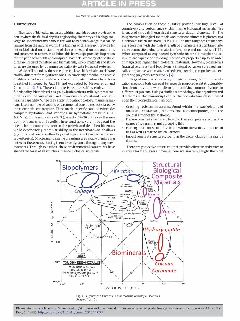
Fig. 1. Toughness as a function of elastiAdapted from [7].
Please cite this article as: S.E. Naleway, et al., Structure andmechanical proEng., C (2015), http://dx.doi.org/10.1016/j.msec.2015.10.033
The combination of these qualities provides for high levels ofcomplexity and performance within marine biological materials. Thisis enacted through hierarchical structural design elements [6]. Thetoughness of biological materials and their constituents is plotted as afunction of the elastic modulus in Fig. 1. The high toughness of biopoly-mers together with the high strength of biominerals is combined intomany composite biological materials (e.g. bone and mollusk shell) [7].When compared to engineered synthetic materials, metals and ce-ramics are capable of providing mechanical properties up to an orderof magnitude higher than biological materials. However, biominerals(natural ceramics) and biopolymers (natural polymers) are mechani-cally comparable with many synthetic engineering composites and en-gineering polymers, respectively [5].
Biological materials can be systematized along different classifi-cationmethods. Naleway et al. [6] recently proposed eight structural de-sign elements as a new paradigm for identifying common features indifferent organisms. Using a similar methodology, the organisms andstructures in this manuscript can be divided into four classes basedupon their biomechanical function:
1. Crushing resistant structures: found within the exoskeletons ofmollusks, crustaceans, diatoms and coccolithophores, and theskeletal armor of the seahorse.
2. Flexure resistant structures: found within sea sponge spicules, thespines of sea urchins and porcupine fish.
3. Piercing resistant structures: found within the scales and scutes offish as well as marine skeletal armors.
4. Impact resistant structures: found in the dactyl clubs of the mantisshrimp.
There are protective structures that provide effective resistance tomultiple forms of stress, however here we aim to highlight the most
c modulus for biological materials.
perties of selected protective systems inmarine organisms, Mater. Sci.
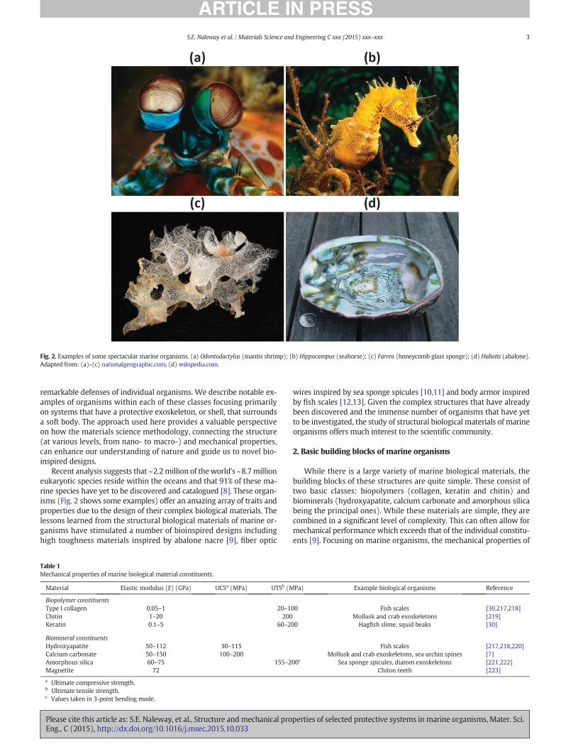
Fig. 2. Examples of some spectacular marine organisms. (a) Odontodactylus (mantis shrimp); (b) Hippocampus (seahorse); (c) Farrea (honeycomb glass sponge); (d) Haliotis (abalone).Adapted from: (a)–(c) nationalgeographic.com, (d) wikipedia.com.
3S.E. Naleway et al. / Materials Science and Engineering C xxx (2015) xxx–xxx
remarkable defenses of individual organisms. We describe notable ex-amples of organisms within each of these classes focusing primarilyon systems that have a protective exoskeleton, or shell, that surroundsa soft body. The approach used here provides a valuable perspectiveon how the materials science methodology, connecting the structure(at various levels, from nano- to macro-) and mechanical properties,can enhance our understanding of nature and guide us to novel bio-inspired designs.
Recent analysis suggests that ~2.2million of theworld's ~8.7millioneukaryotic species reside within the oceans and that 91% of these ma-rine species have yet to be discovered and catalogued [8]. These organ-isms (Fig. 2 shows some examples) offer an amazing array of traits andproperties due to the design of their complex biological materials. Thelessons learned from the structural biological materials of marine or-ganisms have stimulated a number of bioinspired designs includinghigh toughness materials inspired by abalone nacre [9], fiber optic
Table 1Mechanical properties of marine biological material constituents.
Material Elastic modulus (E) (GPa) UCSa (MPa) UTSb (M
Biopolymer constituentsType I collagen 0.05–1 20–1Chitin 1–20 200Keratin 0.1–5 60–2
Biomineral constituentsHydroxyapatite 50–112 30–115Calcium carbonate 50–150 100–200Amorphous silica 60–75 155–2Magnetite 72
a Ultimate compressive strength.b Ultimate tensile strength.c Values taken in 3-point bending mode.
Please cite this article as: S.E. Naleway, et al., Structure andmechanical proEng., C (2015), http://dx.doi.org/10.1016/j.msec.2015.10.033
wires inspired by sea sponge spicules [10,11] and body armor inspiredby fish scales [12,13]. Given the complex structures that have alreadybeen discovered and the immense number of organisms that have yetto be investigated, the study of structural biological materials of marineorganisms offers much interest to the scientific community.
2. Basic building blocks of marine organisms
While there is a large variety of marine biological materials, thebuilding blocks of these structures are quite simple. These consist oftwo basic classes: biopolymers (collagen, keratin and chitin) andbiominerals (hydroxyapatite, calcium carbonate and amorphous silicabeing the principal ones). While these materials are simple, they arecombined in a significant level of complexity. This can often allow formechanical performance which exceeds that of the individual constitu-ents [9]. Focusing on marine organisms, the mechanical properties of
Pa) Example biological organisms Reference
00 Fish scales [30,217,218]Mollusk and crab exoskeletons [219]
00 Hagfish slime, squid beaks [30]
Fish scales [217,218,220]Mollusk and crab exoskeletons, sea urchin spines [7]
00c Sea sponge spicules, diatom exoskeletons [221,222]Chiton teeth [223]
perties of selected protective systems inmarine organisms, Mater. Sci.
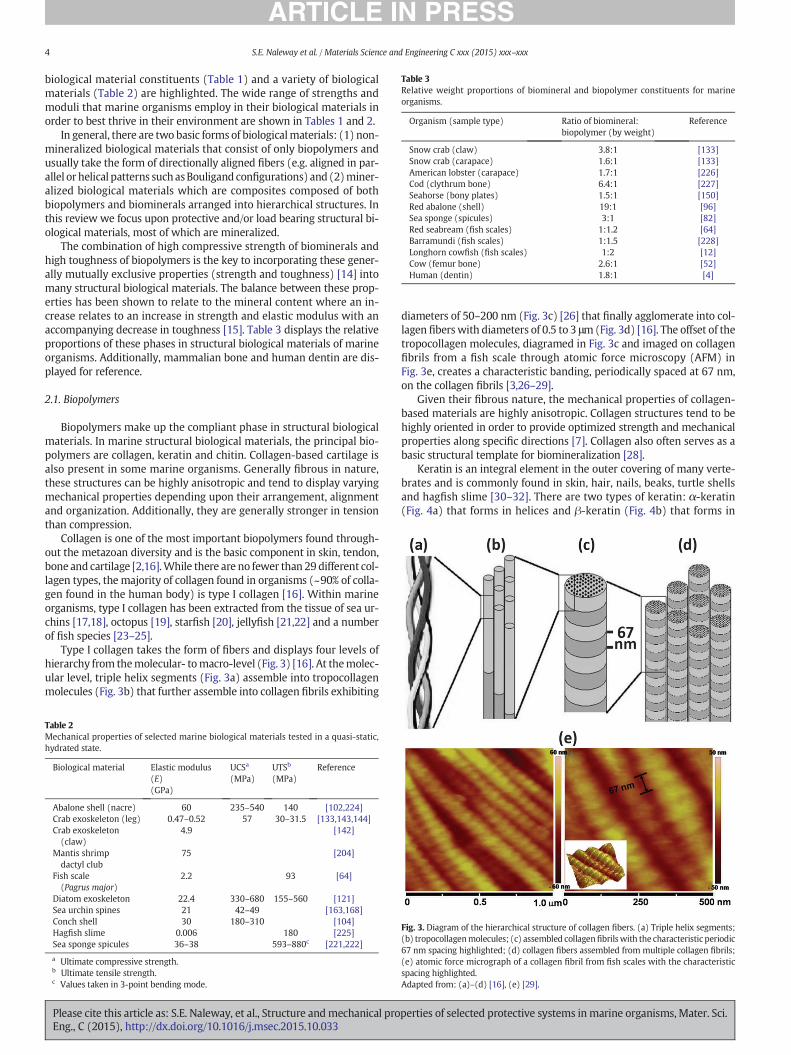
Table 3Relative weight proportions of biomineral and biopolymer constituents for marineorganisms.
Organism (sample type) Ratio of biomineral:biopolymer (by weight)
Reference
Snow crab (claw) 3.8:1 [133]Snow crab (carapace) 1.6:1 [133]American lobster (carapace) 1.7:1 [226]Cod (clythrum bone) 6.4:1 [227]Seahorse (bony plates) 1.5:1 [150]Red abalone (shell) 19:1 [96]Sea sponge (spicules) 3:1 [82]Red seabream (fish scales) 1:1.2 [64]Barramundi (fish scales) 1:1.5 [228]Longhorn cowfish (fish scales) 1:2 [12]Cow (femur bone) 2.6:1 [52]Human (dentin) 1.8:1 [4]
4 S.E. Naleway et al. / Materials Science and Engineering C xxx (2015) xxx–xxx
biological material constituents (Table 1) and a variety of biologicalmaterials (Table 2) are highlighted. The wide range of strengths andmoduli that marine organisms employ in their biological materials inorder to best thrive in their environment are shown in Tables 1 and 2.
In general, there are two basic forms of biologicalmaterials: (1) non-mineralized biological materials that consist of only biopolymers andusually take the form of directionally aligned fibers (e.g. aligned in par-allel or helical patterns suchasBouligand configurations) and (2)miner-alized biological materials which are composites composed of bothbiopolymers and biominerals arranged into hierarchical structures. Inthis review we focus upon protective and/or load bearing structural bi-ological materials, most of which are mineralized.
The combination of high compressive strength of biominerals andhigh toughness of biopolymers is the key to incorporating these gener-ally mutually exclusive properties (strength and toughness) [14] intomany structural biological materials. The balance between these prop-erties has been shown to relate to the mineral content where an in-crease relates to an increase in strength and elastic modulus with anaccompanying decrease in toughness [15]. Table 3 displays the relativeproportions of these phases in structural biological materials of marineorganisms. Additionally, mammalian bone and human dentin are dis-played for reference.
2.1. Biopolymers
Biopolymers make up the compliant phase in structural biologicalmaterials. In marine structural biological materials, the principal bio-polymers are collagen, keratin and chitin. Collagen-based cartilage isalso present in some marine organisms. Generally fibrous in nature,these structures can be highly anisotropic and tend to display varyingmechanical properties depending upon their arrangement, alignmentand organization. Additionally, they are generally stronger in tensionthan compression.
Collagen is one of the most important biopolymers found through-out the metazoan diversity and is the basic component in skin, tendon,bone and cartilage [2,16].While there are no fewer than 29 different col-lagen types, the majority of collagen found in organisms (~90% of colla-gen found in the human body) is type I collagen [16]. Within marineorganisms, type I collagen has been extracted from the tissue of sea ur-chins [17,18], octopus [19], starfish [20], jellyfish [21,22] and a numberof fish species [23–25].
Type I collagen takes the form of fibers and displays four levels ofhierarchy from themolecular- tomacro-level (Fig. 3) [16]. At themolec-ular level, triple helix segments (Fig. 3a) assemble into tropocollagenmolecules (Fig. 3b) that further assemble into collagen fibrils exhibiting
Table 2Mechanical properties of selected marine biological materials tested in a quasi-static,hydrated state.
Biological material Elastic modulus(E)(GPa)
UCSa
(MPa)UTSb
(MPa)Reference
Abalone shell (nacre) 60 235–540 140 [102,224]Crab exoskeleton (leg) 0.47–0.52 57 30–31.5 [133,143,144]Crab exoskeleton(claw)
4.9 [142]
Mantis shrimpdactyl club
75 [204]
Fish scale(Pagrus major)
2.2 93 [64]
Diatom exoskeleton 22.4 330–680 155–560 [121]Sea urchin spines 21 42–49 [163,168]Conch shell 30 180–310 [104]Hagfish slime 0.006 180 [225]Sea sponge spicules 36–38 593–880c [221,222]
a Ultimate compressive strength.b Ultimate tensile strength.c Values taken in 3-point bending mode.
Please cite this article as: S.E. Naleway, et al., Structure andmechanical proEng., C (2015), http://dx.doi.org/10.1016/j.msec.2015.10.033
diameters of 50–200 nm (Fig. 3c) [26] that finally agglomerate into col-lagen fiberswith diameters of 0.5 to 3 μm(Fig. 3d) [16]. The offset of thetropocollagen molecules, diagramed in Fig. 3c and imaged on collagenfibrils from a fish scale through atomic force microscopy (AFM) inFig. 3e, creates a characteristic banding, periodically spaced at 67 nm,on the collagen fibrils [3,26–29].
Given their fibrous nature, the mechanical properties of collagen-based materials are highly anisotropic. Collagen structures tend to behighly oriented in order to provide optimized strength and mechanicalproperties along specific directions [7]. Collagen also often serves as abasic structural template for biomineralization [28].
Keratin is an integral element in the outer covering of many verte-brates and is commonly found in skin, hair, nails, beaks, turtle shellsand hagfish slime [30–32]. There are two types of keratin: α-keratin(Fig. 4a) that forms in helices and β-keratin (Fig. 4b) that forms in
Fig. 3. Diagram of the hierarchical structure of collagen fibers. (a) Triple helix segments;(b) tropocollagenmolecules; (c) assembled collagenfibrilswith the characteristic periodic67 nm spacing highlighted; (d) collagen fibers assembled from multiple collagen fibrils;(e) atomic force micrograph of a collagen fibril from fish scales with the characteristicspacing highlighted.Adapted from: (a)–(d) [16], (e) [29].
perties of selected protective systems inmarine organisms, Mater. Sci.
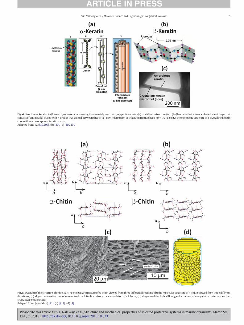
Fig. 5.Diagram of the structure of chitin. (a) Themolecular structure ofα-chitin viewed from three different directions; (b) themolecular structure ofβ-chitin viewed from three differentdirections; (c) aligned microstructure of mineralized α-chitin fibers from the exoskeleton of a lobster; (d) diagram of the helical Bouligand structure of many chitin materials, such ascrustacean exoskeletons.Adapted from: (a) and (b) [41], (c) [211], (d) [4].
Fig. 4. Structure of keratin. (a) Hierarchy ofα-keratin showing the assembly from two polypeptide chains (i) to a fibrous structure (iv); (b)β-keratin that shows a pleated sheet shape thatconsists of antiparallel chains with R-groups that extend between sheets; (c) TEMmicrograph of α-keratin from a sheep horn that displays the composite structure of a crystalline keratincore within an amorphous keratin matrix.Adapted from: (a) [30,209], (b) [30], (c) [30,210].
5S.E. Naleway et al. / Materials Science and Engineering C xxx (2015) xxx–xxx
Please cite this article as: S.E. Naleway, et al., Structure andmechanical properties of selected protective systems inmarine organisms, Mater. Sci.Eng., C (2015), http://dx.doi.org/10.1016/j.msec.2015.10.033
6 S.E. Naleway et al. / Materials Science and Engineering C xxx (2015) xxx–xxx
pleated sheets [30]. The fibrous structure of α-keratin is similar to type Icollagen as both consist of hierarchically assembled molecules, twopolypeptide chains in α-keratin (Fig. 4a) and three tropocollagenmolecules for type I collagen (Fig. 3a). However, once the cells that pro-duce keratin (keratinocytes) assemble, they die, resulting in a non-vascularized “dead” tissue that is in contrast to the extracellular “living”matrix of collagen, intowhich live cells are embedded [30]. Additionally,keratin can be considered a composite material, similar to syntheticsemi-crystalline polymers, consisting of fibers of crystalline keratin re-inforcing an amorphous keratin matrix (Fig. 4c) [33]. While they havedifferent forms, both α-keratin and β-keratin have similar numbers ofpolypeptide chains, suggesting that they both originate from the samemicrofibril structure [34]. Within marine biological materials α-keratinis much more common than β-keratin.
Quite similar to type I collagen, the mechanical properties of fibrousα-keratin are highly anisotropic [35,36]. Given this alignment, α-keratinis capable of much higher mechanical properties parallel to the fiberdirection than the more isotropic β-keratin [30]. Additionally, the com-posite nature of keratin results in a roughly inverse relationship be-tween the stiffness (elastic modulus) and the volume fraction ofamorphous matrix (less-crystalline composite) (i.e. as the content ofthe amorphous keratin increases, the bulk keratin's stiffness decreases)[30,37]. This relationship between crystallinity and elastic modulus isalso common to semi-crystalline synthetic polymers [38,39].
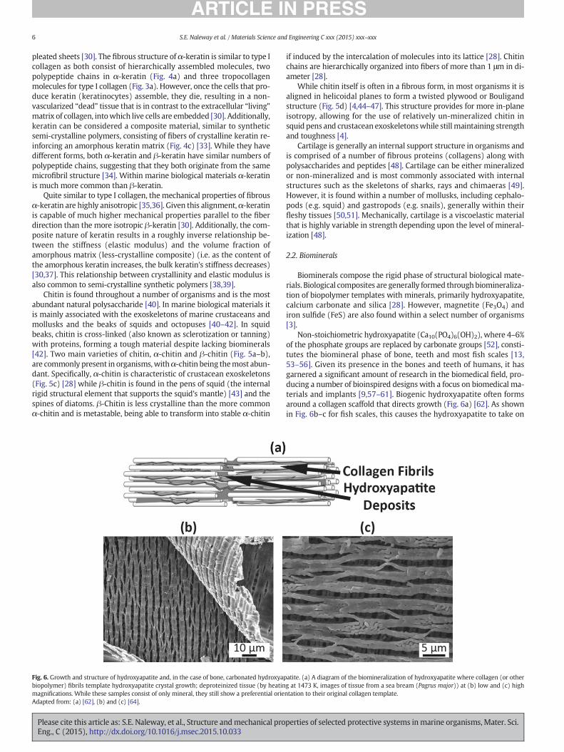
Chitin is found throughout a number of organisms and is the mostabundant natural polysaccharide [40]. In marine biological materials itis mainly associated with the exoskeletons of marine crustaceans andmollusks and the beaks of squids and octopuses [40–42]. In squidbeaks, chitin is cross-linked (also known as sclerotization or tanning)with proteins, forming a tough material despite lacking biominerals[42]. Two main varieties of chitin, α-chitin and β-chitin (Fig. 5a–b),are commonly present in organisms,withα-chitin being themost abun-dant. Specifically, α-chitin is characteristic of crustacean exoskeletons(Fig. 5c) [28] while β-chitin is found in the pens of squid (the internalrigid structural element that supports the squid's mantle) [43] and thespines of diatoms. β-Chitin is less crystalline than the more commonα-chitin and is metastable, being able to transform into stable α-chitin
Fig. 6. Growth and structure of hydroxyapatite and, in the case of bone, carbonated hydroxyapbiopolymer) fibrils template hydroxyapatite crystal growth; deproteinized tissue (by heatinmagnifications. While these samples consist of only mineral, they still show a preferential orieAdapted from: (a) [62], (b) and (c) [64].
Please cite this article as: S.E. Naleway, et al., Structure andmechanical proEng., C (2015), http://dx.doi.org/10.1016/j.msec.2015.10.033
if induced by the intercalation of molecules into its lattice [28]. Chitinchains are hierarchically organized into fibers of more than 1 μm in di-ameter [28].
While chitin itself is often in a fibrous form, in most organisms it isaligned in helicoidal planes to form a twisted plywood or Bouligandstructure (Fig. 5d) [4,44–47]. This structure provides for more in-planeisotropy, allowing for the use of relatively un-mineralized chitin insquid pens and crustacean exoskeletonswhile still maintaining strengthand toughness [4].
Cartilage is generally an internal support structure in organisms andis comprised of a number of fibrous proteins (collagens) along withpolysaccharides and peptides [48]. Cartilage can be either mineralizedor non-mineralized and is most commonly associated with internalstructures such as the skeletons of sharks, rays and chimaeras [49].However, it is found within a number of mollusks, including cephalo-pods (e.g. squid) and gastropods (e.g. snails), generally within theirfleshy tissues [50,51]. Mechanically, cartilage is a viscoelastic materialthat is highly variable in strength depending upon the level of mineral-ization [48].
2.2. Biominerals
Biominerals compose the rigid phase of structural biological mate-rials. Biological composites are generally formed throughbiomineraliza-tion of biopolymer templates with minerals, primarily hydroxyapatite,calcium carbonate and silica [28]. However, magnetite (Fe3O4) andiron sulfide (FeS) are also found within a select number of organisms[3].
Non-stoichiometric hydroxyapatite (Ca10(PO4)6(OH)2), where 4–6%of the phosphate groups are replaced by carbonate groups [52], consti-tutes the biomineral phase of bone, teeth and most fish scales [13,53–56]. Given its presence in the bones and teeth of humans, it hasgarnered a significant amount of research in the biomedical field, pro-ducing a number of bioinspired designs with a focus on biomedical ma-terials and implants [9,57–61]. Biogenic hydroxyapatite often formsaround a collagen scaffold that directs growth (Fig. 6a) [62]. As shownin Fig. 6b–c for fish scales, this causes the hydroxyapatite to take on
atite. (a) A diagram of the biomineralization of hydroxyapatite where collagen (or otherg at 1473 K, images of tissue from a sea bream (Pagrus major)) at (b) low and (c) highntation to their original collagen template.
perties of selected protective systems inmarine organisms, Mater. Sci.
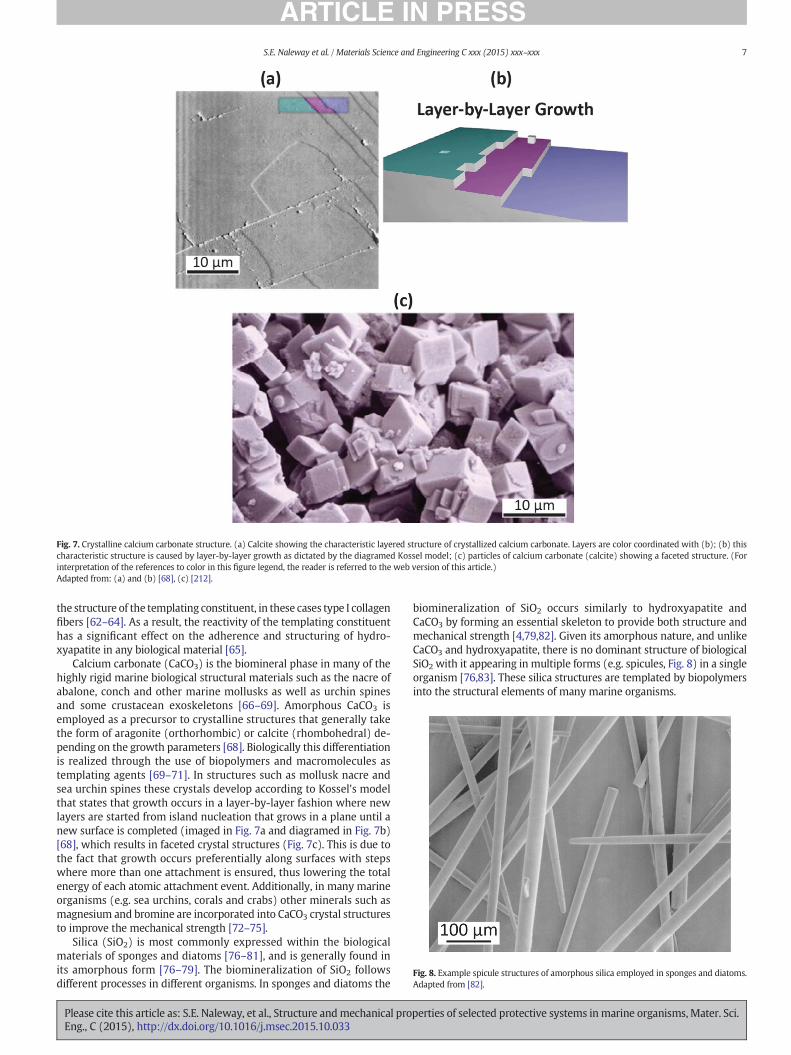
Fig. 8. Example spicule structures of amorphous silica employed in sponges and diatoms.Adapted from [82].
Fig. 7. Crystalline calcium carbonate structure. (a) Calcite showing the characteristic layered structure of crystallized calcium carbonate. Layers are color coordinated with (b); (b) thischaracteristic structure is caused by layer-by-layer growth as dictated by the diagramed Kossel model; (c) particles of calcium carbonate (calcite) showing a faceted structure. (Forinterpretation of the references to color in this figure legend, the reader is referred to the web version of this article.)Adapted from: (a) and (b) [68], (c) [212].
7S.E. Naleway et al. / Materials Science and Engineering C xxx (2015) xxx–xxx
the structure of the templating constituent, in these cases type I collagenfibers [62–64]. As a result, the reactivity of the templating constituenthas a significant effect on the adherence and structuring of hydro-xyapatite in any biological material [65].
Calcium carbonate (CaCO3) is the biomineral phase in many of thehighly rigid marine biological structural materials such as the nacre ofabalone, conch and other marine mollusks as well as urchin spinesand some crustacean exoskeletons [66–69]. Amorphous CaCO3 isemployed as a precursor to crystalline structures that generally takethe form of aragonite (orthorhombic) or calcite (rhombohedral) de-pending on the growth parameters [68]. Biologically this differentiationis realized through the use of biopolymers and macromolecules astemplating agents [69–71]. In structures such as mollusk nacre andsea urchin spines these crystals develop according to Kossel's modelthat states that growth occurs in a layer-by-layer fashion where newlayers are started from island nucleation that grows in a plane until anew surface is completed (imaged in Fig. 7a and diagramed in Fig. 7b)[68], which results in faceted crystal structures (Fig. 7c). This is due tothe fact that growth occurs preferentially along surfaces with stepswhere more than one attachment is ensured, thus lowering the totalenergy of each atomic attachment event. Additionally, in many marineorganisms (e.g. sea urchins, corals and crabs) other minerals such asmagnesium and bromine are incorporated into CaCO3 crystal structuresto improve the mechanical strength [72–75].
Silica (SiO2) is most commonly expressed within the biologicalmaterials of sponges and diatoms [76–81], and is generally found inits amorphous form [76–79]. The biomineralization of SiO2 followsdifferent processes in different organisms. In sponges and diatoms the
Please cite this article as: S.E. Naleway, et al., Structure andmechanical proEng., C (2015), http://dx.doi.org/10.1016/j.msec.2015.10.033
biomineralization of SiO2 occurs similarly to hydroxyapatite andCaCO3 by forming an essential skeleton to provide both structure andmechanical strength [4,79,82]. Given its amorphous nature, and unlikeCaCO3 and hydroxyapatite, there is no dominant structure of biologicalSiO2 with it appearing in multiple forms (e.g. spicules, Fig. 8) in a singleorganism [76,83]. These silica structures are templated by biopolymersinto the structural elements of many marine organisms.
perties of selected protective systems inmarine organisms, Mater. Sci.
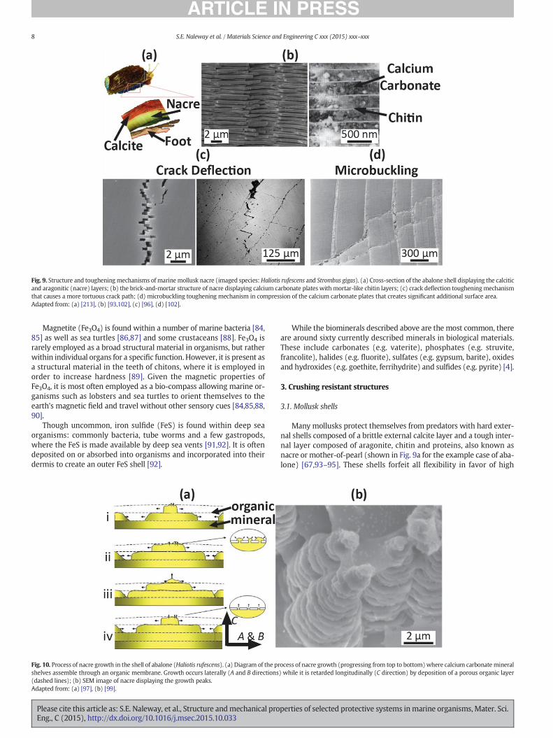
Fig. 9. Structure and tougheningmechanisms of marinemollusk nacre (imaged species: Haliotis rufescens and Strombus gigas). (a) Cross-section of the abalone shell displaying the calciticand aragonitic (nacre) layers; (b) the brick-and-mortar structure of nacre displaying calcium carbonate plates with mortar-like chitin layers; (c) crack deflection tougheningmechanismthat causes a more tortuous crack path; (d) microbuckling toughening mechanism in compression of the calcium carbonate plates that creates significant additional surface area.Adapted from: (a) [213], (b) [93,102], (c) [96], (d) [102].
8 S.E. Naleway et al. / Materials Science and Engineering C xxx (2015) xxx–xxx
Magnetite (Fe3O4) is found within a number of marine bacteria [84,85] as well as sea turtles [86,87] and some crustaceans [88]. Fe3O4 israrely employed as a broad structural material in organisms, but ratherwithin individual organs for a specific function. However, it is present asa structural material in the teeth of chitons, where it is employed inorder to increase hardness [89]. Given the magnetic properties ofFe3O4, it is most often employed as a bio-compass allowing marine or-ganisms such as lobsters and sea turtles to orient themselves to theearth's magnetic field and travel without other sensory cues [84,85,88,90].
Though uncommon, iron sulfide (FeS) is found within deep seaorganisms: commonly bacteria, tube worms and a few gastropods,where the FeS is made available by deep sea vents [91,92]. It is oftendeposited on or absorbed into organisms and incorporated into theirdermis to create an outer FeS shell [92].
Fig. 10. Process of nacre growth in the shell of abalone (Haliotis rufescens). (a) Diagram of the prshelves assemble through an organic membrane. Growth occurs laterally (A and B directions(dashed lines); (b) SEM image of nacre displaying the growth peaks.Adapted from: (a) [97], (b) [99].
Please cite this article as: S.E. Naleway, et al., Structure andmechanical proEng., C (2015), http://dx.doi.org/10.1016/j.msec.2015.10.033
While the biominerals described above are the most common, thereare around sixty currently described minerals in biological materials.These include carbonates (e.g. vaterite), phosphates (e.g. struvite,francolite), halides (e.g. fluorite), sulfates (e.g. gypsum, barite), oxidesand hydroxides (e.g. goethite, ferrihydrite) and sulfides (e.g. pyrite) [4].
3. Crushing resistant structures
3.1. Mollusk shells
Many mollusks protect themselves from predators with hard exter-nal shells composed of a brittle external calcite layer and a tough inter-nal layer composed of aragonite, chitin and proteins, also known asnacre or mother-of-pearl (shown in Fig. 9a for the example case of aba-lone) [67,93–95]. These shells forfeit all flexibility in favor of high
ocess of nacre growth (progressing from top to bottom)where calcium carbonatemineral) while it is retarded longitudinally (C direction) by deposition of a porous organic layer
perties of selected protective systems inmarine organisms, Mater. Sci.
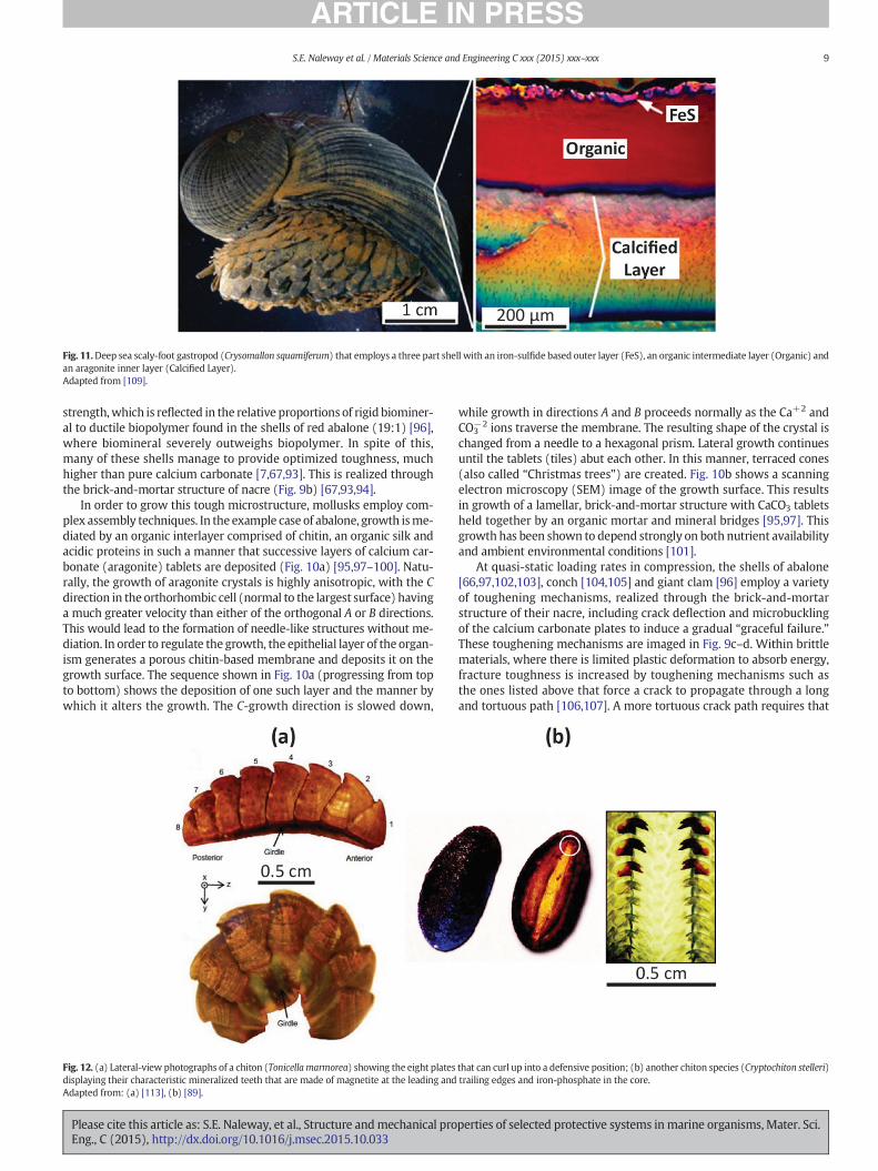
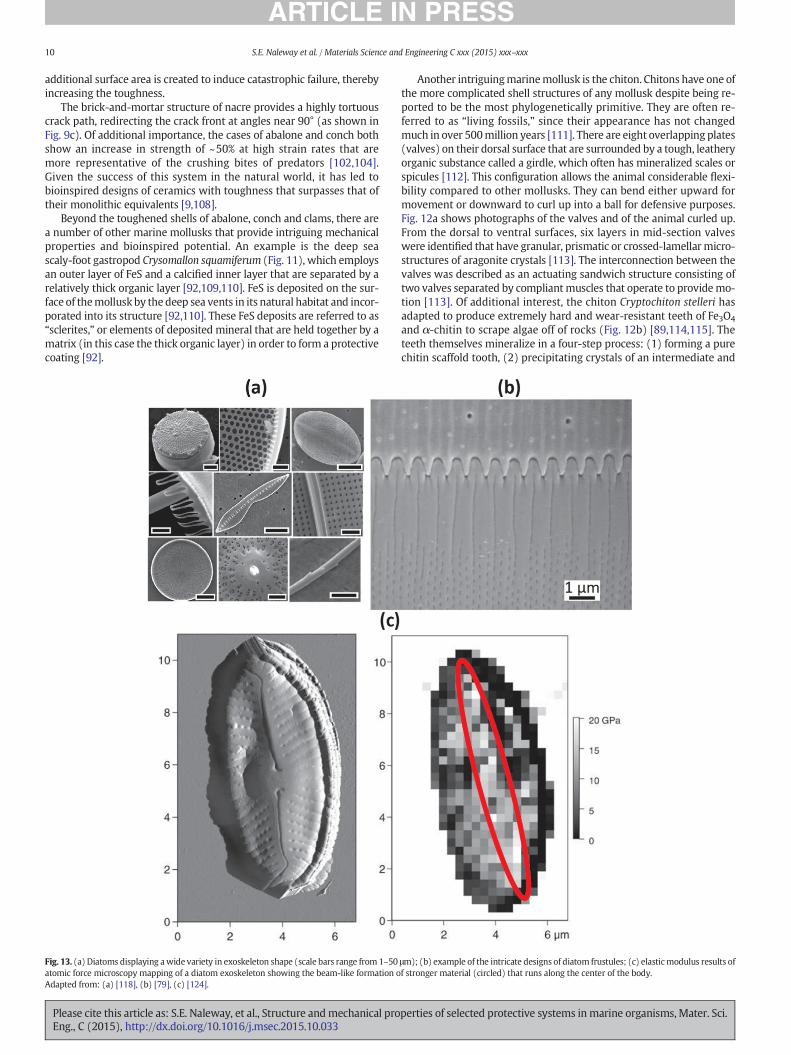
Fig. 11.Deep sea scaly-foot gastropod (Crysomallon squamiferum) that employs a three part shell with an iron-sulfide based outer layer (FeS), an organic intermediate layer (Organic) andan aragonite inner layer (Calcified Layer).Adapted from [109].
9S.E. Naleway et al. / Materials Science and Engineering C xxx (2015) xxx–xxx
strength,which is reflected in the relative proportions of rigid biominer-al to ductile biopolymer found in the shells of red abalone (19:1) [96],where biomineral severely outweighs biopolymer. In spite of this,many of these shells manage to provide optimized toughness, muchhigher than pure calcium carbonate [7,67,93]. This is realized throughthe brick-and-mortar structure of nacre (Fig. 9b) [67,93,94].
In order to grow this tough microstructure, mollusks employ com-plex assembly techniques. In the example case of abalone, growth isme-diated by an organic interlayer comprised of chitin, an organic silk andacidic proteins in such a manner that successive layers of calcium car-bonate (aragonite) tablets are deposited (Fig. 10a) [95,97–100]. Natu-rally, the growth of aragonite crystals is highly anisotropic, with the Cdirection in the orthorhombic cell (normal to the largest surface) havinga much greater velocity than either of the orthogonal A or B directions.This would lead to the formation of needle-like structures without me-diation. In order to regulate the growth, the epithelial layer of the organ-ism generates a porous chitin-based membrane and deposits it on thegrowth surface. The sequence shown in Fig. 10a (progressing from topto bottom) shows the deposition of one such layer and the manner bywhich it alters the growth. The C-growth direction is slowed down,
Fig. 12. (a) Lateral-view photographs of a chiton (Tonicella marmorea) showing the eight platesdisplaying their characteristic mineralized teeth that are made of magnetite at the leading andAdapted from: (a) [113], (b) [89].
Please cite this article as: S.E. Naleway, et al., Structure andmechanical proEng., C (2015), http://dx.doi.org/10.1016/j.msec.2015.10.033
while growth in directions A and B proceeds normally as the Ca+2 andCO3
−2 ions traverse the membrane. The resulting shape of the crystal ischanged from a needle to a hexagonal prism. Lateral growth continuesuntil the tablets (tiles) abut each other. In this manner, terraced cones(also called “Christmas trees”) are created. Fig. 10b shows a scanningelectron microscopy (SEM) image of the growth surface. This resultsin growth of a lamellar, brick-and-mortar structure with CaCO3 tabletsheld together by an organic mortar and mineral bridges [95,97]. Thisgrowth has been shown to depend strongly on both nutrient availabilityand ambient environmental conditions [101].
At quasi-static loading rates in compression, the shells of abalone[66,97,102,103], conch [104,105] and giant clam [96] employ a varietyof toughening mechanisms, realized through the brick-and-mortarstructure of their nacre, including crack deflection and microbucklingof the calcium carbonate plates to induce a gradual “graceful failure.”These toughening mechanisms are imaged in Fig. 9c–d. Within brittlematerials, where there is limited plastic deformation to absorb energy,fracture toughness is increased by toughening mechanisms such asthe ones listed above that force a crack to propagate through a longand tortuous path [106,107]. A more tortuous crack path requires that
that can curl up into a defensive position; (b) another chiton species (Cryptochiton stelleri)trailing edges and iron-phosphate in the core.
perties of selected protective systems inmarine organisms, Mater. Sci.
10 S.E. Naleway et al. / Materials Science and Engineering C xxx (2015) xxx–xxx
additional surface area is created to induce catastrophic failure, therebyincreasing the toughness.
The brick-and-mortar structure of nacre provides a highly tortuouscrack path, redirecting the crack front at angles near 90° (as shown inFig. 9c). Of additional importance, the cases of abalone and conch bothshow an increase in strength of ~50% at high strain rates that aremore representative of the crushing bites of predators [102,104].Given the success of this system in the natural world, it has led tobioinspired designs of ceramics with toughness that surpasses that oftheir monolithic equivalents [9,108].
Beyond the toughened shells of abalone, conch and clams, there area number of other marine mollusks that provide intriguing mechanicalproperties and bioinspired potential. An example is the deep seascaly-foot gastropod Crysomallon squamiferum (Fig. 11), which employsan outer layer of FeS and a calcified inner layer that are separated by arelatively thick organic layer [92,109,110]. FeS is deposited on the sur-face of themollusk by the deep sea vents in its natural habitat and incor-porated into its structure [92,110]. These FeS deposits are referred to as“sclerites,” or elements of deposited mineral that are held together by amatrix (in this case the thick organic layer) in order to form a protectivecoating [92].
Fig. 13. (a) Diatoms displaying awide variety in exoskeleton shape (scale bars range from 1–50atomic force microscopy mapping of a diatom exoskeleton showing the beam-like formation oAdapted from: (a) [118], (b) [79], (c) [124].
Please cite this article as: S.E. Naleway, et al., Structure andmechanical proEng., C (2015), http://dx.doi.org/10.1016/j.msec.2015.10.033
Another intriguingmarinemollusk is the chiton. Chitons have one ofthe more complicated shell structures of any mollusk despite being re-ported to be the most phylogenetically primitive. They are often re-ferred to as “living fossils,” since their appearance has not changedmuch in over 500million years [111]. There are eight overlapping plates(valves) on their dorsal surface that are surrounded by a tough, leatheryorganic substance called a girdle, which often has mineralized scales orspicules [112]. This configuration allows the animal considerable flexi-bility compared to other mollusks. They can bend either upward formovement or downward to curl up into a ball for defensive purposes.Fig. 12a shows photographs of the valves and of the animal curled up.From the dorsal to ventral surfaces, six layers in mid-section valveswere identified that have granular, prismatic or crossed-lamellarmicro-structures of aragonite crystals [113]. The interconnection between thevalves was described as an actuating sandwich structure consisting oftwo valves separated by compliant muscles that operate to providemo-tion [113]. Of additional interest, the chiton Cryptochiton stelleri hasadapted to produce extremely hard and wear-resistant teeth of Fe3O4
and α-chitin to scrape algae off of rocks (Fig. 12b) [89,114,115]. Theteeth themselves mineralize in a four-step process: (1) forming a purechitin scaffold tooth, (2) precipitating crystals of an intermediate and
μm); (b) example of the intricate designs of diatom frustules; (c) elasticmodulus results off stronger material (circled) that runs along the center of the body.
perties of selected protective systems inmarine organisms, Mater. Sci.
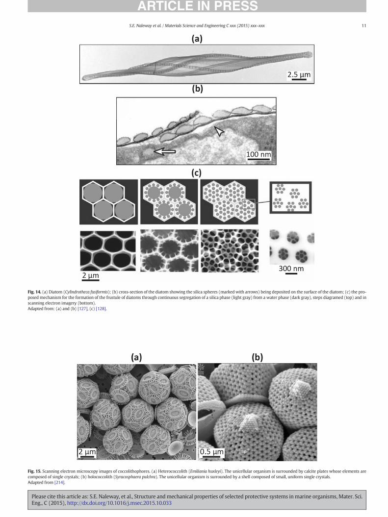
Fig. 15. Scanning electron microscopy images of coccolithophores. (a) Heterococcolith (Emiliania huxleyi). The unicellular organism is surrounded by calcite plates whose elements arecomposed of single crystals; (b) holococcolith (Syracosphaera pulchra). The unicellular organism is surrounded by a shell composed of small, uniform single crystals.Adapted from [214].
Fig. 14. (a) Diatom (Cylindrotheca fusiformis); (b) cross-section of the diatom showing the silica spheres (marked with arrows) being deposited on the surface of the diatom; (c) the pro-posed mechanism for the formation of the frustule of diatoms through continuous segregation of a silica phase (light gray) from a water phase (dark gray), steps diagramed (top) and inscanning electron imagery (bottom).Adapted from: (a) and (b) [127], (c) [128].
11S.E. Naleway et al. / Materials Science and Engineering C xxx (2015) xxx–xxx
Please cite this article as: S.E. Naleway, et al., Structure andmechanical properties of selected protective systems inmarine organisms, Mater. Sci.Eng., C (2015), http://dx.doi.org/10.1016/j.msec.2015.10.033
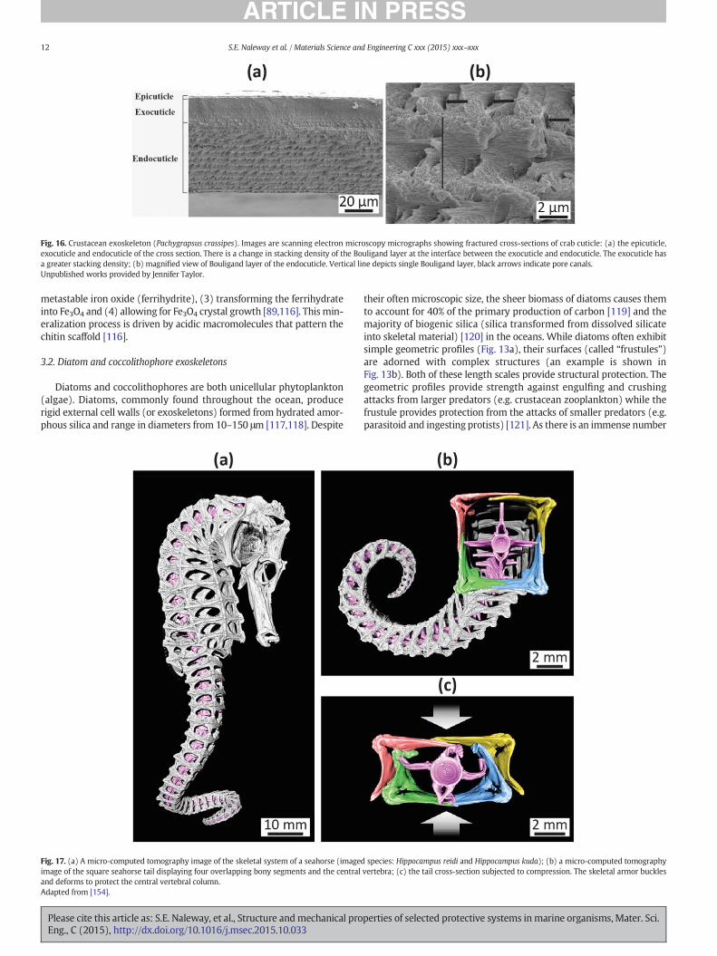
Fig. 16. Crustacean exoskeleton (Pachygrapsus crassipes). Images are scanning electron microscopy micrographs showing fractured cross-sections of crab cuticle: (a) the epicuticle,exocuticle and endocuticle of the cross section. There is a change in stacking density of the Bouligand layer at the interface between the exocuticle and endocuticle. The exocuticle hasa greater stacking density; (b) magnified view of Bouligand layer of the endocuticle. Vertical line depicts single Bouligand layer, black arrows indicate pore canals.Unpublished works provided by Jennifer Taylor.
12 S.E. Naleway et al. / Materials Science and Engineering C xxx (2015) xxx–xxx
metastable iron oxide (ferrihydrite), (3) transforming the ferrihydrateinto Fe3O4 and (4) allowing for Fe3O4 crystal growth [89,116]. This min-eralization process is driven by acidic macromolecules that pattern thechitin scaffold [116].
3.2. Diatom and coccolithophore exoskeletons
Diatoms and coccolithophores are both unicellular phytoplankton(algae). Diatoms, commonly found throughout the ocean, producerigid external cell walls (or exoskeletons) formed from hydrated amor-phous silica and range in diameters from 10–150 μm [117,118]. Despite
Fig. 17. (a) A micro-computed tomography image of the skeletal system of a seahorse (imageimage of the square seahorse tail displaying four overlapping bony segments and the centraland deforms to protect the central vertebral column.Adapted from [154].
Please cite this article as: S.E. Naleway, et al., Structure andmechanical proEng., C (2015), http://dx.doi.org/10.1016/j.msec.2015.10.033
their often microscopic size, the sheer biomass of diatoms causes themto account for 40% of the primary production of carbon [119] and themajority of biogenic silica (silica transformed from dissolved silicateinto skeletal material) [120] in the oceans. While diatoms often exhibitsimple geometric profiles (Fig. 13a), their surfaces (called “frustules”)are adorned with complex structures (an example is shown inFig. 13b). Both of these length scales provide structural protection. Thegeometric profiles provide strength against engulfing and crushingattacks from larger predators (e.g. crustacean zooplankton) while thefrustule provides protection from the attacks of smaller predators (e.g.parasitoid and ingesting protists) [121]. As there is an immense number
d species: Hippocampus reidi and Hippocampus kuda); (b) a micro-computed tomographyvertebra; (c) the tail cross-section subjected to compression. The skeletal armor buckles
perties of selected protective systems inmarine organisms, Mater. Sci.
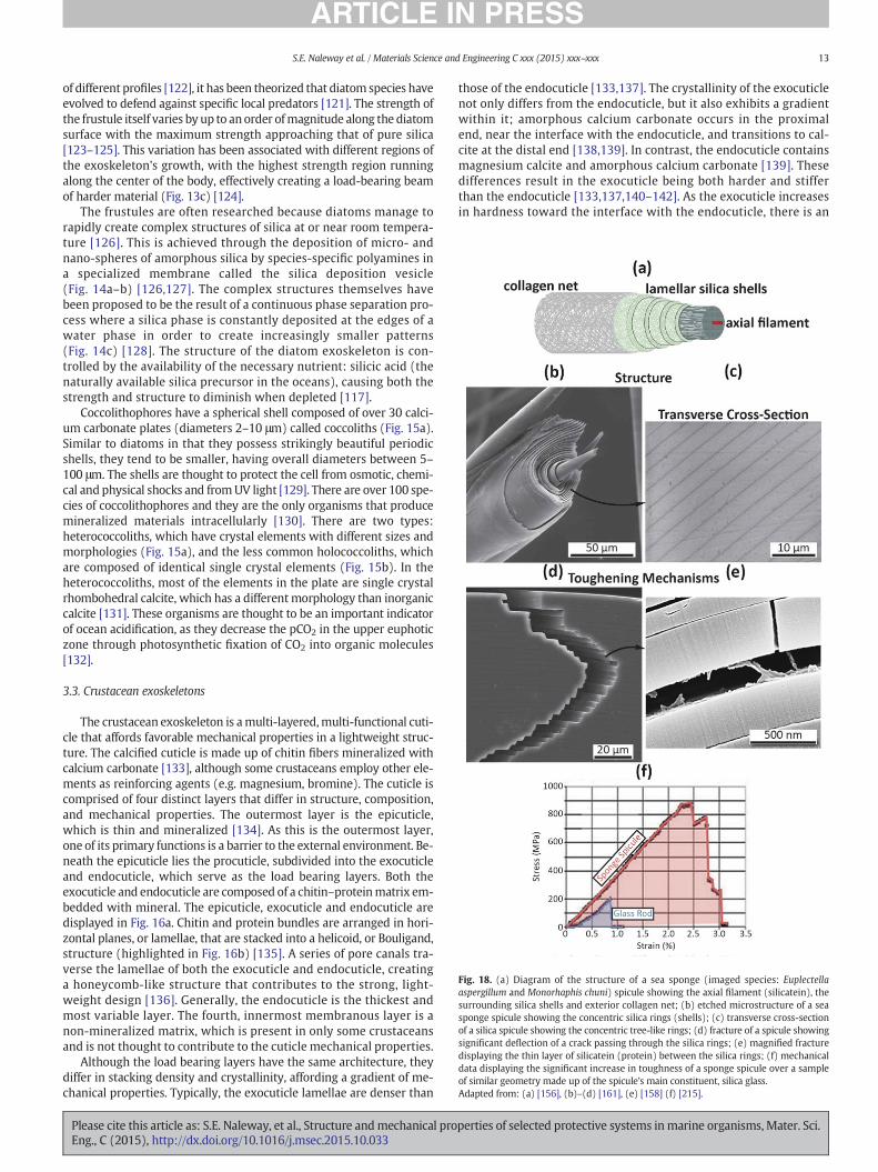
Fig. 18. (a) Diagram of the structure of a sea sponge (imaged species: Euplectellaaspergillum and Monorhaphis chuni) spicule showing the axial filament (silicatein), thesurrounding silica shells and exterior collagen net; (b) etched microstructure of a seasponge spicule showing the concentric silica rings (shells); (c) transverse cross-sectionof a silica spicule showing the concentric tree-like rings; (d) fracture of a spicule showingsignificant deflection of a crack passing through the silica rings; (e) magnified fracturedisplaying the thin layer of silicatein (protein) between the silica rings; (f) mechanicaldata displaying the significant increase in toughness of a sponge spicule over a sampleof similar geometry made up of the spicule's main constituent, silica glass.Adapted from: (a) [156], (b)–(d) [161], (e) [158] (f) [215].
13S.E. Naleway et al. / Materials Science and Engineering C xxx (2015) xxx–xxx
of different profiles [122], it has been theorized that diatom species haveevolved to defend against specific local predators [121]. The strength ofthe frustule itself varies by up to anorder ofmagnitude along the diatomsurface with the maximum strength approaching that of pure silica[123–125]. This variation has been associated with different regions ofthe exoskeleton's growth, with the highest strength region runningalong the center of the body, effectively creating a load-bearing beamof harder material (Fig. 13c) [124].
The frustules are often researched because diatoms manage torapidly create complex structures of silica at or near room tempera-ture [126]. This is achieved through the deposition of micro- andnano-spheres of amorphous silica by species-specific polyamines ina specialized membrane called the silica deposition vesicle(Fig. 14a–b) [126,127]. The complex structures themselves havebeen proposed to be the result of a continuous phase separation pro-cess where a silica phase is constantly deposited at the edges of awater phase in order to create increasingly smaller patterns(Fig. 14c) [128]. The structure of the diatom exoskeleton is con-trolled by the availability of the necessary nutrient: silicic acid (thenaturally available silica precursor in the oceans), causing both thestrength and structure to diminish when depleted [117].
Coccolithophores have a spherical shell composed of over 30 calci-um carbonate plates (diameters 2–10 μm) called coccoliths (Fig. 15a).Similar to diatoms in that they possess strikingly beautiful periodicshells, they tend to be smaller, having overall diameters between 5–100 μm. The shells are thought to protect the cell from osmotic, chemi-cal and physical shocks and fromUV light [129]. There are over 100 spe-cies of coccolithophores and they are the only organisms that producemineralized materials intracellularly [130]. There are two types:heterococcoliths, which have crystal elements with different sizes andmorphologies (Fig. 15a), and the less common holococcoliths, whichare composed of identical single crystal elements (Fig. 15b). In theheterococcoliths, most of the elements in the plate are single crystalrhombohedral calcite, which has a different morphology than inorganiccalcite [131]. These organisms are thought to be an important indicatorof ocean acidification, as they decrease the pCO2 in the upper euphoticzone through photosynthetic fixation of CO2 into organic molecules[132].
3.3. Crustacean exoskeletons
The crustacean exoskeleton is amulti-layered,multi-functional cuti-cle that affords favorable mechanical properties in a lightweight struc-ture. The calcified cuticle is made up of chitin fibers mineralized withcalcium carbonate [133], although some crustaceans employ other ele-ments as reinforcing agents (e.g. magnesium, bromine). The cuticle iscomprised of four distinct layers that differ in structure, composition,and mechanical properties. The outermost layer is the epicuticle,which is thin and mineralized [134]. As this is the outermost layer,one of its primary functions is a barrier to the external environment. Be-neath the epicuticle lies the procuticle, subdivided into the exocuticleand endocuticle, which serve as the load bearing layers. Both theexocuticle and endocuticle are composed of a chitin–proteinmatrix em-bedded with mineral. The epicuticle, exocuticle and endocuticle aredisplayed in Fig. 16a. Chitin and protein bundles are arranged in hori-zontal planes, or lamellae, that are stacked into a helicoid, or Bouligand,structure (highlighted in Fig. 16b) [135]. A series of pore canals tra-verse the lamellae of both the exocuticle and endocuticle, creatinga honeycomb-like structure that contributes to the strong, light-weight design [136]. Generally, the endocuticle is the thickest andmost variable layer. The fourth, innermost membranous layer is anon-mineralized matrix, which is present in only some crustaceansand is not thought to contribute to the cuticle mechanical properties.
Although the load bearing layers have the same architecture, theydiffer in stacking density and crystallinity, affording a gradient of me-chanical properties. Typically, the exocuticle lamellae are denser than
Please cite this article as: S.E. Naleway, et al., Structure andmechanical proEng., C (2015), http://dx.doi.org/10.1016/j.msec.2015.10.033
those of the endocuticle [133,137]. The crystallinity of the exocuticlenot only differs from the endocuticle, but it also exhibits a gradientwithin it; amorphous calcium carbonate occurs in the proximalend, near the interface with the endocuticle, and transitions to cal-cite at the distal end [138,139]. In contrast, the endocuticle containsmagnesium calcite and amorphous calcium carbonate [139]. Thesedifferences result in the exocuticle being both harder and stifferthan the endocuticle [133,137,140–142]. As the exocuticle increasesin hardness toward the interface with the endocuticle, there is an
perties of selected protective systems inmarine organisms, Mater. Sci.
14 S.E. Naleway et al. / Materials Science and Engineering C xxx (2015) xxx–xxx
abrupt change in both hardness and stiffness of more than an orderof magnitude between these two layers [137].
Despite the cuticle being brittle and prone to delamination [133,143,144], the Bouligand structure of the procuticle layers affords sometoughness by crack deflection and bridging, which force cracks to prop-agate in a tortuous, stepwise fashion [142]. The ductile pore canal tu-bules are also thought to enhance toughness, as necking is observedduring tensile testing normal to the cuticle surface [133].
Crustaceans exhibit variation in cuticle mineralization and structure(e.g. layer thickness, stacking density) across species and across func-tional regions within an individual. For example, appendage joints con-tain non-calcified arthrodial membranes that must remain soft andflexible to permit rotation [145]. There are also great differences be-tween the cuticle of the carapace, whichprotects internal organs and re-sists compression, the walking legs, which primarily resist musclecontraction forces, and the claws, which function as weapons and preycapture devices. In crab claws, for example, the exocuticle is 3–4 timesharder than the walking leg exocuticle, and this is primarily due to itsgreater mineral content [133]. The effectiveness of claws in securingprey depends not only on their hardness, but also on their resistanceto cracking and abrasion. In some crabs, such as stone crabs, the clawtips are darkly pigmented and even harder and tougher than non-pigmented areas, possibly due to the increased cross-linking associatedwith tanning and reduced porosity [146]. Other crabs, such as shorecrabs, enhance their claws by adding bromine rich tips that can affordan order of magnitude increase in fracture resistance compared to calci-fied tips [74,75].
The intricacy and variability of the crustacean cuticle is evenmore impressive considering that, unlike mollusk shells that form by
Fig. 19. (a) Hierarchical skeleton macrostructure of the Venus' flower basket sponge (Euplectemploys with diagrams, optical microscopy and scanning electron microscopy images: (b) expredominately on the interior of the lattice and the vertical struts are predominately on the exAdapted from: (a) [158], (b)–(d) [157].
Please cite this article as: S.E. Naleway, et al., Structure andmechanical proEng., C (2015), http://dx.doi.org/10.1016/j.msec.2015.10.033
accretion, crustaceans repeatedly shed and secrete a whole new cuticleduring the process ofmolting. The secretion ofmatrix to form the cuticlelayers and the subsequent deposition of mineral occur rapidly, over thecourse of days, and as frequently as once per week in small juveniles.Cuticle formation is a conserved and intricate process, but can be affect-ed by environmental conditions such as ocean carbon chemistry.
3.4. Seahorse skeleton
Seahorses (Hippocampus) have plated skeletons that cover theirbodies and prehensile tails (Fig. 17a). Unlike most fish, seahorsesswim upright, utilizing their dorsal fin for propulsion and two pectoralfins for maneuverability, resulting in slow swimming velocities[147–149]. Thus, they use their prehensile tails for stability, grippingand holding onto objects such as sea grasses, mangrove roots, andcoral reefs [147]. The tail skeleton is composed of several articulatingsegments arranged into cross-sectional squares, each composed offour bony plates that surround a central vertebra (Fig. 17b) [147–154].These plates are connected by overlapping joints that allow them suffi-cient flexibility for grasping as well as added strength for armored pro-tection [154]. In grasping, the square structure of the tail provides moresurface contact and a mechanism for maintaining organization of thearticulating plates [154]. When the tail (of a deceased seahorse) iscrushed, it can be compressed up to nearly 50% of its original widthbefore fracture of the vertebral column (Fig. 17c) [150]. This unique en-ergy absorption mechanism provides seahorses protection againstpredators, many of whom attack by crushing (e.g. claws of crabs,beaks of sea turtles and birds) [155].
ella aspergillum). Examples of the complex structure of struts that Euplectella aspergillumterior spiraling ridges; (c) horizontal and vertical struts where the horizontal struts areterior of the lattice; (d) diagonal struts.
perties of selected protective systems inmarine organisms, Mater. Sci.
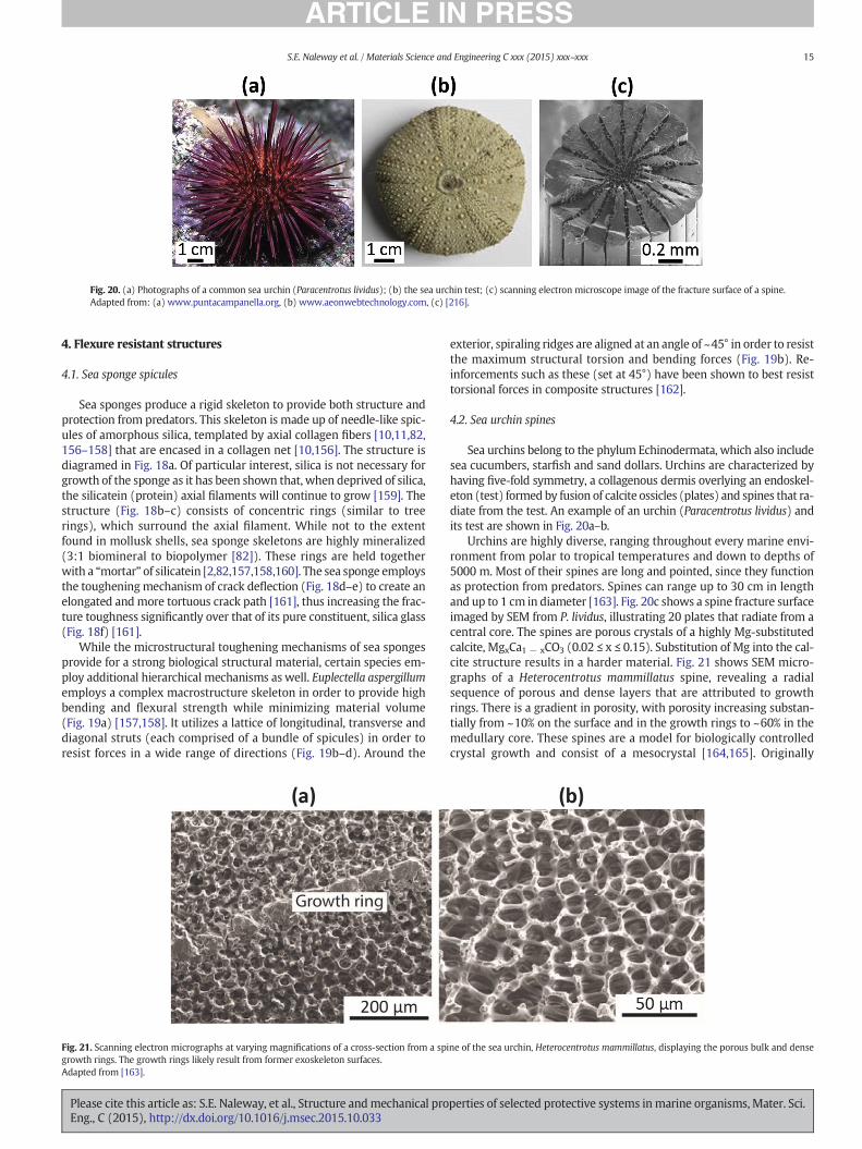
Fig. 20. (a) Photographs of a common sea urchin (Paracentrotus lividus); (b) the sea urchin test; (c) scanning electron microscope image of the fracture surface of a spine.Adapted from: (a) www.puntacampanella.org, (b) www.aeonwebtechnology.com, (c) [216].
15S.E. Naleway et al. / Materials Science and Engineering C xxx (2015) xxx–xxx
4. Flexure resistant structures
4.1. Sea sponge spicules
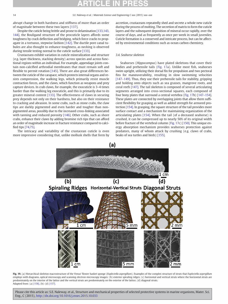
Sea sponges produce a rigid skeleton to provide both structure andprotection from predators. This skeleton is made up of needle-like spic-ules of amorphous silica, templated by axial collagen fibers [10,11,82,156–158] that are encased in a collagen net [10,156]. The structure isdiagramed in Fig. 18a. Of particular interest, silica is not necessary forgrowth of the sponge as it has been shown that, when deprived of silica,the silicatein (protein) axial filaments will continue to grow [159]. Thestructure (Fig. 18b–c) consists of concentric rings (similar to treerings), which surround the axial filament. While not to the extentfound in mollusk shells, sea sponge skeletons are highly mineralized(3:1 biomineral to biopolymer [82]). These rings are held togetherwith a “mortar” of silicatein [2,82,157,158,160]. The sea sponge employsthe tougheningmechanism of crack deflection (Fig. 18d–e) to create anelongated and more tortuous crack path [161], thus increasing the frac-ture toughness significantly over that of its pure constituent, silica glass(Fig. 18f) [161].
While the microstructural toughening mechanisms of sea spongesprovide for a strong biological structural material, certain species em-ploy additional hierarchical mechanisms as well. Euplectella aspergillumemploys a complex macrostructure skeleton in order to provide highbending and flexural strength while minimizing material volume(Fig. 19a) [157,158]. It utilizes a lattice of longitudinal, transverse anddiagonal struts (each comprised of a bundle of spicules) in order toresist forces in a wide range of directions (Fig. 19b–d). Around the
Fig. 21. Scanning electron micrographs at varying magnifications of a cross-section from a spigrowth rings. The growth rings likely result from former exoskeleton surfaces.Adapted from [163].
Please cite this article as: S.E. Naleway, et al., Structure andmechanical proEng., C (2015), http://dx.doi.org/10.1016/j.msec.2015.10.033
exterior, spiraling ridges are aligned at an angle of ~45° in order to resistthe maximum structural torsion and bending forces (Fig. 19b). Re-inforcements such as these (set at 45°) have been shown to best resisttorsional forces in composite structures [162].
4.2. Sea urchin spines
Sea urchins belong to the phylum Echinodermata, which also includesea cucumbers, starfish and sand dollars. Urchins are characterized byhaving five-fold symmetry, a collagenous dermis overlying an endoskel-eton (test) formed by fusion of calcite ossicles (plates) and spines that ra-diate from the test. An example of an urchin (Paracentrotus lividus) andits test are shown in Fig. 20a–b.
Urchins are highly diverse, ranging throughout every marine envi-ronment from polar to tropical temperatures and down to depths of5000 m. Most of their spines are long and pointed, since they functionas protection from predators. Spines can range up to 30 cm in lengthand up to 1 cm in diameter [163]. Fig. 20c shows a spine fracture surfaceimaged by SEM from P. lividus, illustrating 20 plates that radiate from acentral core. The spines are porous crystals of a highly Mg-substitutedcalcite, MgxCa1 − xCO3 (0.02 ≤ x ≤ 0.15). Substitution of Mg into the cal-cite structure results in a harder material. Fig. 21 shows SEM micro-graphs of a Heterocentrotus mammillatus spine, revealing a radialsequence of porous and dense layers that are attributed to growthrings. There is a gradient in porosity, with porosity increasing substan-tially from ~10% on the surface and in the growth rings to ~60% in themedullary core. These spines are a model for biologically controlledcrystal growth and consist of a mesocrystal [164,165]. Originally
ne of the sea urchin, Heterocentrotus mammillatus, displaying the porous bulk and dense
perties of selected protective systems inmarine organisms, Mater. Sci.
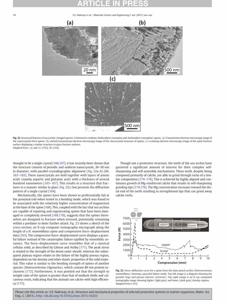
Fig. 23. Force–deflection curve for a spine from the slate pencil urchin (Heterocentrotusmammillatus) showing a graceful failure mode. Top left image is a diagram showing thegrowth rings and porous interior (stereom). Top right image is an X-ray computedtomography image showing higher (light grey) and lower (dark grey) density regions.Adapted from [163].
Fig. 22. Structural features of sea urchin (imaged species: Echinometramathaei, Anthocidaris crassispina and Authoeidaris erassispina) spines. (a) Transmission electronmicroscopy image ofthe nanocrystals from spines; (b) etched transmission electron microscopy image of the mesocrystal structure of spines; (c) scanning electron microscopy image of the spine fracturesurface displaying a similar structure to glass fracture surfaces.Adapted from: (a) and (c) [165], (b) [164].
16 S.E. Naleway et al. / Materials Science and Engineering C xxx (2015) xxx–xxx
thought to be a single crystal [166,167], it has recently been shown thatthe structure consists of periodic and uniform nanocrystals, 30–50 nmin diameter, with parallel crystallographic alignment (Fig. 22a–b) [68,163–165]. These nanocrystals are held together with layers of aminoacids (mainly aspartic and glutamic acid) with a thickness of severalhundred nanometers [165–167]. This results in a structure that frac-tures in a manner similar to glass (Fig. 22c) but presents the diffractionpattern of a single crystal [164].
Mechanically, the spines have been shown to preferentially fail atthe proximal end when tested in a bending mode, which was found tobe associated with the relatively higher concentration of magnesiumat the base of the spine [168]. This, coupledwith the fact that sea urchinsare capable of repairing and regenerating spines that have been dam-aged or completely severed [169,170], suggests that the spines them-selves are designed to fracture when stressed, potentially remainingwithin a predator to deter further attack. Fig. 23 shows a sketch of thecross-section, an X-ray computer tomography micrograph along thelength of a H. mammillatus spine and compressive force–displacementdata [163]. The compressive force–displacement curve displays a grace-ful failure instead of the catastrophic failure typified by monolithic ce-ramics. The force–displacement curve resembles that of a classicalcellular solid, as described by Gibson and Ashby [171]. The peak stressis related to the strength of the dense outer sheath, whereas the subse-quent plateau region relates to the failure of the highly porous region,dependent on the density and other elastic properties of the solidmate-rial. This value is similar to the bending strength of spines of anotherspecies (Heterocentrotus trigonarius), which contain 80 nm protein in-clusions [172]. Furthermore, it was pointed out that the strength toweight ratio of the spines is greater than that of mollusk shells and cal-careous rocks, indicating that the animals use calcite with high efficien-cy [173].
Please cite this article as: S.E. Naleway, et al., Structure andmechanical proEng., C (2015), http://dx.doi.org/10.1016/j.msec.2015.10.033
Though not a protective structure, the teeth of the sea urchin havegarnered a significant amount of interest for their complex self-sharpening and self-assembly mechanisms. These teeth, despite beingcomposed primarily of calcite, are able to grind through rocks of a sim-ilar composition [174–176]. This is achieved by highly aligned and con-tinuous growth of Mg-reinforced calcite that results in self-sharpeninggrinding tips [174,176]. TheMg concentration increases toward the dis-tal end of the teeth resulting in strengthened tips that can grind awaycalcite rocks.
perties of selected protective systems inmarine organisms, Mater. Sci.
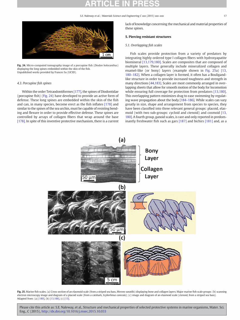
Fig. 24. Micro-computed tomography image of a porcupine fish (Diodon holocanthus)displaying the long spines embedded within the skin of the fish.Unpublished works provided by Frances Su (UCSD).
17S.E. Naleway et al. / Materials Science and Engineering C xxx (2015) xxx–xxx
4.3. Porcupine fish spines
Within the order Tetraodontiformes [177], the spines of Diodontidae(porcupine fish) (Fig. 24) have developed to provide an active form ofdefense. These long spines are embedded within the skin of the fishand can, in many species, become erect as the fish inflates [178] andsimilar to the spines of the sea urchin,must be capable of resisting bend-ing and flexure in order to provide effective defense. These spines arecontrolled by arrays of collagen fibers that wrap around the base[178]. In spite of this inventive protective mechanism, there is a current
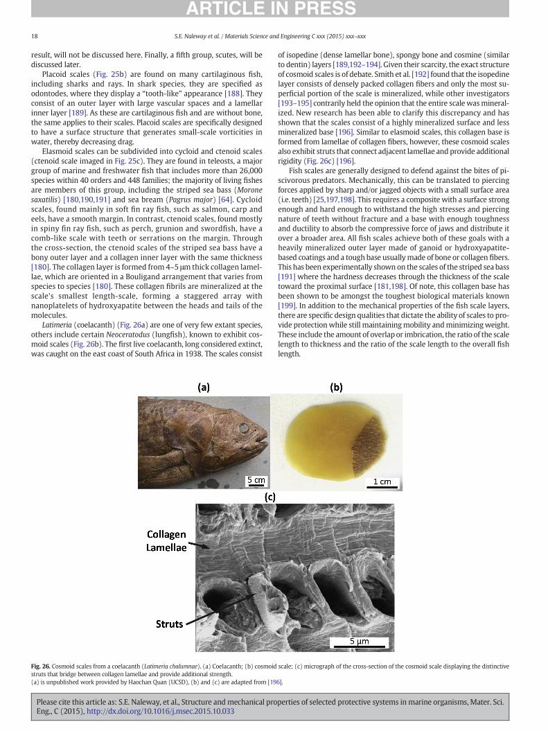
Fig. 25.Marinefish scales. (a) Cross-section of an elasmoid scale (from a striped sea bass,Moroneelectron microscopy image and diagram of a placoid scale (from a catshark, Scyliorhinus caniculAdapted from: (a) [180], (b) [13,188], (c) [13].
Please cite this article as: S.E. Naleway, et al., Structure andmechanical proEng., C (2015), http://dx.doi.org/10.1016/j.msec.2015.10.033
lack of knowledge concerning themechanical andmaterial properties ofthese spines.
5. Piercing resistant structures
5.1. Overlapping fish scales
Fish scales provide protection from a variety of predators byintegrating highly ordered type I collagen fibers with hydroxyapatitebiomineral [13,179,180]. Scales are composites that are composed ofmultiple layers. These generally include mineralized collagen andenamel-like (or bony) layers (example shown in Fig. 25a) [13,180–182]. When a collagen layer is formed, it often has a Bouligand-like structure in order to provide increased toughness and strength inmany directions [44,183]. Scales are most commonly arranged in over-lapping sheets that allow for smoothmotion of the body for locomotionwhile ensuring full coverage for protection from predators [13,180].This overlapping pattern minimizes drag to ease swimming by regulat-ing wave propagation about the body [184–186]. While scales can varygreatly in size, shape and arrangement from species to species, theyhave been classified into three relevant general groups: placoid, elas-moid (with two sub-groups: cycloid and ctenoid) and cosmoid [13,180]. A fourth group, ganoid scales, is rare and only reported in predom-inantly freshwater fish such as gars [187] and bichirs [181] and, as a
saxatilis) displaying bone and collagen layers. Majormarine fish scale groups: (b) scanninga); (c) image and diagram of an elasmoid scale (ctenoid, from a striped sea bass).
perties of selected protective systems inmarine organisms, Mater. Sci.
18 S.E. Naleway et al. / Materials Science and Engineering C xxx (2015) xxx–xxx
result, will not be discussed here. Finally, a fifth group, scutes, will bediscussed later.
Placoid scales (Fig. 25b) are found on many cartilaginous fish,including sharks and rays. In shark species, they are specified asodontodes, where they display a “tooth-like” appearance [188]. Theyconsist of an outer layer with large vascular spaces and a lamellarinner layer [189]. As these are cartilaginous fish and are without bone,the same applies to their scales. Placoid scales are specifically designedto have a surface structure that generates small-scale vorticities inwater, thereby decreasing drag.
Elasmoid scales can be subdivided into cycloid and ctenoid scales(ctenoid scale imaged in Fig. 25c). They are found in teleosts, a majorgroup of marine and freshwater fish that includes more than 26,000species within 40 orders and 448 families; the majority of living fishesare members of this group, including the striped sea bass (Moronesaxatilis) [180,190,191] and sea bream (Pagrus major) [64]. Cycloidscales, found mainly in soft fin ray fish, such as salmon, carp andeels, have a smooth margin. In contrast, ctenoid scales, found mostlyin spiny fin ray fish, such as perch, grunion and swordfish, have acomb-like scale with teeth or serrations on the margin. Throughthe cross-section, the ctenoid scales of the striped sea bass have abony outer layer and a collagen inner layer with the same thickness[180]. The collagen layer is formed from 4–5 μm thick collagen lamel-lae, which are oriented in a Bouligand arrangement that varies fromspecies to species [180]. These collagen fibrils are mineralized at thescale's smallest length-scale, forming a staggered array withnanoplatelets of hydroxyapatite between the heads and tails of themolecules.
Latimeria (coelacanth) (Fig. 26a) are one of very few extant species,others include certain Neoceratodus (lungfish), known to exhibit cos-moid scales (Fig. 26b). The first live coelacanth, long considered extinct,was caught on the east coast of South Africa in 1938. The scales consist
Fig. 26. Cosmoid scales from a coelacanth (Latimeria chalumnae). (a) Coelacanth; (b) cosmoidstruts that bridge between collagen lamellae and provide additional strength.(a) is unpublished work provided by Haochan Quan (UCSD), (b) and (c) are adapted from [19
Please cite this article as: S.E. Naleway, et al., Structure andmechanical proEng., C (2015), http://dx.doi.org/10.1016/j.msec.2015.10.033
of isopedine (dense lamellar bone), spongy bone and cosmine (similarto dentin) layers [189,192–194]. Given their scarcity, the exact structureof cosmoid scales is of debate. Smith et al. [192] found that the isopedinelayer consists of densely packed collagen fibers and only the most su-perficial portion of the scale is mineralized, while other investigators[193–195] contrarily held the opinion that the entire scale wasmineral-ized. New research has been able to clarify this discrepancy and hasshown that the scales consist of a highly mineralized surface and lessmineralized base [196]. Similar to elasmoid scales, this collagen base isformed from lamellae of collagen fibers, however, these cosmoid scalesalso exhibit struts that connect adjacent lamellae and provide additionalrigidity (Fig. 26c) [196].
Fish scales are generally designed to defend against the bites of pi-scivorous predators. Mechanically, this can be translated to piercingforces applied by sharp and/or jagged objects with a small surface area(i.e. teeth) [25,197,198]. This requires a composite with a surface strongenough and hard enough to withstand the high stresses and piercingnature of teeth without fracture and a base with enough toughnessand ductility to absorb the compressive force of jaws and distribute itover a broader area. All fish scales achieve both of these goals with aheavily mineralized outer layer made of ganoid or hydroxyapatite-based coatings and a tough base usuallymade of bone or collagen fibers.This has been experimentally shown on the scales of the striped sea bass[191] where the hardness decreases through the thickness of the scaletoward the proximal surface [181,198]. Of note, this collagen base hasbeen shown to be amongst the toughest biological materials known[199]. In addition to the mechanical properties of the fish scale layers,there are specific design qualities that dictate the ability of scales to pro-vide protectionwhile still maintainingmobility andminimizingweight.These include the amount of overlap or imbrication, the ratio of the scalelength to thickness and the ratio of the scale length to the overall fishlength.
scale; (c) micrograph of the cross-section of the cosmoid scale displaying the distinctive
6].
perties of selected protective systems inmarine organisms, Mater. Sci.
19S.E. Naleway et al. / Materials Science and Engineering C xxx (2015) xxx–xxx
5.2. Marine scutes and skeletal armors
While most fish scales are overlapping, the examples of both fishscutes and marine skeletal armors provide alternative modes of protec-tion. Similar to fish scales, both are composites that consist of hydroxy-apatite and collagen. While still providing effective protection againstpredators, these armors do so in distinct ways.
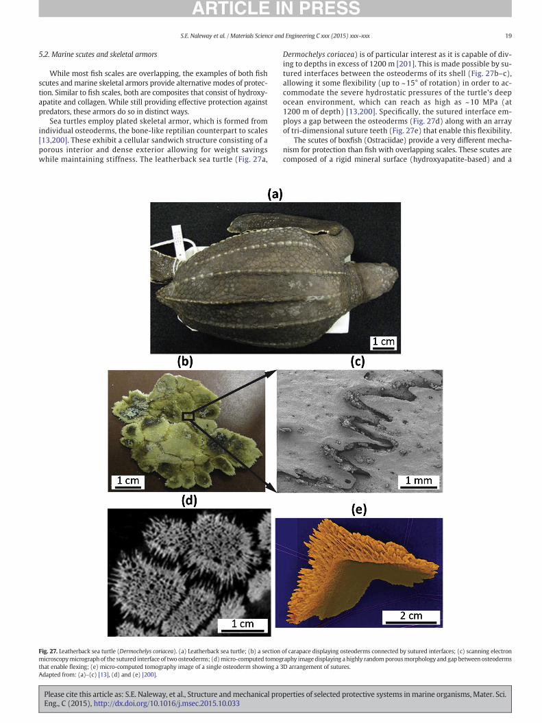
Sea turtles employ plated skeletal armor, which is formed fromindividual osteoderms, the bone-like reptilian counterpart to scales[13,200]. These exhibit a cellular sandwich structure consisting of aporous interior and dense exterior allowing for weight savingswhile maintaining stiffness. The leatherback sea turtle (Fig. 27a,
Fig. 27. Leatherback sea turtle (Dermochelys coriacea). (a) Leatherback sea turtle; (b) a sectionmicroscopymicrograph of the sutured interface of two osteoderms; (d)micro-computed tomogthat enable flexing; (e) micro-computed tomography image of a single osteoderm showing a 3Adapted from: (a)–(c) [13], (d) and (e) [200].
Please cite this article as: S.E. Naleway, et al., Structure andmechanical proEng., C (2015), http://dx.doi.org/10.1016/j.msec.2015.10.033
Dermochelys coriacea) is of particular interest as it is capable of div-ing to depths in excess of 1200 m [201]. This is made possible by su-tured interfaces between the osteoderms of its shell (Fig. 27b–c),allowing it some flexibility (up to ~15° of rotation) in order to ac-commodate the severe hydrostatic pressures of the turtle's deepocean environment, which can reach as high as ~10 MPa (at1200 m of depth) [13,200]. Specifically, the sutured interface em-ploys a gap between the osteoderms (Fig. 27d) along with an arrayof tri-dimensional suture teeth (Fig. 27e) that enable this flexibility.
The scutes of boxfish (Ostraciidae) provide a very different mecha-nism for protection than fish with overlapping scales. These scutes arecomposed of a rigid mineral surface (hydroxyapatite-based) and a
of carapace displaying osteoderms connected by sutured interfaces; (c) scanning electronraphy imagedisplaying a highly randomporousmorphology and gap between osteodermsD arrangement of sutures.
perties of selected protective systems inmarine organisms, Mater. Sci.
20 S.E. Naleway et al. / Materials Science and Engineering C xxx (2015) xxx–xxx
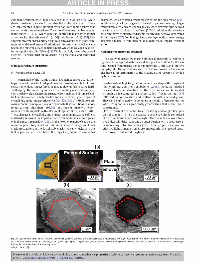
compliant collagen base (type I collagen) (Fig. 28a) [12,202]. Whilethese constituents are similar to other fish scales, the way that theyare implemented is quite different, with non-overlapping scutes thatconnect with sutured interfaces. The ratio of biomineral to biopolymerin the scutes is 1:2 [12] which is in stark contrast to many other dermalarmors such as the seahorse (~2:1) [150] and alligator (~2:1) [203]. Thissuggests an armor based primarily in collagen as opposed tomore com-mon mineral-based armor. Of additional interest, when mechanicallytested, the mineral surface remains intact while the collagen base de-forms significantly (Fig. 28b–c) [12]. While this likely lowers the overallstrength, it ensures that failure occurs in a predictable and controlledmanner.
6. Impact resistant structures
6.1. Mantis shrimp dactyl club
The mandible of the mantis shrimp (highlighted in Fig. 29a) is per-haps the most remarkable adaptation of the crustacean cuticle. It mustresist tremendous impact forces as they rapidly unfurl to strike hard-shelled prey. The impacting surface of the smashingmantis shrimp spe-cies, the dactyl club, changes in structure from an inner bulk area of chi-tin fibers to anouter, heavily calcified surface,with the highest degree ofcrystallinity at the impact surface (Fig. 29b) [204,205]. The bulk area pri-marily contains amorphous calcium carbonate that transitions to amor-phous calcium phosphate [205,206] and then ultimately a hyper-mineralized fluorapatite with calcium phosphate at the surface [204].These changes in crystallinity and mineral result in increasing stiffnessand hardness toward the impact surface, with hardness six times great-er in the impact region [204–206]. Similar to other regions of cuticle, theimpact region is toughened with chitin that absorbs energy and limitscrack propagation. In the dactyl club, cracks typically nucleate in thebulk region and are deflected in the impact region due to a modulus
Fig. 28. (a) Structure of the lateral scutes of the boxfish (Lactoria cornuta), the external surfa(b) fracture of a scute tested in a punchingmode by a hexagonal punch (highlighted); (c) fractubase while the mineral remains relatively intact.Adapted from [12].
Please cite this article as: S.E. Naleway, et al., Structure andmechanical proEng., C (2015), http://dx.doi.org/10.1016/j.msec.2015.10.033
mismatch, which contains cracks mostly within the bulk region [205].In this region, cracks propagate in a helicoidal pattern, creating a largercrack surface area, and are stopped quicklywhen traversing the helicoidtrajectory by an oscillation in stiffness [205]. In addition, this structurehas been shown to effectively disperse the stress pulsewaves generatedduring impact [207]. Combining a hard outer layerwith an inner, energydispersive matrix is reminiscent of human-made impact resistantarmor.
7. Bioinspired materials potential
The study of protective marine biological materials is leading tosignificant bioinspiredmaterials and designs. These allow for the les-sons learned from marine biological materials to affect and improveeveryday life. Though not an extensive list, we provide a few exam-ples here as an introduction to the materials and research providedby bioinspiration.
• Crush resistant, high toughness ceramics based upon the tough andhighly mineralized shells of abalone [9,108]: the nacre-inspiredbrick-and-mortar structure of these ceramics are fabricatedthrough an ice templating process called “freeze casting” [57]followed by compression and infiltration with a second phase.They can be infiltrated with polymers or metals to form compositeswhose toughness is significantly greater than that of their baseconstituents.
• Flexure resistant fiber optics based on strong and tough silica spic-ules of sponges [10,11]: the structure of the spicules is comprisedof three sections: a core with a high refractive index, a low refrac-tive index cylindrical tube and an outer portion with a progressive-ly increasing refractive index [10]. These properties allow foreffective light transmission. More importantly, the layered struc-ture provides enhanced toughness.
ce is mineralized and rigid, but if removed, a base of aligned collagen fibers is revealed;re of the surrounding scutes. In both cases, the facture occurs primarilywithin the collagen
perties of selected protective systems inmarine organisms, Mater. Sci.
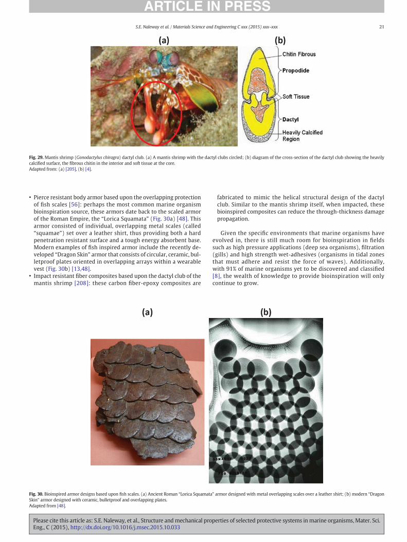
Fig. 29. Mantis shrimp (Gonodactylus chiragra) dactyl club. (a) A mantis shrimp with the dactyl clubs circled; (b) diagram of the cross-section of the dactyl club showing the heavilycalcified surface, the fibrous chitin in the interior and soft tissue at the core.Adapted from: (a) [205], (b) [4].
21S.E. Naleway et al. / Materials Science and Engineering C xxx (2015) xxx–xxx
• Pierce resistant body armor based upon the overlapping protectionof fish scales [56]: perhaps the most common marine organismbioinspiration source, these armors date back to the scaled armorof the Roman Empire, the “Lorica Squamata” (Fig. 30a) [48]. Thisarmor consisted of individual, overlapping metal scales (called“squamae”) set over a leather shirt, thus providing both a hardpenetration resistant surface and a tough energy absorbent base.Modern examples of fish inspired armor include the recently de-veloped “Dragon Skin” armor that consists of circular, ceramic, bul-letproof plates oriented in overlapping arrays within a wearablevest (Fig. 30b) [13,48].
• Impact resistant fiber composites based upon the dactyl club of themantis shrimp [208]: these carbon fiber-epoxy composites are
Fig. 30. Bioinspired armor designs based upon fish scales. (a) Ancient Roman “Lorica SquamataSkin” armor designed with ceramic, bulletproof and overlapping plates.Adapted from [48].
Please cite this article as: S.E. Naleway, et al., Structure andmechanical proEng., C (2015), http://dx.doi.org/10.1016/j.msec.2015.10.033
fabricated to mimic the helical structural design of the dactylclub. Similar to the mantis shrimp itself, when impacted, thesebioinspired composites can reduce the through-thickness damagepropagation.
Given the specific environments that marine organisms haveevolved in, there is still much room for bioinspiration in fieldssuch as high pressure applications (deep sea organisms), filtration(gills) and high strength wet-adhesives (organisms in tidal zonesthat must adhere and resist the force of waves). Additionally,with 91% of marine organisms yet to be discovered and classified[8], the wealth of knowledge to provide bioinspiration will onlycontinue to grow.
” armor designed with metal overlapping scales over a leather shirt; (b) modern “Dragon
perties of selected protective systems inmarine organisms, Mater. Sci.
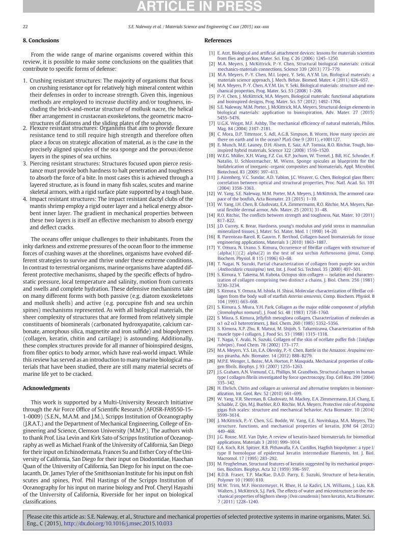
22 S.E. Naleway et al. / Materials Science and Engineering C xxx (2015) xxx–xxx
8. Conclusions
From the wide range of marine organisms covered within thisreview, it is possible to make some conclusions on the qualities thatcontribute to specific forms of defense:
1. Crushing resistant structures: The majority of organisms that focuson crushing resistance opt for relatively high mineral content withintheir defenses in order to increase strength. Given this, ingeniousmethods are employed to increase ductility and/or toughness, in-cluding the brick-and-mortar structure of mollusk nacre, the helicalfiber arrangement in crustacean exoskeletons, the geometric macro-structures of diatoms and the sliding plates of the seahorse.
2. Flexure resistant structures: Organisms that aim to provide flexureresistance tend to still require high strength and therefore oftenplace a focus on strategic allocation of material, as is the case in theprecisely aligned spicules of the sea sponge and the porous/denselayers in the spines of sea urchins.
3. Piercing resistant structures: Structures focused upon pierce resis-tance must provide both hardness to halt penetration and toughnessto absorb the force of a bite. In most cases this is achieved through alayered structure, as is found in many fish scales, scutes and marineskeletal armors, with a rigid surface plate supported by a tough base.
4. Impact resistant structures: The impact resistant dactyl clubs of themantis shrimp employ a rigid outer layer and a helical energy absor-bent inner layer. The gradient in mechanical properties betweenthese two layers is itself an effective mechanism to absorb energyand deflect cracks.
The oceans offer unique challenges to their inhabitants. From theinky darkness and extreme pressures of the ocean floor to the immenseforces of crashing waves at the shorelines, organisms have evolved dif-ferent strategies to survive and thrive under these extreme conditions.In contrast to terrestrial organisms, marine organisms have adapted dif-ferent protective mechanisms, shaped by the specific effects of hydro-static pressure, local temperature and salinity, motion from currentsand swells and complete hydration. These defensive mechanisms takeon many different forms with both passive (e.g. diatom exoskeletonsand mollusk shells) and active (e.g. porcupine fish and sea urchinspines) mechanisms represented. As with all biological materials, thesheer complexity of structures that are formed from relatively simpleconstituents of biominerals (carbonated hydroxyapatite, calcium car-bonate, amorphous silica, magnetite and iron sulfide) and biopolymers(collagen, keratin, chitin and cartilage) is astounding. Additionally,these complex structures provide for all manner of bioinspired designs,from fiber optics to body armor, which have real-world impact. Whilethis reviewhas served as an introduction tomanymarine biologicalma-terials that have been studied, there are still many material secrets ofmarine life yet to be cracked.
Acknowledgments
This work is supported by a Multi-University Research Initiativethrough the Air Force Office of Scientific Research (AFOSR-FA9550-15-1-0009) (S.E.N., M.A.M. and J.M.), Scripps Institution of Oceanography(J.R.A.T.) and the Department of Mechanical Engineering, College of En-gineering and Science, Clemson University (M.M.P.). The authors wishto thank Prof. Lisa Levin and Kirk Sato of Scripps Institution of Oceanog-raphy as well as Michael Frank of the University of California, San Diegofor their input on Echinodermata, Frances Su and Esther Cory of theUni-versity of California, San Diego for their input on Diodontidae, HaochanQuan of the University of California, San Diego for his input on the coe-lacanth, Dr. James Tyler of the Smithsonian Institute for his input on fishscutes and spines, Prof. Phil Hastings of the Scripps Institution ofOceanography for his input on marine biology and Prof. Cheryl Hayashiof the University of California, Riverside for her input on biologicalclassifications.
Please cite this article as: S.E. Naleway, et al., Structure andmechanical proEng., C (2015), http://dx.doi.org/10.1016/j.msec.2015.10.033
References
[1] E. Arzt, Biological and artificial attachment devices: lessons for materials scientistsfrom flies and geckos, Mater. Sci. Eng. C 26 (2006) 1245–1250.
[2] M.A. Meyers, J. McKittrick, P.-Y. Chen, Structural biological materials: criticalmechanics-materials connections, Science 339 (2013) 773–779.
[3] M.A. Meyers, P.-Y. Chen, M.I. Lopez, Y. Seki, A.Y.M. Lin, Biological materials: amaterials science approach, J. Mech. Behav. Biomed. Mater. 4 (2011) 626–657.
[4] M.A. Meyers, P.-Y. Chen, A.Y.M. Lin, Y. Seki, Biological materials: structure and me-chanical properties, Prog. Mater. Sci. 53 (2008) 1–206.
[5] P.-Y. Chen, J. McKittrick, M.A. Meyers, Biological materials: functional adaptationsand bioinspired designs, Prog. Mater. Sci. 57 (2012) 1492–1704.
[6] S.E. Naleway, M.M. Porter, J. McKittrick, M.A. Meyers, Structural design elements inbiological materials: application to bioinspiration, Adv. Mater. 27 (2015)5455–5476.
[7] U.G.K. Wegst, M.F. Ashby, The mechanical efficiency of natural materials, Philos.Mag. 84 (2004) 2167–2181.
[8] C. Mora, D.P. Tittensor, S. Adl, A.G.B. Simpson, B. Worm, How many species arethere on earth and in the ocean? PLoS One 9 (2011), e1001127.
[9] E. Munch, M.E. Launey, D.H. Alsem, E. Saiz, A.P. Tomsia, R.O. Ritchie, Tough, bio-inspired hybrid materials, Science 322 (2008) 1516–1520.
[10] W.E.G. Müller, X.H. Wang, F.Z. Cui, K.P. Jochum, W. Tremel, J. Bill, H.C. Schroder, F.Natalio, U. Schlossmacher, M. Wiens, Sponge spicules as blueprints for thebiofabrication of inorganic–organic composites and biomaterials, Appl. Microbiol.Biotechnol. 83 (2009) 397–413.
[11] J. Aizenberg, V.C. Sundar, A.D. Yablon, J.C. Weaver, G. Chen, Biological glass fibers:correlation between optical and structural properties, Proc. Natl. Acad. Sci. 101(2004) 3358–3363.
[12] W. Yang, S.E. Naleway, M.M. Porter, M.A. Meyers, J. McKittrick, The armored cara-pace of the boxfish, Acta Biomater. 23 (2015) 1–10.
[13] W. Yang, I.H. Chen, B. Gludovatz, E.A. Zimmermann, R.O. Ritchie, M.A. Meyers, Nat-ural flexible dermal armor, Adv. Mater. 25 (2013) 31–48.
[14] R.O. Ritchie, The conflicts between strength and toughness, Nat. Mater. 10 (2011)817–822.
[15] J.D. Currey, K. Brear, Hardness, young's modulus and yield stress in mammalianmineralized tissues, J. Mater. Sci. Mater. Med. 1 (1990) 14–20.
[16] R. Parenteau-Bareil, R. Gauvin, F. Berthod, Collagen-based biomaterials for tissueengineering applications, Materials 3 (2010) 1863–1887.
[17] Y. Omura, N. Urano, S. Kimura, Occurrence of fibrillar collagen with structure of(alpha(1))(2) alpha(2) in the test of sea urchin Asthenosoma ijimai, Comp.Biochem. Physiol. B 115 (1996) 63–68.
[18] T. Nagai, N. Suzuki, Partial characterization of collagen from purple sea urchin(Anthocidaris crassispina) test, Int. J. Food Sci. Technol. 35 (2000) 497–501.
[19] S. Kimura, Y. Takema, M. Kubota, Octopus skin collagen — isolation and character-ization of collagen comprising two distinct a chains, J. Biol. Chem. 256 (1981)3230–3234.
[20] S. Kimura, Y. Omura, M. Ishida, H. Shirai, Molecular characterization of fibrillar col-lagen from the body wall of starfish Asterias amurenis, Comp. Biochem. Physiol. B104 (1993) 663–668.
[21] S. Kimura, S. Miura, Y.H. Park, Collagen as the major edible component of jellyfish(Stomolophus nomural), J. Food Sci. 48 (1983) 1758–1760.
[22] S. Miura, S. Kimura, Jellyfish mesogloea collagen. Characterization of molecules asα1 α2 α3 heterotrimers, J. Biol. Chem. 260 (1985) 5352–5356.
[23] S. Kimura, X.P. Zhu, R. Matsui, M. Shijoh, S. Takamizawa, Characterization of fishmuscle type-I collagen, J. Food Sci. 53 (1988) 1315–1318.
[24] T. Nagai, Y. Araki, N. Suzuki, Collagen of the skin of ocellate puffer fish (Takifugurubripes), Food Chem. 78 (2002) 173–177.
[25] M.A. Meyers, Y.S. Lin, E.A. Olevsky, P.-Y. Chen, Battle in the Amazon: Arapaima ver-sus piranha, Adv. Biomater. 14 (2012) B88–B279.
[26] M.P.E. Wenger, L. Bozec, M.A. Horton, P. Masquida, Mechanical properties of colla-gen fibrils, Biophys. J. 93 (2007) 1255–1263.
[27] J.S. Graham, A.N. Vomund, C.L. Phillips, M. Grandbois, Structural changes in humantype I collagen fibrils investigated by force spectroscopy, Exp. Cell Res. 299 (2004)335–342.
[28] H. Ehrlich, Chitin and collagen as universal and alternative templates in biominer-alization, Int. Geol. Rev. 52 (2010) 661–699.
[29] W. Yang, V.R. Sherman, B. Gludovatz, M. Mackey, E.A. Zimmermann, E.H. Chang, E.Schaible, Z. Qin, M.J. Buehler, R.O. Ritchie, M.A. Meyers, Protective role of Arapaimagigas fish scales: structure and mechanical behavior, Acta Biomater. 10 (2014)3599–3614.
[30] J. McKittrick, P.-Y. Chen, S.G. Bodde, W. Yang, E.E. Novitskaya, M.A. Meyers, Thestructure, functions, and mechanical properties of keratin, JOM 64 (2012)449–468.
[31] J.G. Rouse, M.E. Van Dyke, A review of keratin-based biomaterials for biomedicalapplications, Materials 3 (2010) 999–1014.
[32] E.A. Koch, R.H. Spitzer, R.B. Pithawalla, F.A. Castillos, Hagfish biopolymer: a type I/type II homologue of epidermal keratin intermediate filaments, Int. J. Biol.Macromol. 17 (1995) 283–292.
[33] M. Feughelman, Structural features of keratin suggested by its mechanical proper-ties, Biochim. Biophys. Acta 32 (1959) 596–597.
[34] R.D.B. Fraser, T.P. MacRae, D.A.D. Parry, E. Suzuki, Structure of beta-keratin,Polymer 10 (1969) 810.
[35] M.W. Trim, M.F. Horstemeyer, H. Rhee, H. Le Kadiri, L.N. Williams, J. Liao, K.B.Walters, J. McKittrick, S.J. Park, The effects of water and microstructure on the me-chanical properties of bighorn sheep (Ovis canadensis) horn keratin, Acta Biomater.7 (2011) 1228–1240.
perties of selected protective systems inmarine organisms, Mater. Sci.
23S.E. Naleway et al. / Materials Science and Engineering C xxx (2015) xxx–xxx
[36] L. Tombolato, E.E. Novitskaya, P.-Y. Chen, F.A. Sheppard, J. McKittrick, Microstruc-ture, elastic properties and deformation mechanisms of horn keratin, ActaBiomater. 6 (2010) 319–330.
[37] H. Zahn, J. Fohles, M. Nienhaus, A. Schwan, M. Spel, Wool as a biological compositestructure, Ind. Eng. Chem. Prod. Res. Dev. 19 (1980) 496–501.
[38] S.L. Rosen, Fundamental Principles of Polymeric Materials, 2nd ed. John Wiley &Sons Inc., New York, 1993.
[39] M.A. Kennedy, A.J. Peacock, L. Mandelkern, Tensile properties of crystalline poly-mers — linear polyethylene, Macromolecules 27 (1994) 5297–5310.
[40] M.N.V.R. Kumar, A review of chitin and chitosan applications, React. Funct. Polym.46 (2000) 1–27.
[41] M. Rinaudo, Chitin and chitosan: properties and applications, Prog. Polym. Sci. 31(2006) 603–632.
[42] A. Miserez, D. Rubin, J.H. Waite, Cross-linking chemistry of squid beak, J. Biol.Chem. 285 (2010) 38115–38124.
[43] K.M. Rudall, W. Kenchington, The chitin system, Biol. Rev. 48 (1973) 597–633.[44] Y. Bouligand, Twisted fiberous arrangements in biological materials and cholesteric
mesophases, Tissue Cell 4 (1972) 189–217.[45] M.M. Giraud-Guille, Fine structure of the chitin protein system in the crab cuticle,
Tissue Cell 16 (1984) 75–92.[46] M.M. Giraud-Guille, H. Chanzy, R. Vuong, Chitin crystals in arthropod cuticles re-
vealed by diffraction contrast transmission electron-microscopy, J. Struct. Biol.103 (1990) 232–240.
[47] M.M. Giraud-Guille, Plywood structures in nature, Curr. Opin. Solid StateMater. Sci.3 (1998) 221–227.
[48] H. Ehrlich, Biological Materials of Marine Origin: Springer, 2015.[49] M.N. Dean, A.P. Summers, Mineralized cartilage in the skeleton of chondrichthyan
fishes, Zoology 109 (2006) 164–168.[50] A.G. Cole, B.K. Hall, Cartilage differentiation in cephalopod molluscs, Zoology 112
(2009) 2–15.[51] A.G. Cole, B.K. Hall, The nature and significance of invertebrate cartilages revisited:
distribution and histology of cartilage and cartilage-like tissues within theMetazoa, Zoology 107 (2004) 261–273.
[52] P.-Y. Chen, D. Toroian, P.A. Price, J. McKittrick, Minerals form a continuum phase inmature cancellous bone, Calcif. Tissue Int. 88 (2011) 351–361.
[53] S.V. Dorozhkin, Calcium orthophosphates in nature, biology and medicine, Mate-rials 2 (2009) 399–498.
[54] S. Weiner, H.D. Wangner, The material bone: structure mechanical function rela-tions, Annu. Rev. Mater. Sci. 28 (1998) 271–298.
[55] E.E. Novitskaya, P.-Y. Chen, S. Lee, A.B. Castro-Cesena, G.A. Hirata, V.A. Lubarda, J.McKittrick, Anisotropy in the compressive mechanical properties of bovine corticalbone and the mineral and protein constituents, Acta Biomater. 7 (2011)3170–3177.
[56] W. Yang, I.H. Chen, J. McKittrick, M.A. Meyers, Flexible dermal armor in nature,JOM 64 (2012) 475–485.
[57] M.M. Porter, J. McKittrick, M.A. Meyers, Biomimetic materials by freeze casting,JOM 65 (2013) 720–727.
[58] M. Vallet-Regi, J.M. Gonzalez-Calbet, Calcium phosphates as substitution of bonetissues, Prog. Solid State Chem. 32 (2004) 1–31.
[59] L.L. Hench, Bioceramics — from concept to clinic, J. Am. Ceram. Soc. 74 (1991)1487–1510.
[60] L.L. Hench, Bioceramics, J. Am. Ceram. Soc. 81 (1998) 1705–1728.[61] S. Deville, E. Saiz, R.K. Nalla, A.P. Tomsia, Freezing as a path to build complex com-
posites, Science 311 (2006) 515–518.[62] M.J. Olszta, X.G. Cheng, S.S. Jee, R. Kumar, Y.Y. Kim, M.J. Kaufman, E.P. Douglas, L.B.
Gower, Bone structure and formation: a new perspective, Mater. Sci. Eng. R 58(2007) 77–116.
[63] F. Nudelman, K. Pieterse, A. George, P.H.H. Bomans, H. Friedrich, L.J. Brylka, P.A.J.Hilbers, G. de With, N.A.J.M. Sommerdijk, The role of collagen in bone apatite for-mation in the presence of hydroxyapatite nucleation inhibitors, Nat. Mater. 9(2010) 1004–1009.
[64] T. Ikoma, H. Kobayashi, J. Tanaka, D. Walsh, S. Mann, Microstructure, mechanical,and biomimetic properties of fish scales from Pagrus major, J. Struct. Biol. 142(2003) 327–333.
[65] P. Ducheyne, Q. Qiu, Bioactive ceramics: the effect of surface reactivity on bone for-mation and bone cell function, Biomaterials 20 (1999) 2287–2303.
[66] G. Mayer, Rigid biological systems as models for synthetic composites, Science 310(2005) 1144–1147.
[67] S. Blank, M. Arnoldi, S. Khoshnavaz, L. Treccani, M. Kuntz, K. Mann, G. Grathwohl,M. Fritz, The nacre protein perlucin nucleates growth of calcium carbonate crystals,J. Microsc. (Oxford) 212 (2003) 280–291.
[68] F.C. Meldrum, H. Colfen, Controlling mineral morphologies and structures in bio-logical and synthetic systems, Chem. Rev. 108 (2008) 4332–4432.
[69] G. Wolf, E. Konigsberger, H.G. Schmidt, L.C. Konigsberger, H. Gamsjager, Thermo-dynamic aspects of the vaterite-calcite phase transition, J. Therm. Anal. Calorim.60 (2000) 463–472.
[70] G. Falini, S. Albeck, S. Weiner, L. Addadi, Control of aragonite or calcite polymor-phism by mollusk shell macromolecules, Science 271 (1996) 67–69.
[71] A.M. Belcher, X.H. Wu, R.J. Christensen, P.K. Hansma, G.D. Stucky, D.E. Morse, Con-trol of crystal phase switching and orientation by soluble mollusc-shell proteins,Nature 381 (1996) 56–58.
[72] X. Long, Y.R. Ma, L.M. Qi, Biogenic and synthetic high magnesium calcite — a re-view, J. Struct. Biol. 185 (2014) 1–14.
[73] G. Falini, S. Fermani, M. Gazzano, A. Ripamonti, Biomimetic crystallization of calci-um carbonate polymorphs by means of collagenous matrices, Chem. Eur. J. 3(1997) 1807–1814.
Please cite this article as: S.E. Naleway, et al., Structure andmechanical proEng., C (2015), http://dx.doi.org/10.1016/j.msec.2015.10.033
[74] R.M.S. Schofield, J.C. Niedbala, M.H. Nesson, Y. Tao, J.E. Shokes, R.A. Scott, M.J.Latimer, Br-rich tips of calcified crab claws are less hard but more fracture resis-tant: a comparison of mineralized and heavy-element biological materials, J.Struct. Biol. 166 (2009) 272–287.
[75] B.W. Cribb, A. Rathmell, R. Charters, R. Rasch, H. Huang, I.R. Tibbetts, Structure,composition and properties of naturally occurring non-calcified crustacean cuticle,Arthropod Struct. Dev. 38 (2009) 173–178.
[76] C.C. Perry, T. Keeling-Tucker, Biosilicification: the role of the organic matrix instructure control, J. Biol. Inorg. Chem. 5 (2000) 537–550.
[77] E. Epstein, Silicon: its manifold roles in plants, Ann. Appl. Biol. 155 (2009)155–160.
[78] P.W. Lucas, I.M. Turner, N.J. Dominy, N. Yamashita, Mechanical defences to herbiv-ory, Ann. Bot. 86 (2000) 913–920.
[79] I.C. Gebeshuber, J.H. Kindt, J.B. Thompson, Y. Del Amo, H. Stachelberger, M.A.Brzezinski, G.D. Stucky, D.E. Morse, P.K. Hansma, Atomic force microscopy studyof living diatoms in ambient conditions, J. Microsc. (Oxford) 212 (2003) 292–299.
[80] A.P. Garcia, N. Pugno, M.J. Buehler, Superductile, wavy silica nanostructures in-spired by diatom algae, Adv. Eng. Mater. 13 (2011) B14–B405.
[81] E. Epstein, Silicon, Annu. Rev. Plant Physiol. Plant Mol. Biol. 50 (1999) 641–664.[82] K. Shimizu, J. Cha, G.D. Stucky, D.E. Morse, Silicatein alpha: cathepsin L-like protein
in sponge biosilica, Proc. Natl. Acad. Sci. U. S. A. 95 (1998) 6234–6238.[83] L. Xia, Y. Liu, Z.B. Li, Biosilicification templated by amphiphilic block copolypeptide
assemblies, Macromol. Biosci. 10 (2010) 1566–1575.[84] D. Schuler, R.B. Frankel, Bacterial magnetosomes: microbiology, biomineralization
and biotechnological applications, Appl. Microbiol. Biotechnol. 52 (1999) 464–473.[85] C.T. Lefevre, N. Viloria, M.L. Schmidt, M. Posfai, R.B. Frankel, D.A. Bazylinski, Novel
magnetite-producing magnetotactic bacteria belonging to theGammaproteobacteria, ISME J. 6 (2012) 440–450.
[86] K.J. Lohmann, Magnetic orientation by hatchling loggerheasd sea-turtles (Carettacaretta), J. Exp. Biol. 155 (1991) 37–49.
[87] K.J. Lohmann, C.M.F. Lohmann, Detection of magnetic field intensity by sea turtles,Nature 380 (1996) 59–61.
[88] L.C. Boles, K.J. Lohmann, True navigation and magnetic maps in spiny lobsters, Na-ture 421 (2003) 60–63.
[89] Q.Q. Wang, M. Nemoto, D.S. Li, J.C. Weaver, B. Weden, J. Stegemeier, K.N. Bozhilov,L.R. Wood, G.W. Milliron, C.S. Kim, E. DiMasi, D. Kisailus, Phase transformations andstructural developments in the radular teeth of Cryptochiton stelleri, Adv. Funct.Mater. 23 (2013) 2908–2917.
[90] S.E. Greene, A. Komeili, Biogenesis and subcellular organization of themagnetosome organelles of magnetotactic bacteria, Curr. Opin. Cell Biol. 24(2012) 490–495.
[91] G.W. Luther, T.F. Rozan, M. Taillefert, D.B. Nuzzio, C. Di Meo, T.M. Shank, R.A. Lutz,S.C. Cary, Chemical speciation drives hydrothermal vent ecology, Nature 410(2001) 813–816.
[92] Y. Suzuki, R.E. Kopp, T. Kogure, A. Suga, K. Takai, S. Tsuchida, N. Ozaki, K. Endo, J.Hashimoto, Y. Kato, C. Mizota, T. Hirata, H. Chiba, K.H. Nealson, K. Horikoshi, J.L.Kirschvink, Sclerite formation in the hydrothermal-vent "scaly-foot" gastropod —possible control of iron sulfide biomineralization by the animal, Earth Planet. Sci.Lett. 242 (2006) 39–50.
[93] F. Barthelat, H. Tang, P.D. Zavattieri, C.M. Li, H.D. Espinosa, On the mechanics ofmother-of-pearl: a key feature in the material hierarchical structure, J. Mech.Phys. Solids 55 (2007) 306–337.
[94] H.D. Espinosa, A.L. Juster, F.J. Latourte, O.Y. Loh, D. Gregoire, P.D. Zavattieri, Tablet-level origin of toughening in abalone shells and translation to synthetic compositematerials, Nat. Commun. 2 (2011).
[95] L. Addadi, D. Joester, F. Nudelman, S. Weiner, Mollusk shell formation: a source ofnew concepts for understanding biomineralization processes, Chemistry 12 (2006)980–987.
[96] A.Y.M. Lin, M.A. Meyers, K.S. Vecchio, Mechanical properties and structure ofStrombus gigas, Tridacna gigas, and Haliotis rufescens sea shells: a comparativestudy, Mater. Sci. Eng. C 26 (2006) 1380–1389.
[97] A.Y.M. Lin, P.-Y. Chen, M.A. Meyers, The growth of nacre in the abalone shell, ActaBiomater. 4 (2008) 131–138.
[98] A. Lin, M.A. Meyers, Growth and structure in abalone shell, Mater. Sci. Eng. A 390(2005) 27–41.
[99] M.A. Meyers, C.T. Lim, A. Li, B.R.H. Nizam, E.P.S. Tan, Y. Seki, J. McKittrick, The role oforganic intertile layer in abalone nacre, Mater. Sci. Eng. C 29 (2009) 2398–2410.
[100] M.I. Lopez, P.E.M. Martinez, M.A. Meyers, Organic interlamellar layers, mesolayersand mineral nanobridges: contribution to strength in abalone (Haliotis rufescence)nacre, Acta Biomater. 10 (2014) 2056–2064.
[101] M.I. Lopez, P.-Y. Chen, J. McKittrick, M.A. Meyers, Growth of nacre in abalone: sea-sonal and feeding effects, Mater. Sci. Eng. C 31 (2011) 238–245.
[102] R. Menig, M.H. Meyers, M.A. Meyers, K.S. Vecchio, Quasi-static and dynamic me-chanical response of Haliotis rufescens (abalone) shells, Acta Mater. 48 (2000)2383–2393.
[103] C. Salinas, D. Kisailus, Fracture mitigation strategies in gastropod shells, JOM 65(2013) 473–480.
[104] R. Menig, M.H. Meyers, M.A. Meyers, K.S. Vecchio, Quasi-static and dynamic me-chanical response of Strombus gigas (conch) shells, Mater. Sci. Eng. A 297 (2001)203–211.
[105] S. Kamat, X. Su, R. Ballarini, A.H. Heuer, Structural basis for the fracture toughnessof the shell of the conch Strombus gigas, Nature 405 (2000) 1036–1040.
[106] R.O. Ritchie, Mechanisms of fatigue crack-propagation in metals, ceramics andcomposites — role of crack tip sheilding, Mater. Sci. Eng. A 103 (1988) 15–28.
[107] R.O. Ritchie, Mechanisms of fatigue-crack propagation in ductile and brittle solids,Int. J. Fract. 100 (1999) 55–83.
perties of selected protective systems inmarine organisms, Mater. Sci.
24 S.E. Naleway et al. / Materials Science and Engineering C xxx (2015) xxx–xxx
[108] F. Bouville, E. Maire, S. Meille, B. Van de Moortele, A.J. Stevenson, S. Deville, Strong,tough and stiff bioinspired ceramics from brittle constituents, Nat. Mater. 13(2014) 508–514.
[109] H.M. Yao, M. Dao, T. Imholt, J.M. Huang, K. Wheeler, A. Bonilla, S. Suresh, C. Ortiz,Protection mechanisms of the iron-plated armor of a deep-sea hydrothermalvent gastropod, PNAS 107 (2010) 987–992.
[110] A.Waren, S. Bengtson, S.K. Goffredi, C.L. Van Dover, A hot-vent gastropod with ironsulfide dermal sclerites, Science 302 (2003) 1007.
[111] M.J. Vendrasco, T.E. Wood, B.N. Runnegar, Articulated Palaeozoic fossil with 17plates greatly expands disparity of early chitons, Nature 429 (2004) 288–291.
[112] K. Treves, W. Traub, S. Weiner, L. Addadi, Aragonite formation in the chiton(Mollusca) girdle, Helv. Chim. Acta 86 (2003) 1101–1112.
[113] M.J. Connors, H. Ehrlich, M. Hog, C. Godeffroy, S. Araya, I. Kallai, D. Gazit, M. Boyce,C. Ortiz, Three-dimensional structure of the shell plate assembly of the chitonTonicella marmorea and its biomechanical consequences, J. Struct. Biol. 177(2012) 314.
[114] M. Nemoto, Q.Q. Wang, D.S. Li, S.Q. Pan, T. Matsunaga, D. Kisailus, Proteomic anal-ysis from the mineralized radular teeth of the giant Pacific chiton, Cryptochitonstelleri (Mollusca), Proteomics 12 (2012) 2890–2894.
[115] K.M. Towe, H.A. Lowensta, Ultrastructure and development of iron mineralizationin radular teeth of Cryptochiton stelleri (Mollusca), J. Ultrastruct. Res. 17 (1967)1–13.
[116] L.M. Gordon, J.K. Roman, R.M. Everly, M.J. Cohen, J.J.Wilker, D. Joester, Selective for-mation of metastable ferrihydrite in the chiton tooth, Angew. Chem. 53 (2014)11506–11509.
[117] V. Martin-Jezequel, M. Hildebrand, M.A. Brzezinski, Silicon metabolism in diatoms:implications for growth, J. Phycol. 36 (2000) 821–840.
[118] M. Hildebrand, Diatoms, biomineralization processes, and genomics, Chem. Rev.108 (2008) 4855–4874.
[119] G. Sarthou, K.R. Timmermans, S. Blain, P. Treguer, Growth physiology and fate ofdiatoms in the ocean: a review, J. Sea Res. 53 (2005) 25–42.
[120] P. Treguer, D.M. Nelson, A.J. Van Beenekom, D.J. DeMaster, A. Leynaert, B.Queguiner, The silica balance in the world ocean: a reestimate, Science 268(1995) 375–379.
[121] C.E. Hamm, R. Merkel, O. Springer, P. Jurkojc, C. Maier, K. Prechtel, V. Smetacek, Ar-chitecture and material properties of diatom shells provide effective mechanicalprotection, Nature 421 (2003) 841–843.
[122] H. Hildebrand, C.D. Durselen, D. Kirschtel, U. Pollingher, T. Zohary, Biovolume cal-culation for pelagic and benthic microalgae, J. Phycol. 35 (1999) 403–424.
[123] D. Losic, K. Short, J.G. Mitchell, R. Lal, N.H. Voelcker, AFM nanoindentations of dia-tom biosilica surfaces, Langmuir 23 (2007) 5014–5021.
[124] N. Almqvist, Y. Delamo, B.L. Smith, N.H. Thomson, A. Bartholdson, R. Lal, M.Brzezinski, P.K. Hansma, Micromechanical and structural properties of a pennatediatom investigated by atomic force microscopy, J. Microsc. 202 (2001) 518–532.
[125] G. Francius, B. Tesson, E. Dague, V. Martin-Jezequel, Y.F. Dufrene, Nanostructureand nanomechanics of live Phaeodactylum tricornutum morphotypes, Environ.Microbiol. 10 (2008) 1344–1356.
[126] N. Kroger, R. Deutzmann, C. Bergsdorf, M. Sumper, Species-specific polyaminesfrom diatoms control silica morphology, PNAS 97 (2000) 14133–14138.
[127] N. Kroger, R. Deutzmann, M. Sumper, Polycationic peptides from diatom biosilicathat direct silica nanosphere formation, Science 286 (1999) 1129–1132.
[128] M. Sumper, A phase separation model for the nanopatterning of diatom biosilica,Science 295 (2002) 2430–2433.
[129] J.R. Young, Functions of coccoliths, in: W.G. Siesser, A. Winter (Eds.),Coccolithophores, Cambridge University Press, Cambridge, UK 1994, pp. 63–82.
[130] B.L. Smith, G.T. Paloczi, P.K. Hansma, R.P. Levine, Discerning nature's mechanismfor making complex biocomposite crystals, J. Cryst. Growth 211 (2000) 116–121.
[131] S. Mann, N.H.C. Sparks, Single crystal nature of coccolith elements of the marinealga Emiliania huxleyi as determined by electron diffraction and high resolutionelectron microscopy, Proc. R. Soc. B 234 (1988) 441–453.
[132] M.E. Marsh, Regulation of CaCO3 formation in coccolithophores, Comp. Biochem.Physiol. B 136 (2003) 743–754.
[133] P.-Y. Chen, A.Y.M. Lin, J. McKittrick, M.A. Meyers, Structure and mechanical proper-ties of crab exoskeletons, Acta Biomater. 4 (2008) 587–596.
[134] D.F. Travis, The molting cycle of the spiny lobster, Panulirus argus-Latreille. II. Pre-ecdysial histological and histochemical changes in the hepatopancreas andintegumental tissues, Biol. Bull. 108 (1955) 88–112.
[135] Y. Bouligand, Aspects ultrastructuraux de la calcification chez les Crabes, SeptiemeCongres Internationale de Microscopie Electronique, Gernoble, France, 1970105–106.
[136] H.O. Fabritius, C. Sachs, P.R. Triguero, D. Raabe, Influence of structural principles onthe mechanics of a biological fiber-based composite material with hierarchical or-ganization: the exoskeleton of the lobster Homarus americanus, Adv. Mater. 21(2009) 391–400.
[137] D. Raabe, C. Sachs, P. Romano, The crustacean exoskeleton as an example of astructurally and mechanically graded biological nanocomposite material, ActaMater. 53 (2005) 4281–4292.
[138] R. Dillaman, S. Hequembourg, M. Gay, Early pattern of calcification in the dorsalcarapace of the blue crab, Callinectes sapidus, J. Microsc. 263 (2005) 356–374.
[139] H.O. Fabritius, E.S. Karsten, K. Balasundaram, S. Hild, K. Huemer, D. Raabe, Correla-tion of structure, composition and local mechanical properties in the dorsal cara-pace of the edible crab Cancer pagurus, Z. Krist. Cryst. Mater. 227 (2012) 766–776.
[140] D. Raabe, P. Romano, C. Sachs, H. Fabritius, A. Al-Sawalmih, S. Yi, G. Servos, H.G.Hartwig, Microstructure and crystallographic texture of the chitin-protein networkin the biological composite material of the exoskeleton of the lobster Homarusamericanus, Mater. Sci. Eng. A 421 (2006) 143–153.
Please cite this article as: S.E. Naleway, et al., Structure andmechanical proEng., C (2015), http://dx.doi.org/10.1016/j.msec.2015.10.033
[141] L. Cheng, L.Y. Wang, A.M. Karlsson, Image analyses of two crustacean exoskeletonsand implications of the exoskeletal microstructure on the mechanical behavior, J.Mater. Res. 23 (2008) 2854–2872.
[142] C. Sachs, H. Fabritius, D. Raabe, Experimental investigation of the elastic–plastic de-formation of mineralized lobster cuticle by digital image correlation, J. Struct. Biol.155 (2006) 409–425.
[143] H.R. Hepburn, I. Joffe, N. Green, K.J. Nelson, Mechanical-properties of a crab shell,Comp. Biochem. Physiol. 50 (1975) 551–554.
[144] I. Joffe, H.R. Hepburn, K.J. Nelson, N. Green, Mechanical-properties of a crustaceanexoskelton, Comp. Biochem. Physiol. 50 (1975) 545–549.
[145] C.L. Williams, R.M. Dillaman, E.A. Elliott, D.M. Gay, Formation of the arthrodialmembrane in the blue crab, Callinectes sapidus, J. Morphol. 256 (2003) 260–269.
[146] C.A. Melnick, Z. Chen, J.J. Mecholsky, Hardness and toughness of exoskeleton mate-rial in the stone crab, Menippe mercenaria, J. Mater. Res. 11 (1996) 2903–2907.
[147] S.J. Foster, A.C.J. Vincent, Life history and ecology of seahorses: implications forconservation and management, J. Fish Biol. 65 (2004) 1–61.
[148] T.R. Consi, P.A. Seifert, M.S. Triantafyllou, E.R. Edelman, The dorsal fin engine of theseahorse, Hippocampus 248 (2001) 1.
[149] M.A. Ashley-Ross, Mechanical properties of the dorsal fin muscle of seahorse(Hippocampus) and pipefish (Syngnathus), J. Exp. Zool. 293 (2002) 561–577.
[150] M.M. Porter, E.E. Novitskaya, A.B. Castro-Cesena, M.A. Meyers, J. McKittrick, Highlydeformable bones: unusual deformation mechanisms of seahorse armor, ActaBiomater. 9 (2013) 6763–6770.
[151] M.E. Hale, Functional morphology of ventral tail bending and prehensile abilities ofthe seahorse, Hippocampus kuda, J. Morphol. 227 (1996) 51–65.
[152] T. Praet, D. Adriaens, S. Van Cauter, B. Masschaele, M. De Beule, B. Verhegghe, Inspi-ration from nature: dynamic modelling of the musculoskeletal structure of theseahorse tail, Int. J. Numer. Methods Biomed. Eng. 28 (2012) 1028–1042.
[153] C. Neutens, D. Adriaens, J. Christiaens, B. De Kegel, M. Dierick, R. Boistel, L. VanHoorebeke, Grasping convergent evolution in syngnathids: a unique tale of tails,J. Anat. 224 (2014) 710–723.
[154] M.M. Porter, D. Adriaens, R.L. Hatton, M.A. Meyers, J. McKittrick, Why the seahorsetail is square, Science 349 (2015) 6683 (aaa).
[155] D. Kleiber, L.K. Blight, I.R. Caldwell, A.C.J. Vincent, The importance of seahorses andpipefishes in the diet of marine animals, Rev. Fish Biol. Fish. 21 (2011) 205–223.
[156] W.E.G. Müller, X.H. Wang, K. Kropf, H. Ushijima, W. Geurtsen, C. Eckert, M.N. Tahir,W. Tremel, A. Boreiko, U. Schloßmacher, J.H. Li, H.C. Schröder, Bioorganic/inorganichybrid composition of sponge spicules: matrix of the giant spicules and of thecomitalia of the deep sea hexactinellid Monorhaphis, J. Struct. Biol. 161 (2008)188–203.
[157] J.C. Weaver, J. Aizenberg, G.E. Fantner, D. Kisailus, A. Woesz, P. Allen, K. Fields, M.J.Porter, F.W. Zok, P.K. Hansma, P. Fratzl, D.E. Morse, Hierarchical assembly of the si-liceous skeletal lattice of the hexactinellid sponge Euplectella aspergillum, J. Struct.Biol. 158 (2007) 93–106.
[158] J. Aizenberg, J.C. Weaver, M.S. Thanawala, V.C. Sundar, D.E. Morse, P. Fratzl, Skele-ton of Euplectella sp.: structural hierarchy from the nanoscale to the macroscale,Science 309 (2005) 275–278.
[159] G. Imsiecke, R.T. Steffen, M. Custodio, R. Borojevic,W.Muller, Formation of spiculesby sclerocytes from the freshwater sponge Ephydatia muelleri in short-term cul-tures in vitro, In Vitro Cell. Dev. Biol. Anim. 31 (1995) 528–535.
[160] J.N. Cha, K. Shimizu, Y. Zhou, S.C. Christiansen, B.F. Chmelka, G.D. Stucky, D.E.Morse, Silicatein filaments and subunits from amarine sponge direct the polymer-ization of silica and silicones in vitro, Proc. Natl. Acad. Sci. U. S. A. 96 (1999)361–365.
[161] A. Woesz, J.C. Weaver, M. Kazanci, Y. Dauphin, J. Aizenberg, D.E. Morse, P. Fratzl,Micromechanical properties of biological silica in skeletons of deep-sea sponges,J. Mater. Res. 21 (2006) 2068–2078.
[162] M.M. Porter, L. Meraz, A. Calderon, H. Choi, A. Chouhan, L. Wang, M.A. Meyers, J.McKittrick, Torsional properties of helix-reinforced composites fabricated by mag-netic freeze casting, Compos. Struct. 119 (2015) 174–184.
[163] V. Presser, S. Schultheiss, C. Berthold, K.G. Nickel, Sea urchin spines as a model-system for permeable, light-weight ceramics with graceful failure behavior. PartI. Mechanical behavior of sea urchin spines under compression, J. Bionic Eng. 6(2009) 203–213.
[164] J. Seto, Y.R. Ma, S.A. Davis, F. Meldrum, A. Gourrier, Y.Y. Kim, U. Schilde, M. Sztucki,M. Burghammer, S. Maltsev, C. Jager, H. Colfen, Structure–property relationships ofa biological mesocrystal in the adult sea urchin spine, PNAS 109 (2012)3699–3704.
[165] Y. Oaki, H. Imai, Nanoengineering in echinoderms: the emergence of morphologyfrom nanobricks, Small 2 (2006) 66–70.
[166] J. Aizenberg, J. Hanson, T.F. Koetzle, S. Weiner, L. Addadi, Control of macromoleculedistribution within synthetic and biogenic single calcite crystals, J. Am. Chem. Soc.119 (1997) 881–886.
[167] Y. Politi, T. Arad, E. Klein, S. Weiner, L. Addadi, Sea urchin spine calcite formsvia a transient amorphous calcium carbonate phase, Science 306 (2004)1161–1164.
[168] C. Moureaux, A. Perez-Huerta, P. Compere, W. Zhu, T. Leloup, M. Cusack, P. Dubois,Structure, composition and mechanical relations to function in sea urchin spine, J.Struct. Biol. 170 (2010) 41–49.
[169] P.B. Edwards, T.A. Ebert, Plastic responses to limited food availability and spinedamage in the sea urchin Strongylocentrotus purpuratus (Stimpson), J. Exp. Mar.Biol. Ecol. 145 (1991) 205–220.
[170] P. Dubois, L. Ameye, Regeneration of spines and pedicellariae in echinoderms: a re-view, Microsc. Res. Tech. 55 (2001) 427–437.
[171] L.J. Gibson, M.F. Ashby, Cellular Solids: Structure and Properties, Cambridge Uni-versity Press, 1999.
perties of selected protective systems inmarine organisms, Mater. Sci.
25S.E. Naleway et al. / Materials Science and Engineering C xxx (2015) xxx–xxx
[172] X. Su, S. Kamat, A.H. Heuer, The structure of sea urchin spines, large biogenic singlecrystals of calcite, J. Mater. Sci. 35 (2000) 5545–5551.
[173] J. Weber, R. Greer, B. Voight, E. White, R. Roy, Unusual strength properties of echi-noderm calcite, J. Ultrastruct. Res. 26 (1969) 355–366.
[174] Y. Ma, B. Aichmayer, O. Paris, P. Fratzl, A. Meibom, R.A. Metzler, Y. Politi, L. Addadi,P.U.P.A. Gilbert, S. Weiner, The grinding tip of the sea urchin tooth exhibits exqui-site control over calcite crystal orientation and Mg distribution, PNAS 106 (2009)6048–6053.
[175] Y. Ma, S.R. Cohen, L. Addadi, S. Weiner, Sea urchin tooth design: an “all-calcite”polycrystalline reinforced fiber composite for grinding rocks, Adv. Mater. 20(2008) 1555–1559.
[176] C.E. Killian, R.A. Metzler, Y. Gong, T.H. Churchill, I.C. Olson, V. Trubetskoy, M.B.Christensen, J.H. Fournelle, F. De Carlo, S. Cohen, J. Mahamid, A. Scholl, A. Young,A. Doran, F.H. Wilt, S.N. Coppersmith, P.U.P.A. Gilbert, Self-sharpening mechanismof the sea urchin tooth, Adv. Funct. Mater. 21 (2011) 682–690.
[177] J.C. Tyler, F. Santini, Review and reconstructions of the tetraodonitform fishes fromthe Eocene of Monte Bolca, Italy, with comments on related tertiary taxa, Studi eRicerche sui Giacimenti Terziari di Bolca, Museo Civico di Storia Naturale di Verona,9 2002, pp. 47–119.
[178] E.L. Brainerd, Puffer inflation: functional morphology of postcranial structures inDiodon holocanthus (Tetraodontiformes), J. Morphol. 220 (1994) 243–261.
[179] T. Ikoma, H. Kobayashi, J. Tanaka, D. Walsh, S. Mann, Physical properties of type Icollagen extracted from fish scales of Pagrus major and Oreochromis niloticas, Int.J. Biol. Macromol. 32 (2003) 199–204.
[180] D. Zhu, C.F. Ortega, R. Motamedi, L. Szewciw, F. Vernerey, F. Barthelat, Structureand mechanical performance of a “modern” fish scale, Adv. Eng. Mater. 14(2012) B94–B185.
[181] B.J.F. Bruet, J.H. Song, M.C. Boyce, C. Ortiz, Materials design principles of ancient fisharmour, Nat. Mater. 7 (2008) 748–756.
[182] J.H. Song, C. Ortiz, M.C. Boyce, Threat-protection mechanics of an armored fish, J.Mech. Behav. Biomed. Mater. 4 (2011) 699–712.
[183] E.A. Zimmermann, B. Gludovatz, E. Schaible, N.K.N. Dave, W. Yang, M.A. Meyers,R.O. Ritchie, Mechanical adaptability of the Bouligand-type structure in naturaldermal armour, Nat. Commun. 4 (2013) 2634.
[184] J.H. Long, M.E. Hale, M.J. McHenry, M.W. Westneat, Functions of fish skin: flexuralstiffness and steady swimming of longnose gar Lepisosteus osseus, J. Exp. Biol. 199(1996) 2139–2151.
[185] J.H. Long, B. Adcock, R.G. Root, Force transmission via axial tendons in undulatingfish: a dynamic analysis, Comp. Biochem. Physiol. A Physiol. 133 (2002) 911–929.
[186] J.H. Long, T.J. Koob, K. Irving, K. Combie, V. Engel, N. Livingston, A. Lammert, J.Schumacher, Biomimetic evolutionary analysis: testing the adaptive value of ver-tebrate tail stiffness in autonomous swimming robots, J. Exp. Biol. 209 (2006)4732–4746.
[187] W. Yang, B. Gludovatz, E.A. Zimmermann, H.A. Bale, R.O. Ritchie, M.A. Meyers,Structure and fracture resistance of alligator gar (Atractosteus spatula) armoredfish scales, Acta Biomater. 9 (2013) 5876–5889.
[188] J.Y. Sire, A. Huysseune, Formation of dermal skeletal and dental tissues in fish: acomparative and evolutionary approach, Biol. Rev. 78 (2003) 219–249.
[189] E.S. Goodrich, On the scales of fish living and extinct, and then importance in clas-sification, Proc. Zool. Soc. London 77 (1907) 751–774.
[190] F.J. Vernerey, F. Barthelat, Skin and scales of teleost fish: simple structure but highperformance and multiple functions, J. Mech. Phys. Solids 68 (2014) 66–76.
[191] D.J. Zhu, L. Szewciw, F. Vernerey, F. Barthelat, Puncture resistance of the scaled skinfrom striped bass: collective mechanisms and inspiration for new flexible armordesigns, J. Mech. Behav. Biomed. Mater. 24 (2013) 30–40.
[192] M.M. Smith, M.H. Hobdell, W.A. Miller, Structure of scales of Latimeria chalumnae, J.Zool. 167 (1972) 501–509.
[193] T.O.R. Orvig, Paleohistological notes I. On the structure of the bone tissue in thescales of certain Palaeonisciformes, Arkiv Zool. 10 (1957) 481–490.
[194] G.H. Roux, The microscopic anatomy of the Latimeria scale, S. Afr. J. Med. Sci. 7(1942) 1–18.
[195] E. Jarvik, On some osteolepiform crossopterygians from the Upper Old Red Sand-stone of Scotland, K. Svenska Vetenskapsakad. Handl. 2 (1950) 1–35.
[196] V.R. Sherman, W. Yang, M.A. Meyers, The materials science of collagen, J. Mech.Behav. Biomed. Mater. (2015) (in press).
[197] J.R. Grubich, S. Huskey, S. Crofts, G. Orti, J. Porto, Mega-bites: extreme jaw forces ofliving and extinct piranhas (Serrasalmidae), Sci. Report. (2012).
[198] P.-Y. Chen, J. Schirer, A. Simpson, R. Nay, Y.-S. Lin, W. Yang, M.I. Lopez, J. Li, E.A.Olevsky, M.A. Meyers, Predation versus protection: fish teeth and scales evaluatedby nanoindentation, J. Mater. Res. 27 (2012) 100–112.
[199] A.K. Dastjerdi, F. Barthelat, Teleost fish scales amongst the toughest collagenousmaterials, J. Mech. Behav. Biomed. Mater. (2015) (in press).
[200] I.H. Chen, W. Yang, M.A. Meyers, Leatherback sea turtle shell: a tough and flexiblebiological design, Acta Biomater. (2015) (in press).
[201] J.D.R. Houghton, T.K. Doyle, J. Davenport, R.P. Wilson, G.C. Hays, The role of infre-quent and extraordinary deep dives in leatherback turtles (Dermochelys coriacea),J. Exp. Biol. 211 (2008) 2566–2575.
Please cite this article as: S.E. Naleway, et al., Structure andmechanical proEng., C (2015), http://dx.doi.org/10.1016/j.msec.2015.10.033
[202] L. Besseau, Y. Bouligand, The twisted collagen network of the box-fish scutes, Tis-sue Cell 30 (1998) 251–260.
[203] C.-Y. Sun, P.-Y. Chen, Structural design and mechanical behavior of alligator(Alligator mississippiensis) osteoderms, Acta Biomater. 9 (2013) 9049–9064.
[204] S. Amini, A. Masic, L. Bertinetti, J.S. Teguh, J.S. Herrin, X. Zhu, H.B. Su, A. Miserez,Textured fluorapatite bonded to calcium sulphate strengthen stomatopod raptorialappendages, Nat. Commun. 5 (2014).
[205] J.C.Weaver, G.W. Milliron, A. Miserez, K. Evans-Lutterodt, S. Herrera, I. Gallana,W.J.Mershon, B. Swanson, P. Zavattieri, E. DiMasi, D. Kisailus, The stomatopod dactylclub: a formidable damage-tolerant biological hammer, Science 336 (2012)1275–1280.
[206] J.D. Currey, A. Nash,W. Bonfield, Calcified cuticle in the stomatopod smashing limb,J. Mater. Sci. 17 (1982) 1939–1944.
[207] N. Guarín-Zapata, J. Gomez, N. Yaraghi, D. Kisailus, P.D. Zavattieri, Shear wave fil-tering in naturally-occurring Bouligand structures, Acta Biomater. 23 (2015)11–20.
[208] L.K. Grunenfelder, N. Suksangpanya, C. Salinas, G. Milliron, N. Yaraghi, S. Herrera, K.Evans-Lutterodt, S.R. Nutt, P. Zavattieri, D. Kisailus, Bio-inspired impact-resistantcomposites, Acta Biomater. 10 (2014) 3997–4008.
[209] D.A.D. Parry, A.C.T. North, Hard alpha-keratin intermediate filament chains: sub-structure of the N- and C-terminal domains and the predicted structure and func-tion of the C-terminal domains of Type I and Type II chains, J. Struct. Biol. 122(1998) 67–75.
[210] J. McKittrick, P.-Y. Chen, L. Tombolato, E.E. Novitskaya, M.W. Trim, G.A. Hirata, E.A.Olevsky, M.F. Horstemeyer, M.A. Meyers, Energy absorbent natural materials andbioinspired design strategies: a review, Mater. Sci. Eng. C 30 (2010) 331–342.
[211] D. Raabe, A. Al-Sawalmih, S.B. Yi, H. Fabritius, Preferred crystallographic texture ofalpha-chitin as a microscopic and macroscopic design principle of the exoskeletonof the lobster Homarus americanus, Acta Biomater. 3 (2007) 882–895.
[212] Y.S. Han, G. Hadiko, M. Fuji, M. Takahashi, Factors affecting the phase andmorphol-ogy of CaCO3 prepared by a bubbling method, J. Eur. Ceram. Soc. 26 (2006)843–847.
[213] M.I. Lopez, M.A. Meyers, The organic interlamellar layer in abalone nacre: forma-tion and mechanical response, Mater. Sci. Eng. C 58 (2015) 7–13.
[214] J.R. Young, M. Geisen, L. Cros, A. Kleijne, I. Probert, J.B. Ostergaard, A guide to extantcoccolithophore taxonomy, J. Nanoplankton Res. 1 (2003) 1–132.
[215] G. Mayer, M. Sarikaya, Rigid biological composite materials: structural examplesfor biomimetic design, Exp. Mech. 42 (2002) 395–403.
[216] S. Weiner, L. Addadi, Design strategies in mineralized biological materials, J. Mater.Chem. 7 (1997) 689–702.
[217] B.H. Ji, H.J. Gao, Mechanical properties of nanostructure of biological materials, J.Mech. Phys. Solids 52 (2004) 1963–1990.
[218] I. Jager, P. Fratzl, Mineralized collagen fibrils: a mechanical model with a staggeredarrangement of mineral particles, Biophys. J. 79 (2000) 1737–1746.
[219] J.F.V. Vincent, U.G.K. Wegst, Design and mechanical properties of insect cuticle, Ar-thropod Struct. Dev. 33 (2004) 187–199.
[220] G. De With, H.J.A. Van Dijk, N. Hattu, K. Prijs, Preparation, microstructure and me-chanical properties of dense polycrystalline hydroxy apatite, J. Mater. Sci. 16(1981) 1592–1598.
[221] C. Levi, J.L. Barton, C. Guillemet, E. Lebras, P. Lehuede, A remarkably strong naturalglassy rod— the anchoring spicule of the monorhaphis sponge, J. Mater. Sci. Lett. 8(1989) 337–339.
[222] M. Sarikaya, H. Fong, N. Sunderland, B.D. Flinn, G. Mayer, A. Mescher, E. Gaino, Bio-mimetic model of a sponge-spicular optical fiber — mechanical properties andstructure, J. Mater. Res. 16 (2001) 1420–1428.
[223] P.-Y. Chen, A.Y.M. Lin, A.G. Stokes, Y. Seki, S.G. Bodde, J. McKittrick, M.A. Meyers,Structural biological materials: overview of current research, JOM 60 (2008)23–32.
[224] A.P. Jackson, J.F.V. Vincent, R.M. Turner, The mechanical design of nacre, Proc. R.Soc. B 234 (1988).
[225] D.S. Fudge, J.M. Gosline, Molecular design of the alpha-keratin composite: insightsfrom a matrix-free model, hagfish slime threads, Proc. R. Soc. B 271 (2004)291–299.
[226] D.K. Hayes, W.D. Armstrong, Distribution of mineral material in calcified carapaceand claw shell of the American lobster, Homarus americanus, evaluated by meansof microroentgenograms, Biol. Bull. 121 (1961) 307–315.
[227] L.D. Mkukuma, J.M.S. Skakle, I.R. Gibson, C.T. Imrie, R.M. Aspden, D.W.L. Hukins, Ef-fect of the proportion of organic material in bone on thermal decomposition ofbone mineral: an investigation of a variety of bones from different species usingthermogravimetric analysis coupled to mass spectrometry, high-temperature X-ray diffraction, and Fourier transform infrared spectroscopy, Calcif. Tissue Int. 75(2004) 321–328.
[228] S. Sankar, S. Sekar, R. Mohan, S. Rani, J. Sundaraseelan, T.R. Sastry, Preparation andpartial characterization of collagen sheet from fish (Lates calcarifer) scales, Int. J.Biol. Macromol. 42 (2008) 6–9.
perties of selected protective systems inmarine organisms, Mater. Sci.