Embed Size (px)
Citation preview
Recruitment of the transcriptionalmachinery through GAL11P: structureand interactions of the GAL4dimerization domainPatricia Hidalgo,1,2,4 Aseem Z. Ansari,3,6 Peter Schmidt,1 Brian Hare,1 Natasha Simkovich,3
Susan Farrell,3,5 Eun Ji Shin,3 Mark Ptashne,3,6 and Gerhard Wagner1,7
1Department of Biological Chemistry and Molecular Pharmacology, Harvard Medical School, Boston, Massachusetts 02115,USA; 2Bristol-Myers Squibb Pharmaceutical Research Institute fellow of the Life Sciences Research Foundation, Princeton,New Jersey 08544; 3Department of Molecular and Cellular Biology, Harvard University,Cambridge, Massachusetts 02138, USA
The GAL4 dimerization domain (GAL4-dd) is a powerful transcriptional activator when tethered to DNA in acell bearing a mutant of the GAL11 protein, named GAL11P. GAL11P (like GAL11) is a component of theRNA–polymerase II holoenzyme. Nuclear magnetic resonance (NMR) studies of GAL4-dd revealed anelongated dimer structure with C2 symmetry containing three helices that mediate dimerization viacoiled-coil contacts. The two loops between the three coiled coils form mobile bulges causing a variation oftwist angles between the helix pairs. Chemical shift perturbation analysis mapped the GAL11P-binding site tothe C-terminal helix �3 and the loop between �1 and �2. One GAL11P monomer binds to one GAL4-dddimer rendering the dimer asymmetric and implying an extreme negative cooperativity mechanism.Alanine-scanning mutagenesis of GAL4-dd showed that the NMR-derived GAL11P-binding face is crucial forthe novel transcriptional activating function of the GAL4-dd on GAL11P interaction. The binding of GAL4 toGAL11P, although an artificial interaction, represents a unique structural motif for an activating regioncapable of binding to a single target to effect gene expression.
[Key Words: GAL4; dimerization domain; GAL11P; NMR structure]
Received December 14, 2000; revised version accepted February 20, 2001.
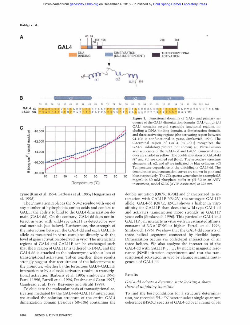
The GAL4 transcriptional activator is required for theregulation of genes involved in galactose and melibiosemetabolism in the yeast Saccharomyces cerevisiae(Johnston 1987). GAL4 has a modular structure in whichdistinct regions of the molecule mediate recognition andbinding to DNA, activation of transcription, and dimer-ization (Fig. 1A; Laughon 1984; Laughon et al. 1984;Keegan et al. 1986; Ma and Ptashne 1987; Lin et al. 1988;Carey et al. 1990). Only the structure of the DNA-bind-ing domain (residues 1–66) has been solved alone in so-lution (Baleja et al. 1992; Kraulis et al. 1992) and in com-plex with DNA (Marmorstein et al. 1992). It contains anN-terminal DNA-recognition domain (residues 7–40), inwhich six cysteines bind two Zn+2 ions. In the free pro-tein in solution, the region from residue 41–66 is un-
structured. In the crystal structure of the complex withDNA, residues 50–63 form a coiled-coil dimerization el-ement that extends perpendicularly away from the DNAhelix (Marmorstein et al. 1992). Nevertheless, GAL41–65 is a monomer in the absence of DNA. Functionalcharacterization of the 881 residue GAL4 protein hasshown that a DNA-independent dimerization domainextends beyond residue 63 comprising the sequence from65 to 94 (Carey et al. 1989); however, no structure of aconstruct containing the complete dimerization domainhas been reported yet. Transcriptional activation func-tion in yeast has been assigned to two regions: the pri-mary activating region comprising residues 768–881 anda weaker one that lies between residues 148–196 (Keeganet al. 1986; Ma and Ptashne 1987; see Fig. 1).GAL450–97 ordinarily mediates dimerization (Carey et
al. 1989). However, the same fragment manifests a noveltranscriptional activating capability in yeast cells carry-ing a single point mutation in the GAL11 protein. Thismutant GAL11P (P standing for transcriptional potentia-tor) and wild-type GAL11 are located in an ∼2-MD com-plex with RNA polymerase and 50 or more polypeptidesin what has been called the RNA–polymerase II holoen-
Present addresses: 4Institut für Physiologie, Rheinisch WestfalischeTechnische Hochschule Aachen, Germany; 5Department of InternalMedicine, University of Michigan Medical Center, Ann Arbor, MI 48109,USA; 6Memorial Sloan-Kettering Cancer Center, New York, NY 10021,USA.7Corresponding author.E-MAIL [email protected]; FAX (617) 432-4383.Article and publication are at www.genesdev.org/cgi/doi/10.1101/gad.873901.
GENES & DEVELOPMENT 15:1007–1020 © 2001 by Cold Spring Harbor Laboratory Press ISSN 0890-9369/01 $5.00; www.genesdev.org 1007
Cold Spring Harbor Laboratory Press on December 4, 2015 - Published by genesdev.cshlp.orgDownloaded from
zyme (Kim et al. 1994; Barberis et al. 1995; Hengartner etal. 1995).The P mutation replaces the N342 residue with one of
any number of hydrophobic amino acids and confers toGAL11 the ability to bind to the GAL4 dimerization do-main (GAL4-dd). On the contrary, GAL4-dd does not in-teract in vitro with wild-type GAL11 as detected by sev-eral methods (see below). Furthermore, the strength ofthe interaction between the GAL4-dd and each GAL11Pallele as measured in vitro correlates directly with thelevel of gene activation observed in vivo. The interactingregions of GAL4 and GAL11P can be exchanged suchthat the P region of GAL11P is tethered to DNA, and theGAL4-dd is attached to the holoenzyme without loss oftranscriptional activation. Taken together, these resultsstrongly suggest that recruitment of the holoenzyme tothe promoter, whether by the fortuitous GAL4–GAL11Pinteraction or by a classic activator, results in transcrip-tional activation (Barberis et al. 1995; Simkovich 1996;Farrell 1996; Farrell et al. 1996; Ptashne and Gann 1997;Gaudreau et al. 1998; Keaveney and Struhl 1998).To elucidate the molecular basis of transcriptional ac-
tivation mediated by the GAL4-dd–GAL11P interaction,we studied the solution structure of the entire GAL4dimerization domain (residues 50–106) containing the
double mutation (Q87R, K90E) and characterized its in-teraction with GAL11P N342V, the strongest GAL11Pallele. GAL4-dd (Q87R, K90E) shows a higher in vitroaffinity for GAL11P than does the wild-type GAL4-ddand activates transcription more strongly in GAL11Pyeast cells (Simkovich 1996). This particular GAL4 andGAL11P pair interacts in vitro with an estimated affinityconstant of 3.3 × 10⁶/M or higher (Farrell et al. 1996;Simkovich 1996). We show that the GAL4-dd consists ofthree helical segments connected by flexible loops.Dimerization occurs via coiled-coil interactions of allthree helices. We also analyze the interaction of theGAL4-dd with GAL11P261–352 by nuclear magnetic reso-nance (NMR) titration experiments and test the tran-scriptional activation in vivo by alanine scanning muta-genesis of GAL4-dd.
Results
GAL4-dd adopts a dynamic state lacking a sharpthermal unfolding transition
To find the best conditions for a structure determina-tion, we recorded 1H–15N heteronuclear single quantumcoherence (HSQC) spectra of GAL4-dd over a range of pH
Figure 1. Functional domains of GAL4 and primary se-quence of the GAL4 dimerization domain (GAL450–106). (A)GAL4 contains several separable functional regions, in-cluding a DNA-binding domain, a dimerization domain,and three activating regions (the activating region between94–106 is nonfunctional in yeast; Simkovich 1996). TheC-terminal region of GAL4 (851–881) recognizes theGAL80 inhibitory protein (not shown). (B) Partial aminoacid sequences of the GAL4-dd and LAC9. Conserved resi-dues are shaded in yellow. The double mutation on GAL4-dd(87 and 90) are colored red (bold). The secondary structureelements, �1, �2, and �3 are indicated by blue cylinders. (C)Temperature dependence of the unfolding of GAL4-dd. Thedenaturation and renaturation curves are shown in pink andblue, respectively. TheCD spectra were taken in a sample 0.5mg/mL in 50 mM phosphate buffer at pH 7.2 in an AVIVinstrument, model 62DS (AVIV Associates) at 222 nm.
Hidalgo et al.
1008 GENES & DEVELOPMENT
Cold Spring Harbor Laboratory Press on December 4, 2015 - Published by genesdev.cshlp.orgDownloaded from
and temperature. The spectra were best in a narrowrange of pH near 7.5 and at 35°C. Variation of the tem-perature between 10°C and 50°C results in large spectralchanges that are different from the kind usually observedfor cooperative thermal unfolding. Although severalsharp signals that were later assigned to the C-terminaltail stayed almost invariant, most other signals changedline shapes with temperature, disappeared, or newly ap-peared in an uncorrelated manner. This indicates tem-perature-dependent noncooperative conformationalchanges. Consistently, the temperature dependence ofthe circular dichroism (CD) spectrum shown in Figure1C lacks a sharp cooperative unfolding transition andresembles the flat melting curves of helix–coil transi-tions in short helical polypeptides (Zimm and Bragg1959). Aware of the dynamic state of the protein, wepursued a characterization of GAL4-dd at 35°C andpH 7.4.
GAL4-dd contains three helices that dimerize viacoiled-coil interactions
Figure 1B shows the amino acid sequence of the GAL4-dd (50–106) aligned with the primary sequence of Lac9,the transcriptional activator that regulates the expres-sion of galactose and lactose genes in Kluveromyces lac-tis (Das and Hollenberg 1982, Salmeron and Johnston1986; Wray et al. 1987). Resonance assignments ofGAL4-dd were pursued with NMR triple-resonance andnuclear Overhauser enhancement (NOE) methods. Ofthe 57 residues, 43 were assigned unambiguously. Fiveresidues at the N terminus (50–54) and four residues atthe C terminus (101 and 103–105) could not be assigned.Flexible tails often remain unassigned in NMR analyses.Furthermore, nine residues from interior sites, subse-quently showed to be located in connecting loop regions(67, 69, 73–77, 84, and 86), could not be identified. Eitherthey were absent or the few unassigned signals showedno spectral properties enabling assignment. The absenceof peaks is consistent with the dynamic state of GAL4-dd mentioned above.Observation of characteristic NOE patterns and analy-
sis of chemical shifts revealed that there are three shorthelices at positions 55–66, 78–84, and 87–97 (Fig. 1B).However, there are no long-range contacts between thethree helices indicating that the structure is extended.The first helix �1 coincides with the terminal helix inthe crystal structure of the complex of GAL41–65 withDNA (Marmorstein et al. 1992).To characterize the mode of dimerization, we followed
a previously described procedure (Walters et al. 1997a) inwhich a uniformly 15N- and 2H-labeled sample wasmixed with unlabeled protein. In H2O solution, all am-ide groups are protonated, and all cross peaks between15N-bound amide protons and aliphatic protons in athree-dimensional 15N-dispersed NOE spectroscopy(NOESY) experiment must be across the dimerizationinterface. In this way, 40 intermonomer NOE contactswere found (involving 16 residues of each monomer), 24for �1, two for the loop between �1 and �2, six for �2, sixfor �3, and two for the region beyond �3.
Structure of the GAL4 dimerization domain
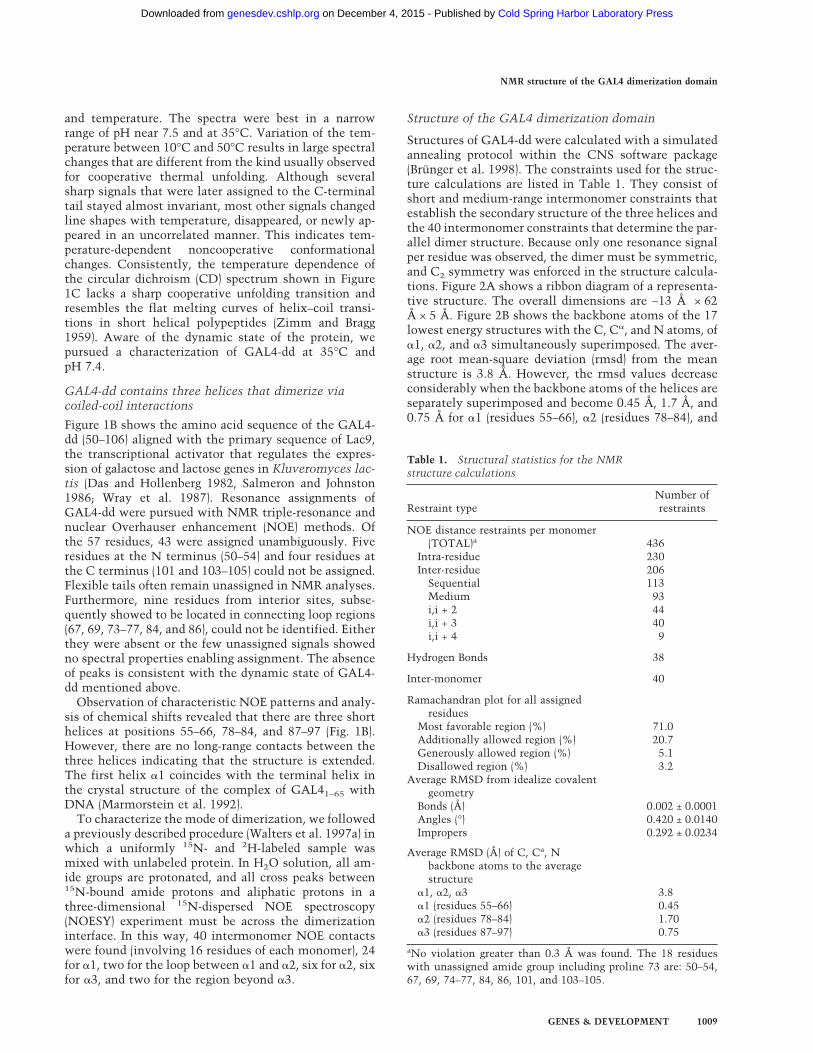
Structures of GAL4-dd were calculated with a simulatedannealing protocol within the CNS software package(Brünger et al. 1998). The constraints used for the struc-ture calculations are listed in Table 1. They consist ofshort and medium-range intermonomer constraints thatestablish the secondary structure of the three helices andthe 40 intermonomer constraints that determine the par-allel dimer structure. Because only one resonance signalper residue was observed, the dimer must be symmetric,and C2 symmetry was enforced in the structure calcula-tions. Figure 2A shows a ribbon diagram of a representa-tive structure. The overall dimensions are ∼13 Å × 62Å × 5 Å. Figure 2B shows the backbone atoms of the 17lowest energy structures with the C, C�, and N atoms, of�1, �2, and �3 simultaneously superimposed. The aver-age root mean-square deviation (rmsd) from the meanstructure is 3.8 Å. However, the rmsd values decreaseconsiderably when the backbone atoms of the helices areseparately superimposed and become 0.45 Å, 1.7 Å, and0.75 Å for �1 (residues 55–66), �2 (residues 78–84), and
Table 1. Structural statistics for the NMRstructure calculations
Restraint typeNumber ofrestraints
NOE distance restraints per monomer(TOTAL)a 436
Intra-residue 230Inter-residue 206Sequential 113Medium 93i,i + 2 44i,i + 3 40i,i + 4 9
Hydrogen Bonds 38
Inter-monomer 40
Ramachandran plot for all assignedresidues
Most favorable region (%) 71.0Additionally allowed region (%) 20.7Generously allowed region (%) 5.1Disallowed region (%) 3.2
Average RMSD from idealize covalentgeometry
Bonds (Å) 0.002 ± 0.0001Angles (°) 0.420 ± 0.0140Impropers 0.292 ± 0.0234
Average RMSD (Å) of C, Ca, Nbackbone atoms to the averagestructure
�1, �2, �3 3.8�1 (residues 55–66) 0.45�2 (residues 78–84) 1.70�3 (residues 87–97) 0.75
aNo violation greater than 0.3 Å was found. The 18 residueswith unassigned amide group including proline 73 are: 50–54,67, 69, 74–77, 84, 86, 101, and 103–105.
NMR structure of the GAL4 dimerization domain
GENES & DEVELOPMENT 1009
Cold Spring Harbor Laboratory Press on December 4, 2015 - Published by genesdev.cshlp.orgDownloaded from
�3 (residues 87–97), respectively (Fig. 2C). �1 and �3 he-lices show the classic coiled-coil conformation. The �3helices are less super-coiled than the �1 pair, however,and their interactions seems to be weaker as fewer in-
terhelix contacts were observed. The short �2 helicespack onto each other with angles >90° in an almost an-tiparallel orientation (see Fig. 2A). A relatively long loopconnects �1 and �2, whereas �2 and �3 are linked by
Figure 2. Three-dimensional structure of the GAL4-dd. (A) A stereoview of the ribbon diagram of a representative structure of theGAL4-dd showing residues H53 to D100. The N and C termini are labeled N and C, respectively. The two monomers are coloreddifferently. Unassigned residues P73 to L77 within the loop connecting �1 and �2 are colored orange. This figure was generated withthe programMOLMOL (Koradi et al. 1996). (B) A stereoview of the backbone atoms from residues L54 to D100 of the 17 lowest energystructures with helices �1, �2, and �3 (blue) simultaneously superimposed. The average backbone RMSD of �1, �2, and �3 to the meanstructure is 3.8 Å. (C) A stereoview of the 17 lowest energy structures whereby the backbone atoms from residues H53 to D100 of eachof the three helices were separately superimposed. For clarity, the loops regions are displayed for only one structure. The backboneatoms of the unassigned P73 to L77 segment are colored in orange as in A. The average backbone RMSD of �1, �2, and �3 to the meanstructure is 0.45, 1.7, and 0.75 Å, respectively. (D) Ensemble of the 17 structures aligned by superimposing the backbone heavy atomsof helix �1.
Hidalgo et al.
1010 GENES & DEVELOPMENT
Cold Spring Harbor Laboratory Press on December 4, 2015 - Published by genesdev.cshlp.orgDownloaded from
only a couple of residues. The longest loop bears astrictly hydrophobic segment from F68 to F72. In con-trast, the subsequent segment from P73 to L77 is pre-dominately hydrophilic. Because there was no observ-able signal for any of the latter residues, we concludethat this segment might undergo slow conformationalexchange.The disorder between the helices, which is apparent in
Figure 2B, is primarily a variation of the amount of twistaround the C2 symmetry axis between the �1–�1�, �2–�2�, and �3–�3� coiled coils. This is shown in Figure 2Din which the structures were aligned by superimposingonly the backbone atoms of the �1 helices. The figureshows that there is variability of orientation around theC2 symmetry axis, of the large loop, the �2 and �3 heli-ces relative to the �1–�1�. The tilt angle between the�1–�1� and �3–�3� coiled coils varies rather little in theensemble of calculated structures. This is reasonable be-cause any permanent tilt would make the dimer asym-metric, which would contradict the symmetric nature ofthe observed NMR spectra. From these considerations,we conclude that the average solution structure ofGAL4-dd is extended as shown in Figure 2A, and thedisorder consists of the amount of twist and presumablya transient variation of the lengths of the helices.
Dimerization interface
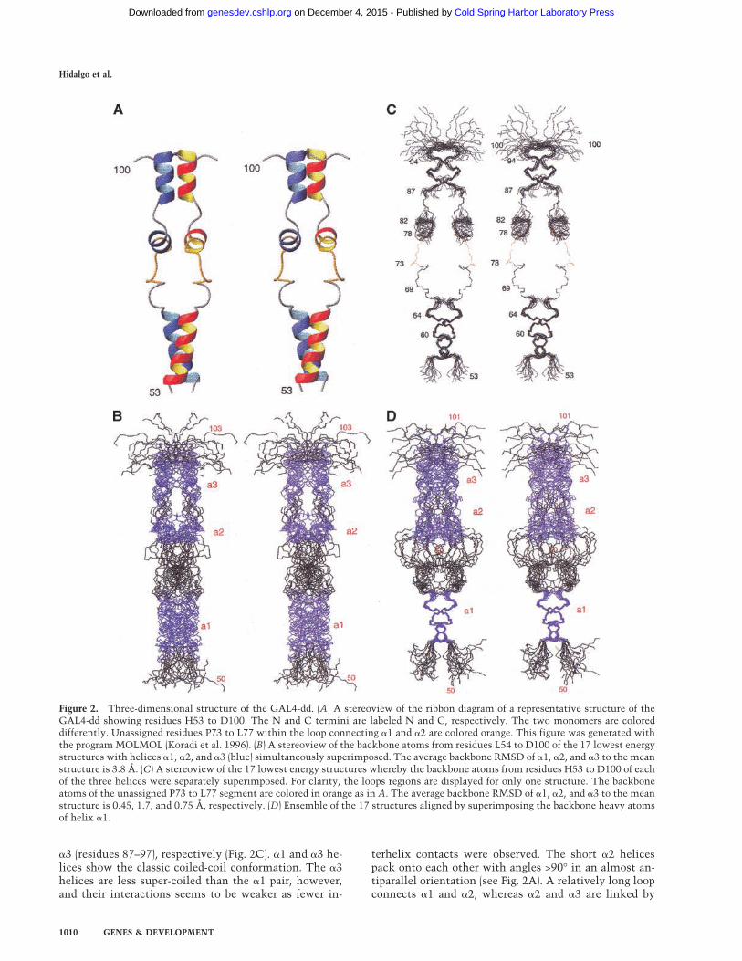
The GAL4-dd dimerizes in solution because of multiplecoiled-coil type interactions between symmetry relatedatoms situated along each monomer. The identificationof the intermonomers contacts forming the dimer inter-face was essential for obtaining the structure of theGAL4 dimer. Intermonomer NOEs were obtained usinga previously described asymmetric deuteration strategy(Walters et al. 1997a).Figure 3A and B shows the superposition of the 17
lowest energy structures with the side chains of residuesforming the dimer interface within �1 and �3 displayed.A projection of the helices onto a wheel diagram isshown in Figure 3C and D. Annotating the heptad repeatsequence characteristic of coiled coils (Lupas 1996) byletters from a to g, �1 contains the sequencea1b1c1d1e1f1g1a2b2c2d2 in which the hydrophobic resi-dues (in boldface) at positions a1, d1, a2, and d2 corre-sponding to the residues L54, V57, L61, and L64, respec-tively, form the core of the dimer interface. The samearrangement was reported for �1 in the crystal structureof GAL4 complexed with DNA (Marmorstein et al.1992). The disruption of the coiled-coil conformation oc-curs at the second half of the second heptad (e2f2g2)where position g2 is occupied by a hydrophobic residue(L67) instead of a hydrophilic one presumably causingthe interruption of the coiled coil.Among the residues located in the loop between �1
and �2, I71 shows intermonomer contacts. In the shorthelix �2, I80, L81, and K82 also show dimer contacts.Finally, in �3, we observe intermonomers contacts forI89, E90, and L93, but the packing of the �3 helices ap-pears to be less tight than in �1. Considering that the
number of observed intermonomer and intramonomerNOE distance restraints was higher for �1 coiled coil, thedifferences may reflect variations in the dynamic behav-ior of the molecule. Therefore, we assayed this possibil-ity by studying the mobility of the GAL4-dd. The dy-namic properties of the GAL4-dd may be relevant for thetranscriptional activation function as will be discussedbelow.
Protein mobility
To understand whether the apparent disorder in someparts of the GAL4-dd structure is due to mobility ratherthan lack of constraints, and to provide a basis for a pos-sible correlation between mobility and activation func-tion, we studied the motional properties of the GAL4-ddby using 15N relaxation experiments. Three relaxationparameters were measured, the nitrogen longitudinal re-laxation rate RN(Nz), the nitrogen transverse relaxationrate RN(Nx,y), and the heteronuclear NOE (XNOE) (Fig.4; Peng and Wagner 1992). The 15N relaxation param-eters are related to the rotational diffusion of the N–Hbond due to overall tumbling and internal motion, and sothey contain information on the molecular mobility of
Figure 3. Dimerization interface within �1 and �3 of GAL4(50–106). (A) A stereoview of the superimposed backbone atomswithin �1 for the 17 lowest energy structures. The side chains ofV57, L61, and L64 forming intermonomer contacts are dis-played in different colors for each monomer. (B) A stereoview ofthe superimposed backbone atoms within �3 for the 17 lowestenergy structures. The side chains of I89, L93 forming inter-monomer contacts and F97 are shown in different colors foreach monomer. (C) Helical wheel representation of �1 with hy-drophobic, polar, and charged residues displayed in red, green.and blue, respectively. (D) Helical wheel representation of �3.Same color code for the residues as C but glycine in black.
NMR structure of the GAL4 dimerization domain
GENES & DEVELOPMENT 1011
Cold Spring Harbor Laboratory Press on December 4, 2015 - Published by genesdev.cshlp.orgDownloaded from
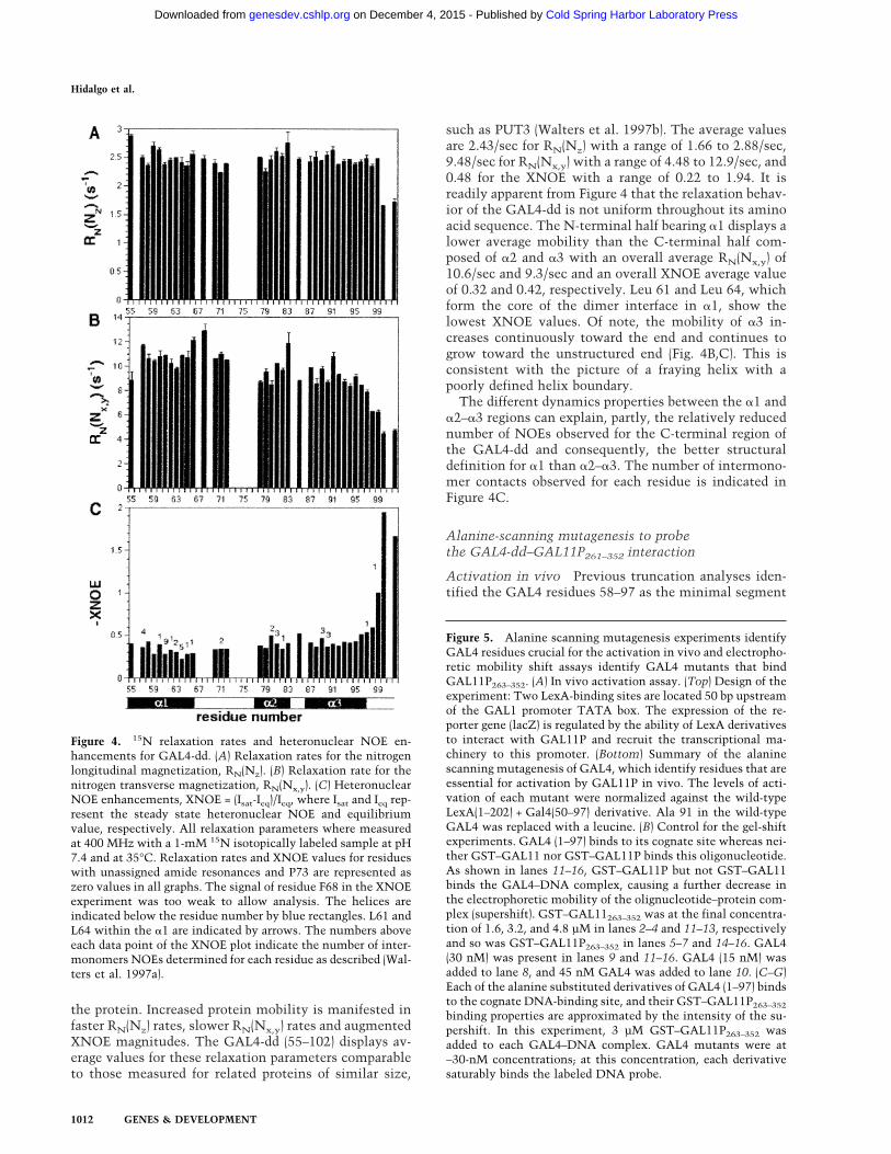
the protein. Increased protein mobility is manifested infaster RN(Nz) rates, slower RN(Nx,y) rates and augmentedXNOE magnitudes. The GAL4-dd (55–102) displays av-erage values for these relaxation parameters comparableto those measured for related proteins of similar size,
such as PUT3 (Walters et al. 1997b). The average valuesare 2.43/sec for RN(Nz) with a range of 1.66 to 2.88/sec,9.48/sec for RN(Nx,y) with a range of 4.48 to 12.9/sec, and0.48 for the XNOE with a range of 0.22 to 1.94. It isreadily apparent from Figure 4 that the relaxation behav-ior of the GAL4-dd is not uniform throughout its aminoacid sequence. The N-terminal half bearing �1 displays alower average mobility than the C-terminal half com-posed of �2 and �3 with an overall average RN(Nx,y) of10.6/sec and 9.3/sec and an overall XNOE average valueof 0.32 and 0.42, respectively. Leu 61 and Leu 64, whichform the core of the dimer interface in �1, show thelowest XNOE values. Of note, the mobility of �3 in-creases continuously toward the end and continues togrow toward the unstructured end (Fig. 4B,C). This isconsistent with the picture of a fraying helix with apoorly defined helix boundary.The different dynamics properties between the �1 and
�2–�3 regions can explain, partly, the relatively reducednumber of NOEs observed for the C-terminal region ofthe GAL4-dd and consequently, the better structuraldefinition for �1 than �2–�3. The number of intermono-mer contacts observed for each residue is indicated inFigure 4C.
Alanine-scanning mutagenesis to probethe GAL4-dd–GAL11P261–352 interaction
Activation in vivo Previous truncation analyses iden-tified the GAL4 residues 58–97 as the minimal segment
Figure 4. 15N relaxation rates and heteronuclear NOE en-hancements for GAL4-dd. (A) Relaxation rates for the nitrogenlongitudinal magnetization, RN(Nz). (B) Relaxation rate for thenitrogen transverse magnetization, RN(Nx,y). (C) HeteronuclearNOE enhancements, XNOE = (Isat-Ieq)/Ieq, where Isat and Ieq rep-resent the steady state heteronuclear NOE and equilibriumvalue, respectively. All relaxation parameters where measuredat 400 MHz with a 1-mM 15N isotopically labeled sample at pH7.4 and at 35°C. Relaxation rates and XNOE values for residueswith unassigned amide resonances and P73 are represented aszero values in all graphs. The signal of residue F68 in the XNOEexperiment was too weak to allow analysis. The helices areindicated below the residue number by blue rectangles. L61 andL64 within the �1 are indicated by arrows. The numbers aboveeach data point of the XNOE plot indicate the number of inter-monomers NOEs determined for each residue as described (Wal-ters et al. 1997a).
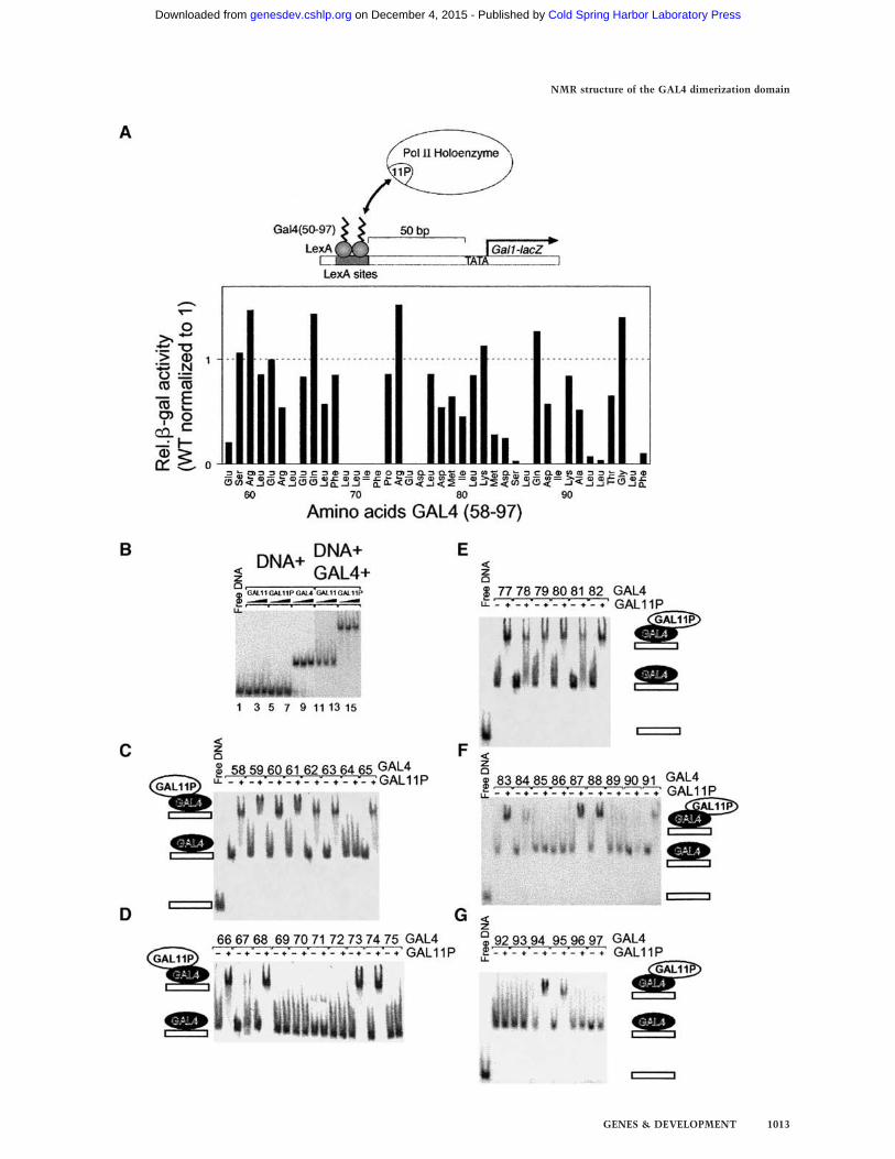
Figure 5. Alanine scanning mutagenesis experiments identifyGAL4 residues crucial for the activation in vivo and electropho-retic mobility shift assays identify GAL4 mutants that bindGAL11P263–352. (A) In vivo activation assay. (Top) Design of theexperiment: Two LexA-binding sites are located 50 bp upstreamof the GAL1 promoter TATA box. The expression of the re-porter gene (lacZ) is regulated by the ability of LexA derivativesto interact with GAL11P and recruit the transcriptional ma-chinery to this promoter. (Bottom) Summary of the alaninescanning mutagenesis of GAL4, which identify residues that areessential for activation by GAL11P in vivo. The levels of acti-vation of each mutant were normalized against the wild-typeLexA(1–202) + Gal4(50–97) derivative. Ala 91 in the wild-typeGAL4 was replaced with a leucine. (B) Control for the gel-shiftexperiments. GAL4 (1–97) binds to its cognate site whereas nei-ther GST–GAL11 nor GST–GAL11P binds this oligonucleotide.As shown in lanes 11–16, GST–GAL11P but not GST–GAL11binds the GAL4–DNA complex, causing a further decrease inthe electrophoretic mobility of the olignucleotide–protein com-plex (supershift). GST–GAL11263–352 was at the final concentra-tion of 1.6, 3.2, and 4.8 µM in lanes 2–4 and 11–13, respectivelyand so was GST–GAL11P263–352 in lanes 5–7 and 14–16. GAL4(30 nM) was present in lanes 9 and 11–16. GAL4 (15 nM) wasadded to lane 8, and 45 nM GAL4 was added to lane 10. (C–G)Each of the alanine substituted derivatives of GAL4 (1–97) bindsto the cognate DNA-binding site, and their GST–GAL11P263–352binding properties are approximated by the intensity of the su-pershift. In this experiment, 3 µM GST–GAL11P263–352 wasadded to each GAL4–DNA complex. GAL4 mutants were at∼30-nM concentrations; at this concentration, each derivativesaturably binds the labeled DNA probe.
Hidalgo et al.
1012 GENES & DEVELOPMENT
Cold Spring Harbor Laboratory Press on December 4, 2015 - Published by genesdev.cshlp.orgDownloaded from
NMR structure of the GAL4 dimerization domain
GENES & DEVELOPMENT 1013
Cold Spring Harbor Laboratory Press on December 4, 2015 - Published by genesdev.cshlp.orgDownloaded from
necessary for transcriptional activation in GAL11P cells(Barberis et al. 1995). To identify individual residueswithin this segment of GAL4 that play a role in tran-scriptional activation in vivo, we performed site-directedalanine substitution mutagenesis of each of the 40 resi-dues (Fig. 5). The mutations were made in the context ofa chimeric protein in which residues 50–97 of GAL4were fused to LexA, a dimeric bacterial DNA-bindingprotein that functions efficiently in yeast. In GAL11Pcells, the LexA–GAL4 (50–97) fusion protein stimulatesthe expression of a reporter gene bearing LexA-bindingsites upstream of the minimal GAL1 promoter (Barberiset al. 1995; Farrell et al. 1996). Thus, it allowed us tocircumvent any effects of alanine substitutions ondimerization and subsequent DNA binding of GAL4 toits cognate sites and identified those residues that spe-cifically effect the ability of GAL4-dd to activate tran-scription. Figure 5A shows a scheme of the experimentaldesign and a summary of the activity of each mutantnormalized to the wild-type LexA–GAL4 (58–97) fusionprotein. The results shown in Fig. 5A identify a cluster ofhydrophobic residues from 69–72 (LLIF), and residuesL64, E75, D76, S85, L86, I89, L92, L93, L96, and F97 asessential for activation in vivo.To ascertain if the loss of activation occurs because of
a loss in interaction, we purified wild-type GAL4 (1–97)and 39 mutant forms each of which bears an alaninemutation at a single position scanning the residues 58–97, except for position 91, which is an alanine in the wildtype and substituted by leucine. The mutant GAL4,D76A, was insoluble and thus refractory to furtheranalysis. The in vitro interaction of these GAL4 deriva-tives with a fragment of GAL11 and GAL11P (residues263–352) expressed as a GST fusion protein was mea-sured in a gel mobility shift assay (Fig. 5B–G). In thisassay, neither GAL11 nor GAL11P bind to DNA bearinga GAL4 DNA-binding site (Fig. 5B, lanes 1–7), but, asexpected, GAL4 (1–97) does (Fig. 5B, lanes 8–10). Con-versely, Figure 5B shows that GAL11P (lanes 14–16),but not GAL11 wild type (lanes 11–13), binds to theDNA-tethered GAL4 as indicated by the supershift ofthe protein–DNA complex. Figure 5(C–G) shows that all39 GAL4 mutants bound the GAL4 DNA site, and 26 ofthese interacted with GAL11P as shown by the super-shift.There is a strong correlation between the ability of
each GAL4 mutant to stimulate activation in vivo asmeasured in Figure 5A and the ability to interact withGAL11P in vitro as measured in Figure 5C–G. Thus, ofthe 39 mutants, those that interacted with GAL11P invitro, all activated in vivo. Moreover, all of the 13 de-rivatives that failed to interact in vitro (positions 64,69–72, 75, 85, 86, 89, 92, 93, 96, and 97) also failed toactivate in vivo. K90A bound Gal11P very weakly invitro but activated well in vivo. Conversely, E58A boundwell in vitro but did not activate well in vivo; however,it was particularly difficult to purify, and thus its inabil-ity to activate transcription might be a consequence ofinstability in vivo.The results from the alanine scanning are elucidated
further by visualizing the crucial mutations (Fig. 7B, be-low) on the GAL4-dd structure (Fig. 1B). Leu 69 to Phe 72map on the hydrophobic part of the loop connecting �1and �2 whereas E75 and D76 are on the hydrophilic sec-tion of the same loop. M83 to L86 constitute the loopbetween �2 and �3, and I89, L92, L93, L96, and F97 are inhelix �3. Leu 64 form the core of �1 dimerization inter-face, and the effect of its mutation is probably purelystructural.
NMR titration of GAL4-dd with GAL11P261–352
To identify the GAL11P-binding site, we titrated 15N-labeled GAL4-dd with unlabeled GAL11P261–352 andmonitored the effect with 1H–15N HSQC spectra. Threeof five spectra recorded in this series are shown in Figure6B (0, 0.5, and 1mMGAL11P). Increasing concentrationsof GAL11P from 0 to 2 mM, in 0.25-mM increments,were added to 1 mM (dimer) 15N-GAL4-dd while mea-suring 1H–15NHSQC spectra. Because only the signals ofprotons covalently attached to 15N nuclei are detected,all resonances observed arise from the 15N-labeledGAL4-dd while the GAL11P peptide is invisible. We ob-tained the following results.GAL11P forms a tight complex with GAL4-dd with
slow exchange kinetics (koff < 5/sec) because addingGAL11P causes disappearance of a subset of the HSQCpeaks and appearance of new peaks proportional to theconcentration of the ligand (Fig. 6B). Addition ofthe wild-type GAL11 peptide under otherwise identicalconditions had no effect on the GAL4-dd resonancesup to a 1:3 ratio of GAL4-dd to wt-GAL11 (data notshown).GAL11P binds as a monomer to the GAL4-dd dimer
because addition of GAL11P to the 1 mM (dimer) sampleof GAL4-dd causes spectral changes up to 1 mM (mono-mer) GAL11P concentration. No significant spectralchanges happen at higher concentration of GAL11P. Therelative concentrations of the GAL4-dd versus GAL11Ppeptide particularly for this assays were verified byamino acid analysis.Formation of the monomer–dimer complex intro-
duces asymmetry into the GAL4-dd dimer (Fig. 6B). Al-though the spectrum of the complex has not been as-signed, a splitting of signals is observed for several resi-dues, most clearly documented for G95, which is theonly glycine and thus well separated from all other sig-nals. The peak at 109.6 ppm 15N chemical shift splits intwo peaks at 108.3 and 110.1 ppm 15N chemical shift.Many other peaks disappear in the titration and numer-ous new signals of half the intensity appear. Thus, intro-duction of asymmetry is manifested in many otherpeaks. However, the new peaks cannot easily be assignedbecause they are far away from those of the free GAL4-dd, and a complete assignment of the complex was notachieved. The assignments for those peaks that clearlydisappear in the complex are highlighted in yellow in theleft panel of Figure 6B; those that remain unchanged arenot.
Hidalgo et al.
1014 GENES & DEVELOPMENT
Cold Spring Harbor Laboratory Press on December 4, 2015 - Published by genesdev.cshlp.orgDownloaded from
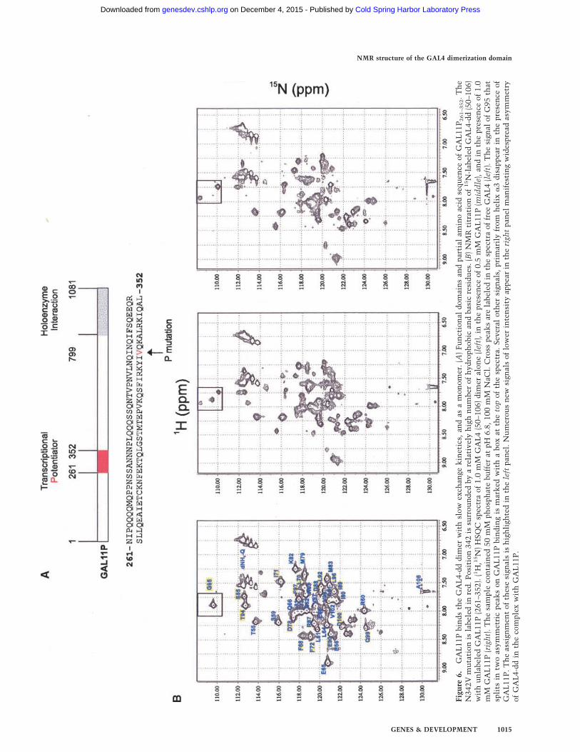
Figu
re6.
GAL11PbindstheGAL4-dddimerwithslow
exchangekinetics,andasamonomer.(A)FunctionaldomainsandpartialaminoacidsequenceofGAL11P261–352.The
N342Vmutationislabeledinred.Position342issurroundedbyarelativelyhighnumberofhydrophobicandbasicresidues.(B)NMRtitrationof15N-labeledGAL4-dd(50–106)
withunlabeledGAL11P(261–352).(1H,15N)HSQCspectraof1.0mMGAL4(50–106)dimeralone(left),inthepresenceof0.5mMGAL11P(middle),andinthepresenceof1.0
mMGAL11P(right).Thesamplecontained50mMphosphatebufferatpH
6.8,100mMNaCl.CrosspeaksarelabeledinthespectraoffreeGAL4(left).ThesignalofG95that
splitsintwoasymmetricpeaksonGAL11Pbindingismarkedwithaboxatthetopofthespectra.Severalothersignals,primarilyfrom
helix
�3disappearinthepresenceof
GAL11P.Theassignmentofthesesignalsishighlightedintheleftpanel.Numerousnewsignalsoflowerintensityappearintherightpanelmanifestingwidespreadasymmetry
ofGAL4-ddinthecomplexwithGAL11P.
NMR structure of the GAL4 dimerization domain
GENES & DEVELOPMENT 1015
Cold Spring Harbor Laboratory Press on December 4, 2015 - Published by genesdev.cshlp.orgDownloaded from
Examination of the chemical shift perturbation ofthe NMR resonances on titration shows that there areno or only very minor changes observed for the entirehelix �1. All residues of the loop connecting �1 and�2 disappear with increasing GAL11P concentration.The helix �2 is only affected at its C-terminal end bythe addition of GAL11P. However, all of the helix �3and the subsequent residues 97–100 disappear or splitin two components. Because the binding of GAL11Ppeptide to GAL4-dd undoubtedly brings G95 to thevicinity of GAL11P, we assumed that the nearby resi-dues whose resonances also are perturbed in the titra-tion experiments also must be in close proximity toGAL11P. However, many of these GAL4 residuesthat participate in the dimerization may play a rolein direct interactions with GAL11P, as observed inthe case of G95. These observations indicate thatGAL11P binds GAL4-dd via the �1–�2 loop and thehelix �3.Figure 7 shows the residues sensitive to GAL11P iden-
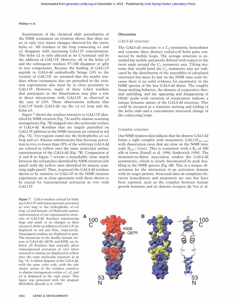
tified by NMR titration (Fig. 7A) and by alanine-scanningmutagenesis (Fig. 7B) mapped onto the molecular surfaceof GAL4-dd. Residues that are largely perturbed onGAL11P addition in the NMR titration are colored in red(Fig. 7A). Two regions stand out: the hydrophobic �1–�2loop and �3. Alanine substitutions that decrease activa-tion in vivo to lower than 10% of the wild-type GAL4-ddare colored in yellow onto the same molecular surfacerepresentation of the GAL4-dd (Fig. 7B). Comparison ofA and B in Figure 7 reveals a remarkably close matchbetween the red patches identified by NMR titration (leftpanel) with the yellow ones identified by alanine scan-ning (right panel). Thus, in general the GAL4-dd residuesshown to be sensitive to GAL11P in the NMR titrationexperiment are in close agreement with those shown tobe crucial for transcriptional activation in vivo withGAL11P.
Discussion
GAL4-dd structure
The GAL4-dd structure is a C2-symmetric homodimerand contains three distinct coiled-coil helix pairs con-nected by mobile loops. The average structure is ex-tended but mobile and poorly defined with respect to thetwist angle around the C2 symmetry axis. Tilting mo-tions that would bend the C2 symmetry axis are indi-cated by the distribution of the ensembles of calculatedstructures but must be fast on the NMR time scale be-cause there is no solid evidence for asymmetry in theNMR spectra of the free GAL4-dd dimer. The roughlylinear melting behavior, the absence of cooperative ther-mal unfolding, and the appearing and disappearing ofHSQC peaks with variation of temperature indicate aunique dynamic nature of the GAL4-dd structure. Thiscould be pictured as a transient melting and folding ofthe helix ends and a concomitant structural change ofthe connecting loops.
Complex structure
OurNMR titration data indicate that the dimeric GAL4-ddforms a tight complex with monomeric GAL11P261–352with dissociation rates that are slow on the NMR timescale (koff < 5/sec). This is consistent with a Kd of 300nM or lower (Farrell et al. 1996; Simkovich 1996). Themonomer-to-dimer association renders the GAL4-ddasymmetric, which is clearly documented by peak dou-bling in the NMR spectra (Fig. 6B). This is a unique ob-servation for the interaction of an activation domainwith its target protein. Structural data on complexes be-tween homodimers and monomers are rare but havebeen reported, such as the complex between humangrowth hormone and its dimeric receptor (de Vos et al.
Figure 7. GAL4 residues critical for bind-ing GAL11P and transcriptional activationin vivo map to the hydrophobic �1–�2loop, �3 and beyond. (A) Molecular surfacerepresentation of one representative struc-ture of GAL4-dd. Residues experiencinglarge and small or no changes in theirchemical shifts on addition of GAL11P aredisplayed in red and blue, respectively.Unassigned residues are displayed in gray.The mutations in the double mutant ver-sion of GAL4-dd, Q87R, and K90E, are la-beled. (B) Residues that critically affecttranscriptional activation in vivo whenmutated to alanine are displayed in yellowonto the same molecular structure as inFig. 7A. A ribbon diagram of the GAL4-dd,with the same color code, with the sidechains atoms of the residues sensitiveto alanine mutagenesis within �1, �2, and�3 is displayed in the right panel. Thisfigure was generated with the programMOLMOL (Koradi et al. 1996).
Hidalgo et al.
1016 GENES & DEVELOPMENT
Cold Spring Harbor Laboratory Press on December 4, 2015 - Published by genesdev.cshlp.orgDownloaded from
1992) or the RNA bacteriophage coat protein–operatorcomplex (Valegard et al. 1994). The symmetry-breakingaspects have been discussed extensively in Bardsley et al.(1998). As discussed for the previous cases, binding of thefirst GAL11P monomer changes the conformation of theGAL4-dd dimer in a way that abrogates affinity for thesecond binding site. This is an extreme case of negativecooperativity.Despite the tight interaction, the complex is in a dy-
namic state. This is concluded from the HSQC spectrumrecorded at the end point of the titration (Fig. 6B, right)and from the spectra recorded with excess of GAL11P(data not shown). These spectra contain far less than thenumber of signals expected for an entirely folded GAL4-dd dimer in complex with the unlabeled GAL11P. Con-versely, an HSQC spectrum of free GAL11P261–352 showsonly 48 weak peptide NH cross peaks (data not shown).Thus, GAL4-dd, GAL11P261–352, and their complex con-tain large portions that undergo apparently slow confor-mational exchange, which would make a more detailedstructural characterization difficult.
GAL4-dd shows a novel transcriptionalactivation motif
The NMR titration and the alanine scanning experi-ments locate the GAL11P-binding site to the loop re-gions, the terminal helix �3, and part of the segmentbeyond �3. In wt-GAL4-dd, these regions bear six nega-tive charges (E75, D76, D78, D84, D88, and D100) bal-anced by three basic side chains (R74, K82, and K90; Fig.1B). The double mutant (Q87R, K90E) that introduces anextra negative charge shows higher affinity to GAL11Pand activates transcription stronger. Thus, the novel ac-tivation region of GAL4-dd shows the feature of highacidity present in other classic activating domains. Intri-guingly, the GAL11P-binding face on GAL4-dd coincideswith part of an acidic segment (residues 75–147) thatpreviously been has shown to activate promoters bearingGAL4-binding sites in HeLa cell extracts (Lin et al.1988). Therefore, GAL11P may use the same activatormotif.In contrast with earlier studies of activating domains,
which all revealed a single helix in the target-bound state(Kussie et al. 1996; Radhakrishnan et al. 1997; Uesugi etal. 1997) the novel activation motif of the GAL4-dd is acoiled-coil dimer supporting two acidic/hydrophobicloops. The mobility experiments revealed that theGAL4-dd (the terminal �3 helix pair) shows enhancedmobility gradually increasing toward the C terminus(Fig. 4). This may be important for activation functionand is consistent with the observation that mutationsstabilizing the �3 dimerization reduce the activation po-tential (A.Z. Ansari and M. Ptashe, unpubl.).The apparent need for weak dimer interaction of the
�3 helices may explain why the double mutant versionof the GAL4-dd studied here is a better activator. Bothresidues R87 and E90 are placed to the same side of thehelix facing away from the dimerization interface. Inter-action between the two side chains may stabilize the
individual helices but not the dimer interaction. This isconsistent with the previously mentioned observationthat stabilizing the dimer reduces the activation func-tion.
The recruitment of GAL11P by GAL4-dd relieson polar and hydrophobic interactions
The clustering of negative charges in the GAL11P-bind-ing site of GAL4-dd suggest an electrostatic interaction.Consistently, the segment flanking the P-mutation inGAL11P has excess positive charge with five basic resi-dues within ±10 positions (Fig. 6A). It needs to be ex-plained, however, why the N342V mutation in GAL11Pgenerates a binding site for the GAL4-dd. The introduc-tion of a hydrophobic residue at position 342 mightcause folding of GAL11P, and the new binding functionmight be a conformational effect rather than a directhydrophobic interaction of V342 with GAL4-dd. How-ever, mutagenesis experiments in which residue at posi-tion 342 in GAL11 has been systematically substitutedwith various residues showed that only hydrophobicsubstitutions confer the transcriptional potentiator phe-notype to GAL11. The different hydrophobic substitu-tions lead to different in vitro binding affinities for GAL4and likewise different degrees of in vivo transcriptionalactivation (Barberis et al. 1995; Farrell 1996; Farrell et al.1996). It is difficult to imagine that such differences inaffinities reflect different degree of structural stabiliza-tion toward permissive binding conformations. The largeeffect of such mutations at position 342 on binding af-finity suggests a direct contact between the residue atthis position with GAL4-dd. Thus, the structure ofGAL4-dd and the features of the binding site suggest thatthe hydrophobic residue V342 and other nearby hydro-phobic groups (Y340, I341, F336, I337, A445, L446, andI449; see Fig. 6A) in GAL11P might interact with thehydrophobic section of the loop connecting �1 and �2 inGAL4-dd. Mutational analysis indicates that in GAL11Pbearing another hydrophobic substitution at position342 (N342I), changing the neighboring hydrophobic resi-due I341 to Asn, reduces transcriptional activation some10-fold. Moreover, adding the change Q343I to thisGAL11 double mutant (N342I–I341N) improves activa-tion by twofold. This observation further support theidea that hydrophobic residues neighboring 342 partici-pate in GAL4–GAL11P interactions.Hydrophobic interactions are usually tighter than hy-
drophilic ones. This might explain the low Kd and theslow dissociation rates, which is unusual for activator–target interactions. For comparison, the VP16 activationdomain is in fast exchange with the bound state, and thehelical structure of the bound conformation was eluci-dated with transferred NOE experiment (Uesugi et al.1997). Typical activators are capable of contacting sev-eral targets to recruit the transcriptional machinery tothe promoter (Ptashne and Gann 1997; Koh et al. 1998and references therein). Thus, rapid exchange may allowthem to recruit and orient the holoenzyme, and othercomponents of the transcriptional machinery appropri-
NMR structure of the GAL4 dimerization domain
GENES & DEVELOPMENT 1017
Cold Spring Harbor Laboratory Press on December 4, 2015 - Published by genesdev.cshlp.orgDownloaded from
ately on the promoter (Kim et al. 1994; Koleske andYoung 1994; Kuras and Struhl 1999; Li et al 1999). Theslow exchange kinetics observed here may be biologi-cally relevant for the newly acquired ability of GAL4-ddto function as an activator in the presence of GAL11P.GAL4-dd can only interact with the P region of GAL11P,thereby recruiting the holoenzyme to the promoter. Thethermodynamic and structural properties of GAL4-ddbinding to GAL11P described in this study are consistentwith the notion that GAL4-dd needs to hold on to theholoenzyme long enough to permit binding of other es-sential components of the transcriptional machinery tothe promoter. However, the significance of a kinetic ver-sus a thermodynamic explanation remains to be tested.Notably, this singular activator-target interaction suf-fices for high levels of transcriptional activation that arecomparable to those elicited by strong natural activators.
Materials and methods
Sample preparation
Dr. R. Marmorstein kindly provided the purification procedurefor GAL450–106. GAL450–106 was used instead of 50–97 becausethe latter expresses poorly in bacteria. GAL450–106 was ex-pressed in BL21-DE(3) pLysS Escherichia coli cells from apRSETA expression vector (Invitrogen). Cells were grown at37°C, induced 3 h with 1 mM IPTG, and harvested by centrifu-gation. Purification was performed through two consecutiveammonium sulfate precipitation with 0.25 and 0.15 g/mL, fol-lowed by ion exchange chromatography on a DEAE–Sepharosecolumn and gel filtration on a Superdex 75 (16/60) column(Amersham Pharmacia Biotech). The fractions containingGAL450–106 were analyzed by SDS-PAGE electrophoresis.
15N-and 15N–13C-labeled GAL4 were obtained by using 15NH4Cl and15NH4Cl,
13C6-D-glucose as the sole nitrogen and nitrogen-car-bon sources in M9 medium, respectively. 2H–15N–labeled pro-tein was produce as described previously (Walters 1997a).The expression of GAL11P261–352 was performed similarly
from a pRSETA vector in BL21-DE(3) pLysS E. coli cells. Puri-fication of GAL11P261–352 was performed by partial fraction-ation with 0.3 g/mL ammonium sulfate followed by ion ex-change chromatography by using SP-Sepharose and gel filtrationon a Superdex-75 column.Verification of the identity of the purified proteins was per-
formed by mass spectrometry and amino acid analysis. TheNMR protein samples ranged from 0.5 to 1 mM concentrationin 50 mM sodium phosphate buffer at pH 7.4.
Alanine scanning mutagenesis
Mutations of the GAL4 segment in the context of LexA(1–202) + Gal4(50–97) were generated by site-directed PCR muta-genesis. The PCR products were subcloned between XbaII–SalIsites in frame with the LexA coding sequence in RJR 238 bear-ing a chromosomal origin of replication, a centromere, and a His3 histidine auxotrophy selection marker. The LexA derivativesare expressed by a strong yeast actin promoter and the transcrip-tional unit ends with a GAL11 terminator. Each of the 40 mu-tants was confirmed by sequencing. Expression was tested in aGAL11P strain from a chromosomally integrated �-gal reportergene bearing two LexA-binding sites 50 bp upstream of theGAL1 promoter TATA box. �-gal assays were performed as de-scribed previously (Barberis et al. 1995; Simkovich 1996).
Electrophoretic mobility shift assay
A double stranded oligonucleotide of 30 bp containing a singleGAL4-binding site (5�-TCCGGAGGACTGTCCTCCGGT-3�)was labeled with 32P at the 5� end. The labeled DNA was incu-bated in a 20-µL reaction volume, with each of the GAL4 mu-tants and with GST–GAL11 or GST–GAL11P in EMSA buffer(20 mM Hepes at pH 7.5, 25 mM NaCl, 5 mM MgCl2, 20 mMZnCl2, 6% glycerol, 200 µg/mL bovine serine albumin, 3 mM�-mercaptoethanol, 2 mg/mL polydIdC). The complexes wereresolved on a 9% polyacrylamide gel in 0.5× Tris-borate run for2.5 h at 100 V at 9°C. The bands were visualized by Phosphor-imaging on a Fuji-Bas PhosphorImager.The GAL4 mutants were expressed in BL21-DE(3) pLysS bac-
terial strain and purified on a SP-Fast Flow Sepharose resin(Pharmacia) as described previously (Ansari et al. 1998). GST–GAL11 or GST–GAL11P (residues 263–352 of GAL11) also wereexpressed in this strain and purified on a glutathione–Sepharose(Pharmacia) column by using standard procedures (Smith andJohnson 1988). Both proteins were dialyzed against the EMSAbuffer.
NMR spectroscopy and assignments
All NMR spectra were recorded on Bruker AMX 500, VarianUnityPlus 400, Varian Unity 500, Inova 500, and UnityPlus 750spectrometers at 35°C. Spectra were processed using FELIX(Molecular Simulations) and analyzed using XEASY (Bartels etal. 1995) on Silicon Graphics workstations. Backbone assign-ments were obtained using double-resonance 15N-dispersedNOESY triple resonance three-dimensional (1H, 15N, and 13C)HNCA and HN(CO)CA spectra. Side-chain resonances were as-signed using 15N- and 13C-dispersed NOESY experiments.HNHA and HNHB experiments were used to assign H� and H�,respectively (Archer et al. 1991; Madsen et al. 1993; Vuister andBax 1993). Intermonomer contacts were obtained as describedpreviously (Walters et al. 1997a) using a 200-msec mixing timefor the 15N-dispersed NOESY. Complete mixing of the two ho-modimers species, unlabeled and 2H–15N–labeled, was achievedby heating the sample for 10 min to 60°C. No mixing was ob-served at room temperature over a period of 1 wk.
NOE and distance restraints
Distance restraints were obtained using a two-dimensionalNOESY spectrum with a mixing time of 80 msec and 15N- and13C-dispersed NOESY (Talluri and Wagner 1996) spectra withmixing times of 120 and 80 msec, respectively. Cross peakswere integrated using the XEASY software package (Bartels etal. 1995). The intensities of NOE cross peaks arising from in-teractions between H� and HN nuclei within helical regionswere used to calibrate the 15N-dispersed NOESY spectrum(Wüthrich 1986). The 13C-dispersed NOESY spectrum was cali-brated by assuming the weakest cross peak corresponding to aninteraction between H� and H�, within well-defined helical el-ements, to be 5 Å. NOE derived restraints were supplementedwith 38 hydrogen bonds per monomer within regular secondarystructural elements. Secondary structural elements were iden-tified based on the H� chemical shift index (Wishart et al. 1992)and NOE connectivity. For each hydrogen bond, two restraintswere used such that the distances between the amide protonand the oxygen and between amide nitrogen and oxygen wererestricted to be 1.8–2.5 Å and 2.5–3.3 Å, respectively. Distancesymmetry restraints for all assigned residues were used to gen-erated a twofold symmetric arrangement of the dimer as de-scribed previously (Brünger 1992).
Hidalgo et al.
1018 GENES & DEVELOPMENT
Cold Spring Harbor Laboratory Press on December 4, 2015 - Published by genesdev.cshlp.orgDownloaded from
Structure calculations
Structure calculations were performed using CNS version 0.5(Brünger et al. 1998) on an R10000 IndigoII Silicon Graphicsworkstation. The molecular dynamics simulated annealing wasperformed using NMR distance restraints and distance symme-try restraints starting from an extended duplicated strand. Withthe exception of the NOESY cross peaks obtained using theGAL4 (50–106) heterodimer and the hydrogen bonds, all NOEswere treated as ambiguous (Nilges 1993). The InsightII program(Molecular Simulations) andMOLMOL (Koradi et al. 1996) wereused for displaying and visual inspection of the three-dimen-sional structures. PROCHECK (Laskowoski et al. 1993) wasused to further assess the quality of the structures. Residues inwhich the amide group was unassigned were dismissed from thestatistics reported for the Ramachandran plot (Table 1). Coor-dinates of the structures have been deposited at the ProteinData Bank.
Relaxation experiments
All relaxation experiments were performed on a Varian Unity-Plus400 spectrometer. The three relaxation parameters, 15Nlongitudinal and transverse relaxation rates and heteronuclearNOE values, were measured using two-dimensional hetero-nuclear pulse sequences with pulsed-field gradients and sensi-tivity enhancement (Dayie and Wagner 1994). The analysis ofthe relaxation data was performed as described previously (PengandWagner 1992). Each peak was integrated by volume by usingXEASY and fitted to single exponential function by using theLevenburg–Marquardt nonlinear least squares method imple-ment in PLOT (New Unit). Uncertainties in the relaxation rateswere estimated with Monte Carlo simulations by using onerepeated time point as described (Peng and Wagner 1992).
Acknowledgments
We are indebted to Dr. Kylie Walters for sharing her valuableexperience in solving dimers structures throughout all thiswork. We are grateful to Dr. Yingxi Lin and Dr. ChristophFahlke for many helpful discussions, to Gregory Heffron, Dr.Hiroshi Matsuo, and Dr. Walfrido Antuch for their assistance inNMR spectroscopy. We thank the Helen Hay Whitney Founda-tion for support to A.Z.A. and Howard HughesMedical Institutefor a predoctoral fellowship to N.S. This work was supported bygrants from the NIH and the NSF.The publication costs of this article were defrayed in part by
payment of page charges. This article must therefore be herebymarked “advertisement” in accordance with 18 USC section1734 solely to indicate this fact.
References
Ansari, A.Z., Reece, R.J., and Ptashne, M. 1998. A transcrip-tional activating region with two contrasting modes of pro-tein interaction. Proc. Natl. Acad. Sci. 95: 13543–13548.
Archer, S.J., Ikura, M., Torchia, D.A., and Bax, A. 1991. Analternative 3D NMR technique for correlating backbone 15Nwith side chain H� resonances in larger proteins. J. Magn.Reson. 95: 636–641.
Baleja, J.D., Marmorstein, R., Harrison, S., and Wagner, G. 1992.Solution structure of the DNA-binding domain of Cd2-GAL4from S. cerevisiae. Nature 356: 450–453.
Barberis, A., Pearlberg, J., Simkovich, N., Farrell, S., Reinagel,P., Bamdad, C., Sigal, G., and Ptashne, M. 1995. Contact
with a component of the polymerase II holoenzyme sufficesfor gene activation. Cell 81: 359–368.
Bardsley, B., Cho, Y.R., Westwell, M.S., and Williams, D.H.1998. Induction of asymmetry into homodimers. Chirality10: 14–23.
Bartels, Ch., Xia, T-H., Billeter, P., Güntert., P., and Wüthrich,K. 1995. The program XEASY for computer-supported NMRspectral analysis of biological macromolecules. J. Biomol.NMR 5: 1–10.
Brünger, A.T. 1992. X-PLOR version 3.1. A system for x-raycrystallography and NMR. Yale University, New Haven,CT.
Brünger, A.T., Adams, P.D., Clore, M.G., DeLano, W.L. Gros, P.,Groesse-Kunstleve, W., Jiang, J.-S., Kuszewski, J., Nilges, M.,Pannu, N.S., et al. 1998. Crystallography and NMR System:A new software suite for macromolecular structure determi-nation. Acta Crystallogr. 54: 905–921.
Carey, M., Kakidani, H., Leatherwood, J., Mostashari, F., andPtashne, M. 1989. An amino-terminal fragment of GAL4binds as a dimer. J. Mol. Biol. 209: 423–432.
Carey, M., Lin, Y., Green, M.R., and Ptashne, M. 1990. Amechanism for synergistic activation of mammalian gene byGAL4 derivatives. Nature 345: 361–364.
Das, S. and Hollenberg, C.P. 1982. A high frequency transfor-mation system for the yeast Kluveromyces lactis. Curr.Genet. 6: 123–128.
Dayie, K.T. and Wagner, G. 1994. Relaxation-rate measure-ments for 15N-1H groups with pulsed-field gradients andpreservation of coherence pathways. J. Magn. Reson. 111:121–126.
de Vos, A.M., Ultsch, M., and Kossiakoff, A.A. 1992. Humangrowth hormone and extracellular domain of its receptor:Crystal structure of the complex. Science 255: 306–312.
Farrell, S. 1996. “GAL11P and GAL4: A novel protein-proteininteraction that activates transcription in yeast.” Ph.D. the-sis, Harvard University, Cambridge, MA.
Farrell, S., Simkovich, N., Wu, Y., Barberis, A., and Ptashne, M.1996. Gene activation by recruitment of the RNA polymer-ase II holoenzyme. Genes & Dev. 10: 2359–2367.
Gaudreau, L., Adam, M., and Ptashne, M. 1998. Activation oftranscription in vitro by recruitment of the yeast RNA poly-merase II holoenzyme. Mol. Cell 1: 913–916.
Hengartner, C.J., Thompson, C.M., Zhang, J., Chao, D.M., Liao,S.M., Koleske, A.J., Okamura, S., and Young, R.A. 1995. As-sociation of an activator with an RNA polymerase II holo-enzyme. Genes & Dev. 9: 897–910.
Johnston, M. 1987. A model fungal regulatory mechanism; thegal genes of Saccharomyces cerevisiae. Microbiol. Rev.51: 458–476.
Keaveney, M. and Struhl, K. 1998. Activator-mediated recruit-ment of the RNA polymerase II machinery is the predomi-nant mechanism for transcriptional activation in yeast.Mol.Cell 1: 917–924.
Keegan, L., Gill, G., and Ptashne, M. 1986. Separation of DNAbinding from the transcription-activating function of a eu-karyotic regulatory protein. Science 231: 669–704.
Kim, Y.J., Bjorklund, S., Li, Y., Sayre, M.H., and Korenberg, R.D.1994. A multiprotein mediator of transcriptional activationand its interaction with the C-terminal repeat domain ofRNA polymerase II. Cell 77: 599–608.
Koh, S.S., Ansari, A.Z., Ptashne, M., and Young, R.A. 1998. Anactivator target in the RNA polymerase II holoenzyme.Mol.Cell 1: 859–904.
Koleske, A.J. and Young, R.A. 1994. An RNA polymerase IIholoenzyme responsive to activators. Nature 368: 466–469.
Koradi, R., Billeter, M., and Wüthrich, K. 1996. MOLMOL: A
NMR structure of the GAL4 dimerization domain
GENES & DEVELOPMENT 1019
Cold Spring Harbor Laboratory Press on December 4, 2015 - Published by genesdev.cshlp.orgDownloaded from
program for display and analysis of macromolecular struc-tures. J. Mol. Graph. 14: 51–55.
Kraulis, P.J., Raine, A.R., Gadhavi, P.L., and Laue, E.D. 1992.Structure of the DNA-binding domain of zinc GAL4.Nature356: 448–450.
Kuras, L. and Struhl, K. 1999. Binding of TBP to promoters invivo is stimulated by activators and requires PolII holoen-zyme. Nature 399: 609–613.
Kussie, P.H., Gorina, S., Marechal, V., Elenbaas, B., Moreau, J.,Levine, A.J., and Pavletich, N.P. 1996. Structure of theMDM2 oncoprotein bound to the p53 tumor suppressortransactivation domain. Science 274: 948–953.
Laskowoski, R.A., MacArthur, M.W., Moss, D.S., and Thorn-ton, J.M. 1993. PROCHECK: A program to check the stere-ochemical quality of protein structures. J. Appl. Cryst.26: 283–291.
Laughon, A. 1984. Primary structure of the Saccharomyces ce-revisiae GAL4 gene. Mol. Cell. Biol. 4: 260–267.
Laughon, A., Driscoll, R., Wills, N., and Gesteland, R.F. 1984.Identification of two proteins encoded by the Saccharomy-ces cerevisiae GAL4 gene. Mol. Cell. Biol. 4: 268–275.
Li, X.-Y., Virbasius, A., Zhu, X., and Green, M.R. 1999. En-hancement of TBP binding by activators and general tran-scription factors. Nature 399: 605–609.
Lin, Y.S., Carey, M.F., Ptashne, M., and Green, M.R. 1988.GAL4 derivatives function alone and synergistically withmammalian activators in vitro. Cell 54: 659–664.
Lupas, A. 1996. Coiled-coils: New structures and new func-tions. Trends Biochem. Sci. 21: 375–382.
Ma, J. and Ptashne, M. 1987. Deletion analysis of GAL4 definestwo transcriptional activating segments. Cell 48: 847–853.
Madsen, J. C., Sørensen, O.W., Sørensen, P., and Poulsen, F.M.1993. Improved pulse sequences for measuring coupling con-stants in 13C, 15N-labeled proteins. J. Biomol. NMR 3: 239–244.
Marmorstein, R., Carey, M., Ptashne, M., and Harrison, S. 1992.DNA recognition by GAL4: Structure of a protein–DNAcomplex. Nature 356: 408–414.
Nilges, M. 1993. A calculation strategy for the structural deter-mination of symmetric dimers by 1H NMR. Proteins17: 297–309.
Peng, J.W. andWagner, G. 1992. Mapping of spectral densities ofN-H bond motions in eglin c using heteronuclear relaxationexperiments. Biochemistry 31: 8571–8586.
Ptashne, M. and Gann, A. 1997. Transcriptional activation byrecruitment. Nature 386: 569–577.
Radhakrishnan, I., Perez-Alvarado, G.C., Parker, D., Dyson,H.J., Montminy, M.R., and Wright, P.E. 1997. Solution struc-ture of the KIX domain of CBP bound to the transactivationdomain of CREB: A model for activator:coactivator interac-tions. Cell 91: 741–752.
Salmeron, Jr., J.M. and Johnston, S.A. 1986. Analysis of the Klu-veromyces lactis positive regulatory gene Lac9 reveals func-tional homology to, but sequence divergence from the Sac-charomyces cerevisiae GAL4 gene. Nucleic Acids Res.14: 7767–7781.
Simkovich, N. 1996. “GAL4: A novel function for the dimer-ization domain.” Ph.D. thesis, Harvard University, Cam-bridge, MA.
Smith, D.B. and Johnson, K.S. 1988. Single-step purification ofpolypeptides expressed in Escherichia coli as fusions withglutathione S-transferase. Gene 67: 31–40
Talluri, S. and Wagner, G. 1996. An optimized 3D NOESY-HSQC. J. Magn. Reson. 112: 200–205.
Uesugi, M., Nyanguile, O., Lu, H., Levine, A.J., and Verdine,G.L. 1997. Induced � helix in the VP16 activation domain
upon binding to a human TAF. Science 277: 1310–1313.Valegard, K., Murray, J.B., Stockley, P.G., Stonehouse, N.J., andLiljas, L. 1994. Crystal structure of an RNA bacteriophagecoat protein–operator complex. Nature 371: 623–626.
Vuister, G.W. and Bax, A. 1993. Quantitative J correlation: Anew approach for measuring homonuclear three bond J(HN/H�) coupling constants in 15N-enriched proteins. J. Am.Chem. Soc. 115: 7772–7777.
Walters, K.L., Matsuo, H., and Wagner, G. 1997a. A simplemethod to distinguish intermonomers NOEs in homodi-meric proteins with C2 symmetry. J. Am. Chem. Soc.119: 5958–5959.
Walters, K.L., Dayie, K.T., Reece, R.J. Ptashne, M., and Wagner,G. 1997b. Structure and mobility of the PUT3 dimer. Nat.Struct. Biol. 4: 744–750.
Wishart, D.S., Sykes, B.D., and Richards, F.M. 1992. The chemi-cal shift index: A fast and simple method for the assignmentof protein secondary structure through NMR spectroscopy.Biochemistry 31: 1647–1651.
Wray, L., Witte, M., Dickinson, R.C., and Riley, M.I. 1987.Characterization of a positive regulatory gene, Lac9, thatcontrols induction of the lactose-galactose regulon of Klu-veromyces lactis. Mol. Cell. Biol. 4: 1985–1998.
Wüthrich, K. 1986. NMR of proteins and nucleic acids. Wiley,New York.
Zimm, B.H. and Bragg, J.K. 1959. Theory of phase transitionbetween helix and random coil in polypeptide chains. J.Chem. Phys. 31: 526–535.
Hidalgo et al.
1020 GENES & DEVELOPMENT
Cold Spring Harbor Laboratory Press on December 4, 2015 - Published by genesdev.cshlp.orgDownloaded from
10.1101/gad.873901Access the most recent version at doi: 2001 15: 1007-1020 Genes Dev.
Patricia Hidalgo, Aseem Z. Ansari, Peter Schmidt, et al. structure and interactions of the GAL4 dimerization domainRecruitment of the transcriptional machinery through GAL11P:
References
http://genesdev.cshlp.org/content/15/8/1007.full.html#ref-list-1
This article cites 46 articles, 11 of which can be accessed free at:
ServiceEmail Alerting
click here.right corner of the article orReceive free email alerts when new articles cite this article - sign up in the box at the top
http://genesdev.cshlp.org/subscriptionsgo to: Genes & Development To subscribe to
Cold Spring Harbor Laboratory Press
Cold Spring Harbor Laboratory Press on December 4, 2015 - Published by genesdev.cshlp.orgDownloaded from

![STUDIES OF THE DROSOPHILA BRAIN USING P[GAL4] ENHANCER …theses.gla.ac.uk/75487/1/13832077.pdf · 2019. 11. 19. · STUDIES OF THE DROSOPHILA BRAIN USING P[GAL4] ENHANCER TRAP LINES](https://img.pdfslide.us/doc/110x75/613774870ad5d2067648a163/studies-of-the-drosophila-brain-using-pgal4-enhancer-2019-11-19-studies-of.jpg)