Embed Size (px)
Citation preview

Structure and Insight into Blue Light-Induced Changes in the BlrP1 BLUFDomain†,‡
Qiong Wu and Kevin H. Gardner*
Departments of Biochemistry and Pharmacology, UniVersity of Texas Southwestern Medical Center,5323 Harry Hines BouleVard, Dallas, Texas 75390-8816
ReceiVed December 8, 2008; ReVised Manuscript ReceiVed January 29, 2009
ABSTRACT: BLUF domains (sensors of blue light using flavin adenine dinucleotide) are a group of flavin-containing blue light photosensory domains from a variety of bacterial and algal proteins. Whilespectroscopic studies have indicated that these domains reorganize their interactions with an internallybound chromophore upon illumination, it remains unclear how these are converted into structural andfunctional changes. To address this, we have solved the solution structure of the BLUF domain fromKlebsiella pneumoniae BlrP1, a light-activated c-di-guanosine 5′-monophosphate phosphodiesterase whichconsists of a sensory BLUF and a catalytic EAL (Glu-Ala-Leu) domain [Schmidt et. al. (2008) J. Bacteriol.187, 4774–4781]. Our dark state structure of the sensory domain shows that it adopts a standard BLUFdomain fold followed by two C-terminal R helices which adopt a novel orientation with respect to therest of the domain. Comparison of NMR spectra acquired under dark and light conditions suggests thatresidues throughout the BlrP1 BLUF domain undergo significant light-induced chemical shift changes,including sites clustered on the �4�5 loop, �5 strand, and R3R4 loop. Given that these changes were observedat several sites on the helical cap, over 15 Å from chromophore, our data suggest a long-range signaltransduction process in BLUF domains.
Sensors of blue light using flavin adenine dinucleotide(FAD) (BLUF domains1) (1) are a recently discovered groupof blue light photoreceptors distributed among a variety ofproteins in bacteria and lower eukaryotes. These proteinsdetect light using FAD chromophores, undergoing a char-acteristic red 10 nm shift of their UV-visible absorptionspectra after blue light illumination (2) to form a signalingstate. Upon returning to dark conditions, the signalingconformation spontaneously decays to the ground-state withkinetics that varies from seconds to minutes (3, 4) in differentproteins. The photoactivated conformations of these proteinsdemonstrate a variety of altered functional properties, includ-ing changes in protein-protein interactions and, ultimately,gene regulation (AppA) (2, 5), adenyl cyclase activity(PAC) (6, 7), and oligomerization state (PixD/PixE) (8).These data clearly indicate that this common input domain,triggered by a common stimulus, can be harnessed to regulatea wide range of biological activities within different proteincontexts. However, the molecular mechanism of this controlis still unclear and remains a topic of significant speculation.
To provide further data to this end, we have undertaken aseries of biophysical studies of BlrP1 (blue light regulatedphosphodiesterase 1) (9), a BLUF-containing protein fromKlebsiella pneumoniae. This soluble, two-domain protein
provides a good model for studying BLUF regulation ofbiological activity, as it avoids issues with unknown effectors(e.g., BlrB) or more complicated proteins (e.g., AppA, PAC).The domain architecture of BlrP1 (Figure 1a) shows anN-terminal photosensory BLUF domain followed by aphosphodiesterase EAL (Glu-Ala-Leu) domain. EAL do-mains regulate cellular levels of cyclic-di-guanosine 5′-monophosphate (GMP), a ubiquitous bacterial second mes-senger, via its destruction in opposition to GGDEF domain-containing diguanylate cyclases that catalyze its production(10-13). The presence of BLUF and EAL domains in BlrP1implies that blue light should influence the activity of thisenzyme, which has recently been demonstrated by a light-induced increase in phosphodiesterase activity (14). However,it remains unclear how light-induced structural changesoriginating in the BLUF domain are transmitted to the EALdomain and influence its catalytic activities. Previous studieson BLUF domains have implicated photochemically triggeredalterations in the surrounding �-sheet and an accompanyingC-terminal helical cap observed in most, but not all, BLUFstructures as the possible interface to regulate the light-mediated signal between the BLUF domain core and itseffectors (8, 15, 16).
To further test the generality of this sheet-and-cap model,we focused our attention on the BlrP1 BLUF domain (BlrP1BLUF). We started by demonstrating that this domain is self-contained, bound a flavin chromophore, and was properlyphotocycled, but only when it contained a 50 residueC-terminal extension in addition to the canonical BLUF R/�core. Using solution NMR spectroscopy, we determined thedark state structure of BlrP1 BLUF, confirming that the
* To whom correspondence should be addressed. Phone: 1-214-645-6365. Fax: 1-214-645-6353. E-mail: [email protected].
† This work was supported by a grant from the Robert A. WelchFoundation (I-1424) to K.H.G.
‡ Coordinates have been deposited with the RCSB PDB (PDB ID2KB2).
1 Abbreviations: BLUF domain, sensor for blue light using FAD;BlrP1, blue light tegulated phosphodiesterase 1.
Biochemistry 2009, 48, 2620–26292620
10.1021/bi802237r CCC: $40.75 2009 American Chemical SocietyPublished on Web 02/03/2009
C-terminal extension adopted a helical conformation asanticipated. However, this helical cap packed against theBLUF core in a novel way compared to existing BLUFdomain structures (17-19), suggesting some degree ofplasticity among family members. Comparing NMR spectraacquired under dark and light conditions, we found that BlrP1BLUF exhibited more significant light-dependent chemicalshift changes than corresponding BLUF domains from BlrB(15) and AppA (20), particularly involving a cluster ofresidues on the �4�5 loop, �5 strand, and R3R4 loop. Further,we observed remote light-induced chemical shift changes atseveral sites on the helical cap (over 15 Å from chro-mophore), consistent with long-range conformational changes.Finally, we examined the properties of BlrP1 BLUF pointmutants, targeting residues implicated in flavin bindingand this possible signal transduction pathway. We demon-strated that aliphatic residues on the �-sheet (I39 and L41)provided an important structural anchor between the BLUFcore and the helical cap and that changes here perturb light-induced signaling. These observations support the proposedsignaling pathway: FAD to �-sheet, then to the helical cap,and finally to the downstream effectors.
MATERIALS AND METHODS
Protein Expression and Purification. DNA fragmentencoding for BlrP1 residues 1-96 and 1-145 were poly-merase chain reaction amplified from the full length blrPsequence (9) and subcloned into the pHis-G�1-parallel
expression vector (21). Escherichia coli BL21(DE3) cellstransformed with this plasmid were grown in M9 mediacontaining 1 g/L 15NH4Cl for U-15N samples and supple-mented with 3 g/L 13C6-glucose for U-15N/13C labeledsamples. Cultures were grown at 37 °C until reaching A600
∼ 0.7 and then induced overnight at 20 °C by adding 0.5mM isopropyl �-D-thiogalactoside (IPTG). Cells were har-vested by centrifugation and the resulting pellets resuspendedin 50 mM Tris (pH 8.0) and 100 mM NaCl buffer and lysedby extrusion. Lysates were clarified by centrifugation at12 000g for 30 min. Supernatants were loaded into a Ni2+-nitrilotriacetic acid (Ni2+-NTA) column in the same buffer(supplemented with 25 mM imidazole to reduce nonspecificbinding), allowing the rapid affinity purification of His-G�1tagged BlrP1 BLUF by eluting with 250 mM imidazole in50 mM Tris (pH 8.0), 100 mM NaCl, and 5 mM �-mercap-toethanol buffer. After exchanging the eluted proteins into50 mM Tris (pH 7.0), 30 mM NaCl, and 5 mM �-mercap-toethanol buffer, the His-G�1 tag was cleaved by adding 1mg His6 tobacco etch virus (His6-TEV) protease (22) per 30mg of protein. The His-G�1 tag and His6-TEV protease wereremoved from BlrP1 BLUF by passing over Ni2+-NTA again.Purified proteins were concentrated to 50 µM for UV-visibleabsorbance spectroscopy experiments and either 100 µM (fordark vs light NMR experiments) or 600 µM (for chemicalshift assignment and nuclear Overhauser effect spectrometry(NOESY) experiments) for solution NMR experiments.
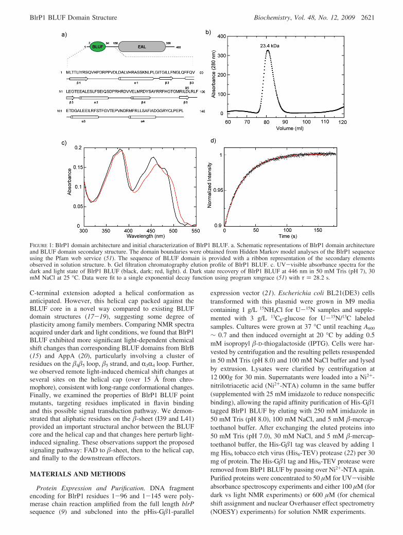
FIGURE 1: BlrP1 domain architecture and initial characterization of BlrP1 BLUF. a. Schematic representations of BlrP1 domain architectureand BLUF domain secondary structure. The domain boundaries were obtained from Hidden Markov model analyses of the BlrP1 sequenceusing the Pfam web service (51). The sequence of BLUF domain is provided with a ribbon representation of the secondary elementsobserved in solution structure. b. Gel filtration chromatography elution profile of BlrP1 BLUF. c. UV-visible absorbance spectra for thedark and light state of BlrP1 BLUF (black, dark; red, light). d. Dark state recovery of BlrP1 BLUF at 446 nm in 50 mM Tris (pH 7), 30mM NaCl at 25 °C. Data were fit to a single exponential decay function using program xmgrace (51) with τ ) 28.2 s.
BlrP1 BLUF Domain Structure Biochemistry, Vol. 48, No. 12, 2009 2621

UV-Visible Absorbance Spectroscopy. All UV-visibleabsorbance spectra were acquired using a Cary50 (Varian)spectrophotometer. Samples were photoactivated using aphotographic flash. The subsequent recovery of the dark stateform of the flavin cofactor was monitored by the changes inthe absorbance at 446 nm after illumination, recording A446
every 1 s with an integration time of 0.1 s. Rates weredetermined by averaging the fit of four separate measure-ments to a single exponential decay. A circulating water bathwas used for temperature control of the sample in a quartzcuvette. Experiments were conducted at 25 °C in 50 mMTris (pH 7) and 30 mM NaCl in H2O.
Site-Directed Mutagenesis. Point mutations of the BlrP1BLUF domain were generated using QuickChange (Strat-agene) from wild-type DNA and primers including thedesired mutations (I39L, I39V, I39Y, L41T, L41V, L41Y,Q49N, Q49L, T90W, T90S, T90Y, T90A, M92V, M92I,F112Y, F112L, V117F, and V117L). All changes wereverified by DNA sequencing. Transformation, protein induc-tion, and purification were performed as described above forwild-type.
NMR Spectroscopy. All NMR experiments were performedat 25 °C on Varian Inova 500, 600, and 800 MHz spec-trometers, using nmrPipe for data processing (23) andNMRview for analysis (24). Dark state backbone chemicalshift assignments were obtained using standard 3D 15N-editedtriple resonance experiments including HNCACB, CBCA-(CO)NH, and HNCO spectra (25). Sidechain chemical shiftswere assigned from HCCH total correlation spectroscopy(HCCH-TOCSY), HC(CO)NH-TOCSY, and C(CO)NH-TOCSY data, complemented for aromatic ring assignmentsby HBCB(CGCD)HD (26) and aromatic 13C/1H hetero-nuclear single quantum coherence (HSQC) data from auniformly 13C-,15N-labeled sample and 2D double-quantum-filtered correlation spectroscopy (DQF-COSY), TOCSY (τm
) 30 ms), and NOESY (τm ) 50 ms) spectra of an unlabeledsample in 99.9% D2O. Simultaneous 15N-, 13C-edited NOESY(τm ) 120 ms) data were used to facilitate the chemical shiftassignment at all stages and to obtain distance restraints forstructure calculation. Backbone dihedral angle restraints wereobtained from TALOS analysis (27) of backbone 15N, 13C,and 1H chemical shifts.
To obtain NMR spectra of photoexcited BlrP1 BLUFsamples, we generated blue light from a 5 W Coherent Inova-90C argon laser running in single wavelength mode at 457nm, directing the output from this laser into a quartz fiberoptic inserted into the NMR sample (21). Power levels atthe end of this fiber were 90 mW as measured before eachexperiment. 15N/1H and constant time 13C/1H HSQC spectraof the light state of BlrP1 BLUF were recorded by precedingeach transient in the NMR experiment with a 300 ms laserpulse during the recycle delay (total 1060 ms delay).
As a result of its relatively rapid dark state recovery rate(τ ∼ 28 s at 25 °C) and significant optical density at 457nm in both dark and light states, a substantial amount oflight is needed to populate the BlrP1 BLUF photoproductstate. Unfortunately, photobleaching precludes the possibilityof full chemical shift assignments of the BlrP1 BLUF lightstate using standard triple resonance methods. Thus, all thelight state chemical shift assignments of BlrP1 BLUF weremade by simply pairing the nearest crosspeaks in dark andlight state spectra where such pairings were unambiguous
in well-resolved portions of both spectra. This was possiblefor 47 of 137 backbone amides and 36 of 93 methyl groups.
Structure Calculation. Following the chemical shift as-signment, we used ARIA2 (ambiguous restraints for iterativeassignment) (28) to assign NOEs from 15N-, 13C-editedNOESY data in an automated manner. BlrP1 BLUF struc-tures were determined with a mix of automated and manualassignment of NOESY spectra. Given our observations andothers (15-17, 19) that the adenine ring of FAD is highlysolvent-exposed and does not make significant contacts withthe protein, we simply modeled the FAD chromophore as aflavin mononucleotide (FMN). FMN topology and parameterfiles were obtained from the HIC-Up database (29), and asingle FMN molecule was placed in the starting coordinates30 Å from the BlrP1 BLUF to avoid biasing the structurecalculation. Backbone dihedral angle restraints were createdfrom TALOS (27) analysis, using bounds set to two timesthe standard deviation of the TALOS predictions andenforcing a minimum of (30°. Hydrogen bond restraintswere established by examining the conformations of residuesin regular secondary structure (as established by patterns ofNOEs and TALOS-generated φ, ψ restraints) in initialstructure ensembles and implementing H-bond restraintswhen HNsOdC distances and angles were consistent withH-bonding. Of 500 structures in the final iteration of therefinement, the 20 lowest energy structures were analyzedusing routines in ARIA2 and PROCHECK (30). Coordinatesof BlrP1 BLUF have been deposited in the Protein Data Bankunder accession code 2KB2.
RESULTS
Solution Structure of BlrP1 BLUF. In our previous studieson BlrB, a 140 amino acid residue protein which containsonly a canonical mixed R/� BLUF domain and a pair ofC-terminal helices, we demonstrated that this C-terminalextension is essential for the structural integrity and properphotocycling of the domain (15). To test the generality ofthis finding for other BLUF domains, we generated twoBlrP1 constructs at different lengths: BlrP1(1-96), whichincluded only a BLUF domain, and BlrP1(1-145), whichincluded the BLUF and approximately 50 C-terminal residuesfrom the linker between the BLUF and the EAL domains.Consistent with our experiences with comparable constructsfor BlrB (15), the shorter construct suffered poor expressionand low yield, did not appear to bind FAD, and precipitatedduring purification. In contrast, the longer construct, elutedas an elongated stable monomer at an apparent molecularmass of 23.4 kDa (Figure 1b), showed a 10 nm characteristicred shift of BLUF domains in the UV-visible absorbancespectrum upon light illumination (Figure 1c) with exponentialdark state recovery kinetics (τ ∼ 28 s) as monitored by theabsorbance of the flavin chromophore at 446 nm (Figure 1d).Further, BlrP1(1-145) (hereafter referred to as BlrP1 BLUF)displayed a well-dispersed 15N/1H HSQC spectrum, was verystable in solution during the course of multiday 3D NMRexperiments, and was generally better suited for in-depthstructural and biophysical studies.
Using solution NMR spectroscopy, we determined thestructure of BlrP1 using over 4000 geometric constraints,including interproton distances, dihedral angles, and hydro-gen bonds (Table 1). All of these data are well-satisfied by
2622 Biochemistry, Vol. 48, No. 12, 2009 Wu and Gardner

the 20 lowest-energy structures, which form a high precisionensemble (backbone pairwise root-mean-square deviation(rmsd) of 0.15 ( 0.05 Å for ordered regions in residues 2-9,21-34, 37-42, 47-70, 75-82, 92-96, 102-113, and124-135) as shown in Figure 2a. The BLUF domain ofBlrP1 has five � strands and four R helices ordered as�1R1�2�3R2�4�5R3R4 which can be broken into two substruc-tures: the BLUF core domain (�1R1�2�3R2�4�5) and a
C-terminal cap domain (R3R4). The isoalloxazine ring of FADis sandwiched between R1 and R2 helices inside the BLUFdomain, leaving the adenosine monophosphate moiety solvent-exposed and flexible (Figure 2). The BlrP1 BLUF domainsurrounds the FAD isoalloxazine ring of BlrP1 BLUF witha set of conserved residues among BLUF domains, settingup hydrogen bonds involving Asn34 (to the FAD N3position) and Gln52 (to the FAD O4 and the Tyr 10 Oη).Similar hydrogen bond networks were also observed in otherBLUF domain proteins (17, 19, 31, 32). Notably, we didnot observe any issue with intermediate exchange broadeningof signals from residues in either the �3 or the �5 strands asobserved with AppA (20) or BlrB (15), providing qualitativeevidence for differences in the stability of the protein/flavincomplex in the dark state among different BLUF domains.
While the structure of BLUF core of BlrP1 is similar toother BLUF domain structures, the last two R helices(R3, R4) adopt a rather different orientation (17-20, 31, 33).Compared to other BLUF domain structures, the BlrP1BLUF helical cap is rotated ∼90 degrees with respect to thecentral � strands, making the two cap helices almost parallelwith, rather than perpendicular to, the �-sheet (Figure 2;Figure 1 of Supporting Information). The conserved residuesT90 (W for most BLUF domains at that position) and M92are located at the end of �4�5 loop with T90 pointing outand M92 pointing in the chromophore binding pocket,consistent with the orientations of the corresponding residuesin most published BLUF structures (17-19). A notableexception for this is the AppA BLUF domain, whichtypically has the conserved tryptophan (W104) buried in thehydrophobic core near the chromophore-binding pocket(20, 31, 32).
Light-Induced Changes in BLUF Domain of BlrP1. Tocharacterize the influence of blue light illumination on proteinstructure and dynamics of BlrP1 BLUF, we used solutionNMR spectroscopy to investigate light-activated changes atthe atomic level. Both dark and light NMR spectra of BlrP1BLUF (Figure 3) were monitored at backbone amide andside chain methyl groups using 15N/1H and 13C/1H HSQCspectra, respectively. Unlike previous NMR studies of BLUFdomains (e.g., BlrB (15), AppA (20)), crosspeaks in thesespectra of BlrP1 BLUF display significant chemical shiftchanges after light excitation without widespread linebroadening as seen for BlrB and AppA. This indicates thatthe BlrP1 BLUF light state has a different degree ofconformational change or rates of conformational exchangethan these other two BLUF domains, despite using the samephotochemistry to initiate the changes. To systematicallyquantitate these light-activated chemical shift changes, weused minimum chemical shift difference analyses (34) ondark and light BlrP1 BLUF 15N/1H HSQC spectra. By thisapproach, peak locations from these spectra were comparedto obtain the minimum chemical shift changes for each darkstate peak to its nearest light state neighbor by the relation-ship ∆δmcs ) min[((1HN ∆ppm)2 + (0.17(15N ∆ppm)2))1/2]and plotted as a function of residue number (Figure 4a). Thisapproach conservatively estimates the degree of chemicalshift changes caused by illumination, even in spectral regionswith significant chemical shift overlap. Not surprisingly, weobserved relatively large light-activated chemical shiftchanges in the vicinity of the FAD chromophore (Figure 5a;Figure 3 of Supporting Information), on account of the
Table 1: Statistics for BlrP1 BLUF Solution Structure Determination
list of constraints number of constraints
NOE distance restraints 4138unambiguous 2720ambiguous 1418
hydrogen bond restraints 77dihedral angle (θ, φ)
restraints from TALOS168
Structure Analysisaverage number of NOE
violations greater than 0.5 Å0
average number of dihedralviolations greater than 5°
0
geometric analysis of 2°structured residuesrmsd to mean 0.15 ( 0.02 Å (backbone)
0.45 ( 0.06 Å (all heavy atoms)Procheck 73.4% most favored region
24.0% allowed region2.3% generally allowed region0.4% disallowed region
FIGURE 2: Solution structure of BlrP1 BLUF. a. Ensemble of the20 lowest energy structures of BlrP1 BLUF, calculated as indicatedin the text, with the FMN chromophore shown in stick representa-tion. b. Ribbon diagram of the structure with the lowest energy inthe ensemble shown in a. The canonical BLUF domain is coloredgreen, and the C-terminal cap is colored blue. Secondary structureelements are as labeled.
BlrP1 BLUF Domain Structure Biochemistry, Vol. 48, No. 12, 2009 2623

significant light-induced changes to the electronic structureof the isoalloxazine ring (35). However, we also noted thatsites exhibiting above average light-induced minimumchemical shift changes clustered in a few interesting areas,specifically the �4 and �5 strands, the loop between them,and the loop between R3 and R4 (Figure 4b). More specif-ically, several sites (A105, I109, L110, F112, S113, G116,V117, and T118) over 15 Å from the chromophore displayedlarge light-induced chemical shift changes in both 15N/1Hand 13C/1H HSQC spectra (Figure 5a; Figure 3 of SupportingInformation), indicating that the light-activated signal around
the chromophore somehow involves these sites, possibly bya specific signal transduction pathway, and then likely tothe EAL domain. This is consistent with other studiessuggesting that these regions probably play an important rolein activating and transforming the light-induced signal inBLUF domains (33, 36, 37). More evidence along this lineis the Cε methyl group of M92, which shows the biggestchemical shift change (>2 ppm) after light excitation,suggesting that light significantly alters the chemical envi-ronment at this site (15). Yuan et al. recently proposed the�4�5 loop and the C-terminal R helices as the interface forPixD oligomerization when its interaction partner PixE ispresent under dark conditions. Major conformational changeson the interface after light illumination result in the disas-sembly of the PixD10-PixE5 complex (8).
Of particular interest were residues at the �-sheet interfacebetween the BLUF domain and the C-terminal cap, includingseveral (I39/L40/L41) which showed light-induced chemicalshift changes in both 15N/1H and 13C/1H HSQC spectra(Figure 5a; Figure 3 of Supporting Information). The methylgroups of I39 and L41 point to the opposite side of the�-sheet as the FAD chromophore and are in close contact
FIGURE 3: 15N/1H and 13C/1H HSQC spectra of BlrP1 BLUF underdark and light conditions. a. Dark (black) vs light (red) 15N/1Hspectra of BlrP1 BLUF. b. Methyl region of dark (black) vs light(red) constant time 13C/1H HSQC spectra of BlrP1 BLUF. BlrP1BLUF underwent significant chemical shift changes upon lightillumination shown by both backbone 15N/1H and methyl side chain13C/1H HSQC spectra. Sites with relatively large chemical shiftchanges are labeled with the assignments where available.
FIGURE 4: Minimum chemical shift difference analysis on dark andlight states of BlrP1 BLUF. a. Minimum chemical shift differencesfrom 15N/1H HSQC spectra obtained in the dark and light statesfor BlrP1 BLUF. Secondary structure elements are as shown ontop of the figure. Red dashed line indicates the noise level (0.065ppm). b. Minimum chemical shift differences between these twospectra are mapped onto the solution structure of BlrP1 BLUF witha color gradient encoding the magnitude of the shift change.
2624 Biochemistry, Vol. 48, No. 12, 2009 Wu and Gardner

with the residues on the last two R helices as validated bystrong NOE peaks between I39 and F112, I39 and L129,L41 and F132, and L41 and I133 in 13C-edited NOESYspectra. These hydrophobic interactions appear to serve asan anchor between the sheet and the cap, locking the latterinto the proper position and protecting the �-sheet fromsolvent exposure. On the basis of the destabilizing effectsof deleting the helical cap from the BLUF domains of eitherBlrP1 or BlrB (15), we believe such protection is essentialfor many BLUF domains to maintain their structural andfunctional properties. In contrast, changes at L40 wereexpected given that it is located at the same side of FAD,∼3.5 Å away from the chromophore (and has NOE peakswith the FAD methyl C7a group). The fact that these threeresidues exhibit such significant and concerted light-depend-ent effects strongly supports the speculation that the light-activated signal may be passed from FAD to the �-sheet,then to the helical cap, and eventually to the downstream
effectors by the interactions between these residues with thechromophore and with the C-terminal cap.
BlrP1 BLUF Mutants: BLUF Core, Q49, T90, and M92.Our BlrP1 BLUF structure and light-dependent changessuggested several important residues in its FAD binding,structural stability, and photocycling. To investigate theseresidues in more detail, we made a series of point mutantsin the BLUF R/� core (Q49, T90, and M92), in the anchorcoupling the core BLUF fold to the C-terminal helical cap(I39 and L41), and in the cap itself (F112 and V117) (Figure5b; Figure 2 of Supporting Information). All of these residuesare highly conserved, are critical for chromophore bindingin other BLUF domains, or showed significant NMRchemical shift changes upon illumination. We evaluated theFAD-binding abilities and photochemical properties of thesemutants using UV-visible absorbance spectroscopy (Table1, Supporting Information), while we assayed their structuraleffects by comparing dark and light state 15N/1H HSQCspectra of the mutants with corresponding wild-type BlrP1BLUF.
Our data indicate that two highly conserved residues inthe BLUF core, Q49 and M92, are absolutely essential forFAD binding and photocycling. These sites were intolerantof any of the changes we made, as all mutants for these tworesidues completely abolished BlrP1 BLUF FAD bindingcapacity and corresponding photocycling, consistent with theresults regarding their important roles in the structure andsignaling of other BLUF domains (15, 31, 33, 38).
The third residue we selected for mutagenesis in the BLUFcore was T90, which has been implicated in playing a centralrole in signal transduction mechanism together withM92 (33, 39). Notably, this �5 strand position is highlyconserved as a Trp residue in most BLUF domains (Figure 1,Supporting Information) but has been replaced with a Thrin BlrP1 BLUF and an Ala in the similar E. coli YcgFprotein. All four T90 mutants that we examined still boundFAD and properly photocycled, albeit with ∼1.1-2.5-foldslower dark state recovery rates compared with wild-typeBlrP1 BLUF. These changes and corresponding differencesin 15N/1H HSQC spectra were relatively subtle for T90A andT90S but were more dramatic for T90Y and T90W. Ofparticular interest is a comparison of dark versus light NMRspectra of T90W, which revealed that the light states of thewild-type and T90W BlrP1 BLUF exhibit somewhat differentproperties. Notably, the T90W light state spectrum showssignificant line broadening in addition to the chemical shiftchanges observed for the wild-type protein (Figure 4,Supporting Information). Further, the newly generated indole15Nε/1Hε crosspeak of T90Wswhich is unique in the 15N/1H HSQC spectrum, as the wild-type BlrP1 BLUF domainlacks any Trp residuessdid not show any chemical shiftchange after light excitation (Figure 6c). These data stronglyindicate that this indole ring does not undergo any light-driven transient exposure/burial, as has been speculated asapossiblesignalingmechanismforotherBLUFdomains(33,39).While these data have been obtained on a BlrP1 BLUF pointmutant, they are consistent with steady-state fluorescenceresults for the corresponding W104 of AppA in both wild-type and W64F backgrounds (40). Notably, the AppA W104position is tolerant of point mutations even though the sidechain is internally directed and near the FAD isoalloxazinering (41). Masuda et al. observed similar results in PixD,
FIGURE 5: a. Light-perturbed sites in BlrP1 BLUF. Residues withlight-dependent chemical shift change on methyl sites in 13C/1Hdark versus light spectra are represented as sticks and labeled withresidue numbers; the methyl sites are shown as red spheres. b.Mutated sites on BlrP1 BLUF. Mutated amino acid sites of BlrP1BLUF on the structure: Ile39, Leu41, Gln49, Thr90, Met92, Phe112,and Val117. Sites are shown as gold sticks and labeled with residuenumbers.
BlrP1 BLUF Domain Structure Biochemistry, Vol. 48, No. 12, 2009 2625

finding that the M93A mutant of PixD (equivalent to BlrP1M92) is light insensitive with biological activities incompat-ible with those of the light-adapted wild-type. However,W91A (T90 in BlrP1) retained light sensitivity and biologicalfunction (38). Putting these data together, it is clear that T90is more tolerant of substitution, suggesting that it plays less
of a role in domain stability and, potentially, signal trans-duction than other highly conserved residues.
BlrP1 BLUF Mutants: Anchor Between the BLUF Coreand the Helical Cap, I39 and L41. As noted in our solutionstructure, I39 and L41 both project from the BLUF domain�-sheet toward the R3/R4 helical cap and show significant
FIGURE 6: Light-dependent effect on the BlrP1 BLUF mutants. a. Overlay of 15N/1H HSQC spectra of BlrP1 BLUF mutants, residueswith significant chemical shift changes in both of the mutants are as labeled. Wild-type (black), I39L (blue), and L41V (red). b. Sitesshowing consistent chemical shift changes in 15N/1H spectra for three mutants (L41V/I39L/I39V) are mapped on BlrP1 BLUF structure.These sites are F112, S113, T114, T118, and V121. c. Indole region of dark and light 15N/1H spectra of wild-type (black, red) andT90W mutant (cyan, pink) for BlrP1 BLUF. d. Residue 41 in dark and light 15N/1H HSQC spectra of wild-type (black, red) and L41Vmutant (cyan, blue) of BlrP1 BLUF. e. Methyl groups of I109.Cδ1 and A105.C� in dark (black) and light (red) 13C/1H HSQC spectraof wild-type BlrP1 BLUF. f. Methyl groups of I109.Cδ1 and A105.C� in dark (cyan) and light (pink) 13C/1H HSQC spectra of BlrP1BLUF L41V mutant.
2626 Biochemistry, Vol. 48, No. 12, 2009 Wu and Gardner

light-induced chemical shift changes, suggesting that theymay couple the BLUF core and helical cap structurestogether. Of the six mutations we generated at either I39 orL41, three (I39Y, L41Y, and L41T) lost the ability to bindFAD and precipitated during purification. The remainingthree mutants (I39V, I39L, and L41V) all still bound FADand exhibited the characteristic light-induced 10 nm red shiftin their visible absorbance spectra. Supporting our findingsthat deletion of the helical caps destabilizes the BLUF coredomains of BlrP1 BLUF (vide supra) and the BLUF-onlyprotein BlrB (15), these data suggest that the specific packingof the BLUF �-sheet and the R helical cap is essential forFAD binding and domain stability.
Examination of the dark state 15N/1H HSQC spectra ofI39V, I39L, and L41V mutants and the wild-type proteinrevealed that the point mutations induced chemical shiftchanges not only near the mutated residues but also at longdistances involving residues in the helical cap. These long-range changes involved sites in the R3R4 loop (F112, S113,T114, T119, and V121) and the adjacent �4�5 loop (G89),with consistent chemical shift changes among all threemutants (I39L and L41V data shown in Figure 6a,b; I39Vin Figure 5, Supporting Information). We speculate that theconsistency of the observed chemical shift changes amongthe three mutants could reflect a cooperative movement ofthe helical cap in response to the different length of themutated side chains.
Analogous comparisons of the effects of the I39V, I39L,and L41V mutations on light state 15N/1H spectra providedus with evidence that these sites might be involved in thetransmission of conformational changes between the BLUFcore and the helical cap. Notably, these mutations interferedwith the interaction between the BLUF core and the helicalcap as exhibited by smaller light-dependent chemical shiftchanges in the mutants. An example of this is provided bythe backbone amide of V41, the mutated site in the L41Vpoint mutant, which showed a greatly reduced light-inducedchemical shift change in 15N/1H HSQC spectra and significantpeak heterogeneity in the light state when compared to wild-type BlrP1 BLUF (Figure 6d). Notably, the light state 15N/1H HSQC spectra of all three mutants exhibited greater linebroadening than we observed for the wild-type BlrP1 BLUF(Figure 6, Supporting Information), suggesting that thesealterations perturbed the degree of conformational change(or exchange kinetics) associated with the light state.
We found similar effects in 13C/1H HSQC spectra, wherethe methyl groups of helical cap residues A105, I109, andL110 showed long-range light-dependent chemical shiftchanges for wild-type BlrP1 BLUF but much smaller effectsfor L41V (Figure 6e,f) where the dark and light state peaksare essentially overlapped. Unfortunately, resonances fromL110 fall into a crowded region of the 13C/1H HSQCspectrum of L41V BlrP1 BLUF, complicating its assignment.
BlrP1 BLUF Mutants: Helical Cap, F112 and V117. Giventhat F112 and V117 are on the helical cap, far from the FADchromophore, their light-dependent chemical shift changesstrongly suggest that conformational changes are somehowpropagated to these sites after illumination. To probe thiseffect, we made a total of four mutations (F112Y, F112L,V117L, and V117F) at these two sites. All four bound FAD,photocycled properly, and had similar dark regeneration ratesas wild-type. While the light state 15N/1H spectra of these
mutants generally shared similar patterns of light-inducedchemical shift changes as wild-type, they also exhibitdramatic line broadening and accompanying reductions inpeak intensities. Overall, substituting the two residues onthe helical cap with other types of amino acids clearly didnot interrupt the photochemistry and the interaction betweenchromophore and BlrP1 BLUF. However, the mutantsdisplay abnormal spectral behavior compared with wild-typeprotein in an amino acid type independent manner.
DISCUSSION
What defines a BLUF domain? Following the initialproposal of a conserved ∼90 amino acid mixed R/� domainarchitecture from sequence homology (1), subsequent bio-physical and biochemical studies have shown that this coreBLUF domain is often associated with a helical C-terminalstructural element (17-19). Truncation studies have furthershown that these caps are essential for the stability of BLUFdomains and FAD binding, demonstrating their necessity(15, 16). Extending this to a broader group of BLUF domainswith secondary structure prediction (Figure 2, SupportingInformation), we suggest that this is likely an integral elementof many further BLUF domains, regardless of whether theyoriginate from small proteins that consist solely of a BLUFdomain (“BLUF-only proteins”) or from larger proteinscontaining additional domains. This conservation implies ageneral role for the C-terminal helical cap, possibly protectingthe �-sheet interface from solvent or facilitating a mechanismto propagate signals from the BLUF photosensory domainto the downstream effector.
Given this generality, it is interesting that the helical capsadopt different orientations with respect to the central �-sheetin different BLUF domains. This is best demonstrated witha structure-based superposition of five BLUF domainstructures (17-19, 31) (Figure 1, Supporting Information)based on the conserved R/� secondary structure elements inthe core. Notably, the helical caps of the three BLUF-onlyproteins are oriented almost perpendicular to the �-sheet,while BlrP1 BLUF (a component of a multidomain protein)has a helical cap that is almost parallel to the �-sheet. Thisis supported by a recently determined X-ray diffractionstructure of full length BlrP1, the BLUF domain of whichis very similar (1.1 Å CR rmsd) to our structure (14), rulingout artifacts caused by either protein truncation or experi-mental approach. The different orientation of the helical capmay indicate a possible mechanism in regulating BLUF-containing proteins with otherwise very different biologicalfunctions.
Despite extensive structural and photochemical studies onBLUF domains (15, 17-19, 31, 35), their signaling mech-anisms and its generality remains largely unknown. To date,it is widely accepted that a critical glutamine (Q49 in BlrP1)flips by 180° upon illumination, playing a central role inrearranging the protein/chromophore hydrogen-bonding net-work conserved among BLUF domains (31, 35, 42). In thisstudy, our data support the important structural roles ofseveral conserved residues Q49 and M92 in establishing thelight-activatedstateaswithotherBLUFdomains(18-20,31,33).However, the light state 15N/1H HSQC spectrum of BlrP1BLUF is markedly different from those of other BLUFsystems (e.g., AppA BLUF (20) and BlrB (15)) in that
BlrP1 BLUF Domain Structure Biochemistry, Vol. 48, No. 12, 2009 2627

photoactivation is accompanied by peak shifting rather thanbroadening. This is consistent with a greater degree ofstructural change or different kinetics of exchange acrossmultiple protein conformations in the transient BlrP1 BLUFphotoexcited states and also supports some diversity ofsignaling state structures and kinetics among BLUF domains(43). Although all BLUF domains display the same charac-teristic 10 nm red shift in the visible absorbance spectrumof the FAD chromophore upon illumination, the structuraland kinetic properties of the subsequent conformationalchange may vary among BLUF domains. Here we observethat the �4�5 loop, �5 strand, and R3R4 loop exhibit significantlight-dependent conformational changes, consistent withobservations from AppA BLUF (20), PixD (19), and BlrB(15). Further, residues T90 and M92 (in BlrP1 BLUF), whichare the subject of intense debate regarding their roles in signalactivation process for BLUF domains, are located on the �5
strand (33, 37, 39) and show significant light-activatedchanges. Combining these observations and prior work raisesthe possibility that, after blue light illumination, the helicalcap of BlrP1 BLUF undergoes some kind of reorientationprocess, and that might be associated with the conformationalchanges of the �5 strand and the two loops.
Of particular interest on the �-sheet of BlrP1 BLUF aretwo methyl-containing residues, I39 and L41. These are bothlocated in the �2 strand, next to the �5 strand in our solutionstructure. Our data clearly demonstrate that the side chainsof these two residues are essential for the BLUF domaincore to hold onto the helical cap and to maintain the FAD-binding capacity. Are they important in transmitting the light-induced signal as well? Limited evidence from the dark andlight 13C/1H HSQC spectra of L41V shows that the light-induced chemical shift changes for residues A105 and I109of wide-type do not exist in this mutant, suggesting that themutation on this site interrupts the light-activated signalpropagation in some way.
An interesting comparison is provided by another classof flavin-bound blue light photoreceptors, the LOV (Light-Oxygen-Voltage) domains. LOV and BLUF are both smallflavin-based sensing units with similar, but not identical, R/�folds. Although both types of domains use similar chro-mophores (FMN versus FAD) to sense blue light, they relyon different photochemistries that are either solely nonco-valent (BLUF domains) or result in the generation of newprotein/flavin covalent bonds (LOV domains (44)). Intrigu-ingly, LOV domains also utilize R helices outside thecanonical domain boundaries, as shown with AVena satiVaphototropin1 LOV2 (at both N- and C-termini) (21, 45) andNeurospora crassa Vivid (at N-terminus) (46). These helicescover the otherwise solvent-exposed �-sheet faces for bothdomains and are displaced or reoriented by light-inducedconformational changes after illumination, analogously to therelationship between the BLUF domain and the cap regions.More broadly, the �-sheet interface appears to be importantfor a wide array of inter- and intramolecular interactions invaried biological systems that use the related PAS (Period-ARNT-singleminded) fold. These include the �-sheet inter-faces of the HIF-2R and ARNT PAS-B domains that areused for PAS/PAS heterodimerization in the mammalianhypoxia response pathway (47-49) and the homodimeric�-sheet interface of the KinA PAS-A domain involved inBacillus subtilis sporulation (50). Taken together, these data
suggest that small sensor units may share interesting commonfeatures in signal sensing and transduction despite theirdifferences in their sequence, structure, and functions.
ACKNOWLEDGMENT
We thank Ilme Schlichting and Mark Gomelsky forproviding materials and information in advance of publication.
SUPPORTING INFORMATION AVAILABLE
Additional data and analyses in one table and six figures.This material is available free of charge via the Internet athttp://pubs.acs.org.
REFERENCES
1. Gomelsky, M., and Klug, G. (2002) BLUF: A novel FAD-bindingdomain involved in sensory transduction in microorganisms. TrendsBiochem. Sci. 27, 497–500.
2. Gomelsky, M., and Kaplan, S. (1998) AppA, a redox regulator ofphotosystem formation in Rhodobacter sphaeroides 2.4.1, is aflavoprotein. Identification of a novel FAD binding domain. J. Biol.Chem. 273, 35319–35325.
3. Kraft, B. J., Masuda, S., Kikuchi, J., Dragnea, V., Tollin, G.,Zaleski, J. M., and Bauer, C. (2003) Spectroscopic and mutationalanalysis of the blue-light photoreceptor AppA: a novel photocycleinvolving flavin stacking with an aromatic amino acid. Biochemistry42, 6726–6734.
4. Zirak, P., Penzkofer, A., Schiereis, T., Hegemann, P., Jung, A.,and Schlichting, I. (2006) Photodynamics of the small BLUFprotein BlrB from Rhodobacter sphaeroides. J. Photochem. Pho-tobiol., B 83, 180–194.
5. Masuda, S., and Bauer, C. (2002) AppA is a blue light photore-ceptor that antirepresses photosynthesis gene expression in R.Sphaeroides. Cell (Cambridge, MA, U.S.) 110, 613–623.
6. Iseki, M., Matsunaga, S., Murakami, A., Ohno, K., Shiga, K.,Yoshida, K., Sugai, M., Takahashi, T., Hori, T., and Watanabe,M. (2002) A blue-light-activated adenylyl cyclase mediates pho-toavoidance in Euglena gracilis. Nature 415, 1047–1051.
7. Yoshikawa, S., Suzuki, T., Watanabe, M., and Iseki, M. (2005)Kinetic analysis of the activation of photoactivated adenylyl cyclase(PAC), a blue-light receptor for photomovements of Euglena.Photochem. Photobiol. Sci. 4, 727–731.
8. Yuan, H., and Bauer, C. E. (2008) PixE promotes dark oligomer-ization of the BLUF photoreceptor PixD. Proc. Natl. Acad. Sci.U.S.A. 105, 11715–11719.
9. Tyagi, A., Penzkofer, A., Griese, J., Schlichting, I., Kirienko, N. V.,and Gomelsky, M. (2008) Photodynamics of blue-light-regulatedphosphodiesterase BlrP1 protein from Klebsiella pneumoniae andits photoreceptor BLUF domain. Chem. Phys. 354, 130–141.
10. Christen, M., Christen, B., Folcher, M., Schauerte, A., and Jenal,U. (2005) Identification and characterization of a cyclic di-GMP-specific phosphodiesterase and its allosteric control by GTP. J. Biol.Chem. 280, 30829–30837.
11. Jenal, U., and Malone, J. (2006) Mechanisms of cyclic-di-GMPsignaling in bacteria. Annu. ReV. Genet. 40, 385–407.
12. Paul, R., Weiser, S., Amiot, N. C., Chan, C., Schirmer, T., Giese,B., and Jenal, U. (2004) Cell cycle-dependent dynamic localizationof a bacterial response regulator with a novel di-guanylate cyclaseoutput domain. Genes DeV. 18, 715–727.
13. Schmidt, A. J., Ryjenkov, D. A., and Gomelsky, M. (2005) Theubiquitous protein domain EAL is a cyclic diguanylate-specificphosphodiesterase: enzymatically active and inactive EAL domains.J. Bacteriol. 187, 4774–4781.
14. Barends, T. R. M., Hartmann, E., Griese, J., Beitlich, T., Kirienko,N. V., Ryjenkov, D. A., Reinstein, J., Shoeman, R. L., Gomelsky,M., and Schlichting, I. (2009) Structure and mechanism of abacterial light-regulated cyclic nucleotide phosphodiesterase, inpress.
15. Wu, Q., Ko, W. H., and Gardner, K. H. (2008) Structuralrequirements for key residues and auxiliary portions of a BLUFdomain. Biochemistry 47, 10271–10280.
16. Schreoder, C., Werner, K., Otten, H., Kratzig, S., Schwalbe, H.,and Essen, L. (2008) Influence of a joining helix on the BLUFdomain of the YcgF photoreceptor from Escherichia coli. Chem-BioChem 9, 2463–2473.
2628 Biochemistry, Vol. 48, No. 12, 2009 Wu and Gardner

17. Jung, A., Domratcheva, T., Tarutina, M., Wu, Q., Ko, W. H.,Shoeman, R. L., Gomelsky, M., Gardner, K. H., and Schlichting,I. (2005) Structure of a bacterial BLUF photoreceptor: insights intoblue light-mediated signal transduction. Proc. Natl. Acad. Sci.U.S.A. 102, 12350–12355.
18. Kita, A., Okajima, K., Morimoto, Y., Ikeuchi, M., and Miki, K.(2005) Structure of a cyanobacterial BLUF protein, Tll0078,containing a novel FAD-binding blue light sensor domain. J. Mol.Biol. 349, 1–9.
19. Yuan, H., Anderson, S., Masuda, S., Dragnea, V., Moffat, K., andBauer, C. (2006) Crystal structures of the Synechocystis photore-ceptor Slr1694 reveal distinct structural states related to signaling.Biochemistry 45, 12687–12694.
20. Grinstead, J. S., Hsu, S. T., Laan, W., Bonvin, A. M., Hellingwerf,K. J., Boelens, R., and Kaptein, R. (2006) The solution structureof the AppA BLUF domain: Insight into the mechanism of light-induced signaling. ChemBioChem 7, 187–193.
21. Harper, S. M., Neil, L. C., and Gardner, K. H. (2003) Structuralbasis of a phototropin light switch. Science 301, 1541–1544.
22. Blommel, P. G., and Fox, B. G. (2007) A combined approach toimproving large-scale production of tobacco etch virus protease.Protein Expression Purif. 55, 53–68.
23. Delaglio, F., Grzesiek, S., Vuister, G. W., Zhu, G., Pfeifer, J., andBax, A. (1995) NMRPipe: a multidimensional spectral processingsystem based on UNIX pipes. J. Biomol. NMR 6, 277–293.
24. Johnson, B. A. (2004) Using NMRView to visualize and analyzethe NMR spectra of macromolecules. Methods Mol. Biol. (Totowa,NJ, U.S.) 278, 313–352.
25. Sattler, M., Schleucher, J., and Griesinger, C. (1999) Heteronuclearmultidimentional NMR experiments for the structure determinationof proteins in solution employing pulsed field gradients. Prog. Nucl.Magn. Reson. Spectrosc. 34, 93–158.
26. Yamazaki, T., Forman-Kay, J. D., and Kay, L. E. (1993) Two-dimensional NMR experiments for correlating carbon-13 � andproton δ/ε chemical shifts of aromatic residues in 13C-labeledproteins via scalar couplings. J. Am. Chem. Soc. 115, 11054–11055.
27. Cornilescu, G., Delaglio, F., and Bax, A. (1999) Protein backboneangle restraints from searching a database for chemical shift andsequence homology. J. Biomol. NMR 13, 289–302.
28. Linge, J. P., O’Donoghue, S. I., and Nilges, M. (2001) Antomatedassignment of ambiguous nuclear overhauser effects with ARIA.Methods Enzymol. 339, 71–90.
29. Kleywegt, G. J. (2007) Crystallographic refinement of ligandcomplexes. Acta Crystallogr., Sect. D 63, 94–100.
30. Morris, A. L., MacArthur, M. W., Hutchinson, F. G., and Thornton,J. M. (1992) Stereochemical quality of protein structure corrdinates.Protein Expression Purif. 12, 345–365.
31. Anderson, S., Dragnea, V., Masuda, S., Ybe, J., Moffat, K., andBauer, C. (2005) Structure of a novel photoreceptor, the BLUFdomain of AppA from Rhodobacter sphaeroides. Biochemistry 44,7998–8005.
32. Grinstead, J. S., Avila-Perez, M., Hellingwerf, K. J., Boelens, R.,and Kaptein, R. (2006) Light-induced flipping of a conservedglutamine sidechain and its orientation in the AppA BLUF domain.J. Am. Chem. Soc. 128, 15066–15067.
33. Jung, A., Reinstein, J., Domratcheva, T., Shoeman, R. L., andSchlichting, I. (2006) Crystal structures of the AppA BLUF domainphotoreceptor provide insights into blue light-mediated signaltransduction. J. Mol. Biol. 362, 717–732.
34. Farmer, B. T., Constantine, K. L., Goldfarb, V., Friedrichs, M. S.,Wittekind, M., Yanchunas, J., Jr., Robertson, J. G., and Mueller,L. (1996) Localizing the NADP+ binding site on the MurB enzymeby NMR. Nat. Struct. Biol. 3, 995–997.
35. Gauden, M., van Stokkum, I. H., Key, J. M., Luhrs, D. C., vanGrondelle, R., Hegemann, P., and Kennis, J. T. (2006) Hydrogen-bond switching through a radical pair mechanism in a flavin-bindingphotoreceptor. Proc. Natl. Acad. Sci. U.S.A. 103, 10895–10900.
36. Masuda, S., Hasegawa, K., Ishii, A., and Ono, T. A. (2004) Light-induced structural changes in a putative blue-light receptor with anovel FAD binding fold sensor of blue-light using FAD (BLUF);Slr1694 of synechocystis sp. PCC6803. Biochemistry 43, 5304–5313.
37. Masuda, S., Tomida, Y., Ohta, H., and Takamiya, K. (2007) Thecritical role of a hydrogen bond between Gln63 and Trp104 in theblue-light sensing BLUF domain that controls AppA activity. J.Mol. Biol. 368, 1223–1230.
38. Masuda, S., Hasegawa, K., Ohta, H., and Ono, T. (2008) Crucialrole in light signal transduction for the conserved Met93 of theBLUF protein PixD/Slr1694. Plant Cell Physiol. 49, 1600–1609.
39. Masuda, S., Hasegawa, K., and Ono, T. A. (2005) Tryptophan atposition 104 is involved in transforming light signal into changesof beta-sheet structure for the signaling state in the BLUF domainof AppA. Plant Cell Physiol. 46, 1894–1901.
40. Toh, K. C., van Stokkum, I. H., Hendriks, J., Alexandre, M. T.,Arents, J. C., Perez, M. A., van Grondelle, R., Hellingwerf, K. J.,and Kennis, J. T. (2008) On the signaling mechanism and theabsence of photoreversibility in the AppA BLUF domain. Biophys.J. 95, 312–321.
41. Gauden, M., Grinstead, J. S., Laan, W., van Stokkum, I. H., Avila-Perez, M., Toh, K. C., Boelens, R., Kaptein, R., van Grondelle,R., Hellingwerf, K. J., and Kennis, J. T. (2007) On the role ofaromatic side chains in the photoactivation of BLUF domains.Biochemistry 46, 7405–7415.
42. Unno, M., Masuda, S., Ono, T. A., and Yamauchi, S. (2006)Orientation of a key glutamine residue in the BLUF domain fromAppA revealed by mutagenesis, spectroscopy, and quantum chemi-cal calculations. J. Am. Chem. Soc. 128, 5638–5639.
43. Bonetti, C., Mathes, T., van Stokkum, I. H., Mullen, K. M., Groot,M. L., van Grondelle, R., Hegemann, P., and Kennis, J. T. (2008)Hydrogen bond switching among flavin and amino acid side chainsin the BLUF photoreceptor observed by ultrafast infrared spec-troscopy. Biophys. J. 95, 4790–4802.
44. Salomon, M., Christie, J. M., Knieb, E., Lempert, U., and Briggs,W. R. (2000) Photochemical and mutational analysis of the FMN-binding domains of the plant blue light receptor, phototropin.Biochemistry 39, 9401–9410.
45. Halavaty, A. S., and Moffat, K. (2007) N- and C-terminal flankingregions modulate light-induced signal transduction in the LOV2domain of the blue light sensor phototropin 1 from Avena sativa.Biochemistry 46, 14001–14009.
46. Zoltowski, B. D., Schwerdtfeger, C., Widom, J., Loros, J. J., Bilwes,A. M., Dunlap, J. C., and Crane, B. R. (2007) Conformationalswitching in the fungal light sensor Vivid. Science 316, 1054–1057.
47. Erbel, P. J., Card, P. B., Karakuzu, O., Bruick, R. K., and Gardner,K. H. (2003) Structural basis for PAS domain heterodimerizationin the basic helix--loop--helix-PAS transcription factor hypoxia-inducible factor. Proc. Natl. Acad. Sci. U.S.A. 100, 15504–15509.
48. Scheuermann, T. H., Tomchick, D. R., Machius, M., Guo, Y.,Bruick, R. K., and Gardner, K. H. (2009) Artificial ligand bindingwithin the HIF2 alpha PAS-B domain of the HIF2 transcriptionfactor. Proc. Natl. Acad. Sci. U.S.A. 106, 450–455.
49. Yang, J., Zhang, L., Erbel, P. J., Gardner, K. H., Ding, K., Garcia,J. A., and Bruick, R. K. (2005) Functions of the Per/ARNT/Simdomains of the hypoxia-inducible factor. J. Biol. Chem. 280,36047–36054.
50. Lee, J., Tomchick, D. R., Brautigam, C. A., Machius, M., Kort,R., Hellingwerf, K. J., and Gardner, K. H. (2008) Changes at theKinA PAS-A dimerization interface influence histidine kinasefunction. Biochemistry 47, 4051–4064.
51. http://plasma-gate.weizmann.ac.il/Grace/ (accessed Jan 1, 2008).
BI802237R
BlrP1 BLUF Domain Structure Biochemistry, Vol. 48, No. 12, 2009 2629