Embed Size (px)
Citation preview

Clays and Clay Minerals, Vol. 33, No. 6, 473-482, 1985.
STRUCTURE A N D GROWTH MECHANISM OF GLAUCONITE AS SEEN BY HIGH-RESOLUTION TRANSMISSION ELECTRON MICROSCOPY
MARC AMOURIC
Centre de Recherche sur les M6canismes de la Croissance Cristalline CNRS--Campus de Luminy, Case 913, 13288 Marseille cedex 09, France
CLAUDE PARRON
Laboratoire de Grologie Dynamique et Laboratoire associ6 au CNRS no. 132, Facult6 des Sciences Saint-Jrr6me, 13397 MarseiUe cedex 13, France
Abstract--The internal fabric of glauconite pellets has been studied by high-resolution transmission electron microscopy (HRTEM) for a better understanding of the glanconitization process. Typical "la- mellae" which make up the glauconite pellets showed a spindle-like arrangement of layered crystallite
~ ckets. Three main mineral phases were detected: (1) well-ordered glauconite sensu stricto (d(001 ) = 10 generally in the middle of the spindles; (2) a poorly ordered and undetermined layered-phase " X "
with d(001) ~ 12.5 A, usually sandwiching glauconite such that the interface between the two materials is very sharp; and (3) a noncrystalline or gel-like phase located between the lamellae. A 14-A smectite- like phase was rarely observed at the periphery of some grains. The glauconite crystallites clearly showed characteristic growth features (e.g., growth steps), whereas the unknown phase X exhibited destabilization characteristics. A structural analysis of the pure glauconite indicates that this dioctahedral mica was present in the IMd (disordered), 1M, and, to a much lesser extent, 2M~ polytypic forms. HRTEM revealed no interlayering of glauconite with the other layered phases. Rather, it appeared to have formed by a layer-growth mechanism at the expense of the unknown phase X which apparently converted into non- crystalline matter before converting to glauconite. The precursor function of the interlamellae "gel" phase during the evolutive process of glauconitization is not understood.
Key Words--Glauconite, Growth mechanism, High-resolution transmission electron microscopy, Mica, Polytype.
R~sumr--La structure interne de grains de glauconie provenant de roches srdimentaires palrocrnes de C6te d'Ivoire, a 6t6 6tudire par la microscopic 61ectronique en transmission ~t haute rrsolution (METHR) afro de mieux comprendre le processus de glauconitogenrse. Les lamelles typiques, observres au micro- scope 61ectronique ~ balayage, qui composent la glauconite, rrvrlent en METHR une organisation en fuseaux ou navettes constiturs de paquets de cristallites h structure en feuillets. Trois phases principales ont pu 6tre drtectres: (1) La glanconite s.s. bien ordonn~e (d(001) = 10 A) au coeur des fuseaux; (2) une phase mal ordonnre et indrtermin6e (phase X), telle que (d(001) ~ 12,5/~), entoure communrment les paquets de cristallites de glauconite; et (3) une phase non cristallisre, ressemblant h u n gel, localisre entre les lamelles. Une phase ~ 14 ~ de type smectitique a pu 6tre rarement observ6e ~ la p6riph6rie de certalns grains.
L'analyse structurale des cristallites de glauconite indique que ce mica diocta6drique pr6sente le plus souvent les forrnes polytypiques 1M et IMd et moins fr6quemment la forme 2M1 raise en 6vidence ici pour la premi6re fois. Concemant les relations entre les diffgrentes phases observ6es, la METHR ne r6v61e aucune interstratification des feuillets de glauconite avec les autres phases. Les cristallites de glauconite montrent clairement des figures caract6ristiques de croissance (gradins par exemple) aux endroits ou la phase X pr6sente des caract~res de d6stabilisation, tels que sa transformation en un mat6riau pauvrement cristallis6 ou amorphe. Ainsi l'6tude en METHR des grains de glauconie r6v~le que la glanconitisation constitue un processus 6volutif au cours duquel le premier stade cristallis6 semble repr6senter par la formation, pent ~tre h paRir d 'un gel, d 'une phase X en feuillets fi 12,5/~ (smectite Fe ou nontronite?). La cristallisation de la phase glauconitique, par un m6canisme de croissance par couches, suit la d6sta- bilisation (amorphisation) de la phase h 12,5 A. Ces nouvelles observations militent fortement pour la th6orie de la n6oformation de la glauconite plut6t que pour la "layer-lattice theory".
I N T R O D U C T I O N
G r e e n g lauconi t ic pel lets f o u n d in m a r i n e s e d i m e n - tary rocks show cons ide rab le mine ra log ica l var iab i l i ty . Acco rd ing to X - r a y p o w d e r d i f f rac t ion a n d c h e m i c a l data , t he d o m i n a n t m i n e r a l in g lancon i te gra ins is gen- eral ly t h o u g h t to be a n i ron- r ich , m ixed - l aye r mine ra l ,
s t ruc tura l ly s imi la r to in te r s t ra t i f i ed i l l i t e / smect i t e a n d m a d e up o f h igh-charge , mica- l ike , n o n - e x p a n d a b l e layers (10 ,~) a n d low-charge, smect i te- l ike , e x p a n d a b l e layers ( 1 4 - 1 5 ~k)(Warshaw, 1957; Burst , 1958a, 1958b; Hower , 1961; B e n t o r a n d Kas tne r , 1965).
Based on the Fe con ten t , Ve lde a n d O d i n (1975)
Copyright �9 1985, The Clay Minerals Society 473

474 Amouric and Parron Clays and Clay Minerals
Figure 1. Photomicrograph of the dark-green glaucortitic rock made up essentially of various size glauconite pellets, com- monly showing internal random aggregate texture and fibro- radiated rims (polarized light).
distinguished illi te/smectite mixed-layer material from glauconite/smectite mixed-layer material and suggest- ed that the latter forms a continuous solid solution between smectite and glauconite. The mineralogical diversity o f glauconite appears, in any case, to be di- rectly related to the proport ion o f smectite-like layers, which progressively decreases during the glauconiti- zation process (Hower, 1961; Thompson and Hower, 1975). The end-member of this process is glauconite sensu stricto, i.e., a Fe-K-rich, dioctahedral clay-size mica. The mixed-layer model has been rejected by oth- er authors who suggested that the mineralogical and chemical diversity of glauconites results from varia- tions of cation distr ibution and hydrat ion state in a smectite-like structure evolving towards a mica (Odin, 1975), or in the mica structure itself (Kohler, 1980).
To document interstratification phenomena in glau- conite minerals and to produce addit ional information about the glauconitization process, high-resolution transmission electron microscopy (HRTEM) has been carried out to define the microstrnctures in glauconite pellets from Paleocene marine rocks of the Ivory Coast.
EXPERIMENTAL PROCEDURES
Samples
The glauconite pellets analyzed belong to Paleocene formations of the Eboinda region, in the eastern part of the sedimentary basin o f the Ivory Coast (Charpy and Nahon, 1978; Nahon et aL, 1980; Patron and Na- hon, 1980). These formations contain south to north lateral facies changes. Limestones containing phos- phatic and glauconitic pellets change within a distance of 30 km into partly bituminous, glauconitic black shales which become progressively more clastic to the north where sands and conglomerates overlie the Precam- brian basement. The glauconites examined were col- lected from these black shales. The shales consist of
alternating, mil l imeter to centimeter thick, layers of black clay and of lenses of dark-green glauconite ad- mixed locally with minute crystals of pyrite.
Examination under the petrographic microscope shows that the rock contains abundant, round, green glauconitic pellets (Figure 1) which make up as much as 90% of the rock and which show a random, mi- croaggregate internal structure and, commonly, fibro- radiated rims. The grains are brown where they are stained by organic matter. The matr ix of the glauconite pellets consist of clay minerals oriented parallel to the bedding, and relatively rare angular grains of quartz usually <0.2 m m in size. Clusters of pyrite crystals are locally abundant in the matr ix and replace many glau- conite pellets, thereby suggesting that the pyrite is later than the various types of glauconite.
Methods
Sample preparation. Fract ionat ion and purification of the green pellets were made as follows: first, the sedi- ment was washed on a 40-#m screen to separate the green pellets from their matrix. The pellets were screened further, and the 125-315-t~m size fraction, the richest in pellets, was treated with an electromag- netic separator. A final concentration o f the darkest green grains was carried out by hand-picking from the most paramagnetic fraction.
X-ray powder diffraction. X-ray powder diffraction (XRD) data were obtained using CoKa radiat ion (50 kV, 40 mA) and a Philips PW 1130 diffractometer. The X R D patterns were made on random powder mounts, the quickest and most informative technique relating to the degree ofcrystal l ini ty ofglauconite min- erals (Odin, 1975).
Chemical analysis. Chemical analyses for major ele- ments were carried out on two purified fractions of glauconite pellets (125-160 #m and 160-200/zm) hay- hag the same high paramagnetic susceptibility and dark- green color. Samples were heated for one night at 100~ and then for 1 hr at 1000~ to determine absorbed and structural water, respectively. Si and Ti were measured by colorimetric methods, the former after alkaline fu- sion and the latter after dissolution by H F and HC104. Fe 2+ was measured by volumetric methods using KECr207. The other elements were determined using a Varian A-A 775 atomic absorption spectrophotome- ter.
Scanning electron microscopy. A Je01JSM 35 CF scan- ning electron microscope (SEM) was used to observe the internal texture of some highly paramagnetic, dark- green glauconite pellets, previously broken.
High-resolution transmission electron microscopy. Be- cause high-resolution transmission electron micros- copy (HRTEM) requires a min imum thickness of the specimen (typically < 150 ~) parallel to the electron

Vol. 33, No. 6, 1985 Electron microscopy of glauconite 475
Figure 2.
o
g
4 0 35 3 0 25 I 1 _ _ I . . . . 1
2 0 15 10 5 " 2 0 I ~ . . . . . . . . . . . . . . . . . . . J - . . . . . . 1
X-ray powder diffraction pattern on random powder of purified dark-green glauconitic pellets (CoKa radiation).
beam, ex-situ and in-situ glauconite grain slices were prepared using ul tramicrotoming and ion-thinning techniques. The ul tramicrotoming technique was first adapted to clay minerals by Eberhart and Triki (1972) and Tchoubar et al. (1973). The glauconite grains selected were embedded in Araldi te resin, and the grain-bearing rods were then sectioned using a LKB ultramicrotome equipped with a d iamond knife. Only sections below 500/~ in thickness were transferred to c a r b o n - c o a t e d c o p p e r grids. A d r a w b a c k o f th is technique is that the orientation o f the grains or of their constituent crystallites cannot be controlled dur- ing preparation; however, the highly abundant and ran- domly oriented crystallites commonly offered adequate diffraction orientation to characterize the principal mineral phases. Observations parallel to the phyllo- silicate layers, found to be particularly useful in struc- tural analysis of micas by Amour ic et al. (1978) and Amouric and Barormet (1983), were thus made. Even where the ul tramicrotoming technique did not preserve the grain-matrix textural relationships, it proved to be the best method to detect noncrystalline phases.
The ion-thinning technique (Phakey et aL, 1972; Oertel et al., 1973; Paulus et aL, 1975) made use of ordinary petrographic thin sections, 30 ~m thick, as starting material. Copper grids, 2-3 m m in diameter, were glued to the rock slice on areas rich in glauconite pellets, previously selected with the petrographic mi- croscope. The glauconite area-grid pairs were then re- moved from the glass slide and submit ted to double- gun argon ion-mill ing at low beam-incidence angles. At the end o f the milling process, the ion accelerating voltage was lowered to minimize ion-beam damage on the specimen. The latter was finally coated with a thin carbon layer ( ~ 100 ,~ thick). TEM observations were made on the thinnest areas of the preparat ion next to holes. Orientation problems concerning mineral grains were the same as those discussed above for the ultra- microtoming technique because no preferred orienta- t ion was present in the rock.
H R T E M imaging conditions. A JEM 100C electron microscope, equipped with a fixed specimen stage and an objective lens pole piece with a spherical coefficient Cs ~ 1.7 m m was used in the study. All micrographs were recorded using bright-field illumination. Reflec- tions passing through a 40-~m objective aperture cen- tered on the incident 000 beam at 100 kV contributed to the images. The point- to-point resolution was there- fore constrained to -> 3/~. Images were selected from experimental through-focus series recorded in the 800- 1200-/~ range of underfocusing. The best imaging con- ditions for glauconite were defined by referring to pre- vious image simulation in micas (Amouric et aL, 1981). Inasmuch as quick recording of images was needed to avoid severe electron-beam damage to the specimen, tilting procedures were avoided. As a result, one-di- mensional images were mainly analyzed in this work; however, most images showed 00l lattice fringes of the principal mineral phases present in the glanconite pel- lets.
RESULTS
Mineralogical and chemical results
The XRD pattern (Figure 2) reveals a monomin- eralic composit ion for the purified glauconite mineral. The basal lattice spacing (d(001) ~ 10/~), the sharp and symmetrical peaks at 4.53 (020), 3.33 (003), and 2.50 ]k (130) and the 111, 112, and 112 reflections are evidence indicating an ordered 1Mmica- l ike structure (Yoder and Eugster, 1955; Burst, 1958a; Bentor and Kastner, 1965; Odin, 1975) which characterizes glau- conite sensu stricto. A noticeable broadening o f the 111, 112 and 112 reflections, however, indicates a sig- nificant number o f stacking faults within this average 1M structure. Moreover, a low-angle broadening of the 10-/k peak suggests the presence of a material having layer spacings > 10 A mixed with glauconite.
The chemical analyses of the two samples o f different sized pellets (Table 1) are similar. Both fall within the

476 Amouric and Parron Clays and Clay Minerals
3
0
(.~
0
&
=
N o
M
e~
E
c5 c5 O O
o.
o ~
c x l
O oo o~
tt~ tr O O
O O
p~ p~
O tt~ cq
O O
cq O oo cq t 'q e~
c q
O O _ _ ~
cq ~o
0 o o o
c5c5 I I
c5r I I
O O I I
e e ~ I-
c5c5
e e ~
O O �9 ~
~ - r
O O
O e q
O O
O c q o.o. r162
o . o O O
~ O O
t ~ t t ~ �9 o
O O
~ c q r ' ~ O ~ e q O O
O O
I '~OO
O O
c q c q �9 ~
O O
~ r
Figure 3. Scanning electron micrographs showing internal aspects ofglauconite pellets: (a) typical lamellar arrangement of dark-green pellets; (b) loose stacking aspect of lighter green patches in dark-green pellets.
glauconite compositional range (see compilation by McRae, 1972; Odin, 1975; Kohler, 1980). According to the curves reported by Manghnani and Hower (1964), McRae and Lambert (1968), and Veldc and Odin (1975), the K20 content near 7.5~ indicates that the pellets contain fewer than 10%0 expandable layers. The number of K atoms (0.709-0.721) and the total layer charge (0.88-0.89), on the basis o f 22 oxygens (Table 1), confirm the few (5-10%) expandable layers present (cf. Cimbalnikova, 1971a, 1971b; Katsnel'son et al., 1978). Table 1 also provides evidence for an almost perfect dioctahedral nature o f the mica.
The dark-green pellets of glauconite displayed a

Vol. 33, No. 6, 1985 Electron microscopy ofglauconite 477
Figure 4. One-dimensional lattice fringe image of a disor- dered glauconite crystallite. White bars underline the spacing sequence of the aperiodic fringes of this 13-layered mica. Microtomed specimen.
Figure 5. One-dimensional lattice fringe image of a disor- dered glauconite crystallite. White bars underline the ape- riodic fringes spacing sequence of this 8-layered mica. Mi- crotomed specimen.
widespread and characteristic lamellar texture by SEM (Figure 3a). The lamellae, about 3 #m in width and 500 ~ in thickness, are arranged in packets which show a fine interlamellar porosity. This kind of lamellar tex- ture was reported by Odin (1974, 1975) as indicative of evolved and highly-evolved grains. Particle size and arrangement differed from one pellet to another as well as within the same pellet. Such changes were reflected as minor variations in the color of the pellets, i.e., lighter green patches corresponded to the loosest stacked lamellae (cf. Figures 3a and 3b).
High-resolution transmission electron microscopy results
Polytypism in glauconite crystallites. Under precise im- aging conditions, HRTEM allowed the detailed stack- hag sequences of the phyllosilicate layers along c* to be seen. Thus, identification of the different polytypic structures that characterize these minerals, crystallite by crystallite, was possible. To check the X RD results, the grains richest in glauconite were sectioned with the ultramicrotome. Several polytypes of glauconite were observed. Figures 4 and 5 are 00l lattice fringe images of a 13-layer and an 8-layer mica, respectively. The thickness of the specimen and the departure from an ideal zone-axis alignment prevented a detailed analysis o f the stacking sequences being made; however, the fringe contrasts were modulated along c* due to the excitation of "forbidden" diffusion streaks along 00l systematics. As shown by Iijima and Buseck (1978), fringes of equal contrast correspond to layers with the same orientation, thereby allowing repeat distances to be measured and stacking faults to be revealed. The darkest fringes are marked by white bars in Figures 4 and 5 where the 10-, 20-, 30-, and 40-A spacings are clearly visible. No stacking periodicity was noted in these sequences which appeared as disordered (1Md) polytypes. This type of aperiodic sequence was com- mouly observed. Figures 6a and 6b are quasi-structure images of an 1 l-layer crystaUite. Visible white dots,
corresponding to the interlayer channels in the struc- ture (Iijima and Buseck, 1978; Amouric et al., 1978), are superimposed with no lateral shift along c* across the crystal. Hence, this well-ordered structure appears to be a pure I M polytype viewed along [100] or [i00] (Amouric et al., 1978).
Figure 6c shows the corresponding characteristic electron diffraction pattern, in which the 00l and 021, 05~ 1 reflections contributed to the image. Statistically, this 1M structure is as abundant as the 1Md structure. Figure 7 illustrates a polytypic sequence rarely ob- served in this study. The contrast enhancement o f the bright fringes every two layers, likely due to the ex- cessive thickness of the specimen, underlines the stack- ing sequence of this structure along c*. A four-times repeated 20-A periodicity is visible. This specimen is probably a 2M~ polytype o f glauconite.
All ordered and disordered, coherent, individual crystallites analyzed by HRTEM in this study consti- tute, from a polytype point of view, well-structured zones made up exclusively of 10-A monolayers. These features indicate that the principal phase present is pure glauconite, and that this glauconite may exhibit dif- ferent, simple-mica polytypic structures.
Relations of different phases in glauconite grains. A wide field micrograph (Figure 8), taken at a mean direct magnification o f 100,000, shows the intimate structure of characteristic lamellae o f "glauconite" similar to those observed by SEM. In fact, these lamellae appear to consist of a spindle-shaped arrangement of layered crystallite packets. The packets themselves appear to be loosely packed spindles welded to each other by a gel-like noncrystalline material (g) (see also Figure 12a).
Very poorly crystalline matter (A), locally displaying vanishing, unequally spaced linear contrast, coats some spindles (Figure 9), but also exists inside the spindles (Figures 9 and 12a). The spindles themselves are main- ly formed by strongly diffracting, well-organized crys- tallites ofglauconite sensu stricto (G). They show a 10-

478 Amouric and Parron Clays and Clay Minerals
Figure 6. Structure images of an 1 I-layered glauconite crystallite with a 1M sequence viewed along [100] or [i00]. (a) Left part and (b) fight part of this crystallite showing, throughout, its perfect 1M polytypic sequence. (c) Corresponding electron diffraction pattern. Microtomed specimen.
regular basal spacing and are arranged in sub-grain mosaics with their basal planes parallel or at low angles to each other (Figures 10 and 12a). Each coherent glau- conite crystallite consists of a lath commonly 50-150 A wide and about ten t imes longer (Figures 10 and 12a).
A poorly organized layered phase (X) (Figures 9, 11, 12a, 12b, 13, and 14) was noted int imately associated with the glauconite. As shown in Figures 9, 11, 12a, and 12b, its poorly organized sheet structure is revealed by blurred, locally interrupted and wavy, lattice fringe contrasts. The mean basal spacing is ~ 12.5 A (Figures 11 and 12b). The lattice fringes (X) are parallel to those of adjacent glauconite crystallites (G). Because phase X is poorly crystalline and sensitive to the electron beam, every diffraction pattern recorded after the di- rect space image shows only glauconite reflections. The 12.5-A phase sandwiches (Figures 11 and 12b), or, much less commonly, is sandwiched by the glauconite phase (Figures 9 and 13).
The first o f these two situations deserves part icular attention with regard to the phase X-glauconite inter- faces. As clearly shown by Figures 11 and 12b, the interfaces are sharp and smooth. Near the edges of glauconite crystals (G), sharp (growth) steps can be seen (Figure 12b) where the transit ion with the X-phase structure is present. At each giauconite step the 10-,~ lattice fringes appear to be separated from those o f the X-phase matr ix by a quasi-amorphous region (A), sev- eral tens o f ~Uagstrom units in width, into which the 12.5-/~ fringes (X) vanish progressively. This transit ion is also clearly depicted in Figure 14 which shows an elevated step o f glauconite, and in the lower part o f Figure 13. The second situation is well illustrated in Figure 13 which shows a poorly organized zone (X) vertically squeezed in between glauconite crystallites labeled G 1 and G2. The zone appears to consist of a "relic" of the X-phase isolated during the apparent
Figure 7. One-dimensional lattice fringe image of a thick section of glauconite. White bars underline the regular 2M1 stacking sequence of this small mica. Microtomed specimen.
Figure 8. Transmission electron micrograph showing inti- mate structure of "glauconite" lamellae. Note spindle-like arrangement of layered crystallite packets and completely noncrystalline zone (g), having a gel-like appearance, between the packets. Ion-thinned specimen.

Vol. 33, No. 6, 1985 Electron microscopy of glauconite 479
Figure 9. Lattice fringe image of spindle-shaped crystallite packets forming "glauconite" lamellae. Very poorly crystal- line matter (A) may coat spindles or be located inside spindles. G = well-organized and contrasted crystallites of glauconite showing 10-A fringes. X = poorly organized and contrasted layered phase. Ion-thinned specimen.
lateral development of the glauconite. In terms of re- acting phases, such microstructures strongly suggest that evolving glauconite is layer-by-layer solid-state growth at the expense of the X-phase matrix.
Noncrystalline areas, similar to the one labelled (g) in Figure 8, can be observed between lamellae. They usually exhibit a mottled appearance and never appear to develop at the expense of the other phases under beam exposure of the specimen. The analysis of such a zone (g) (Figure 12a) reveals no structural organi- zation resembling the phases described above. Neither vanishing Bragg nor lattice fringes nor Moir6 pattern were detected inside this noncrystalline phase. Also, no specific corresponding diffraction pattern could be recorded. Nevertheless, the observed contrast (Figure
Figure 10. Lattice fringe image of spindles mainly formed by glauconite crystaUites sensu stricto the basal planes of which are parallel to or at low angles to each other. Crystallites are much more extended in width than in thickness. X = poorly organized phase with ~12.5-/~ basal spacing. Microtomed specimen.
Figure 11. Lattice fringe image of poorly-organized X-phase (d(001) ~ 12.5 /~) commonly sandwiching well-organized glauconite (G) (d(001) ~ 10/~) in the spindles. 00l lattice fringes of X-phase are blurred and discontinuous. Note the sharp and smooth phase X-glauconite interface. Microtomed specimen.
12a) is significant and suggests the presence of rela- tively heavy atoms. Such noncrystalline areas resemble a gel; their interlamellar location and wide extension distinguish them from the (A) zones which appeared to be present inside all of the lamellae and which have the characteristics of an X-phase decomposition prod- uct.
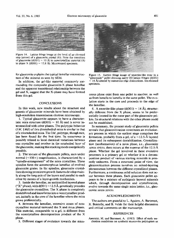
Still another phase has been detected at the periphery of some grains and appears to be a smectite-like struc- ture. Figure 15 shows such a typical sheet-structure stuffed with edge-dislocations, the discontinuous and wavy 00l planes of which yield a d(001) of ~ 14 ,~. These features strongly resemble those which charac- terize the smectic crystals described by De Gennes (1974). The textural relations between this phase and the others is, unfortunately, still unknown.
DISCUSSION
Assuming that only n (2~r/3) (where n = 0, 1, or 2) rotations are possible in the stacking of successive monolayers of mica, the purest glauconite pellets stud- ied show that glauconite appears to adopt three poly- typic basic structures, 1Md, 1M, and 2M1. The last polytype was found here for the first time, as a result of HRTEM. All previous studies of this dioctahedral mica reported only the 1M and 1 M d forms. The 1M form is the stable form; the second form represents a disordered unstable form (Wise and Eugster, 1964; Ernst, 1963; Burst, 1958a). According to Appelo (1978), the cationic composition of its octahedral sheet (pres-

480 Amouric and Parron Clays and Clay Minerals
Figure 13. Lattice fringe image of X-phase sandwiched by glauconite. This phase-X "relic" has probably been isolated during the lateral development of glauconite crystals Gl and Gz. Note the lateral transition glauconite (G3) to zone A to phase X in lower part of the illustration. Arrow shows a growth step. Microtomed specimen.
Figure 12. Lattice fringe images of: (a) two close lamellae showing different structural stages separated by a noncrystal- line zone strongly resembling a gel (g). G = glauconite, X = phase X, A = very poorly crystalline zones regarded as a decomposition product of the X-phase. (b) Glauconite crystal (G) showing sharp growth steps (arrowed). Adjacent to glau- conite steps, note the transition with the X-phase via a very poorly crystalline zone (A). Note also the sharp phase X-glauconite interface and phase X sandwiching the glauco- nite. Microtomed specimen.
ence of larger cations such as Fe 3+) causes glauconite to display a polytypic behavior similar to that oftrioc- tahedral micas. Direct lattice imaging suggests that the IMd sequence is related to a pure glauconite structure and not to an interstratified one as was previously as- sumed (see McRae, 1972).
From a statistical point of view, the frequency of occurrence of these polytypes is 1 m ~ 1Md >> 2M~. So-called unstable forms (1Md, 2M0 are relatively abundant and coexist, even in the same pellet, with the stable form 1M. The rarity of the 2M~ sequence may signify that this form, supposed to be energetically un- stable from a structural point of view, is uncommon in separate glauconite crystallites. This scarcity also explains why the 2MI form has never been detected by XRD.
Glauconite crystallites visualized and characterized by means of their basal planes (e.g., d(001) ~ 10 ]k) are generally located in a central position inside the spindles. All are well-contrasted, exhibit a regular structural organization, and are clearly individual forms.
Growth-steps and important lateral developments are common and are indicative of a fresh phase being gen- erated by a layer-growth mechanism already reported for hydrothermally grown micas (Baronnet, 1974; Bar- onnet et al., 1976).
This mechanism of glauconite formation, by nu- cleation and pure growth phenomena, discards the "layer-lattice theory" (Burst, 1958a, 1958b; Hower, 1961), for the phase X to glauconite transformation. The layer-lattice model involves a simple rearrange- ment in the solid state with incorporation of Fe and K in a smectite-like structure. This structure changes to- wards a micaceous phase, through smectite/mica mixed- layered intermediates. In the present study, the X-phase (d(001) ~ 12.5 ~k), a non-mixed-layered phase, which is significantly different from the 14-A smectite-like structure found here, forms before the mica appears. Its behavior contrasts with that of the glauconite. The X-phase shows all the characteristics of a progressive destabilization ending with its reduction to a noncrys- talline phase. These noncrystalline zones generally mark a front of evolution between the X-phase and the well- structured micaceous phase which grows at the expense of X. Furthermore, the two different phases typically show a parallel or pseudo-parallel orientation of their basal planes. This arrangement can be explained by the fact that the destructuring of the X-phase is easier along the long axis of the layers. Thus, the mica-growth takes place via the noncrystalline zones, in front of the de- stabilized X-phase. In this way, a global conservation of the spindle and lamellar shapes is possible during the evolution towards a glauconite-mica mineral.
The observed X-phase may correspond to the non- tronitic phase reported by several authors on the basis of XRD and chemical analyses to occur during glau- conitization (see, for example: Wermund, 1961; Por- renga, 1967; Martin, 1972; Giresse and Odin, 1973; Velde, 1976; Odom, 1976). Complementary chemical microanalyses, now in progress, may answer this ques- tion. The revealed layer-growth mechanism observed
Vol. 33, No. 6, 1985 Electron microscopy of glauconite 481
Figure 14. Lattice fringe image at the level of an elevated step (arrow) of a glauconite crystal (G). Note the transition of glauconite (d(001) ~ 10 A) to noncrystalline material (A) to phase X (d(001) ~ 12.8 A). Microtomed specimen.
for glauconite explains the typical lamellar microstruc- ture of the mineral as seen by SEM.
In addition, the gel-like material commonly sur- rounding the composite glauconite-X phase lamellae and the apparent transit ional relationship between the gel and X, suggest that the X-phase may have formed from this gel.
CONCLUSIONS
In this work, new results about the structure and genesis of glauconite minerals have been obtained by high-resolution transmission electron microscopy.
1. Typical glauconite appears to have a character- istic mica structure (d(001) ~ l0 ~ ) and is never in- terstratified with other phases. The polytypic behavior (1M, 1 M d ) of this dioctahedral mica is similar to that o fa trioctahedral mica. The 2M, polytype, though rare, has been found for the first t ime. Its occurrence is possibly related to local chemical variations between one crystallite and another in the octahedral layer of the glauconite, making this stacking mode energetically possible.
2. The texture o f the glauconite pellets, seen under normal (~ 100 x) magnification, is characterized by a "spindle-arrangement" of the mica crystallites. These spindles form the automorphous lamellae of evolved glauconite grains. In the spindles, glauconite crystal- lites showing prominent growth features develop main- ly along the long axis of the layers and parallel to each other by means of a layer-growth mechan i sm .
3. Inside the lamellae, an unidentified layered phase ("X" phase), with d(001) ~ 12.5 Zk, genetically precedes the glauconite crystallites. The X phase is completely destabilized and transforms into a noncrystalline prod- uct, initially in the core of the lamellae where the mica grows preferentially.
4. Between the lamellae, extensive zones of non- crystalline material surround the X and mica phases. This material, comparable to a gel, is different from the noncrystalline decomposi t ion product of the X phase.
5. Different stages o f evolution towards the mica-
Figure 15. Lattice fringe image of smectite-like zone in a "glauconite" pellet showing many 00l lattice fringes (d(001) ~ 14 .~) altered by numerous edge dislocations. Ion-thinned specimen.
ceous phase exist from one pellet to another, as well as from lamena to lamella in the same pellet. The evo- lution starts in the core and proceeds to the edge of the lamellae.
6. A smectite-like phase (d(001) ~ 14 A), structur- ally different from the X phase, seems to he prefer- entially located in the outer part of the glauconite pel- lets. Its structural relations with the other phases could not be established.
In summary, the present study of glauconite pellets reveals that glauconitization constitutes an evolution- ary process in which the earliest stage comprises the formation, probably from a gel, o f a ~ 12.5-,~ layered phase and its subsequent destabilization. Crystalliza- t ion (neoformation) of a mica phase, i.e., glauconite sensu stricto, then occurs at the expense of the 12.5-,~ phase. Whether the gel involved in these evolutive processes is a pr imary gel or whether it is a decom- posit ion product of various starting minerals is pres- ently unknown. From a structural point of view, the glauconitization process involves no mixed-layering phenomenon between the different crystallized phases. Furthermore, a continuous solid solution does not oc- cur between these phases. Each glauconite pellet ap- pears to be a mixture of authigenic layered minerals which, through decomposi t ion and crystallization, evolve towards the same single mica lattice, i.e., glan- conite sensu stricto.
A C K N O W L E D G M E N T S
The authors are grateful to L. Aguirre, A. Baronnet, R. Boistelle, and B. Velde for their helpful discussion and critical comments on the manuscript.
REFERENCES
Amouric, M. and Baronnet, A. (1983) Effect of early nu- cleation conditions on synthetic muscovite polytypism as

482 Amouric and Parron Clays and Clay Minerals
seen by high resolution transmission electron microscopy: Phys. Chem. Miner. 9, 146-159.
Amouric, M., Baronnet, A., and Finck, C. (1978) Polyty- pisme et d6sordre dans les micas diocta6driques synth6- tiques. Etude par imagerie de r6seau: Mat. Res. Bull. 13, 627-634.
Amouric, M., Mercuriot, G., and Barormet, A. (1981) On computed and observed HRTEM images of perfect mica polytypes: Bull. Mineral 104, 298-313.
Appelo, C. A.J. (1978) Aspects of mica-related clay min- erals in hydrogeochemistry: Ph.D. thesis, Univ. Amster- dam, 75 pp.
Baronnet, A. (1974) Etude en microscopie 61ectronique des premiers stades de croissance d'un mica synth6tique, la phlogopite hydroxyl6e: High Temp. High Press. 6, 193- 198.
Baronnet, A., Amouric, M., and Chabot, B. (1976) M6ca- nismes de croissance, polytypisme et polymorphisme de la muscovite hydroxyl6e synth6tique: J. Cryst. Growth 32, 37- 59.
Bentor, Y. K. and Kastner, M. (1965) Notes on the min- eralogy and origin of glauconite: J. Sed. Petrol 35, 155- 166.
Burst, J. F. (1958a) Mineralogical heterogeneity in "glau- conite" pellets: Amer. Mineral 43, 481-497.
Burst, J. F. (1958b) "Glauconite" pellets: their mineral na- ture and applications to stratigraphic interpretations: Bull. Amer. Assoc. Pet. GeoL 42, 310-327.
Charpy, N. and Nahon, D. (1978) Contribution h l'6tude lithostratigraphique et chronostratigraphique du Tertiaire du Bassin de C6te d'Ivoire: Rapp. D~p. Sc. Terre, Univ. Abidjian 18, 33 pp.
Cimbhlnikov6, A. (1971a) Chemical variability and struc- tural heterogeneity ofglauconites: Amer. Mineral 56, 1385- 1392.
Cimbhlnikov~, A. (1971b) Influence of 10 /~-14 /~ inter- layering on the layer charge of glauconites: Amer. Mineral 56, 1393-1398.
De Gennes, P. G. (1974) The Physics of Liquid Crystals: Oxford Univ. Press, Oxford, 320 pp.
Eberhart, J. P. and Triki, R. (1972) Description d'une tech- nique permettant d'obtenir des coupes minces de min6raux argileux par ultramicrotomie. Application ~ l'6tude des mi- n6raux argileux interstratifi6s: J. Microscop. 15, 111-120.
Ernst, W. G. (1963) Significance of phengitic micas from low-grade schists: Amer. Mineral. 48, 1357-1373.
Giresse, P. and Odin, G.S. (1973) Nature min6ralogique et odgine des glauconies du plateau continental du Gabon et du Congo: Sedimentology 20, 457-488.
Hower, J. (1961) Some factors concerning the nature and origin of glauconite: Amer. Mineral 46, 313-334.
Iijima, S. and Buseck, P. R. (1978) Experimental study of disordered mica structure by HREM: Acta Crystallogr. A34, 709-719.
Katsnel'son, Y. Y., Nyrkov, A. A., and Yakushev, V.V. ( 1978) Structural and chemical characteristics of glauconite as its qualitative indicators: LithoL Min. Res. 13, 324-335.
Kohler, E. E. (1980) The occurrence and properties ofglau- conites in Mesozoic and Cenozoic sediments in north- western and southern Germany: GeoL Jb. D39, 115-136.
McRae, S. G. (1972) Glauconite: Earth-Sci. Rev. 8, 397- 440.
McRae, S. G. and Lambert, J. L.M. (1968) A study of some glanconites from Cretaceous and Tertiary formations in southeast England: Clay Mineral 7, 431-440.
Manghnani, M.H. andHower, J. (1964) Glauconites:cation exchange capacities and infrared spectra: Amer. Mineral 49, 586-598.
Martin, L. (1972) Etude des "faecal-pellets" min6ralis6s des s6diments du plateau continental de C6te d'Ivoire: Cah. ORSTOM, s~r. G~oL 4, 105-120.
Nahon, D., Carozzi, A. V., and Parron, C. (1980) Lateritic weathering as a mechanism for the generation of ferrugi- nous oo'ids: a t. Sed. Petrol 50, 1287-1298.
Odin, G. S. (1974) Application de la microseopie 61ectro- nique par r6flexion 6 l'6tude des min6ratLX argileux: exemple des min6ranx des glanconies: Tray. Lab. MieropaL, Univ. Paris 3, 297-313.
Odin, G. S. (1975) Les glauconies: constitution, formation, age: Ph.D. thesis, Univ. Paris, 277 pp.
Odom, I. E. (1976) Microstructure, mineralogy and chem- istry of Cambrian galuconite pellets and glauconite, central U.S.A.: Clays & Clay Minerals 24, 232-238.
Oertel, G., Curtis, C. D., and Phakey, P. P. (1973) A trans- mission electron microscope and X-ray diffraction study in muscovite and chlorite: Mineral Mag. 39, 176-188.
Patron, C. and Nahon, D. (1980) Red bed genesis by lateritic weathering of glanconitic sediments: J. GeoL Soc. London 137, 689-693.
Paulus, M., Dubon, A., and Etienne, J. (1975) Application of ion thinning to the study of the structure of argillaceous rocks by transmission electron microscopy: Clays & Clay Minerals 20, 193-197.
Phakey, P. P., Curtis, C. D., and Oertel, G. (1972) Trans- mission electron microscopy of fine-gained phyllosilicates in ultra-thin rock sections: Clays & Clay Minerals 20, 193- 197.
Porrenga, D.H. (1967) Glauconite and chamosite as depth indicators in the marine environment: Marine GeoL 5, 495- 501.
Tchoubar, C., Rauturcau, M., and Clinard, C. (1973) Tech- nique d'inclusion appliqu6e ~ l'6tude des silicates lamel- laires et fibreanx: J. Microsc. 18, 147-157.
Thompson, G. R. and Hower, J. (1975) The mineralogy of glauconite: Clays & Clay Minerals 23, 289-300.
Velde, B. (1976) The chemical evolution ofglanconite pel- lets as seen by microprobe determinations: Mineral Mag. 40, 753-760.
Velde, B. and Odin, G.S. (1975) Further information related to the origin ofglauconite: Clays & Clay Minerals 23, 376- 381.
Warshaw, C.M. (1957) The mineralogy ofglauconite: Ph.D. thesis, Pennsylvania State Univ., University Park, Penn- sylvania, 155 pp.
Wermund, E. G. (1961) Glauconite in early Tertiary sedi- ments of Gulf Coastal Province: Bull. Amer. Assoc. Pet. GeoL 45, 1667-1696.
Wise, W. S. and Eugster, H. P. (1964) Celadonite: synthesis, thermal stability and occurrence: Amer. Mineral 49, 1031- 1083.
Yoder, H. S. and Eugster, H.P. (1955) Synthetic and natural muscovites: Geochim. Cosmochim. Acta 8, 225-280.
(Received 24 April 1984; accepted 10 March 1985; Ms. 1357)