Embed Size (px)
Citation preview
Structure and function of nucleic acids.

Heat.
Heat flows through the boundary of the system because there exists a temperature difference between the system and surroundings.
C – heat capacity, can be measured by putting known amount of heat into the system and measuring the T-difference.
Cdtdq

Work.
P-V work:
dVPAdxPdw exex
gas
Pex
x
P
V
P1,V1
P3,V3
P2,V2

The first law of thermodynamics.
The energy of isolated system is constant:
E – state function, energy change is the same regardless how this change was produced:
wqE
12
2
1
EEdEEE
E
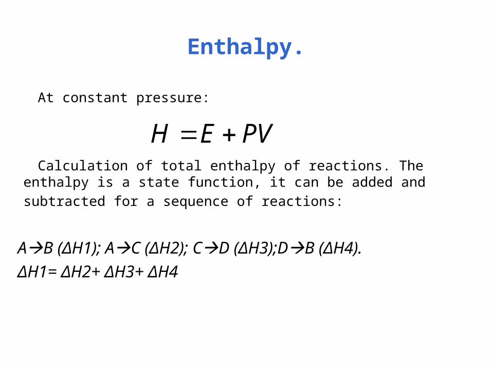
Enthalpy.
At constant pressure:
Calculation of total enthalpy of reactions. The enthalpy is a state function, it
can be added and subtracted for a sequence of reactions:
AB (ΔH1); AC (ΔH2); CD (ΔH3);DB (ΔH4).
ΔH1= ΔH2+ ΔH3+ ΔH4
PVEH

Entropy.
What defines the direction of spontaneous change?
Statistical aspect of entropy:
Ex: rearranging three balls:
123 132 213 231 312 321
Spontaneous process will go in the direction of increasing the probability W.
WKS ln
)/ln( if VVnRS
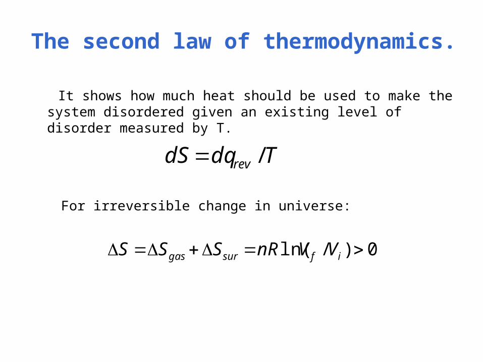
The second law of thermodynamics.
It shows how much heat should be used to make the system disordered given an existing level of disorder measured by T.
For irreversible change in universe:
TdqdS rev /
0)/ln( ifsurgas VVnRSSS

Free energy.
Gibbs free energy at P= const:
G = H –TS
At equilibrium ΔG = 0 and:
K – equilibrium constant
Standard state: at T = 25C and P = 1 atmosphere.
KRTG ln0
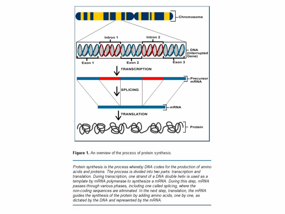

DNA structure.
History:• 1868 Miescher – discovered nuclein
• 1944 Avery – experimental evidence that DNA is constituent of genes.
• 1953 Watson&Crick – double helical nature of DNA.
• 1980 X-ray structure of more than a full turn of B-DNA.
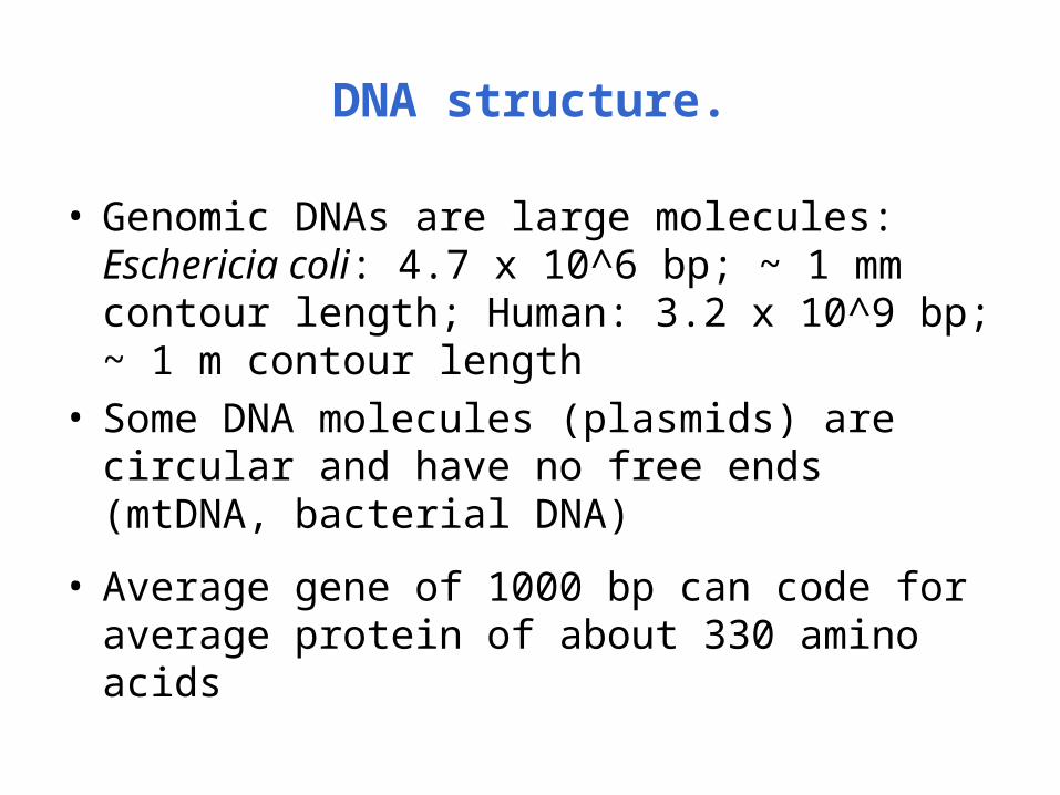
DNA structure.
• Genomic DNAs are large molecules:Eschericia coli: 4.7 x 10^6 bp; ~ 1 mm contour length; Human: 3.2 x 10^9 bp; ~ 1 m contour length
• Some DNA molecules (plasmids) are circular and have no free ends (mtDNA, bacterial DNA)
• Average gene of 1000 bp can code for average protein of about 330 amino acids

Five types of bases.

Nucleotides and phosphodiester bond.
Phosphodiester bond
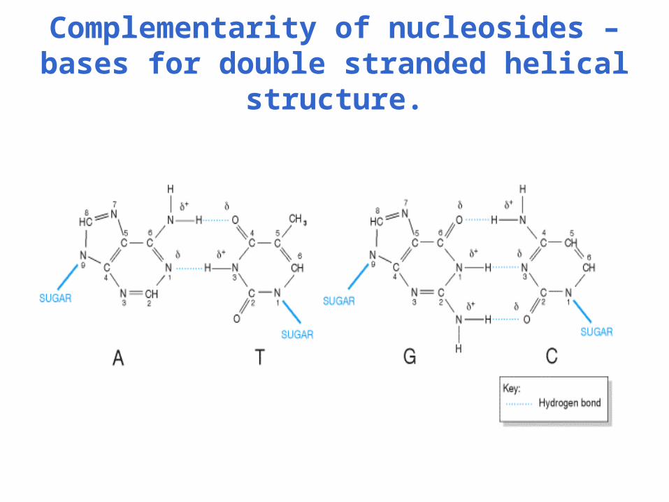
Complementarity of nucleosides – bases for double stranded helical structure.

Chargaff’s rule.
• Amounts of G = C, A = T.• Most DNAs obey this rule – double starnded,
few RNAs obey this rule – single stranded.• Why RNAs do not reach the maximum of base
pairing?
- RNA contains not equal amount of A/U and G/C.
- RNA contain modified bases which prevent base pairing

Double helical structure of DNA.
A- and B-DNA – right-handed helix, Z-DNA – left-handed helix
B-DNA – fully hydrated DNA in vivo,10 base pairs per turn of helix
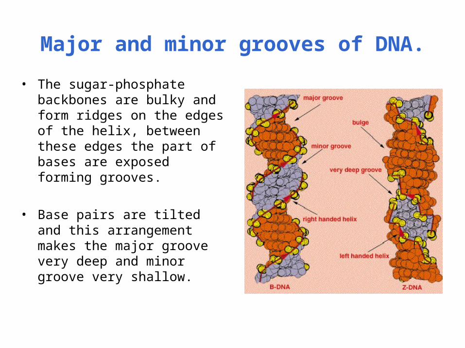
Major and minor grooves of DNA.
• The sugar-phosphate backbones are bulky and form ridges on the edges of the helix, between these edges the part of bases are exposed forming grooves.
• Base pairs are tilted and this arrangement makes the major groove very deep and minor groove very shallow.

Hydration of B-DNA.
From R. Dickerson, Structure & Expression

Major and minor grooves of DNA.
Copyright © Ramaswamy H. Sarma 1996
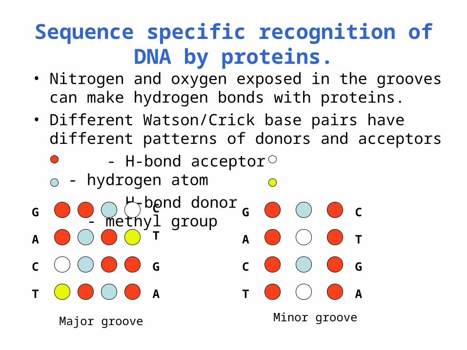
Sequence specific recognition of DNA by proteins.
• Nitrogen and oxygen exposed in the grooves can make hydrogen bonds with proteins.
• Different Watson/Crick base pairs have different patterns of donors and acceptors
- H-bond acceptor - hydrogen atom
- H-bond donor - methyl group
G
A
C
T
C
T
G
A
G
A
C
T
C
T
G
A
Major groove Minor groove
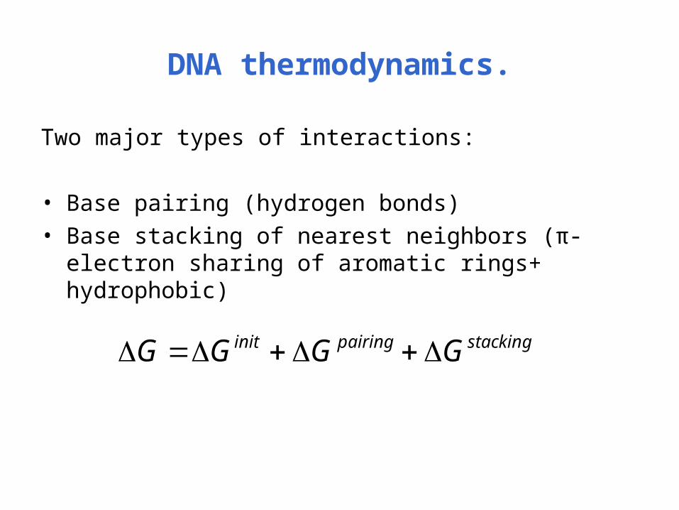
DNA thermodynamics.
Two major types of interactions:
• Base pairing (hydrogen bonds)• Base stacking of nearest neighbors (π-electron sharing
of aromatic rings+ hydrophobic)
stackingpairinginit GGGG
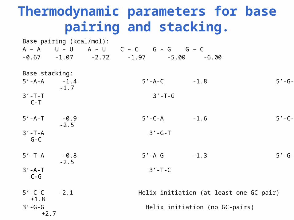
Thermodynamic parameters for base pairing and stacking.
Base pairing (kcal/mol):A – A U – U A – U C – C G – G G – C-0.67 -1.07 -2.72 -1.97 -5.00 -6.00
Base stacking:5’-A-A -1.4 5’-A-C -1.8 5’-G-A -1.73’-T-T 3’-T-G 3’-C-T
5’-A-T -0.9 5’-C-A -1.6 5’-C-G -2.53’-T-A 3’-G-T 3’-G-C
5’-T-A -0.8 5’-A-G -1.3 5’-G-C -2.5 3’-A-T 3’-T-C 3’-C-G
5’-C-C -2.1 Helix initiation (at least one GC-pair) +1.83’-G-G Helix initiation (no GC-pairs) +2.7

Classwork 1.
Calculate ΔG for DNA stabilization:
5’-T-A-C-T-G-3’
3’-A-T-G-A-C-5’
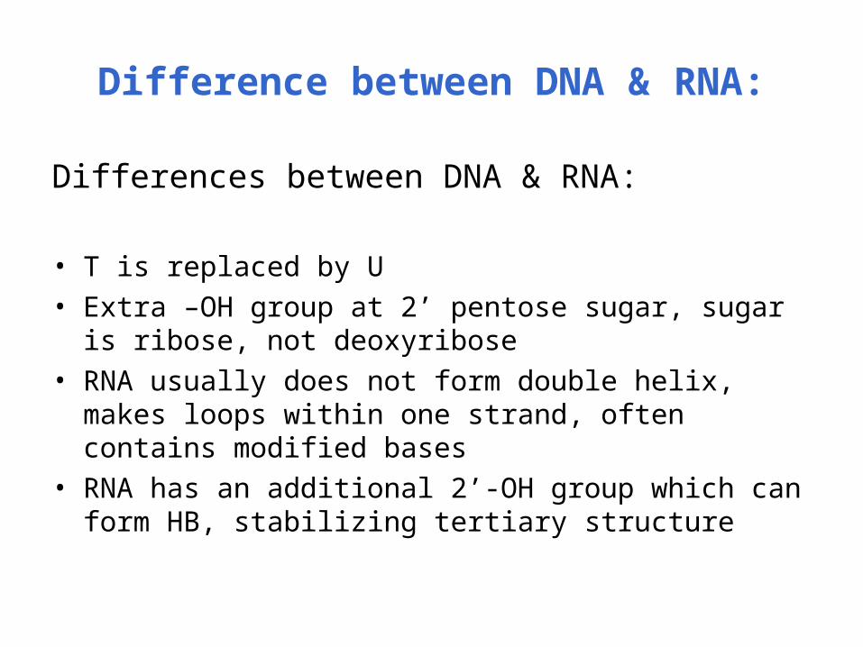
Difference between DNA & RNA:
Differences between DNA & RNA:
• T is replaced by U• Extra –OH group at 2’ pentose sugar, sugar is ribose,
not deoxyribose• RNA usually does not form double helix, makes loops
within one strand, often contains modified bases • RNA has an additional 2’-OH group which can form HB,
stabilizing tertiary structure

RNA functions.
• Information decoding (mRNA)
• Information transfer (tRNA)
• Structural molecule (rRNA)
• Catalytic function (ribozymes)
• Regulatory function

Types of RNA secondary structures.
• hairpin loop, (i ,j) defines a hairpin loop if i and j are paired and k is a free base, i < k < j.
• stacked loop – if i and j are paired and (i+1) and (j-1) are paired.
• internal loop – two closing base pairs and all bases between them are free.
• multi-branched loop or multi-loop – loop has at least three closing base pairs.
• pseudoknot – (i,j,i’,j’) defines pseudoknot if i and j are pairs, i’ and j’ are pairs and i < i’ <j < j’.
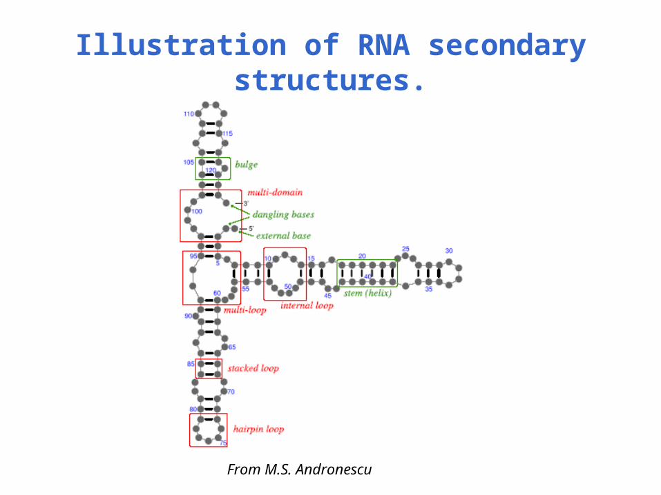
Illustration of RNA secondary structures.
From M.S. Andronescu

Tertiary structure of rRNA.

Information decoding (mRNA).

Information transfer (tRNA).
Characteristic properties:
- Each tRNA is specific for one of the amino acids.
- Forms a lot of tertiary interactions (Ex: tRNA for Phe contains 20 base pairs which form 52 HBs and 40 HBs from tertiary interactions).
- Most of tRNA structure is common between the species.

Structural molecule (rRNA).
Ribosome of prokaryotes: 3 RNA molecules and 55 proteins. Small subunit controls tRNA interactions with mRNA;large subunit controls catalysis.

RNA catalytic properties: ribozymes.
• RNA of self-splicing group I introns, contain 4 sequence elements and form specific secondary structures
• RNA self-splicing group II introns
• RNA from viral and plant satellite RNAs
• Ribosomal RNAs

Classwork 2.
1. Go to http://ndbserver.rutgers.edu/.
2. Select Crystal structure of B-DNA, resolution >=2 Angstroms.
3. Select Crystal structure of single-stranded RNA with mismatch base pairing with resolution >= 2 Angstroms.

Classwork 3: DNA topoisomerase.
1. Go to http://ndbserver.rutgers.edu/.
2. Select a structure for DNA – topoisomerase complex
3. Look at the complex using CN3D and answer the questions:
- What type of groove does the topoisomerase bind to?
- What types of secondary structure are involved in the interaction between DNA and protein?
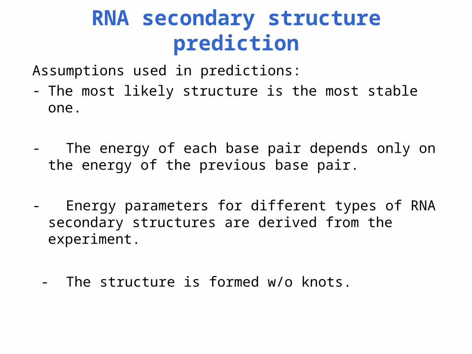
RNA secondary structure prediction
Assumptions used in predictions:- The most likely structure is the most stable one.
- The energy of each base pair depends only on the energy of the previous base pair.
- Energy parameters for different types of RNA secondary structures are derived from the experiment.
- The structure is formed w/o knots.
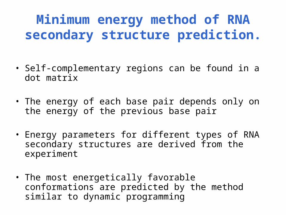
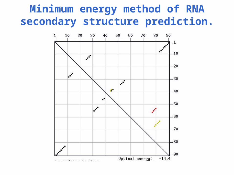
Minimum energy method of RNA secondary structure prediction.
• Self-complementary regions can be found in a dot matrix
• The energy of each base pair depends only on the energy of the previous base pair
• Energy parameters for different types of RNA secondary structures are derived from the experiment
• The most energetically favorable conformations are predicted by the method similar to dynamic programming
Minimum energy method of RNA secondary structure prediction.
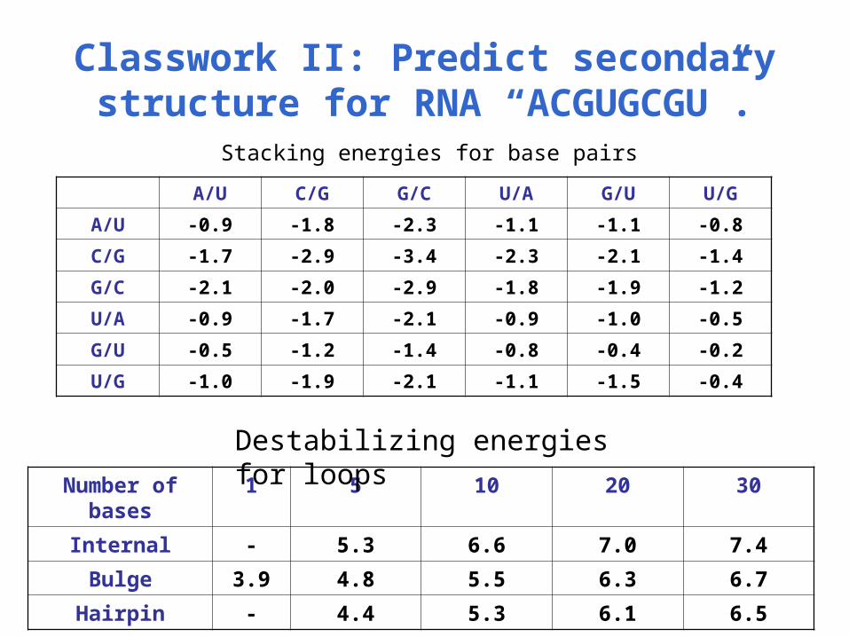
Classwork II: Predict secondary structure for RNA “ACGUGCGU”.Stacking energies for base pairs
A/U C/G G/C U/A G/U U/G
A/U -0.9 -1.8 -2.3 -1.1 -1.1 -0.8
C/G -1.7 -2.9 -3.4 -2.3 -2.1 -1.4
G/C -2.1 -2.0 -2.9 -1.8 -1.9 -1.2
U/A -0.9 -1.7 -2.1 -0.9 -1.0 -0.5
G/U -0.5 -1.2 -1.4 -0.8 -0.4 -0.2
U/G -1.0 -1.9 -2.1 -1.1 -1.5 -0.4
Number of bases
1 5 10 20 30
Internal - 5.3 6.6 7.0 7.4
Bulge 3.9 4.8 5.5 6.3 6.7
Hairpin - 4.4 5.3 6.1 6.5
Destabilizing energies for loops

Sequence covariation method.
Some positions from different species can covary because they are involved in pairing
fm(B1) - frequences in column m;
fn(B2) – frequences in column n;
fm,n(B1,B2) – joint frequences of two nucleotides in two columns.
Seq 1 ---G------C---
Seq 2 ---G------C---
Seq 3 ---A------T---
Seq 4 ---T------A---
))()(/(),( 2121, BfBfBBf nmnm
Ribozymes.
• RNA of self-splicing group I introns, contain 4 sequence elements and form specific secondary structures
• RNA self-splicing group II introns
• RNA from viral and plant satellite RNAs
• Ribosomal RNAs
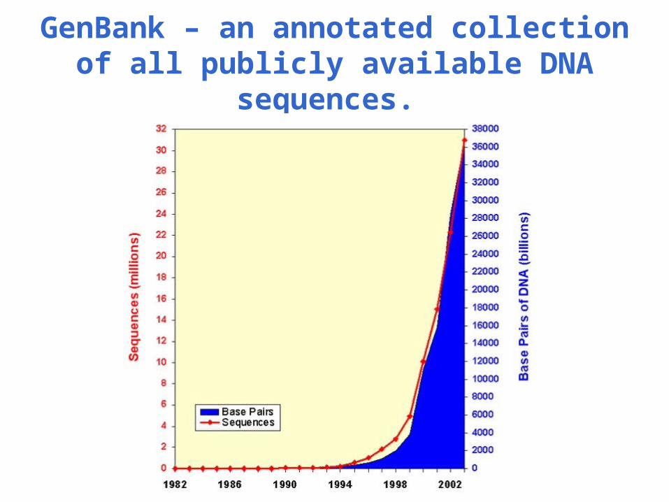
GenBank – an annotated collection of all publicly available DNA sequences.
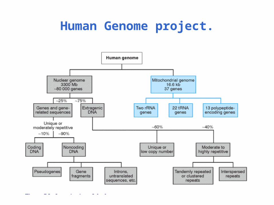
Human Genome project.