Embed Size (px)
Citation preview

HAL Id hal-03335234httpshalarchives-ouvertesfrhal-03335234
Submitted on 6 Sep 2021
HAL is a multi-disciplinary open accessarchive for the deposit and dissemination of sci-entific research documents whether they are pub-lished or not The documents may come fromteaching and research institutions in France orabroad or from public or private research centers
Lrsquoarchive ouverte pluridisciplinaire HAL estdestineacutee au deacutepocirct et agrave la diffusion de documentsscientifiques de niveau recherche publieacutes ou noneacutemanant des eacutetablissements drsquoenseignement et derecherche franccedilais ou eacutetrangers des laboratoirespublics ou priveacutes
Distributed under a Creative Commons Attribution| 40 International License
Structure and dynamics of the quaternary hunchbackmRNA translation repression complex
Jakub Macošek Bernd Simon Johanna-Barbara Linse Pravin Kumar AnkushJagtap Sophie Winter Jaelle Foot Karine Lapouge Kathryn Perez Mandy
Rettel Miloš Ivanović et al
To cite this versionJakub Macošek Bernd Simon Johanna-Barbara Linse Pravin Kumar Ankush Jagtap Sophie Winteret al Structure and dynamics of the quaternary hunchback mRNA translation repression complexNucleic Acids Research Oxford University Press 2021 49 (15) pp8866-8885 101093nargkab635hal-03335234
Nucleic Acids Research 2021 1httpsdoiorg101093nargkab635
Structure and dynamics of the quaternary hunchbackmRNA translation repression complexJakub Macosek 1 Bernd Simon1 Johanna-Barbara Linse2Pravin Kumar Ankush Jagtap 1 Sophie L Winter 1 Jaelle Foot1 Karine Lapouge3Kathryn Perez3 Mandy Rettel4 Milos T Ivanovic2 Pawel Masiewicz1 Brice Murciano1Mikhail M Savitski4 Inga Loedige1 Jochen S Hub2 Frank Gabel5 andJanosch Hennig 16
1Structural and Computational Biology Unit European Molecular Biology Laboratory Heidelberg Heidelberg 69117Germany 2Theoretical Physics and Center for Biophysics Saarland University Saarbrucken 66123 Germany3Protein Expression and Purification Core Facility European Molecular Biology Laboratory HeidelbergHeidelberg 69117 Germany 4Proteomics Core Facility European Molecular Biology Laboratory HeidelbergHeidelberg 69117 Germany 5Institut Biologie Structurale University Grenoble Alpes CEA CNRS Grenoble 38044France and 6Chair of Biochemistry IV Biophysical Chemistry University of Bayreuth Universitatsstrasse 30 95447Bayreuth Germany
Received May 11 2021 Revised July 05 2021 Editorial Decision July 08 2021 Accepted July 27 2021
ABSTRACT
A key regulatory process during Drosophila devel-opment is the localized suppression of the hunch-back mRNA translation at the posterior which givesrise to a hunchback gradient governing the forma-tion of the anterior-posterior body axis This sup-pression is achieved by a concerted action of BrainTumour (Brat) Pumilio (Pum) and Nanos Each pro-tein is necessary for proper Drosophila developmentThe RNA contacts have been elucidated for the pro-teins individually in several atomic-resolution struc-tures However the interplay of all three proteinsduring RNA suppression remains a long-standingopen question Here we characterize the quater-nary complex of the RNA-binding domains of BratPum and Nanos with hunchback mRNA by combin-ing NMR spectroscopy SANSSAXS XLMS with MDsimulations and ITC assays The quaternary hunch-back mRNA suppression complex comprising theRNA binding domains is flexible with unoccupiednucleotides functioning as a flexible linker betweenthe Brat and Pum-Nanos moieties of the complexMoreover the presence of the Pum-HDNanos-ZnFcomplex has no effect on the equilibrium RNA bind-
ing affinity of the Brat RNA binding domain This isin accordance with previous studies which showedthat Brat can suppress mRNA independently and isdistributed uniformly throughout the embryo
INTRODUCTION
One of the key processes during Drosophila development isthe formation of body axes in the embryo This is achievedby RNA localization and spatially restricted translation (1)which results in protein gradients along which the axesare established (2) Proper anterior-posterior axis formationis governed by localization of two maternally transcribedgenes The first bicoid mRNA localizes to the anterior poleof the oocyte (3) whereas the second nanos mRNA local-izes to the posterior pole of the oocyte (4) Translation ofbicoid and nanos mRNA then results in opposing gradientsof Bicoid and Nanos proteins which in turn control trans-lation of another maternally supplied mRNAndashndashhunchbackmRNA (5ndash8) Hunchback mRNA is distributed uniformlyand Nanos suppresses the mRNA translation at the pos-terior whereas Bicoid activates it at the anterior The re-sulting anterior-posterior gradient of Hunchback then en-sures proper development of abdominal and thorax struc-tures (910)
Nanos is thus the trans-acting molecule in the posteriorsuppression of hunchback mRNA translation The related
To whom correspondence should be addressed Tel +49 6221 387 8552 Email janoschhennigembldePresent addressesJakub Macosek Biozentrum University of Basel Basel 4056 SwitzerlandInga Loedige IRI Life Sciences Humboldt University Berlin 10115 GermanyMilos Ivanovic Department of Biochemistry University of Zurich 8057 Zurich Switzerland
Ccopy The Author(s) 2021 Published by Oxford University Press on behalf of Nucleic Acids ResearchThis is an Open Access article distributed under the terms of the Creative Commons Attribution-NonCommercial License(httpcreativecommonsorglicensesby-nc40) which permits non-commercial re-use distribution and reproduction in any medium provided the original workis properly cited For commercial re-use please contact journalspermissionsoupcom
Dow
nloaded from httpsacadem
icoupcomnaradvance-articledoi101093nargkab6356331677 by C
EA user on 06 September 2021
2 Nucleic Acids Research 2021
cis-acting elements are located within the 3prime untranslated re-gion (UTR) of the mRNA and are called Nanos ResponseElements (NREs) (11) There are two NREs (NRE1 andNRE2) that each contain two conserved sequences calledBoxA (upstream) and BoxB (downstream) (Figure 1A)Nanos contains a zinc finger domain (Nanos-ZnF Figure1B) that binds the NREs (12) but requires the trans-actingmolecules Pumilio and Brain Tumor (Brat) for suppres-sion of hunchback mRNA translation (1314) Pumilio andNanos have been long established as trans-acting elementsin the suppression of hunchback mRNA translation and arecent study revealed the underlying mechanism of theirconcerted recognition of hunchback mRNA (Figure 1B)(15) Pumilio features a Pumilio homology domain (Pum-HD) which is a sequence-specific single-stranded RNAbinding domain (RBD) that binds to a site partially over-lapping with BoxB of NREs (16ndash19) In contrast no spe-cific motif in hunchback mRNA is recognized by Nanos-ZnF which binds the RNA nevertheless with high affinitybut presumably with low sequence specificity (1220) How-ever Nanos-ZnF and Pum-HD together with hunchbackmRNA form a high affinity ternary protein-RNA complex(15) Ternary complex formation triggers RNA specificityin Nanos-ZnF for bases adjacent to the binding site of Pum-HD and further enhances the affinity of Pum-HD for itsbinding site While localization of Nanos at the posteriorensures the spatial restriction for hunchback mRNA sup-pression Pumilio conveys specificity for hunchback mRNAbinding Thus the concerted recognition of the RNA byPumilio and Nanos simultaneously allows the spatially con-trolled suppression of hunchback mRNA during Drosophiladevelopment
Brain Tumour has been identified as another trans-actingmolecule in the suppression of hunchback mRNA transla-tion more recently (14) The NCL-1 HT2A and LIN-41domain of Brat (Brat-NHL) was shown to interact specifi-cally with a motif in BoxA of NREs (Figure 1C) and Bratmutants induce similar developmental defects in flies asPumilio or Nanos mutants (21ndash23) Like Pumilio Brat isuniformly distributed throughout the embryo (14) but howit contributes to the suppression of hunchback mRNA byPumilio and Nanos remains unclear (24) despite a wealthof available experimental data on the relationship of Brat-NHL Pum-HD and Nanos-ZnF First published data sug-gest that Brat Pumilio and Nanos form a quaternary com-plex with hunchback mRNA in order to suppress its transla-tion in yeast four-hybrid and pull-down assays Brat-NHLPum-HD and Nanos-ZnF were all necessary to form a sta-ble protein-RNA complex (14) In line with that Pum-HDwas reported to increase the affinity of Brat-NHL for hunch-back mRNA (22) This would explain why Brat mutationsinduce phenotypes similar to those of Pumilio and Nanosmutants Moreover a vast majority of these mutants indi-rectly implied quaternary complex formation as they abro-gated complex formation in yeast four-hybrid assays How-ever it became clear later that the point mutations substi-tuted residues necessary for RNA binding of Brat-NHL(22) so the observed effect is likely due to abrogated RNAbinding and not necessarily due to abrogation of quater-nary complex formation Furthermore gene reporter as-says in Dmel2 cells show that removing Brat-NHL has lit-
tle effect on suppression of a reporter gene in the pres-ence of Pumilio and Nanos and that conversely Brat cansuppress the mRNA translation independently in their ab-sence (2223) In summary it remains unclear whether BratPumilio and Nanos suppress hunchback mRNA transla-tion cooperatively in a single quaternary protein-RNA com-plex or whether Brat suppresses hunchback mRNA inde-pendently of Pumilio and Nanos To determine if the sup-pression activities are cooperative or independent a combi-nation of both functional and biophysical insights into theprocess is required Since there is already a wealth of func-tional data we obtained the biophysical results needed to fillthe existing gap in our understanding of hunchback mRNAtranslation suppression
We carried out an extensive structural biophysicaland computational investigation of Pum-HD Brat-NHLNanos-ZnF and their complex with hunchback mRNANRE2 (hb complex) First we analysed the influence ofPum-HD on the RNA binding of Brat-NHL using isother-mal titration calorimetry (ITC) and showed that they do notbind RNA cooperatively Moreover Brat-NHL and Nanos-ZnF do not show any signs of interaction in vitro in theabsence or presence of RNA and cooperativity in RNAbinding could not be observed between Brat-NHL and thePum-HDNanos-ZnF complex Nevertheless Brat-NHLPum-HD and Nanos-ZnF assemble all on a single NRE2in a stable stoichiometric quaternary protein-RNA com-plex However our data reveals that the complex in con-text of a single NRE2 and isolated RNA binding domainsis largely flexible and that Brat-NHL in the complex movesindependently of Pum-HD and Nanos-ZnF To investi-gate the extent of flexibility of the complex we used un-restrained molecular dynamics (MD) simulation validatedagainst cross-linkingmass spectrometry small-angle X-rayand neutron scattering data Collectively our results de-scribe a complex in which the unoccupied nucleotides ofthe NRE2 RNA function as a flexible linker between theBrat-NHL and Pum-HD-Nanos-ZnF moieties of the com-plex reminiscent of beads on a string Moreover this studyhighlights the importance of combining a multitude ofstructure analysis methods with molecular dynamics sim-ulations to obtain reliable atomistic ensembles of dynamicprotein-RNA complexes
MATERIALS AND METHODS
Protein expression and purification
The Pum-HD and Brat-NHL constructs used in this studycomprise residues 1093ndash1426 and 756ndash1037 of the respec-tive proteins (Figure 1B and C) which are connected bya short linker containing a Usp2cc cleavage site to a His6-Ubiquitin tag in pHUE plasmid (2526) The expression wasdone as previously described (23) but Escherichia coli BL-21(DE3) Rosetta cells were used The cells were then lysedfirst by incubation for 20 min on ice in lysis buffer (50 mMTris 1 M NaCl 5 glycerol 10 mM imidazole pH 80) with1 mgml lysozyme 1 gml DNase I 2 gml Rnase A andprotease inhibitor tablets (Roche) and then by sonication at4C The lysate was then cleared by centrifugation (18 000g 4C 1 h) and purified using a HisTrap HP 5 ml column
Dow
nloaded from httpsacadem
icoupcomnaradvance-articledoi101093nargkab6356331677 by C
EA user on 06 September 2021
Nucleic Acids Research 2021 3
NRE2 - BoxA +
Kd1 = 10 plusmn 03 nMN1 = 10 plusmn 001Kd2 = 05 plusmn 002 μMN2 = 11 plusmn 001
BoxB+
Kd = 8 plusmn 2 nMN = 11 plusmn 001
mRNA Pumilio
NRE2 (23-mer)
1 3350ORF NRE1 NRE2
5rsquo UTR 3rsquo UTR
Brain tumour
BoxA BoxB
A B C
NRE1+NRE2 5degC
13 12 11ω 2 - 1H (ppm)
13
12
11
ω1
- H
(pp
m) NRE1+NRE2 20degC
NRE1+NRE2 + Pum-HD 20degC
Brat-NHL
UUG UUG U GUA CAU AUUG UUG UCUUG UUG UCG AAA AU UCG AAA AUU GUA CAU AAUUG UUG UCG AAA AUU GUA CAU AA
BoxABoxBBoxAextNRE2-BoxBNRE2-BoxANRE2
D
E FITC experimental setup
Protein
RNAproteinRNA-protein
Nanos-ZnF Pum-HD
BoxA+
Kd = 11 plusmn 03 μMN = 08 plusmn 003
NRE2+
Kd = 04 plusmn 01 μMN = 11 plusmn 002
G
NRE2+
Kd1 = 2 plusmn 08 nMN1 = 09 plusmn 001Kd2 = 2 plusmn 01 μMN2 = 14 plusmn 001
BoxA+
No binding
BoxAext +
No binding
NRE2 - BoxB+
Kd = 12 plusmn 008 μMN = 11 plusmn 001
H
NRE2 +
No binding
+
No binding
+
No binding
NRE2-+
Kd = 09 plusmn 02μMN = 10 plusmn 002
NRE2-+
Kd = 09 plusmn 03 μMN = 10 plusmn 002
NRE2-+
Kd = 14 plusmn 06 μMN = 10 plusmn 002
I
Figure 1 The molecules involved in suppression of hunchback mRNA translation and their interactions (A) Two cis-acting elements controlling thesuppression are located within the 3prime UTR and called Nanos Response Element 1 and 2 (NRE1 and NRE2) The original definitions for BoxA and BoxBare underlined however we rather refer to the cognate RNA motifs throughout BoxA UUGUUG for Brat-NHL and BoxB UGUACAUA for Pum-HDThe trans-acting proteins used in this study are (B) the HD domain of Pumilio (Pum-HD grey) and the ZnF domain of Nanos (Nanos-ZnF pink NEDNanos effector domain the resulting complex is shown below (PDB ID 5KL1)) and (C) the NHL domain of Brain tumour (Brat-NHL green B B-boxdomain CC coiled-coil domain PDB ID 4ZLR) (D) A list of RNAs used in this study for ITC experiments and complex formation The shading of theRNA sequences indicates the binding site of the domain matching the colour based on the previously published structures of Brat-NHL-RNA complex (22)and Pum-HD-Nanos-ZnF-RNA complex (15) shown in (B) and (C) RNA comprising NRE2 indicated in the lower part of the left panel (NRE2 RNA)the protein domains and the published high-resolution structures (B and C) were used in this study for experimental measurements or computationalmodelling (E) NRE2 was shown to form a stem loop (22) which is melted by addition of Pum-HD Here we show by 1H-2D NOESY (right panel) thatthe Pum-HD domain is even able to disrupt base pairing in a larger NRE1 + NRE2 construct as imino NOEs disappear with increasing temperature andaddition of Pum-HD (F) Scheme of the experimental ITC setup RNA is always in the cell and protein in the syringe Preformed RNA-protein complexesare in the cell All values shown in ITC plots are the dissociation constants derived from fitting the individual experiment and the resulting fitting errorBrat-NHL-RNA (BoxA and NRE2) interaction studied by ITC NRE2 binds slightly stronger likely due to avidity effects The measurement reveals a11 interaction with a Kd of around 1 M Replicates are listed in Table 1 and shown in Supplementary Figure S1 (also for following panels) (G) ITCexperiments investigating RNA binding of Pum-HD by testing different RNA lengths Pum-HD binds with low nanomolar affinity to its cognate sequenceBoxB It does not bind to BoxA or extension of BoxA However a weak second binding is upstream of the BoxB motif (H) ITC of proteins only showingthat they do not interact in absence of RNA (I) ITC of Brat-NHL titrated to preformed protein-RNA complexes The affinity of Brat-NHL to NRE2does not increase in presence of Pum-HD Nanos-ZnF or both indicating that Brat-NHL does not bind RNA cooperatively with the other two proteins(Supplementary Figure S2E)
Dow
nloaded from httpsacadem
icoupcomnaradvance-articledoi101093nargkab6356331677 by C
EA user on 06 September 2021
4 Nucleic Acids Research 2021
(GE Healthcare) and a 10ndash100 mM imidazole linear gra-dient followed by elution with 250 mM imidazole whichwere created by mixing the lysis buffer with 50 mM Tris 1 MNaCl 5 glycerol 1 M imidazole pH 80 Pooled fractionsof the proteins were then cleaved by His6-tagged Usp2ccat 4C overnight during dialysis in dialysis buffer (50 mMTris 150 mM NaCl 1 mM DTT pH 74) Pum-HD wasthen separated from the tag by reverse affinity chromatog-raphy and in the final step Pum-HD was further purified bysize-exclusion chromatography (SEC) using HiLoad 16600Superdex 75 column (GE Healthcare) in 50 mM Tris 150mM NaCl 1 mM DTT pH 74 buffer For Brat-NHL serialreverse affinity and heparin chromatography (HiTrap HPHeparin 5ml column - GE Healthcare) followed the affinitychromatography The sample was loaded on the columnsconnected in series after equilibrating the columns in dialy-sis buffer Brat-NHL was eluted using a 150 mM to 15 MNaCl gradient The Nanos-ZnF plasmid used in this studycontains residues 301ndash392 of Nanos (Figure 1B) connectedvia a linker with SenP2 cleavage site to a His6-SUMO tag inpETM11-SUMO3GFP vector (EMBL Protein Expressionand Purification Core Facility) Nanos-ZnF was expressedby transforming the plasmid to E coli BL-21(DE3) Rosettacells growing the cells at 37C while shaking until OD600 of06ndash10 followed by addition of IPTG to 03 mM final con-centration and further overnight incubation at 16C whileshaking Nanos-ZnF was then purified as described abovefor Brat-NHL except that in the first affinity chromatog-raphy step the basic lysis buffer was 50 mM Tris 500 mMNaCl 50 M ZnSO4 10 mM imidazole pH 80 and the tagwas cleaved off using His6-tagged SenP2
Isotopic labelling
To obtain diversely isotopically labelled proteins the cellswere grown in M9 minimal media and the expression andpurification followed the standard protocols outlined aboveunless stated otherwise In order to obtain 15N-labelledNanos-ZnF and Brat-NHL the expression was done us-ing 15NH4Cl as the sole nitrogen source In order to ob-tain various degrees of 2H-labelling cells were grown asfollows a 5 ml overnight culture of H2O M9 minimal me-dia with adequate isotopes was spun down resuspendedin 100 ml of D2O M9 minimal media with adequate iso-topes grown to an OD600 of sim06 at 37C while shakingthen diluted to 500 ml final volume with D2O M9 minimalmedium The perdeuterated Brat-NHL and Pum-HD forSANS measurements were obtained by expressing the pro-teins in D2O M9 minimal media with 2H-glucose as the solecarbon source For NMR relaxation and 1H13C HMQC ex-periments Brat-NHL was expressed either uniformly 2H-13C- and 15N-labelled (for free form and RNA-bound mea-surements) or uniformly 2H- and 15N-labelled (in the hbcomplex) and in both cases with C1 of isoleucines and oneof the methyl groups of valines and leucines 1H13C labelledand the other methyl groups 2H- 12C-labelled The uni-form labelling was achieved by expression in D2O M9 min-imal media with 15NH4Cl as the sole nitrogen source and2H- or 2H13C-glucose as the sole carbon souce and adding2-keto-33-d2-1234-13C-butyrate and 2-keto-3-methyl-d3-
3-d1-1234-13C-butyrate one hour prior induction as de-scribed previously (27)
Hb complex formation
To form the hb complex the purified Pum-HD Brat-NHL and Nanos-ZnF proteins were incubated with a 23nucleotide-long NRE2 RNA encompassing the NRE2 inthe 3prime UTR of hunchback mRNA of the sequence 5prime UUGU-UGUCGAAAAUUGUACAUAA 3prime (Microsynth Figure1A) The complex was formed by incubating NRE2 RNAwith Pum-HD Nanos-ZnF and Brat-NHL in a 1112 mo-lar ratio respectively to account for the lower affinity ofBrat-NHL towards the RNA Pum-HD Nanos-ZnF andNRE2 RNA were diluted to 10 M whereas Brat-NHLwas diluted to 20 M Pum-HD and Nanos-ZnF were thenmixed with the RNA incubated on ice for 15 minutes priorto addition of Brat-NHL and incubation overnight at 4CThe mixture was then concentrated by reducing the vol-ume from 48 ml to 1 ml using a 3 kDa cutoff concen-trator and the hb complex was purified by SEC using aHiLoad 16600 Superdex 200 pg column (GE Healthcare)The identity of the hb complex peak was confirmed by SDS-PAGE UV absorption measurement at 260 nm and size-exclusion chromatography-coupled multiangle laser lightscattering (SEC-MALLS)
Size exclusion chromatography-multi-angle laser light scat-tering (SEC-MALLS)
100 l of hb complex (30 mgml) were injected onto a Su-perdex 200 10300 GL gel-filtration column (Cytiva) in 50mM Tris 150 mM NaCl 1 mM DTT pH 74 buffer at roomtemperature The column was coupled to a MALS system(MiniDAWN and Optilab Wyatt Technology) Data wereanalysed using the Astra 7 software (Wyatt Technology)Measurements were performed in duplicates
Isothermal titration calorimetry
All isothermal titration calorimetry (ITC) measurementswere done on a MicroCal PEAQ-ITC instrument (Malvern)at 20C in 50 mM Tris 150 mM NaCl 05 mM TCEP pH74 buffer Diluted RNAs were snap-cooled before the mea-surements by incubating at 65C shaking for 5 minutes andquickly cooling on ice for 20 minutes The proteins werethen concentrated and both the protein and RNA solutionwere degassed The diluted RNA solution was then addedto the cell and the concentrated protein solution was titratedto the RNA from the syringe Each titration comprised ei-ther 13ndash24 injections always with an 04 l initial injectionfollowed by 15 or 3 l injections The number and volumeof injections was selected to optimize the signal with respectto enthalpy change The sample was stirred at 750 rpm in-strument feedback was set to high the reference power wasset to 10 cals and the delays were set to 60 second initialdelay followed by 150 second delays The specifics about in-jections and concentrations in individual titrations are listedin Table 1
Dow
nloaded from httpsacadem
icoupcomnaradvance-articledoi101093nargkab6356331677 by C
EA user on 06 September 2021
Nucleic Acids Research 2021 5
Tab
le1
Sum
mar
yof
ITC
mea
sure
men
tsA
llm
easu
rem
ents
wer
epe
rfor
med
at20
CB
oxB
refe
rsto
the
cogn
ate
RN
Am
otif
ofP
um-H
D(U
GU
AC
AU
A)a
ndB
oxA
refe
rsto
the
cogn
ate
RN
Am
otif
ofB
rat-
NH
L(U
UG
UU
G)
not
toth
eor
igin
alde
finit
ion
outl
ined
inF
igur
e1A
Exp
nam
eR
eplic
ates
Con
csy
ring
e(
M)
Con
cce
ll(
M)
N(s
ites
)N
(err
or)
Kd
(M
)K
d(
Me
rror
)
Ha
(kca
lm
ol)
ndashT
Sa
(kca
lm
ol)
Inje
ctio
nno
In
ject
ion
vol
(l)
Show
nin
figur
e
Pum
-HD
+B
oxB
350
(Pum
-HD
)1)
25
(Box
B)
2)amp
3)5
(Box
B)
11
11
11
001
00
130
005
000
20
0066
0
0081
000
20
004
000
1746
0plusmn
30
340
241
51
S1
Pum
-HD
+B
oxA
230
0(P
um-H
D)
30(B
oxA
)N
obi
ndin
g(o
rto
ow
eak
tobe
asse
ssed
byIT
C)
182
1S1
Pum
-HD
+B
oxA
extb
230
0(P
um-H
D)
30(B
oxA
)N
obi
ndin
g(o
rto
ow
eak
tobe
asse
ssed
byIT
C)
182
1S1
Pum
-HD
+N
RE
2m
inus
Box
Bc
230
0(P
um-H
D)
15(N
RE
2m
inus
Box
B)
11
12
000
80
028
12
10
008
00
2-1
345
plusmn13
510
05
1824
21
S1
Pum
-HD
+N
RE
2m
inus
Box
Ad
250
(Pum
-HD
)5
(NR
E2
min
usB
oxA
)N
1=
11
10
N1
=0
005
001
Kd1
=0
010
011
Kd1
=0
0003
0
0006
Site
1ndash2
15
plusmn0
7Si
te1
107
231
5
N2
=0
91
1N
2=
000
60
01K
d2=
032
05
5K
d2=
003
00
2Si
te2
minus78
3plusmn
81
Site
269
1P
um-H
D+
NR
E2
230
0(P
um-H
D)
20(N
RE
2)N
1=
09
10
N1
=0
010
002
Kd1
=0
002
001
5K
d1=
000
08Si
te1
ndash13
5plusmn
03
Site
16
815
24
21
S1
N2
=1
41
4N
2=
001
00
1K
d2=
20
13
Kd2
=0
10
003
Site
2ndash1
63
plusmn0
5Si
te2
109
2B
rat-
NH
L+
Box
A3
300
(Bra
t-N
HL
)15
ndash30
(Box
A)
08
09
11
003
00
20
021
11
00
80
30
30
2-1
55
plusmn0
87
418
21
S1B
rat-
NH
L+
NR
E2
330
0ndash37
5(B
rat-
NH
L)
25ndash3
0(N
RE
2)1
11
11
00
020
02
003
037
03
21
20
10
10
2-7
7plusmn
045
04
182
1S1
Nan
osZ
nF+
NR
E2
725
0(N
anos
ZnF
)20
ndash25
(NR
E2)
No
bind
ing
(or
too
wea
kto
beas
sess
edby
ITC
)18
21
S1B
rat-
NH
L+
NR
E2
Pum
-HD
425
0ndash30
0(B
rat-
NH
L)
20ndash3
0(N
RE
2P
um-H
D)
10
10
10
11
002
ndash00
33
32
90
91
20
1ndash1
4ndash9
1plusmn
034
10
182
1S1
Bra
t-N
HL
+N
RE
2N
anos
ZnF
525
0ndash30
0(B
rat-
NH
L)
7ndash30
(NR
E2
Nan
osZ
nF)
10
10
10
11
002
ndash00
45
42
30
90
40
20
1ndash0
4ndash1
01
plusmn0
61
318
21
S1
Bra
t-N
HL
+N
RE
2N
anos
ZnF
P
um-H
D6
150ndash
375
(Bra
t-N
HL
)10
ndash25
(NR
E2
Nan
osZ
nF
Pum
-HD
)
10
11
09
08
07
09
002
ndash00
71
43
55
93
81
62
80
2ndash0
5-4
85
plusmn10
324
514
31
S1
Bra
t-N
HL
+P
um-H
D2
467
(Bra
t-N
HL
)30
(Pum
-HD
)N
obi
ndin
g(o
rto
ow
eak
tobe
asse
ssed
byIT
C)
1318
31
S1B
rat-
NH
L+
Nan
osZ
nF3
375
(Bra
t-N
HL
)25
(Nan
osZ
nF)
No
bind
ing
(or
too
wea
kto
beas
sess
edby
ITC
)13
31
S1
a
H(e
ntha
lpy
chan
ge)
and
ndashT
S(e
ntro
pych
ange
)is
give
nas
aver
age
over
allr
eplic
ates
bB
oxA
ext
(UU
GU
UG
UC
)ha
stw
om
ore
base
sto
the
3prime com
pare
dto
Box
A(U
UG
UU
G)
c NR
E2
min
usB
oxB
isth
esa
me
sequ
ence
asth
e23
-mer
NR
E2
exce
ptth
atth
eco
gnat
eP
um-H
Dm
otif
has
been
rem
oved
res
ulti
ngin
the
follo
win
gse
quen
ceU
UG
UU
GU
CG
AA
AA
U
dN
RE
2m
inus
Box
Ais
the
sam
ese
quen
ceas
the
23-m
erN
RE
2ex
cept
that
the
cogn
ate
Pum
-HD
mot
ifha
sbe
enre
mov
edr
esul
ting
inth
efo
llow
ing
sequ
ence
UC
GA
AA
AU
UG
UA
CA
UA
A(A
llus
edse
quen
ces
are
show
nin
Fig
ure
1D)
Dow
nloaded from httpsacadem
icoupcomnaradvance-articledoi101093nargkab6356331677 by C
EA user on 06 September 2021
6 Nucleic Acids Research 2021
Small-angle scattering
Small-angle X-ray scattering (SAXS) data were acquired atthe P12 beamline at DESY Hamburg Germany using SECcoupled online to the beamline and a MALLS instrumentin parallel The measurement was done in a 50 mM Tris 150mM NaCl 1 mM DTT 3 glycerol pH 74 buffer at 25CAn Agilent BioInert HPLCFPLC system equipped withSuperdex 200 Increase 10300 GL column (GE Healthcare)was used for the SEC The hb complex at 72 mgml concen-tration was injected and run at 06 mlmin flowrate SAXSmeasurements were then done over a 1 second exposure pe-riod per frame using X-rays with 123 A wavelength and Pi-latus 6M at 3 m detector distance The scattering curve wasthen obtained by averaging frames of the SEC peak with aconsistent radius of gyration (Rg) and subtracting averagedbuffer signal from frames in a baseline region of the SECrun before and after the hb complex peak
For small-angle neutron scattering (SANS) measure-ments samples of the hb complex with varying subunit-selective perdeuteration of the protein components weremeasured at concentrations ranging from 37 to 5 mgml in200 l of 50 mM Tris 150 mM NaCl 1 mM DTT pH 74buffer in Hellmareg 100QS quartz cuvette at 20C The sam-ples were generally measured at three different percentagesof D2O content in the bufferndashndashat 0 D2O at D2O percent-age close to 1H protein match point (sim42) and at D2O per-centage close to the 1H RNA match point (sim63) Matchpoints of each sample were calculated using SASSIE-web(httpssassie- webchemutkedusassie2)
The following samples were measured as standards aboroncadmium sample for 5 min an empty cell for 10 min-utes and 0 42 and 63 D2O buffers each for 20 min-utes The samples of the hb complex were then measuredfor 60 minutes each Transmission of each sample was mea-sured to precisely determine the D2O content of the bufferThe measurements were done at the D22 instrument at ILLGrenoble France using a neutron wavelength of 6 A later-ally shifted detector and a detector and collimator distanceof 4 m respectively The samples are listed in detail in Table2 The SANS data have been aqcuired at ILL as part of thebeam time allocation group BAG-8-36
All experimental scattering curves were buffer subtractedand initial points with beamstop shadow were removedThe data was processed and analysed using various soft-ware from the ATSAS package (28) specifically PRIMUS(29) CRYSON (30) CRYSOL (31) or EOM (32) as well asusing ScAtter (33)
Cross-linkingmass-spectrometry
Cross-linking of the hb complex was done by adapting apreviously published protocol (34) The complex was cross-linked via lysine amines by a mixture of 1H- and 2H- la-belled disuccinimidyl suberate (DSS) in 20 mM HEPES 150mM NaCl 1 mM DTT pH 74 buffer First 15 mgml hbcomplex was cross-linked by 02 and 1 mM DSS The cross-linking was done once using 02 mM DSS and in three bio-logical replicates using 1 mM DSS The reaction was incu-bated while shaking at 37C for 30 min and then quenchedby addition of NH4HCO3 to 50 mM final concentrationThe cross-linked complex was then digested by incubating
with LysC (Wako) at 1100 proteaseprotein ratio at 37Cfor 35 h and with Trypsin at 150 proteaseprotein ratio at37C overnight The digested peptides were then desaltedusing OASISreg HLB Elution Plate and enriched by SECin 30 (vv) acetonitrile (ACN) and 01 (vv) trifluoroacetic acid (TFA) using a Superdex Peptide PC 3230 col-umn (GE Healthcare) After evaporating to dryness anddissolving in 4 (vv) ACN in 1 (vv) formic acid (FA) thesamples were analysed by liquid chromatographyndashcoupledtandem mass spectrometry (LCndashMSMS) using a nanoAc-quity UPLC system (Waters) connected online to an LTQ-Orbitrap Velos Pro instrument (Thermo) LC was done us-ing a BEH300 C18 nanoAcquity UPLC column (Waters)with a 3 to 85 (vv) gradient of ACN in 01 (vv)FA The MSMS was done using a top-20 strategy - byfirst acquiring survey MS scans in mz range of 375ndash1600mz in the Orbitrap (resolution of 30 000) and fragment-ing the top 20 of the most abundant ions per full scan bycollision-induced dissociation (CID with 40 normalizedcollision energy) and analysing them in the LTQ Chargestates 1 2 and unknown were rejected to focus the acqui-sition on larger cross-linked peptides and dynamic exclu-sion was set to 60 s Ion target values for full scans andfor MSMS scans were 1000000 (for 500 ms max fill time)and 10 000 (for 50 ms max fill time) respectively The LCndashMSMS was done in two technical duplicates Cross-linkingof 15 mgml complex with 02 and 1 mM DSS produceda high number of multimeric intradomain cross-links soeventually the cross-linking was repeated in two biologi-cal replicates using 095 mgml hb complex and 05 mMDSS The cross-linking digestion and peptide separationwas done as described above but the mass spectrometryanalysis was done using UltiMatetrade 3000 RSLCnano system(Thermo Fisher Scientific) directly coupled to an OrbitrapFusion Lumos (Thermo Fisher Scientific) Dried peptideswere dissolved in 4 (vv) ACN in 1 (vv) FA and thenLC was done using -Precolumn C18 PepMap 100 trap-ping cartridge and nanoEase MZ HSS T3 column in 005(vv) TFA with a 2 to 85 (vv) gradient of ACN in 01(vv) FA Full scans were acquired with an mz range of375ndash1600 mz at 120 000 resolution For peptide fragmentspectra the quadrupole window was set to 08 mz and thepeptides were fragmented by CID (35 normalized colli-sion energy) Charge states 3ndash7 were selected and dynamicexclusion was set to 60 s Ion target values for full scans andfor MSMS scans were 200 000 (for 250 ms max fill time)and 20 000 (for 100 ms max fill time) respectively The spec-tra were assigned using xQuest and the posterior probabil-ities were calculated using xProphet (34) The results werethen filtered using the following parameters FDR of 005min delta score of 095 ndash4 to 7 ppm tolerance window andid-score higher than 25 The two biological replicates of 095mgml hb complex cross-linked at 05 mM DSS yielded 20unique interdomain cross-links (Supplementary Table S2)
NMR spectroscopy
All of the NMR experiments were measured on a BrukerAvance III NMR spectrometer operating at a magneticfield strength corresponding to an 800 MHz proton Lar-mor frequency equipped with a Bruker TXI cryo-probe
Dow
nloaded from httpsacadem
icoupcomnaradvance-articledoi101093nargkab6356331677 by C
EA user on 06 September 2021
Nucleic Acids Research 2021 7
Table 2 List of samples for small-angle X-ray and neutron scattering measurements
Labelling
Sample Brat-NHL Pum-HD Nanos-ZnF NRE2 RNA Concentration Buffer D2O
SAXS 1H 1H 1H 1H na 01B1P1N 0 D2O 1H 1H 1H 37 mgml 01B1P1N 66 D2O 1H 1H 1H 37 mgml 661B2P2N 0 D2O 1H 2H 2H 50 mgml 01B2P2N 41 D2O 1H 2H 2H 43 mgml 412B2P1N 0 D2O 2H 2H 1H 40 mgml 02B2P1N 33 D2O 2H 2H 1H 40 mgml 332B2P1N 62 D2O 2H 2H 1H 40 mgml 62
head The measurements were done at 25C in 50 mMTris 150 mM NaCl 1 mM DTT pH 74 buffer All experi-ments except Brat-NHL backbone assignment experimentswere recorded using apodization weighted sampling (35)Backbone resonance assignment of 2H- 15N- 13C-labelledBrat-NHL was achieved to a completion of 77 (exclud-ing prolines) using TROSY-based 1H15N-HSQC HNCACBCA(CO)NH and HNCACB triple resonance experi-ments (36ndash38) NMR interaction studies were performed asfollows 260 M 15N Nanos-ZnF was titrated by unlabelledBrat-NHL to a molar ratio of 115 with an intermediatestep at 11 molar ratio 170 M 15N Brat-NHL was titratedby unlabelled Nanos-ZnF to a 11 molar ratio with no inter-mediate steps The experiments were monitored by record-ing a 1H15N-HSQC at each step The methyl-HMQC spec-tra comparison of Brat-NHL was obtained by recording a1H13C-HMQC For free Brat-NHL methyl-HMQC spec-tra and 15N T1 and T2 relaxation experiments were mea-sured on a 915 M 1H2H-methyl ILV 2H13C15N-labelledBrat-NHL sample (ILV isoleucine leucine valine) The fol-lowing delays were used for the T1 20 50 100 150 400500 650 800 1000 and 1200 ms with the 20 and 150 msdelays measured in duplicates 125 25 375 50 75 125and 150 ms delays were used for T2 experiment and the25 ms delay was measured twice For Brat-NHL bound toRNA the methyl-HMQC spectra were acquired on a 325M 11 mixure of 1H2H-methyl ILV 2H13C15N-labelledBrat-NHL with NRE2 RNA For Brat-NHL in the hb com-plex the methyl-HMQC 15N T1 and T2 relaxation mea-surements were measured on a 65 M hb complex reconsti-tuted using 2H-labelled Pum-HD unlabelled Nanos-ZnF1H2H13C12C-methyl ILV 2H15N labelled Brat-NHL andNRE2 RNA For the relaxation experiments the followingdelays were used 20 150 400 800 and 1200 ms with the150 ms delay measured in duplicates for the T1 experimentand 125 25 50 75 and 125 ms with the 25 ms delay mea-sured in duplicates for the T2 experiment
All spectra were processed using NMRPipe (39) andanalysed using CARA (httpcaranmrch) CcpNmr Anal-ysis (40) or Sparky (41) Peak fitting error estimation andexponential fitting for the relaxation experiments was doneusing PINT (4243) The experimental rotational correla-tion times were calculated according to Equation (1)
τc = 14πν
radic6
T1
T2minus 7 (1)
where is the Larmor frequency in Hz T1 is the 15N spin-lattice relaxation time and T2 is the 15N spin-spin relaxationtime (44) The theoretical rotational correlation times werecalculated from atomic structures using the ELM modulein ROTDIF 3 (4546)
Rigid body modelling
The computational modelling of the hb complex was donewith Crystallography amp NMR System (CNS) software(4748) adapting a previously published protocol (4950)The starting model of the complex was generated from thepublished structures of Brat NHL-RNA complex (PDB4ZLR) (22) and Pum HD-Nanos ZnF-RNA complex(PDB 5KL1) (15) by threading the individual complexeson a single NRE2 RNA containing both binding sitesand the connecting nucleotides A pool of models of thecomplex was then generated by modified protocols withinARIA (51) First the conformation of the NRE2 RNA nu-cleotides which are not bound in either of the publishedRNA complexes was randomized The conformation of theproteins and the RNA elucidated in the published struc-tures were then kept fixed throughout the modelling Therandomized structures were minimized unrestrained or withXLMS derived distances employing standard simulatedannealing protocols within ARIA The pool of unrestrainedmodels was then generated by 40000 MD steps of 6 fs eachwhereas the pool of models with XLMS as distance re-straints was generated by 120000 MD steps of 2 fs eachUnrestrained modelling generated 5055 models modellingwith XLMS data generated 4572 models The XLMSdata were put in as 31 A lysine-lysine CndashC upper distancelimits with log harmonic potential (52) Each model wasthen fitted against the experimental data using CRYSON(30) and CRYSOL (31) then for each modelling an ensem-ble of best fitting models was selected according to a crite-rion of 2 values of the fits of the model to each experimen-tal curve In case of unrestrained modelling the criterionwas 023 quantile of the lowest 2 values whereas for mod-elling with XLMS data the criterion used was 03 quantileof the lowest 2 values
All-atom molecular dynamics simulations
Eight different conformations of the protein-RNA complexwere taken from the rigid-body modelling and henceforthused as starting conformations for all-atom explicit-solvent
Dow
nloaded from httpsacadem
icoupcomnaradvance-articledoi101093nargkab6356331677 by C
EA user on 06 September 2021
8 Nucleic Acids Research 2021
simulations MD simulations were carried out with the Gro-macs software (53) version 20196 Interactions of the pro-tein and the RNA were described with the Amber99sbwsforce-field and the TIP4P-2005s water model was used (54)Each of the eight initial conformations was placed in a do-decahedral simulation box where the distance between theprotein to the box edges was at least 20 nm The boxeswere filled with 184665 water molecules and 17 sodiumions were added to neutralize the systems In total the simu-lation systems contained 751 094 atoms The energy of eachsimulation system was minimized within 400 steps with thesteepest descent algorithm Subsequently the systems wereequilibrated for 100 ps with harmonic position restraintsapplied to the backbone atoms of the proteins (force con-stant 1000 kJ molminus1nmminus2) Finally each of the eight replicaswas simulated for 110 ns without any restraints The temper-ature was kept at 298 K using velocity rescaling (τ = 01 ps)(55) The pressure was controlled at 1 bar with the Berend-sen (τ = 1 ps) (56) and with the Parrinello-Rahman barostat(τ = 5 ps) (57) during equilibration and production simu-lations respectively The geometry of water molecules wasconstrained with the SETTLE algorithm (58) and LINCSwas used to constrain all other bond lengths (59) Hydrogenatoms were modeled as virtual sites allowing a time step of4 fs Lennard-Jones potentials with a cut-off at 1nm wereused to describe dispersive interactions and short-range re-pulsion Electrostatic interactions were computed with thesmooth particle-mesh Ewald method (6061) Visual inspec-tion of the simulations revealed that the RNAndashprotein con-tacts were stable throughout the simulations
Explicit-solvent SAXSSANS calculations
The SAXS and SANS calculations were performed withan in-house modification of Gromacs 20188 as also im-plemented by our webserver WAXSiS (62ndash64) The im-plementation and tutorials are available at httpsbiophysuni-saarlanddesoftwarehtml Simulation frames from thetime interval between 30 and 110 ns were used forSAXSSANS calculations A spatial envelope was builtaround all solute frames from all eight replicas of theprotein-RNA complex Solvent atoms inside the envelopecontributed to the calculated SAXSSANS curves The dis-tance between the protein-RNA complex and the envelopesurface was at least 10 nm such that all water atoms of thehydration shell were included The buffer subtraction wascarried out using 783 simulations frames of a pure-watersimulation box which was simulated for 110 ns and whichwas large enough to enclose the envelope The orientationalaverage was carried out using 1700 q-vectors for each ab-solute value of q and the solvent electron density was cor-rected to the experimental value of 334 enm3 as describedpreviously (62) During SANS calculations the perdeutera-tion conditions and D2O concentrations were taken accord-ing to the experimental conditions Here we assigned themean neutron scattering length to all potentially deuteratedhydrogen atoms as described previously (65) This proto-col leads to a constant offset in the SANS curves that isabsorbed into a fitting parameter (see below) To comparethe experimental with the calculated SAXSSANS curveswe fitted the experimental curve via Iexpfit(q) = fmiddot(Isam(q) ndash
middotIbuf(q) + c) by minimizing the chi-square with respect tothe calculated curve Here the factor f accounts for the over-all scale and the offset c takes the uncertainties from thebuffer subtraction and the incoherent scattering in SANSexperiments into account No fitting parameters owing tothe hydration layer or excluded solvent were used imply-ing that also the radius of gyration was not adjusted by thefitting parameters
RESULTS
Brat-NHL and Pum-HDNanos-ZnF bind NRE2 indepen-dently
We employed ITC to test whether Brat-NHL binds RNAcooperatively with Pum-HD Nanos-ZnF or both We fo-cused on the relevant region of NRE2 which includes theearlier identified RNA binding motifs BoxA and BoxB (Fig-ure 1A) which are supposedly recognized by Brat-NHL(Figure 1B) and Pum-HDNanos-ZnF (Figure 1C) re-spectively However later studies showed that the cognatemotifs for both domains within NRE2 are shifted beingUUGUUG for Brat-NHL (22) and UGUACAUA for Pum-HD (66) to which we refer to in the following also as BoxAand BoxB (Figure 1D) The longest RNA used in this studyis a 23-mer which we refer to as NRE2 This RNA consistsof BoxA and BoxB with the additional eight bases in be-tween which harbour the AAU motif bound by Nanos-ZnFin the presence of Pum-HD (Figure 1D) The remaining fivebases are presumably unbound The 23-mer NRE2 has beenshown previously to form a small stem-loop covering partsof BoxA and BoxB by base pairing and unfolds upon addi-tion of Pum-HD (22) Using 1H-2D NOESY experimentswe can confirm that an extended RNA comprising NRE1and NRE2 forms a stem-loop and that Pum-HD can like-wise unwind this structure upon binding (Figure 1E) Theimino-NOE cross peaks clearly indicate base pairing andare weakened upon temperature increase and vanish uponaddition of Pum-HD demonstrating that Pum-HD unfoldsRNA secondary structure We then performed a series ofITC experiments where the RNA is provided in the cellwhether alone or as preformed protein-RNA complexesand the titrant in the syringe is always one of the three pro-teins (Figure 1F) First we confirmed that Brat-NHL bindsto BoxA with low micromolar affinity (around 1 M Fig-ure 1F and Supplementary Figure S1A) The affinity in-creases slightly by around two-fold to the 23-mer NRE2likely due to avidity effects as the three bases 3prime to the BoxAmotif (UCG) would permit sliding to an almost optimalBoxA motif downstream (UUGUCG as in NRE1 Figure1F and Supplementary Figure S1B) This is in agreementwith a previous study where EMSA showed a Kd of around1 M between Brat-NHL and the 23-mer NRE2 RNA (22)In the same study an RNA optimized for crystallizationbound with a Kd of 40 nM This increased affinity resultsfrom avidity effects as a poly(U) sequence following theUUGUUG motif allows the NHL domain to slide to adja-cent bases up to equimolar amounts of protein and RNAUpon addition of excess of Brat-NHL two NHL domainsare bound to one RNA molecule Next we titrated Pum-HD to NRE2 and observed two binding events where the
Dow
nloaded from httpsacadem
icoupcomnaradvance-articledoi101093nargkab6356331677 by C
EA user on 06 September 2021
Nucleic Acids Research 2021 9
tighter binding is difficult to resolve due to the high affin-ity (around 2 nM Figure 1G) The second weaker bind-ing event has a dissociation constant of around 2 M Totest whether NRE2 indeed consists of two binding sites forPum-HD we divided the RNA into several fragments Firstwe could confirm that the stronger binding site is BoxB ashas been demonstrated earlier (66) (around 8 nM Figure1G and Supplementary Figure S1C) Interestingly Pum-HD does not bind at all to BoxA or to an extended BoxA(BoxAext Figure 1G and Supplementary Figure S1D) butit does bind to an NRE2 sequence shortened by the cog-nate Pum-HD motif BoxB (NRE2-BoxB) with low micro-molar affinity (around 1 M Figure 1G and Supplemen-tary Figure S1D) This corresponds to the second weakerbinding event observed with NRE2 Consequently titrat-ing to an RNA which corresponds to NRE2 without BoxAwe again observed two binding events where the first is inthe low nanomolar range (around 10 nM) corresponding toPum-HD binding to the BoxB motif and a second weakerevent in the low micromolar range of Pum-HD bindingto bases located between BoxA and BoxB (UCGAAAAUFigure 1G and Supplementary Figure S1E) This segmentalso contains the Nanos-ZnF binding motif (AAU Figure1D) However Nanos-ZnF does not bind NRE2 in absenceof Pum-HD or Brat-NHL (Figure 1H and SupplementaryFigure S1F) confirming the dependency of Nanos-ZnF onPum-HD for RNA binding (15)
Prior to assessing cooperativity of Brat-NHL with Pum-HD andor Nanos-ZnF for RNA binding we also testedwhether direct protein-protein interactions exist in absenceof RNA Neither titrating Brat-NHL to Pum-HD nor toNanos-ZnF lead to any heat changes (Figure 1H and Sup-plementary Figure S1F)
Finally we preformed NRE2-Pum-HD NRE2-Nanos-ZnF and NRE2-NanosZnFPum-HD complexes in the cellto test whether the affinity of Brat-NHL towards protein-bound RNA increases in comparison to free NRE2 (Figure1I and Supplementary Figure S1GndashI) No change in affinitycould be observed and it remains at low micromolar affin-ity across all experiments These data clearly demonstratethat Brat-NHL binds RNA independently of Pum-HD andNanos-ZnF
Of note titrating Brat-NHL to NRE2 or the preformedNRE2-Nanos-ZnF complex resulted in positive enthalpychanges at later titration points as opposed to all other titra-tions including Brat-NHL titrated to the preformed NRE2-Pum-HD complex We speculate that this is due to openingof the stem loop of NRE2 only at higher concentrations ofBrat-NHL as the majority of the Brat-NHL binding motifis accessible also in the base paired state Nanos-ZnF on thecontrary does not bind to this RNA in absence of other pro-teins whereas Pum-HD opens the base pairs upon binding
Although we could not observe any cooperativity be-tween Brat-NHL and Pum-HD andor Nanos-ZnF inRNA binding by ITC we tested for a possible weak in-teraction between Nanos-ZnF and Brat-NHL by NMRspectroscopy Typically NMR titrations are applicable toa very broad range of interaction affinities from tight toweak or transient interactions so we selected this methodto avoid missing a potential interaction by probing a lim-ited Kd range We expressed both Brat-NHL and Nanos-
ZnF 15N-labelled as well as unlabelled and then titrated ei-ther 15N-labelled protein by their unlabelled counterpartwhile monitoring the titration by 1H15N HSQC spectra(Figure 2) Neither of the two NMR titrations revealed anysigns of interaction We did not observe any changes ofpeak positions that would be indicative of changes of thelocal chemical environment accompanying an interactionor changes in peak intensity reflecting changes of molecu-lar tumbling upon complex formation It should be notedthat the Nanos-ZnF construct is one third of the molecularweight of the Brat-NHL construct (32 kDa) so a 4-fold in-crease of the apparent molecular weight upon complex for-mation should change molecular tumbling of Nanos-ZnFto a degree that obvious changes of peak intensities in NMRshould be observed
Pum-HD Nanos-ZnF and Brat-NHL assemble on a singleNRE2 RNA
To ensure that all three proteins and hunchback mRNAform a quaternary hb complex NRE2 RNA was incubatedwith Pum-HD Nanos-ZnF and Brat-NHL in a 1112 ra-tio and purified as described in the methods section The ex-pected molecular weight of an equimolar quaternary com-plex of the NRE2 RNA with the three domains is approx-imately 878 kDa The SEC chromatogram showed a ma-jor peak with a maximum at an elution volume correspond-ing to approximately 77 kDa based on the molecular weightstandard (Gel filtration markers kit for MWs 12ndash200 kDaSigma Figure 3A) The peak contains all three protein do-mains as visible by SDS-PAGE (Figure 3B) and the NRE2RNA which is evidenced by a high ratio between UV ab-sorption at wavelengths 260 and 280 nm (sim18) The peakwas then analysed by SEC-MALLS to gain further insightsinto the stoichiometry of the complex Notably MALLSreveals a monodisperse particle with a molecular weight of858 plusmn 05 kDa (Figure 3C D) Collectively these resultsconfirm that the four components form a stable 1111 hbcomplex of Pum HD Nanos ZnF Brat NHL and NRE2RNA
Having confirmed the presence of a biochemically sta-ble quaternary protein-RNA complex we then used NMRspectroscopy in order to probe whether transient con-tacts can be observed between Brat-NHL-NRE2 and Pum-HDNanos-ZnF-NRE2 moieties within the hb complex Asthe size of the complex exceeds the sensitivity of standardbiomolecular NMR we used a sample of the complex con-taining unlabelled Nanos-ZnF and RNA 2H-labelled Pum-HD and 1H13C-methyl-ILV 2H 15N-labelled Brat-NHL(Brat-NHL in the complex) and recorded 1H13C TROSY-HMQC spectra to compensate for line broadening by in-creased transverse relaxation due to slow molecular tum-bling We compared the methyl region from samples of freeand RNA-bound 1H13C-methyl-ILV 2H13C15N-labelledBrat-NHL and of NRE2 RNA-bound Brat-NHL and Brat-NHL within the hb complex (Figure 3E F) All sampleswould only provide signal from Brat-NHL but would re-port on the residue-wise chemical environment of Brat-NHL in its free form bound to RNA and in the hb complexA comparison of the spectra of free and RNA-bound Brat-NHL shows obvious chemical shift perturbations (CSPs)
Dow
nloaded from httpsacadem
icoupcomnaradvance-articledoi101093nargkab6356331677 by C
EA user on 06 September 2021
10 Nucleic Acids Research 2021
90
90
85
85
80
80
75
75
70
70
65
65
130 130
125 125
120 120
115 115
110 110
105 105free Nanos-ZnFNanos-ZnF + Brat-NHL (115)
δ 2 -
13C
(ppm
)
δ1 - 1H (ppm)
A
δ1 - 1H (ppm)
δ 2 -
13C
(ppm
)
10
10
9
9
8
8
7
7
130 130
125 125
120 120
115 115
110 110
105 105
free Brat-NHLBrat-NHL + Nanos-ZnF (11)
B
Figure 2 Brat-NHL and Nanos-ZnF do not interact in the absence of RNA NMR titrations were performed to test if Brat-NHL and Nanos-ZnF interactin vitro in the absence of RNA (A) Overlay of the 1H15N HSQC spectra of 15N labelled Nanos-ZnF in the free form and with unlabelled Brat-NHL addedto 115 Nanos-ZnFBrat-NHL molar ratio (B) Overlay of the 1H15N HSQC spectra of 15N labelled Brat-NHL in the free form and with unlabelled Nanos-ZnF added to 11 Brat-NHLNanos-ZnF molar ratio The spectrum of the respective free proteins is always rendered with a lower contour limit so it isvisible that two spectra are overlaid and no chemical shift perturbations or signal intensity decrease is observed in either of the experiments
confirming the interaction between Brat-NHL and RNA(Figure 3E) However a comparison of the spectra of RNA-bound Brat-NHL and Brat-NHL in the complex reveals nofurther CSPs (Figure 3F) suggesting that Brat-NHL doesnot form any additional interaction surfaces in the hb com-plex with another protein or RNA component This fur-ther confirms the absence of additional protein-protein con-tacts between Brat-NHL and Pum-HDNanos-ZnF or al-tered Brat-NHL-RNA contacts when part of the complexThis also suggests that both moieties (Brat-NHL and Pum-HDNanos-ZnF) may behave like beads on a string whichwould allow independent movement of both parts with re-spect to each other
Structural characterization of the hb complex
Although Brat-NHL binds RNA independently of Nanos-ZnFPum-HD we nevertheless set out to obtain a struc-tural model of the hb complex This would provide impor-tant information whether preferred orientations betweenthe proteins exist or which conformation space would besterically excluded Initially we set out to investigate the hbcomplex using X-ray crystallography We tested an exten-sive set of commercial crystallization screens covering vari-ous crystallization conditions (PEGs salts PEG smears al-ternative polymer precipitants etc) at a wide range of hbcomplex concentrations and multiple temperatures In ad-dition we designed and tested custom screens around thepublished crystallization conditions of Pum-HD Nanos-ZnF and Brat-NHL The extensive testing did not yieldany diffracting crystals of the hb complex Lastly carrier-driven crystallization using a Pum-HD fused to an MBP-tag also failed likely because the modification of Pum-HDinterfered with complex formation We then adopted an al-ternative integrative approach to obtain a structural modelof the hb complex which combines small-angle X-ray and
neutron scattering (SAXSSANS) NMR spectroscopycross-linkingmass-spectrometry (XLMS) available crys-tal structures of Brat-NHL-RNA complex and Pum-HD-Nanos-ZnF-RNA complex (1522) as well as molecular dy-namics simulations
First SAXS data indicate a structured hb complex withan Rg of sim374 A according to Guinier analysis (Figure 4ASupplementary Figure S2A Supplementary Table S1) In-direct Fourier transformation results in an asymmetric dis-tance distribution function (P(r)) with a maximum at sim30A that smears out through an additional peak to a maxi-mum particle dimension (Dmax) of sim130 A (Figure 4B) Thisis smaller than the sum of Dmax of the components (Sup-plementary Figure S2B) The P(r) of the Brat-NHL-RNAcomplex is a single approximately symmetrical peak with amaximum at sim25 A and a Dmax of sim50 A The P(r) of thePum-HD-Nanos-ZnF-RNA complex shows a more com-plex distribution with a maximum at sim22 A and a longsmeared tail with an additional peak and a Dmax of sim90A To learn whether the system possesses some degree offlexibility the SAXS data were also plotted as a dimen-sionless Kratky plot (Figure 4C) This plot reveals approxi-mately a Gaussian peak which is typical for a folded glob-ular biomolecule However the position of the maximumof the peak deviates from a position expected for an idealglobular folded protein indicating either a deviation froma globular shape or some degree of flexibility In SANSmeasurements subunit-selective perdeuteration combinedwith varying D2O concentration in the buffer is used duringcontrast matching to determine further structural param-eters (eg centre-of-mass distances between the subunits)(65) which are useful in data-driven structure modelling ofprotein-RNA complexes (5067ndash69) Here we used severaldifferentially subunit-selectively perdeuterated hb complexsamples (Table 2) We obtained in total seven SANS scat-
Dow
nloaded from httpsacadem
icoupcomnaradvance-articledoi101093nargkab6356331677 by C
EA user on 06 September 2021
Nucleic Acids Research 2021 11
A B
D
E F
C140000
120000
100000
80000
60000
40000
20000
025 27 29 31 33 35 37
00
-02
10
08
06
04
02
time (min)
arbi
trary
uni
ts
mol
ar m
ass
(Da)
Calculated MW (Da) Brat-NHL 3192545 Nanos-ZnF 1055010 Pum-HD 3837209 NRE2 RNA 695040 Total 8779804
SEC Elution vol (ml) 749 MW (Da) ~77000
MALLS MW (Da) 85800 plusmn 514 MWMn 1 plusmn 003
MW LSUVRI
Figure 3 Reconstitution of the hb complex and its NMR characterization To reconstitute the hb complex purified Pum-HD Nanos-ZnF and Brat-NHLwere mixed with NRE2 RNA as described in Material and Methods Size-exclusion chromatography (SEC) was then used to purify the complex (A) Thegrey shaded area indicates the analysed fractions of the hb complex peak (B) SDS-PAGE of the selected fractions from SEC to confirm that the major peakconsists of the hb complex (B) (C) SEC-MALLS-based mass determination (D) confirmed the composition of the hb complex The lines in (C) show thefollowing Molecular weight (MW black) refractive index (RI blue) light scattering (LS red) and UV in green MwMn in (D) reflects the polydispersityof the sample and a value of 1 corresponds to a monodisperse sample The MW estimated from SEC is based on a molecular weight standard (Gel filtrationmarkers kit for MWs 12ndash200 kDa Sigma) (E) Overlay of the methyl region of 1H13C HMQC spectra of Brat-NHL in its free form (Free Brat-NHL)and Brat-NHL with NRE2 RNA (at 11 stoichiometric ratio) Red arrows are used to highlight larger chemical shift perturbations (F) Overlay of themethyl region of 1H13C HMQC spectra of Brat-NHL with NRE2 RNA (at 11 stoichiometric ratio) and of Brat-NHL in the hb complex (Brat-NHL inthe complex) No chemical shift perturbations are seen between these two states indicating that no further contacts are formed between Brat-NHL andPum-HD-Nanos-ZnF
Dow
nloaded from httpsacadem
icoupcomnaradvance-articledoi101093nargkab6356331677 by C
EA user on 06 September 2021
12 Nucleic Acids Research 2021
Brat-NHL(SAS major ensemble)
Brat-NHL(SAS minor ensemble)
Nanos-ZnF
Pum-HD
hb complex
hb complexideal maximum (folded globular)
1)
4
3
2
1
0
-1
-2
-30 005 01 015 02
A B
C
D
2
E0 30 60 90
0 100 200 300 400
0 200 400 600
0 10 20 30
0 50 100 150 200
0 3 6 9
0 25 50 75
0 25 50 75
Figure 4 Characterization of the hb complex by small-angle scattering (SAS) (A) Scattering curves of the hb complex The curve labelled hb complexSAXS corresponds to the scattering curve obtained for fully protonated hb complex using small-angle X-ray scattering (SAXS) coupled online to SECThe remaining curves correspond to the scattering curves of hb complex obtained using small-angle neutron scattering (SANS) Each curve was measuredon a distinct sample of the hb complex that varied in perdeuteration of the components of the complex (Table 1) The perdeuteration is always indicatedin the label - 1 stands for a fully protonated component whereas 2 indicates a perdeuterated complex B P and N stands for Brat-NHL Pum-HD andNanos-ZnF respectively NRE2 RNA was always protonated and all curves shown here were measured at varying concentrations of D2O in the buffer asstated in the legend (B) Distance distribution function of the hb complex obtained from indirect Fourier transformation of the SAXS scattering curve (C)Normalized Kratky plot of the SAXS scattering curve The indicated ideal maximum corresponds to an expected maximum for a folded globular protein(103) (D) Distribution of 2 values of fits of the pool of models generated to model hb complex based on SAS data To model the hb complex 5055 randommodels were generated and theoretical scattering curves of each model at each condition were then back-calculated and fitted against the experimentaldata using CRYSOL and CRYSON The best fitting models were selected as the models in the top 022 quantile of 2 values of fits of each curve andare highlighted in the distribution as either the purple or cyan dots according to whether they fall into the major or minor cluster (E) Ensemble of hbcomplex models fitting the SAS data best The models fall into two clusters representing two distinct conformations The first major cluster is shown withBrat-NHL in shades of purple and contains eleven models with Brat-NHL close to the C-terminus of Nanos-ZnF The second minor cluster is shownwith Brat-NHL in shades of cyan and includes four models with Brat-NHL close to the N-terminus of Nanos-ZnF All models in the ensembles are alwayssuperimposed on Pum-HD and Nanos-ZnF
tering curves on a fully protonated hb complex hb complexwith perdeuterated Pum-HD and Nanos-ZnF and hb com-plex with perdeuterated Brat-NHL and Pum-HD (Figure4A)
To search for a potential structure of the hb complex de-scribed by the experimental SAXSSANS data we gener-ated a pool of sim5000 models by unrestrained molecular dy-namics (MD) in CNS (4770) using NRE2 RNA the struc-ture of the BratndashNHLndashRNA complex (PDB ID 4ZLR)(22) and Pum-HD-Nanos-ZnF-RNA complex (PDB ID5KL1) (15) During structure calculation the unboundNRE2 RNA nucleotides (7ndash11) were randomized and theknown structures preserved including protein-RNA con-tacts The pool of structures covers a large conformationalspace which we then aimed to reduce comparing the back-calculated scattering curve of each model with the exper-imental data using CRYSOL and CRYSON (3031) Foreach experimental curve we set a cut-off criterion of 023quantile of lowest 2 values of the fit between the exper-imental and back-calculated scattering (Figure 4D Sup-plementary Figure S3A C) and then searched for modelsthat fulfil the criterion for each experimental curve (SAXSand SANS) This yielded an ensemble of fifteen modelswhich fall into two clusters representing two alternative con-formations that seem to fit the data equally well (Figure4E) These two alternative conformations were present even
when a stricter cut-off criterion was chosen Two distinctconformations fitting the data equally well could be a conse-quence of either the general low resolution of the method orconformational heterogeneity of the complex The low res-olution would allow two distinct conformations to fit wellif the shapes of the conformations are roughly symmetric(this is an intrinsic limitation of the methods and has beenextensively described elsewhere (71ndash73)) In case of con-formational heterogeneity the measured scattering curveis a linear sum of the scattering of individual conforma-tions weighted by their population In case of equally popu-lated conformations fitting each conformation individuallyto the experimental data would result in equally good fitsTo resolve this we acquired more data using XLMS
The cross-linking reaction described in the methods sec-tion was optimized to 05 mM DSS on 095 mgml hbcomplex DSS cross-links the terminal side-chain amines oflysines and the linker arm allows distances of up to 114A to be cross-linked resulting in an allowed distance be-tween the C of linked residues of up to 24 A A toler-ance of 2ndash6 A is usually assumed as it could be shownthat even larger distances are sometimes cross-linked (74)For structure calculations distances between 26ndash30 A aretherefore used for CndashC pairs Our experiment yieldedtwenty inter-molecular cross-links with an id-score higherthan 25 (Figure 5A Supplementary Table S2) Eight of the
Dow
nloaded from httpsacadem
icoupcomnaradvance-articledoi101093nargkab6356331677 by C
EA user on 06 September 2021
Nucleic Acids Research 2021 13
A B
Brat-NHL(SAS+XLMS ensemble)
Nanos-ZnF
Pum-HD
C
DBrat-NHL(SAS major ensemble)
Brat-NHL(SAS minor ensemble)
Brat-NHL(SAS+XLMS ensemble)
Brat-NHL(SAS-MD ensemble)
E
--Satisfied cross-links 412 212 412
089-9775SAXS 203 196 117135-9991B1P1N 0 D2O 182 166 178133-14181B1P1N 66 D2O 155 177 202
1228-261661B2P2N 0 D2O 1958 1977 25641746-35101B2P2N 41 D2O 2010 1961 21423359-644802B2P1N 0 D2O 4925 7576 69413150-440552B2P1N 33 D2O 4756 6877 6258422-119162B2P1N 62 D2O 882 1286 1492
RangeData 3543 (A) 4014 (B) 4876 (C)Model
Model A Model B Model C
20 30 60 90 120
0 100 200 300 400
0 200 400 600
0 10 20 30
0 50 100 150 200
0 3 6 9 12
0 25 50 75
0 25 50 75
Figure 5 Modelling of the hb complex by combined use of SAScross-linking mass spectrometry and SAS-driven MD (A) Cross-links of the hb complexobtained from the XLMS experiments Inter-molecular cross-links are indicated by lines in shades of orange Grey loops indicate intra-molecular cross-links Only cross-links with an id-score higher than 25 are shown (B) Distribution of 2 values of fits of the pool of models generated to model hb complexbased on SAS and XLMS data To model the hb complex 4572 models were generated and theoretical scattering curves of each model at each conditionwere then back-calculated and fitted against the experimental data using CRYSOL and CRYSON (3031) The models in the top 030 quantile of 2 valuesof each curve were selected and then this pool was further restricted to only the models in the top 010 quantile of the lowest distance restraint violationenergy The selected ensemble is highlighted in the distribution as magenta dots (C) Three hb complex models derived using a combination of the SAS andXLMS data All three adopting a similar conformation with Brat-NHL close to the N-terminus of Nanos-ZnF Satisfied cross-links are indicated in greenlines and unsatisfied in blue (D) 2 values against all SAS data for each model shown in C) In the last row the number of satisfied cross-links is shown Thelast column indicates the range of 2 values for all models of the total ensemble (E) hb complex models obtained by all-atom MD simulations of whichBrat-NHL domains are shown as yellow spheres representing the centre-of-mass of the domain This is overlayed with representatives of the ensemblefiltered against SAS data alone (see Figure 4E) and filtered against SAS and XLMS (see C)) All models in the ensembles are always superimposed onPum-HD and Nanos-ZnF
cross-links were between Pum-HD and Nanos-ZnF leav-ing twelve cross-links between Brat-NHL and Pum-HD orNanos-ZnF These were then used as lysinendashlysine distancerestraints with a log-harmonic potential in the same MDprotocol as described above to generate another pool ofsim4500 models The pool of models was then reduced asabove but with a cut-off criterion of 03 quantile of low-est 2 for each curve (Figure 5B Supplementary FigureS3B C) Finally an ensemble was obtained from the re-stricted pool by selecting only the models in 01 quantile ofthe lowest energy of XLMS distance restraints violationsThe ensemble comprised three models in a conformation
similar to the conformation of the minor cluster describedabove except that Brat-NHL was rotated by about 45 (Fig-ure 5C D) However even the model with the least viola-tions of XLMS distance restraints satisfies a maximum offour of the twelve cross-links simultaneously (Figure 5Cmodel C) and visual inspection of other models revealedthat some cross-links can only be satisfied exclusively Suchresults would be expected for a flexible system where thereis no fixed position of Brat-NHL relative to the Pum-HD-Nanos-ZnF moiety
To test whether a continuous structural ensemble is com-patible with the SAXSSANS data and to obtain an atom-
Dow
nloaded from httpsacadem
icoupcomnaradvance-articledoi101093nargkab6356331677 by C
EA user on 06 September 2021
14 Nucleic Acids Research 2021
istic model of the overall complex in solution we usedall-atom MD simulations with the Amber99sbwsTIP4P-2005s force field which has been refined to balance protein-protein versus protein-water interactions (54) To improvethe sampling of the conformational space we simulated 8replicas for 110 ns where each replica started from a dif-ferent conformation taken from the rigid-body simulationsNo bias or restraint was applied In the simulations the con-formational space adopted by Brat-NHL relative to Pum-HD-Nanos-ZnF is characterized by an arch-shaped distri-bution in which Brat-NHL (i) forms only occasional con-tacts with Pum-HD-Nanos-ZnF and (ii) takes various ro-tational states relative to Pum-HD (Figure 5E) SAXS andSANS curves were computed from a total of 6408 simula-tion frames using explicit-solvent calculations taking atom-istic representations for the hydration layer and excludedsolvent into account The experimental curves Iexp(q) werefitted to the calculated curve via Iexpfit(q) = fmiddot(Isam(q) ndashmiddotIbuf(q) + c) but no parameters related to the hydrationlayer or excluded solvent were adjusted Remarkably wefound nearly quantitative agreement between the calculatedand experimental SAXS curves (reduced 2 = 218) andreasonable agreement for the SANS curves (SupplementaryFigure S3D E) without the need to reweight the ensem-ble or couple the simulation to the experimental data high-lighting the quality of the applied force field and suggest-ing that the accessible conformational space was reasonablywell sampled For comparison we also used the establishedensemble optimization method (EOM) (3275) EOM se-lected an ensemble of 7 models (Supplementary Figure S4)using the SAXS curve and the pool of random models gen-erated previously for the modelling using only SAS dataThe EOM ensemble has an Rflex of 883 (Rflex of the pool895) and R of 526 which shows that restricting a poolof approximately 5000 randomly generated models to an en-semble of 7 models with the use of experimental SAXS datadoes not reduce the represented conformational space at allThese results suggest that the hb complex adopts a contin-uous heterogenous ensemble in solution
To further validate this assumption experimentally weused the perdeuterated 15N13C-labelled samples from Fig-ure 3D and E in 1H15N T1 and T2 relaxation experimentsThe relaxation experiments were acquired on free Brat-NHL and on Brat-NHL in the complex Brat NHL is a disc-shaped sim32 kDa -propeller domain whereas the mea-sured molecular weight of the hb complex is sim85 kDa Thusforming the hb complex should be accompanied by a drasticchange of relaxation properties of Brat-NHL if the com-plex tumbles as one entity However the acquired relax-ation parameters reveal that neither R1 (1T1) nor R2 (1T2)changes drastically between free Brat-NHL and Brat-NHLin the complex (Figure 6A and B) We used these values tocalculate the rotational correlation time ( c) which is thetime it takes the average molecule to rotate one radian insolution and so it reports on the overall tumbling of themolecule and allows apparent molecular weight estimation(assuming isotropic tumbling) Brat-NHL has a theoreti-cally predicted c of 153 ns whereas the predicted c ofthe hb complex is 54 ns (with a standard deviation of 15ns assuming the whole complex tumbles jointly) Both val-ues were predicted using ROTDIF 3 (4546) The c of the
hb complex was calculated as a median value of the fifteenmodels generated by modelling using only the small-anglescattering data (described above) The experimentally mea-sured c of free Brat-NHL of 190 plusmn 43 ns matches thepredicted value well Strikingly the measured c of Brat-NHL in the complex was 156 plusmn 38 ns which is similarto the value of free Brat-NHL and is far below the pre-dicted value for a rigid hb complex (Figure 5C) The elevated c of free Brat-NHL compared to Brat-NHL in the com-plex may result from weak unspecific multimerization offree Brat-NHL Nevertheless these c values confirm thatthe hb complex is largely flexible and that Brat-NHL tum-bles independently of the Pum-HD-Nanos-ZnF moiety Insuch a scenario the measured small-angle scattering curvewould be an average over all conformations and the cross-links would arise from stochastic encounters of the domainspossibly yielding conflicting data Additionally we wouldnot expect any further cooperativity or interaction upon re-cruitment of Brat-NHL to the complex
Taken all together our data clearly show that a stablequaternary 1111 complex of Pum-HD Nanos-ZnF Brat-NHL and NRE2 RNA forms However the RNA bindingof Brat-NHL is completely independent of Pum-HD andNanos-ZnF and no additional interaction between Brat-NHL and the Pum-HD-Nanos-ZnF moiety is observedThe resulting hb complex is flexible and the flexibility is pre-sumably only limited by steric restrictions of the conforma-tion of unbound nucleotides between the respective proteinbinding sites in NRE2 RNA
DISCUSSION
ProteinndashRNA complexes play key roles at any stage of geneexpression and underlying molecular mechanisms of theirfunction have been elucidated for large machines like ribo-somes RNA polymerases or the spliceosome (76ndash78) How-ever gt1000 proteins were identified to bind mRNA (7980)In these cases our understanding of molecular mechanismsof the function of those proteins is mostly limited to insightsfrom structures of individual RNA binding proteins (RBPs)bound to a short cognate RNA (81) However these RBPsdo not function in isolation Only a few studies showed ex-amples of cooperative RNA-recognition by multiple RBPsresulting in increased affinity and specificity (8283) How-ever other mechanisms may be possible and detailed mech-anistic understanding of complexes beyond those of indi-vidual RBPs is essential to formulate general molecularmechanisms governing the mRNA interactome Sheddinglight into simultaneous RNA-recognition by multiple RBPsis often made difficult by the transient and dynamic natureof those complexes Addressing this challenge often requiresan integrative approach combining multiple methods (84)In this study we used such an approach to obtain resultsallowing us to provide proper interpretation of previouslypublished data and ultimately clarify our understanding ofthe structure and dynamics of the quaternary protein-RNAcomplex that suppresses hunchback mRNA translation
In case of Pumilio and Nanos there is now sufficient evi-dence to conclude that these two proteins indeed act jointly(15) but how Brat-NHL fits into the picture remained un-clear Several conclusions can be drawn from our results
Dow
nloaded from httpsacadem
icoupcomnaradvance-articledoi101093nargkab6356331677 by C
EA user on 06 September 2021
Nucleic Acids Research 2021 15
1900 plusmn 433
1555 plusmn 376
1534 5397
0
20
40
60
τ c(n
s)
Experimental Theoretical
free
Brat-N
HL
Brat-N
HL in
the
com
plex
Brat-N
HL
hb co
mple
x
C
00
05
10
15R
1(s
-1)
0 20 40 60 80 100 120 140 160 180 200 220 240 260 280
A
Brat-NHL in the complexfree Brat-NHL
0
20
40
60
80
R2
(s-1
)
0 20 40 60 80 100 120 140 160 180 200 220 240 260 280
Residue
B
Figure 6 Flexibility of the hb complex (A) 15N Spin-lattice relaxation rate (R1) of Brat-NHL R1 was measured for both Brat-NHL in its free form (FreeBrat-NHL) and in the hb complex with all the other components of the complex unlabelled (Brat-NHL in the complex) Values related to free Brat-NHLare displayed in green whereas values related to Brat-NHL in the complex are displayed in orange Measured values are plotted as dots with error barshorizontal full line shows a median and the shaded horizontal bar indicates a range of two standard deviations of the median (B) 15N Spinndashspin relaxationrates (R2) of Brat-NHL R2 was measured on the same samples as R1 and labelling corresponds to (A) (C) Rotational correlation time ( c) of Brat-NHLThe left panel of the plot indicates experimentally determined c values calculated based on measured R1 and R2 values The right panel shows theoreticallyexpected c values calculated from the crystal structure of Brat-NHL or an average of theoretically expected c calculated for the ensemble of fifteen modelsof the hb complex modelled using the small-angle scattering data (Figure 4E)
First our binding assays reconcile the published results onRNA-binding by Pum-HD and Brat-NHL (Figures 1 2Table 1 and Supplementary Figure S1) Pum-HD was con-firmed to bind BoxB with high affinity (16) and Brat-NHLbinds with low micromolar affinity to BoxA (Figure 1F G)Interestingly Pum-HD also binds to nucleotides located inbetween BoxA and BoxB part of which is bound by Nanos-ZnF within the hb complex (Figure 1G) Our binding as-says show that this secondary binding of Pum-HD is oflower affinity and comparable to the binding of Brat-NHLto BoxA (sim1 M Kd)
Furthermore we show that incubating the RNA withPum-HD Nanos-ZnF or both does not enhance the Brat-NHL-RNA interaction (Figure 1H) The previously re-ported enhancement was based purely on EMSA experi-ments which are not quantitative equilibrium assays (22)Moreover the reported effect was very mild and likely in theerror range of the method We further confirm that Pum-HD indeed unfolds the secondary structure of the RNA(Figure 1E) However we find no evidence that it enhancesthe BratndashNHLndashRNA interaction and find no reasons to as-sume so particularly considering that the reported Brat-NHL binding site lies predominantly outside of the sec-ondary structure of the RNA (Figure 1A) In conclusion
we show that Pum-HD and Nanos-ZnF have no significanteffect on the binding of BratndashNHL to the RNA suggest-ing that Pum-HDNanos-ZnF and Brat-NHL recognizethe hunchback mRNA neither competitively nor coopera-tively but independently Although the hb complex formswe did not detect any interaction by NMR between the twoindependent moieties of this complex (Figures 2 3 and 6)
The binding sites of Brat-NHL and Pum-HD withNanos-ZnF are separated by four nucleotides so if thereis no cooperativity in the RNA-binding and Pum-HD un-folds the secondary structure of the RNA there should beno constraints on these four nucleotides The unconstrainednucleotides should then give the hb complex flexibility al-lowing free movement of the Pum-HD-Nanos-ZnF andBrat-NHL moieties We therefore derived an atomistic con-formationally free ensemble from unbiased all-atom MDsimulations which quantitatively agreed with the SAXSSANS and XL-MS data suggesting that the hb complexis indeed largely flexible (Figure 5E) Our NMR relaxationmeasurements then confirm the flexibility as we see nodifference between the rotational correlation time of Brat-NHL in its free form and in the hb complex (Figure 6)
To put our findings into context with a previously es-tablished model and mutational analysis we provided a
Dow
nloaded from httpsacadem
icoupcomnaradvance-articledoi101093nargkab6356331677 by C
EA user on 06 September 2021
16 Nucleic Acids Research 2021
Table 3 Previously tested point mutations of each protein component within the hb complex their phenotype and effect on complex formation
Protein Mutation Phenotype Suggested molecular reasonaActual effect on complexformationb Reference
Brat-NHL Y829A R847AR875A
No abdominalsegmentation
Interferes with Pum binding(Yeast-four-hybrid assay)
Interferes with direct RNAbinding
(85)
H802L Neoplastic larvalbrain No abdominalsegmentation
Interferes with another proteinfactor
May affect RNA bindingindirectly
(1486)
G774D No abdominalsegmentation
Interferes with Pum binding Not clear mutations are on theside and bottom surface distalfrom Pum-HDNanos-ZnF
(1487)
G860D Neoplastic larvalbrain
Interferes with another proteinfactor
(86)
Pum-HD G1330D G1330Y No abdominalsegmentation
Interferes with direct Bratinteraction no translationrepression would clash withNHL E782 and D1021
Not clear located on Pufrepeat 7 remote from Nanosand Brat
(14)
C1365R T1366D No abdominalsegmentation
Interferes with direct Bratinteraction no translationrepression
Not clear located on Pufrepeat 7 remote from Nanosand Brat
(14)
Nanos-ZnF M379K No abdominalsegmentation
Interferes with direct Bratinteraction no translationrepression
Interferes with Pum bindinginstead
(14)
F321A N325AY352A T366AK368Q Y369A
Reduced translationrepression
Interferes with Nos-RNAinteraction withinPum-Nanos-RNA complex
Correct (15)
aThis column shows the molecular explanation of the phenotype with regards to hb complex formation as stated in the original publication (last column)bIf available this column shows the actual reason for why complex formation is perturbed This reason is based on crystal structures published in Weidmannet al and Loedige et al (1522) and is discussed in context of the hb complex model presented here
table with mutations reported to have a phenotypic ef-fect and abolish complex formation (Table 3) All muta-tions are located in the RNA binding domains used in thisstudy so they do not give insight into the importance ofregions outside these boundaries on complex formationOf note until Loedige et al and Weidmann et al pub-lished their crystal structures of Brat-NHL-UUGUUG andthe Pum-HD-Nanos-ZnF-RNA complex (1522) it was as-sumed that Brat-NHL does not bind RNA directly butPumilio Edwards et al (85) substituted three Brat-NHLresidues (Y829 R847 R875) to alanines which resultedin the absence of abdominal segmentation and they con-cluded that these mutations interfere with a direct Pum-HD-Brat-NHL interaction inferred from yeast-four-hybridassays However the later crystal structure revealed that allthree residues are directly involved in RNA binding andcan therefore not contact Pum-HD All three residues arelocated on the top surface of Brat-NHL and Edwards etal provided a structural model of a Pum-HD-Brat-NHLcomplex based on their mutational analysis (85) The lo-cation of Brat-NHL with respect to Pum-HD is similar toour model as it interacts with Pum-HDrsquos Puf repeat 8 withits top surface In our model the top surface is also ori-ented towards this end of Pum-HD however in betweenis the RNA and Nanos-ZnF Another Brat-NHL muta-tion H802L causing a neoplastic larval brain is also lo-cated on the top surface and may interfere with RNA bind-ing rather than the suggested interference of interactionwith another unknown protein factor (1486) Two furthermutations (G774D and G860D) causing the same pheno-types were suggested to interfere with binding of Pumilioor another protein factor (148687) These mutations arelocated on the side and bottom surface distal from RNA
and Pum-HD Thus these residues may interfere with an-other protein or instead decrease protein stability Four mu-tations on Pum-HD have been reported to cause a defectin abdominal segmentation (G1330D G1330Y C1365Rand T1366D) which was explained by disrupting a directBrat interaction (14) However all mutations are locatedon the Puf repeat 7 which has been shown later to be in-volved in the interaction with Nanos-ZnF (15) These couldalso cause Pumilio to unfold as the Puf repeat interac-tions are important for the integrity of this domain TheNanos-ZnF mutations M379K causing defects in abdom-inal segmentation was also thought to affect a direct Bratinteraction (14) but it was similarly shown later that thismutation interferes with the Nanos-ZnF-Pum-HD interac-tion (15) All these mutations are therefore not evidenceof Brat-NHL-Pum-HD or Brat-NHL-Nanos-ZnF contactsand therefore do not contradict the validity of our ensemblemodel
In summary our data clearly show that the RBD of Bratand RBDs of Pumilio and Nanos interact with hunchbackRNA independently Considering that the proteins are ableto suppress translation independently (21ndash248889) thiscould indicate that the suppression of hunchback mRNA byBrat is functionally separate of the suppression by Pumiliowith Nanos The independent action of both entities is to acertain degree in disagreement with an earlier study whereBrat-NHL binding was concluded to be dependent on di-rect and joint interactions with Pum-HD and Nanos-ZnF(14) However Sonoda and Wharton used a Nanos-ZnFconstruct considerably longer (288ndash401) than in this study(301ndash392) The former is not suitable for biophysical stud-ies as it shows two bands on a gel (14) Nonetheless thepossibility remains that residues responsible for coopera-
Dow
nloaded from httpsacadem
icoupcomnaradvance-articledoi101093nargkab6356331677 by C
EA user on 06 September 2021
Nucleic Acids Research 2021 17
tivity are located in these extensions Also the RNA usedin the study was longer and Brat binding might be thus de-pendent on PumNos binding to remove secondary struc-ture in which the Brat binding motif could be hidden (22)On the other hand a later study could show that Bratis able to repress translation independent of at least Pum(23) Furthermore studies could show on a transcriptome-wide level that Pumilio and Brat have less joint mRNA tar-gets than mRNAs which are bound by both proteins in-dividually (212489) This suggests that Pumilio and Bratlargely function independently Of course with regards tohunchback mRNA it remains to be seen if regions out-side of the RNA binding domains tested here may con-tribute to cooperative RNA binding The affinity of Brat-NHL for UUGUUG with a dissociation constant of 1M is rather weak However it is in the range of or evenstronger than other classical RBDs (eg RRMs CSDs etc)(9091) Also single RBDs rarely act alone but are oftenpart of larger mRNP complexes which achieve a muchstronger binding by cooperativity thus efficiently repress-ing translation (158292) Similarly Brat-NHL althoughindependent of Pum-HDNanos-ZnF could cooperativelybind together with another set of RBPs Direct interactionswith components of the microRNA pathway (93) or thedeadenylation machinery (94ndash99) have been suggested inDrosophila and human This could be yet another optionof achieving a cooperative effect in RNA binding and ef-ficient translational control The translation of the mater-nal hunchback mRNA is suppressed from early embryogen-esis to maternal-to-zygotic transition (MZT) (100) DuringMZT the maternally supplied mRNA is degraded by ma-ternal and zygotic activities so the embryo switches froma maternal to a zygotic genome (101) mRNA decay is of-ten coupled to translation suppression and in fact Brat-mediated translation suppression was shown to control thematernal activity in RNA decay during MZT (21) Bratis distributed uniformly throughout the embryo (14) butNanos is expressed in a gradient (4) If hunchback mRNAtranslation suppression by Brat and by the Pumilio-Nanospair are separate the suppression by Pumilio and Nanosmight serve to establish the gradient of Hunchback inearly embryogenesis while the suppression by Brat mightserve to control the maternal activity in RNA decay duringMZT
Considering all of these findings there are two majorquestions to answer in order to gain a definitive and full un-derstanding of the mechanism of hunchback mRNA trans-lation suppression First it is crucial to extend this bio-physical investigation ultimately to full-length proteins andRNA covering both NREs to test if Brat and Pumilio withNanos still bind the RNA independently especially consid-ering that Brat has a dimeric coiled-coil domain (102) Sec-ondly functional experiments are necessary to decipher ifthe suppression activities of Brat and Nanos are separateThe suppression of hunchback mRNA translation is one ofthe best studied cases of localized translation suppressionduring development so elucidating its mechanism fully willnot only provide insights into the function of the specifictrans-acting molecules but will also give clues about generalmechanisms of localized translation suppression during de-velopment
DATA AVAILABILITY
All data will be made available upon request including thestructure ensemble of the complex model The SAXS datahas been submitted to the SASBDB with following identi-fier SASDKR7
SUPPLEMENTARY DATA
Supplementary Data are available at NAR Online
ACKNOWLEDGEMENTS
We thank the EMBLDESY Hamburg PETRA-III (P12beamline) and the ILL (D22 beamline) local contact forsupport (Anne Martel) This work was supported by anEmmy-Noether Fellowship (HE 7291 1) and the Prior-ity Program SPP1935 (EP373-1 3-2) of the DeutscheForschungsgemeinschaft (DFG) and the EMBL
FUNDING
Emmy-Noether Fellowship and a priority program (toJH) Deutsche Forschungsgemeinschaft (DFG) [HE7291 1 EP373-1 3-2] Funding for open access chargeDeutsche Forschungsgemeinschaft JBL and JSH weresupported by the DFG [HU 19713-1]Conflict of interest statement None declared
REFERENCES1 JohnstoneO and LaskoP (2001) Translational regulation and
RNA localization in Drosophila oocytes and embryos Annu RevGenet 35 365ndash406
2 Nusslein-VolhardC FrohnhoferHG and LehmannR (1987)Determination of anteroposterior polarity in Drosophila Science238 1675ndash1681
3 BerlethT BurriM ThomaG BoppD RichsteinS FrigerioGNollM and Nusslein-VolhardC (1988) The role of localization ofbicoid RNA in organizing the anterior pattern of the Drosophilaembryo EMBO J 7 1749ndash1756
4 WangC and LehmannR (1991) Nanos is the localized posteriordeterminant in Drosophila Cell 66 637ndash647
5 DrieverW and Nusslein-VolhardC (1989) The bicoid protein is apositive regulator of hunchback transcription in the earlyDrosophila embryo Nature 337 138ndash143
6 HulskampM SchroderC PfeifleC JackleH and TautzD(1989) Posterior segmentation of the Drosophila embryo in theabsence of a maternal posterior organizer gene Nature 338629ndash632
7 IrishV LehmannR and AkamM (1989) The Drosophilaposterior-group gene nanos functions by repressing hunchbackactivity Nature 338 646ndash648
8 SchroderC TautzD SeifertE and JackleH (1988) Differentialregulation of the two transcripts from the Drosophila gapsegmentation gene hunchback EMBO J 7 2881ndash2887
9 LehmannR and Nusslein-VolhardC (1987) hunchback a generequired for segmentation of an anterior and posterior region of theDrosophila embryo Dev Biol 119 402ndash417
10 StruhlG JohnstonP and LawrencePA (1992) Control ofDrosophila body pattern by the hunchback morphogen gradientCell 69 237ndash249
11 WhartonRP and StruhlG (1991) RNA regulatory elementsmediate control of Drosophila body pattern by the posteriormorphogen nanos Cell 67 955ndash967
12 CurtisD TreiberDK TaoF ZamorePD WilliamsonJR andLehmannR (1997) A CCHC metal-binding domain in Nanos isessential for translational regulation EMBO J 16 834ndash843
Dow
nloaded from httpsacadem
icoupcomnaradvance-articledoi101093nargkab6356331677 by C
EA user on 06 September 2021
18 Nucleic Acids Research 2021
13 MurataY and WhartonRP (1995) Binding of pumilio to maternalhunchback mRNA is required for posterior patterning inDrosophila embryos Cell 80 747ndash756
14 SonodaJ and WhartonRP (2001) Drosophila brain tumor is atranslational repressor Genes Dev 15 762ndash773
15 WeidmannCA QiuC ArvolaRM LouTF KillingsworthJCampbellZT Tanaka HallTM and GoldstrohmAC (2016)Drosophila Nanos acts as a molecular clamp that modulates theRNA-binding and repression activities of Pumilio Elife 5 e17096
16 GuptaYK LeeTH EdwardsTA EscalanteCRKadyrovaLY WhartonRP and AggarwalAK (2009)Co-occupancy of two Pumilio molecules on a single hunchbackNRE RNA 15 1029ndash1035
17 WhartonRP SonodaJ LeeT PattersonM and MurataY(1998) The Pumilio RNA-binding domain is also a translationalregulator Mol Cell 1 863ndash872
18 ZamorePD BartelDP LehmannR and WilliamsonJR (1999)The PUMILIO-RNA interaction a single RNA-binding domainmonomer recognizes a bipartite target sequence Biochemistry 38596ndash604
19 ZamorePD WilliamsonJR and LehmannR (1997) The Pumilioprotein binds RNA through a conserved domain that defines a newclass of RNA-binding proteins RNA 3 1421ndash1433
20 HashimotoH HaraK HishikiA KawaguchiS ShichijoNNakamuraK UnzaiS TamaruY ShimizuT and SatoM (2010)Crystal structure of zinc-finger domain of Nanos and its functionalimplications EMBO Rep 11 848ndash853
21 LaverJD LiX RayD CookKB HahnNA Nabeel-ShahSKekisM LuoH MarsolaisAJ FungKY et al (2015) Braintumor is a sequence-specific RNA-binding protein that directsmaternal mRNA clearance during the Drosophilamaternal-to-zygotic transition Genome Biol 16 94
22 LoedigeI JakobL TreiberT RayD StotzM TreiberNHennigJ CookKB MorrisQ HughesTR et al (2015) Thecrystal structure of the NHL domain in complex with RNA revealsthe molecular basis of drosophila brain-tumor-mediated generegulation Cell Rep 13 1206ndash1220
23 LoedigeI StotzM QamarS KramerK HennigJ SchubertTLofflerP LangstG MerklR UrlaubH et al (2014) The NHLdomain of BRAT is an RNA-binding domain that directly contactsthe hunchback mRNA for regulation Genes Dev 28 749ndash764
24 ArvolaRM WeidmannCA Tanaka HallTM andGoldstrohmAC (2017) Combinatorial control of messenger RNAsby Pumilio Nanos and Brain Tumor Proteins RNA Biol 141445ndash1456
25 CatanzaritiAM SobolevaTA JansDA BoardPG andBakerRT (2004) An efficient system for high-level expression andeasy purification of authentic recombinant proteins Protein Sci 131331ndash1339
26 BakerRT CatanzaritiAM KarunasekaraY SobolevaTASharwoodR WhitneyS and BoardPG (2005) Usingdeubiquitylating enzymes as research tools Methods Enzymol 398540ndash554
27 TugarinovV and KayLE (2003) Ile Leu and Val methylassignments of the 723-residue malate synthase G using a newlabeling strategy and novel NMR methods J Am Chem Soc 12513868ndash13878
28 FrankeD PetoukhovMV KonarevPV PanjkovichATuukkanenA MertensHDT KikhneyAG HajizadehNRFranklinJM JeffriesCM et al (2017) A comprehensive dataanalysis suite for small-angle scattering from macromolecularsolutions J Appl Crystallogr 50 1212ndash1225
29 KonarevP VolkovV SokolovaA KochM and SvergunD(2003) PRIMUS a Windows PC-based system for small-anglescattering data analysis J Appl Crystallogr 36 1277ndash1282
30 SvergunDI RichardS KochMH SayersZ KuprinS andZaccaiG (1998) Protein hydration in solution experimentalobservation by x-ray and neutron scattering Proc Natl Acad SciUSA 95 2267ndash2272
31 SvergunD BarberatoC and KochM (1995) CRYSOL - aprogram to evaluate x-ray solution scattering of biologicalmacromolecules from atomic coordinates J Appl Crystallogr 28768ndash773
32 TriaG MertensHD KachalaM and SvergunDI (2015)Advanced ensemble modelling of flexible macromolecules usingX-ray solution scattering IUCrJ 2 207ndash217
33 ForsterS ApostolL and BrasW (2010) Scatter software for theanalysis of nano- and mesoscale small-angle scattering J ApplCrystallogr 43 639ndash646
34 LeitnerA WalzthoeniT and AebersoldR (2014) Lysine-specificchemical cross-linking of protein complexes and identification ofcross-linking sites using LC-MSMS and the xQuestxProphetsoftware pipeline Nat Protoc 9 120ndash137
35 SimonB and KostlerH (2019) Improving the sensitivity ofFT-NMR spectroscopy by apodization weighted sampling JBiomol NMR 73 155ndash165
36 SalzmannM PervushinK WiderG SennH and WuthrichK(1998) TROSY in triple-resonance experiments new perspectives forsequential NMR assignment of large proteins Proc Natl Acad SciUSA 95 13585ndash13590
37 PervushinK RiekR WiderG and WuthrichK (1997)Attenuated T2 relaxation by mutual cancellation of dipole-dipolecoupling and chemical shift anisotropy indicates an avenue to NMRstructures of very large biological macromolecules in solution ProcNatl Acad Sci USA 94 12366ndash12371
38 SattlerM SchleucherJ and GriesingerC (1999) Heteronuclearmultidimensional NMR experiments for the structure determinationof proteins in solution employing pulsed field gradients Prog NuclMagn Reson Spectrosc 34 93ndash158
39 DelaglioF GrzesiekS VuisterGW ZhuG PfeiferJ and BaxA(1995) NMRPipe a multidimensional spectral processing systembased on UNIX pipes J Biomol NMR 6 277ndash293
40 VrankenWF BoucherW StevensTJ FoghRH PajonALlinasM UlrichEL MarkleyJL IonidesJ and LaueED(2005) The CCPN data model for NMR spectroscopy developmentof a software pipeline Proteins 59 687ndash696
41 LeeW TonelliM and MarkleyJL (2015) NMRFAM-SPARKYenhanced software for biomolecular NMR spectroscopyBioinformatics 31 1325ndash1327
42 NiklassonM OttenR AhlnerA AndresenC SchlagnitweitJPetzoldK and LundstromP (2017) Comprehensive analysis ofNMR data using advanced line shape fitting J Biomol NMR 6993ndash99
43 AhlnerA CarlssonM JonssonBH and LundstromP (2013)PINT a software for integration of peak volumes and extraction ofrelaxation rates J Biomol NMR 56 191ndash202
44 FushmanD WeisemannR ThuringH and RuterjansH (1994)Backbone dynamics of ribonuclease T1 and its complex with2primeGMP studied by two-dimensional heteronuclear NMRspectroscopy J Biomol NMR 4 61ndash78
45 RyabovYE GeraghtyC VarshneyA and FushmanD (2006) Anefficient computational method for predicting rotational diffusiontensors of globular proteins using an ellipsoid representation J AmChem Soc 128 15432ndash15444
46 BerlinK LonghiniA DayieTK and FushmanD (2013)Deriving quantitative dynamics information for proteins and RNAsusing ROTDIF with a graphical user interface J Biomol NMR 57333ndash352
47 BrungerAT (2007) Version 12 of the Crystallography and NMRsystem Nat Protoc 2 2728ndash2733
48 BrungerAT AdamsPD CloreGM DeLanoWL GrosPGrosse-KunstleveRW JiangJS KuszewskiJ NilgesMPannuNS et al (1998) Crystallography amp NMR system a newsoftware suite for macromolecular structure determination ActaCrystallogr D Biol Crystallogr 54 905ndash921
49 SimonB MadlT MackerethCD NilgesM and SattlerM(2010) An efficient protocol for NMR-spectroscopy-based structuredetermination of protein complexes in solution Angew Chem IntEd Engl 49 1967ndash1970
50 LapinaiteA SimonB SkjaervenL Rakwalska-BangeMGabelF and CarlomagnoT (2013) The structure of the box CDenzyme reveals regulation of RNA methylation Nature 502519ndash523
51 RiepingW HabeckM BardiauxB BernardA MalliavinTEand NilgesM (2007) ARIA2 automated NOE assignment and dataintegration in NMR structure calculation Bioinformatics 23381ndash382
Dow
nloaded from httpsacadem
icoupcomnaradvance-articledoi101093nargkab6356331677 by C
EA user on 06 September 2021
Nucleic Acids Research 2021 19
52 NilgesM BernardA BardiauxB MalliavinT HabeckM andRiepingW (2008) Accurate NMR structures through minimizationof an extended hybrid energy Structure 16 1305ndash1312
53 AbrahamM MurtolaT SchulzR PallS SmithJ HessB andLindahlE (2015) GROMACS high performance molecularsimulations through multi-level parallelism from laptops tosupercomputers SoftwareX 1-2 19ndash25
54 BestRB ZhengW and MittalJ (2014) Balanced proteinndashwaterinteractions improve properties of disordered proteins andnon-specific protein association J Chem Theory Comput 105113ndash5124
55 BussiG DonadioD and ParrinelloM (2007) Canonical samplingthrough velocity rescaling J Chem Phys 126 014101
56 BerendsenH PostmaJ VangunsterenW DinolaA and HaakJ(1984) Molecular-dynamics with coupling to an external bath JChem Phys 81 3684ndash3690
57 ParrinelloM and RahmanA (1981) Polymorphic transitions insingle-crystals - a new molecular-dynamics method J Appl Phys52 7182ndash7190
58 MiyamotoS and KollmanP (1992) Settle - an analytical version ofthe shake and rattle algorithm for rigid water models J ComputChem 13 952ndash962
59 HessB (2008) P-LINCS a parallel linear constraint solver formolecular simulation J Chem Theory Comput 4 116ndash122
60 EssmannU PereraL BerkowitzM DardenT LeeH andPedersenL (1995) A smooth particle mesh ewald method J ChemPhys 103 8577ndash8593
61 DardenT YorkD and PedersenL (1993) Particle mesh ewald - anNLOG(N) method for ewald sums in large systems J Chem Phys98 10089ndash10092
62 KnightCJ and HubJS (2015) WAXSiS a web server for thecalculation of SAXSWAXS curves based on explicit-solventmolecular dynamics Nucleic Acids Res 43 W225ndashW230
63 ChenPC ShevchukR StrnadFM LorenzC KargeLGillesR StadlerAM HennigJ and HubJS (2019) Combinedsmall-angle X-ray and neutron scattering restraints in moleculardynamics simulations J Chem Theory Comput 15 4687ndash4698
64 ChenPC and HubJS (2014) Validating solution ensembles frommolecular dynamics simulation by wide-angle X-ray scattering dataBiophys J 107 435ndash447
65 JacrotB (1976) The study of biological structures by neutronscattering from solution Rep Prog Phys 39 911ndash953
66 WangX McLachlanJ ZamorePD and HallTM (2002)Modular recognition of RNA by a human pumilio-homologydomain Cell 110 501ndash512
67 SonntagM JagtapPKA SimonB AppavouMS GeerlofAStehleR GabelF HennigJ and SattlerM (2017) Segmentaldomain-selective perdeuteration and small-angle neutron scatteringfor structural analysis of multi-domain proteins Angew Chem IntEd Engl 56 9322ndash9325
68 HennigJ WangI SonntagM GabelF and SattlerM (2013)Combining NMR and small angle X-ray and neutron scattering inthe structural analysis of a ternary protein-RNA complex J BiomolNMR 56 17ndash30
69 CapelMS EngelmanDM FreebornBR KjeldgaardMLangerJA RamakrishnanV SchindlerDG SchneiderDKSchoenbornBP and SillersIY (1987) A complete mapping of theproteins in the small ribosomal subunit of Escherichia coli Science238 1403ndash1406
70 BrungerAT AdamsPD CloreGM DeLanoWL GrosPGrosse-KunstleveRW JiangJS KuszewskiJ NilgesMPannuNS et al (1998) Crystallography amp NMR system A newsoftware suite for macromolecular structure determination ActaCrystallogr D Biol Crystallogr 54 905ndash921
71 GabelF (2012) A simple procedure to evaluate the efficiency ofbio-macromolecular rigid-body refinement by small-anglescattering Eur Biophys J 41 1ndash11
72 GabelF SimonB NilgesM PetoukhovM SvergunD andSattlerM (2008) A structure refinement protocol combining NMRresidual dipolar couplings and small angle scattering restraints JBiomol NMR 41 199ndash208
73 GabelF SimonB and SattlerM (2006) A target function forquaternary structural refinement from small angle scattering andNMR orientational restraints Eur Biophys J 35 313ndash327
74 MerkleyED RysavyS KahramanA HafenRP DaggettV andAdkinsJN (2014) Distance restraints from crosslinking massspectrometry mining a molecular dynamics simulation database toevaluate lysine-lysine distances Protein Sci 23 747ndash759
75 BernadoP MylonasE PetoukhovMV BlackledgeM andSvergunDI (2007) Structural characterization of flexible proteinsusing small-angle X-ray scattering J Am Chem Soc 1295656ndash5664
76 CramerP ArmacheKJ BaumliS BenkertS BruecknerFBuchenC DamsmaGE DenglS GeigerSR JasiakAJ et al(2008) Structure of eukaryotic RNA polymerases Annu RevBiophys 37 337ndash352
77 YusupovaG and YusupovM (2017) Crystal structure ofeukaryotic ribosome and its complexes with inhibitors PhilosTrans R Soc Lond B Biol Sci 372 20160184
78 WilkinsonME CharentonC and NagaiK (2020) RNA splicingby the spliceosome Annu Rev Biochem 89 359ndash388
79 GerstbergerS HafnerM and TuschlT (2014) A census of humanRNA-binding proteins Nat Rev Genet 15 829ndash845
80 HentzeMW CastelloA SchwarzlT and PreissT (2018) A bravenew world of RNA-binding proteins Nat Rev Mol Cell Biol 19327ndash341
81 ChenY and VaraniG (2005) Protein families and RNArecognition FEBS J 272 2088ndash2097
82 HennigJ MilittiC PopowiczGM WangI SonntagMGeerlofA GabelF GebauerF and SattlerM (2014) Structuralbasis for the assembly of the Sxl-Unr translation regulatorycomplex Nature 515 287ndash290
83 HennigJ GebauerF and SattlerM (2014) Breaking theprotein-RNA recognition code Cell Cycle 13 3619ndash3620
84 Dimitrova-PaternogaL JagtapPKA ChenPC and HennigJ(2020) Integrative structural biology of Protein-RNA complexesStructure 28 6ndash28
85 EdwardsTA WilkinsonBD WhartonRP and AggarwalAK(2003) Model of the brain tumor-Pumilio translation repressorcomplex Genes Dev 17 2508ndash2513
86 AramaE DickmanD KimchieZ ShearnA and LevZ (2000)Mutations in the beta-propeller domain of the Drosophila braintumor (brat) protein induce neoplasm in the larval brain Oncogene19 3706ndash3716
87 HarrisRE PargettM SutcliffeC UmulisD and AsheHL(2011) Brat promotes stem cell differentiation via control of abistable switch that restricts BMP signaling Dev Cell 20 72ndash83
88 WeidmannCA and GoldstrohmAC (2012) Drosophila Pumilioprotein contains multiple autonomous repression domains thatregulate mRNAs independently of Nanos and brain tumor MolCell Biol 32 527ndash540
89 GerberAP LuschnigS KrasnowMA BrownPO andHerschlagD (2006) Genome-wide identification of mRNAsassociated with the translational regulator PUMILIO in Drosophilamelanogaster Proc Natl Acad Sci USA 103 4487ndash4492
90 DaubnerGM CleryA and AllainFH (2013) RRM-RNArecognition NMR or crystallography and new findings CurrOpin Struct Biol 23 100ndash108
91 CorleyM BurnsMC and YeoGW (2020) How RNA-Bindingproteins interact with RNA Molecules and mechanisms Mol Cell78 9ndash29
92 HennigJ and SattlerM (2015) Deciphering the protein-RNArecognition code combining large-scale quantitative methods withstructural biology Bioessays 37 899ndash908
93 NeumullerRA BetschingerJ FischerA BushatiNPoernbacherI MechtlerK CohenSM and KnoblichJA (2008)Mei-P26 regulates microRNAs and cell growth in the Drosophilaovarian stem cell lineage Nature 454 241ndash245
94 KomoriH GoldenKL KobayashiT KageyamaR and LeeCY(2018) Multilayered gene control drives timely exit from the stem cellstate in uncommitted progenitors during Genes Dev 32 1550ndash1561
95 WredenC VerrottiAC SchisaJA LieberfarbME andStricklandS (1997) Nanos and pumilio establish embryonicpolarity in Drosophila by promoting posterior deadenylation ofhunchback mRNA Development 124 3015ndash3023
96 Van EttenJ SchagatTL HritJ WeidmannCA BrumbaughJCoonJJ and GoldstrohmAC (2012) Human Pumilio proteins
Dow
nloaded from httpsacadem
icoupcomnaradvance-articledoi101093nargkab6356331677 by C
EA user on 06 September 2021
20 Nucleic Acids Research 2021
recruit multiple deadenylases to efficiently repress messenger RNAsJ Biol Chem 287 36370ndash36383
97 WeidmannCA RaynardNA BlewettNH Van EttenJ andGoldstrohmAC (2014) The RNA binding domain of Pumilioantagonizes poly-adenosine binding protein and acceleratesdeadenylation RNA 20 1298ndash1319
98 EnweremIII ElrodND ChangCT LinA JiP BohnJALevdanskyY WagnerEJ ValkovE and GoldstrohmAC (2021)Human Pumilio proteins directly bind the CCR4-NOT deadenylasecomplex to regulate the transcriptome RNA 27 445ndash464
99 ArvolaRM ChangCT BuytendorpJP LevdanskyYValkovE FreddolinoPL and GoldstrohmAC (2020) Uniquerepression domains of Pumilio utilize deadenylation and decappingfactors to accelerate destruction of target mRNAs Nucleic AcidsRes 48 1843ndash1871
100 TautzD and PfeifleC (1989) A non-radioactive in situhybridization method for the localization of specific RNAs inDrosophila embryos reveals translational control of thesegmentation gene hunchback Chromosoma 98 81ndash85
101 TadrosW and LipshitzHD (2009) The maternal-to-zygotictransition a play in two acts Development 136 3033ndash3042
102 LiuC ShanZ DiaoJ WenW and WangW (2019) Crystalstructure of the coiled-coil domain of Drosophila TRIM proteinBrat Proteins 87 706ndash710
103 Receveur-BrechotV and DurandD (2012) How random areintrinsically disordered proteins A small angle scatteringperspective Curr Protein Pept Sci 13 55ndash75
Dow
nloaded from httpsacadem
icoupcomnaradvance-articledoi101093nargkab6356331677 by C
EA user on 06 September 2021
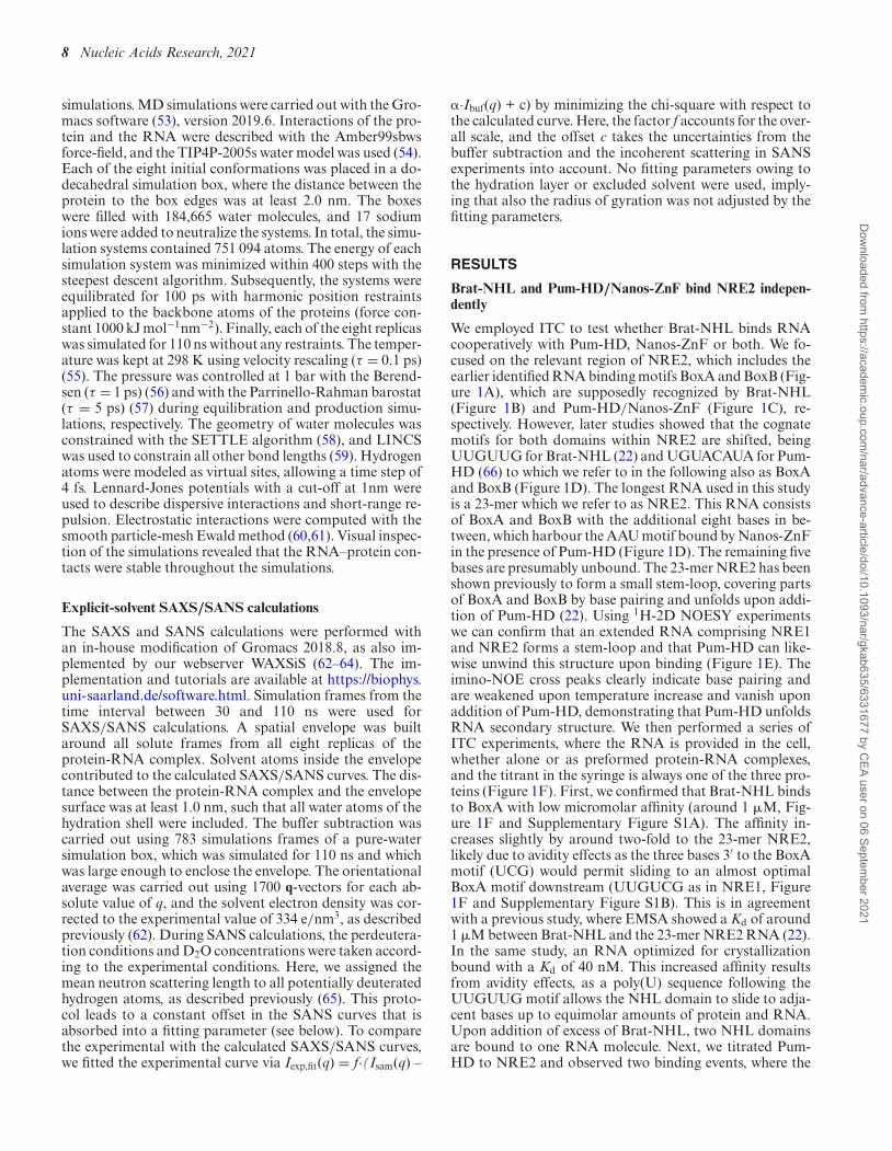