Embed Size (px)
Citation preview

STRUCTURAL STUDIES ON ALKALOIDS
FROM PERIPENTADENIA MEARSII AND
HEDYCARYA ANGUSTI FOLIA ·
by
):r-
"'cf .; -� (°" ':,r-<Y vyY c<v
� (� e Y.A. Geewananda P. Gunawardana B.Sc.(Hons.),
M.Sc. University of Sri Lanka
Submitted in fulfilment of the requirements
for the degree of Doctor of Philosophy
UNIVERSITY OF TASMANIA
HOBART
MARCH., 1983 to ,.be._ \
( co"' f.e , ( cJ l Q 8 4 )
Except as stated therein this thesis contains no material
which has been accepted for the award of any other degree or
diploma in any university, and, to the best of my knowledge and
beli ef, this thesis contains no copy or paraphrase of material
previously published or written by another person, except
when due reference is made in the text of this thesis.
Y.A.G.P. Gunawardana

CONTENTS page
Acknowledgement
Abstract iv
Chapter 1 Introduction.
Alkaloids of the family Elaeocarpaceae 1
Chapter 2 Peripentadenine, the major alkaloid of
Peripentadenia mearsii 10
Chapter 3 NMR spectral assignments of peripentadenine
and its derivatives 31
Chapter 4 Synthesis of peripentadenine and
N[3-(dimethylamino)propyl]hexanamide 53
Chapter 5
Minor alkaloids of the bark extract of
P. mearsii: dinorperipentadenine,
peripentamine andAehydroperipentamine
66
Chapter 6 Constituents of the leaf extract of
P. mearsii: PLM2 and PLM3 90
Chapter 7 Minor alkaloids of uncertain structure 114
Chapter 8 Biogenetic considerations 129
Chapter 9 Experimental for:
Chapter 2 143
Chapter 4
Chapter 5 169
Chapter 6 175
Chapter 7 180
References 185

11
page
Chapter 10
Alkaloids of Hedycarya angustifbZia
191
References
214
Appendix
Alkaloids of Macadamia integrifblia
217

ACKNOWLEDGEMENTS
I wish to express my deepest appreciation and gratitude for
the invaluable supervision, guidance and encouragement given by
Dr. I.R.C. Bick throughout this study.
I am most grateful to Dr. J.A. Lamberton (CSIRO, Melbourne)
for his useful discussions and valuable suggestions and also for the
provision of the bark alkaloid extract and leaves of P. mearsii and
authentic samples of several alkaloids.
I also wish to acknowledge the help given by Drs. A.J. Blackman,
E.J. Browne and J.B. Bremner.
My thanks are likewise due to Prof. A.H. White (University of
Western Australia) for X-ray crystallographic studies, Dr. A.J. Jones
(National NMR Centre, Canberra) for 270 MHz PMR and 13C NMR spectra,
Mr. R. Thomas for recording all the 100 MHz PMR spectra, Dr. G. Lukacs
(CNRS, Gif-sur-Yvette) for 400 MHz PMR spectra and the 13C NMR spectrum
of dehydroperipentamine, Dr. A. Campbell (Otago University, New Zealand)
for microanalysis and Dr. E. Gellert (University of Wollongong) for an
authentic sample of cinnamolaurine.
I am most grateful to Mr. J. Bignall, Mr. M. Power and
Mr. N.W. Davies (Central Science Laboratory) for determining all the
mass spectral data and GC-MS identification of dimethylsulphone,
eudesmol and methyl hexanoate.
•My sincere thanks to Mrs. B. Thomson for carefully and tirelessly
typing this thesis and Mrs. H. Hen for preparing the diagrams.
The financial assistance of a University Postgraduate Research
Scholarship is gratefully acknowledged.
Finally, I would like to thank my wife for her continuous support
throughout this period.

iv
ABSTRACT
A detailed phytochemical investigation of the alkaloids of two
plant species: Peripentadenia mearsii (Elaeocarpaceae) and
Hedycarya angustifblia (Monimiaceae) has been undertaken.
P. mearsii was found to be a rather rich source of alkaloids:
nearly thirty different alkaloids were detected in the bark and leaf
extracts. A total of twenty bases and three non-alkaloidal compounds
were isolated during this investigation.
The structural elucidation of the major alkaloid, peripentadenine,
was carried out by spectroscopic methods and by degradation. Detailed
PMR and 13C NMR spectral assignments of peripentadenine and some of
its twenty derivatives or degradation products are described.
Approaches to the synthesis of EZaeocarpus alkaloids are reviewed.
The structure of peripentadenine was finally confirmed by two different
syntheses.
Structures of three minor bases from the bark: dinorperipentadenine,
peripentamine and dehydroperipentamine were also established.
Dinorperipentadenine was synthesised, and the other two bases were
converted to one of the Hofmann degradation products of peripentadenine.
Tentative structures have been assigned for three other bases
(PBXM2, PLM2 and PLM3) and partial structural analysis was carried
out on two further bases (PBVMD and PLM4).
The identities of the non-alkaloidal compounds isolated were
established by spectroscopy as dimethylsulphone, methylgallate and
2-hydroxy-6-methylacetophenone.
The remaining twelve bases were isolated in minute amounts,
and only their mass spectra were recorded.

Possible biosynthetic origins for Peripentadenia bases have been
discussed. The presence of a three-carbon unit between two
nitrogens was considered to be an interesting structural feature of
these alkaloids and a list of compounds with similar features has
been compiled.
Nine alkaloids including four new compounds were isolated from
Hedycaria angustifoZia. The new compounds include a benzyl-
isoquinoline whose structure was established by X-ray crystallography
(by Professor Allan H. White, University of Western Australia), a
tetrahydrobenzylisoquinoline, a phenanthrene and a dehydraporphine.
The known compounds isolated are all aporphines. Two non-alkaloidal
compounds, 2,3-camphanediol and eudesmol, were also isolated from the
same plant.
The isolation of the alkaloids of Macademia integrifolia was
also attempted but a successful method could not be developed.
CHAPTER 1
Alkaloids of the family Elaeocarpaceae
The history of the elaeocarpaceous alkaloids is a comparatively
short one. Since the isolation of the first alkaloid, (±) elaeocarpine
(I), from a New Guinea species, E. polyductylus Shltr., by Johns and
Lamberton1 , in 1968, nearly 50 new alkaloids have been described from
this family.
The family Elaeocarpaceae consists of 7 genera and about 350
species 2 of which 14 have so far been shown to contain alkaloids.
The alkaloids described belong to two main classes, namely indoles
and indolizidines. The indoles are practically confined to the genus
Aristotelia3, while the indolizidines are found in the genus
EZaeocarpus. An Aceratium species also has been found to contain
alkaloids4
.
During the present investigation of the only Peripentadenia
species, a distinctly different type of alkaloid was encountered.
This type appears to bear some structural and possibly biogenetic
relationship to the indolizidine alkaloids, and therefore this
description will be restricted to EZaeocarpus alkaloids.
Two reviews on Elaeocarpus alkaloids have appeared5,6
. The
alkaloids can be divided into three groups: C16 alkaloids, C12
alkaloids with one nitrogen, and C12 alkaloids with two nitrogens.
In addition to the indolizidines, a single indole alkaloid,
elaeocarpidine (XVIII)7, has also been isolated. For description
purposes the indolizidine nucleus can be visualized as being
substituted at position 8, and the substituent groups include 2-hydroxy-
6-methyl benzoyl and its dihydro derivative, n-butanoyl, and
hydroxybutanoyl. In a majority of compounds, position 7 bears an

2
oxygen which forms a pyran ring system with the 8 substituent. In some
cases this C-7 oxygen has been replaced with a nitrogen. In others
where this third heterocyclic ring is absent, position 7 may bear a
hydroxy function, or may form a C7, C8 double bond.
The variations in the substituents together with the three chiral
centres (C7, C8 and C9) give rise to a whole range of different
compounds. The twenty Elaeocarpus alkaloids described and their
botanical distribution are given in Chart I.
Key to Chart 1.
A - E. altisectus Schltr. 8
B - E. densiflorus Knuth7,9
C - E. doZychostyZus Schltr.10,11
D - E. ganitrus Roxb • 12,13
E - E. kinensis Schltr. 14'15
F - E. poZyductyZus Schltr. 1 ' 16
G - E. sphericus (Gaertn.) K. Schum8'17
3
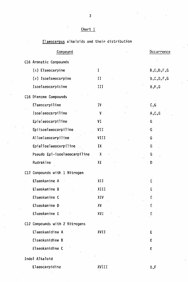
Chart 1
Elaeocarpus alkaloids and their distribution
Compound
Occurrence
C16 Aromatic Compounds
(±) Elaeocarpine
B,C,D,F,G
(±) Isoelaeocarpine
II
B,C,D,F,G
Isoelaeocarpicine III
B,F,G
C16 Dienone Compounds
Elaeocarpiline IV C,G
Isoelaeocarpiline V A,C,G
Epielaeocarpiline VI
Epiisoelaeocarpiline VII
Alloelaeocarpiline VIII
Epialloelaeocarpiline IX
Pseudo Epi-isoelaeocarpiline X
Rudrakine XI
C12 Compounds with 1 Nitrogen
Elaeokanine A XII
Elaeokanine B XIII
Elaeokanine C XIV
Elaeokanine D XV
Elaeokanine E XVI
C12 Compounds with 2 Nitrogens
Elaeokanidine A XVII
Elaeokanidine B
Elaeokanidine C
Indol Alkaloid
Elaeocarpidine XVIII B,F

(±) Elaeocarpine (I)
7R, 8R, 9R
H3
HO
OH
(+) Isoelaeocarpicine (III)
C16 Dienone alkaloids
CHln H v
(+) Elaeocarpiline (IV)
7R, 8R, 9R, 16S
,H30 H• As-
(-) Isoelaeocarpiline (V)
7R, 8S, 9S, 16S
H CH3 O H H
C16 Aromatic alkaloids
(±) Isoelaeocarpine (II)
(-) Epielaeocarpiline (VI)
(+) Epiisoelaeocarpiline (VII)
7S, 8S, 9S, 16S
7S, 8R, 9R, 16S

CH 0
:03 H
a
Alloelaeocarpiline (VIII) 7R, 8R, 9S, 16S
5
(+) Epialloelaeocarpiline (IX)
7S, 8S, 9R, 16S
HO
(+) Pseudoepiisoelaeocarpiline (X) Rudrakine (XI)
C12 alkaloids
Elaeokanine A (XII) Elaeokanine B (XIII)
Elaeokanine C (XIV)

6
H H a H --, 0 H -3 s
CH3
Elaeokanine D (XV)
H
_ N H
CH3
Elaeokanidine A (XVII)
Indole alkaloid
Elaeocarpidine (XVIII)
Elaeokanine E (XVI)

0
7
1.1 C16 Aromatic Alkaloids
The structure of (±) elaeocarpine (I), together with its relative
stereochemistry, was established by X-ray crystal structure analysis'.
The absolute stereochemistry was revealed at a later stage when a
related compound, (-) isoelaeocarpiline (V) was oxidised to S(-)
methyl succinic acid7
.
The structure of other Elaeocarpus alkaloids have been established
mainly from extensive PMR analysis, and confirmed by chemical
transformation into known compounds; in some cases by synthesis.
(-) Isoelaeocarpine (II) was shown to be the C7 epimer of
elaeocarpine (1) by PMR comparison: the coupling constant between
the C7-C8 protons is 11 Hz in (I) where they are trans ( . . C7a, C8B)
as compared to 2.5 Hz in the case of (II), which implies a cis
configuration 16 . The C8-C9 and C1-C9 proton couplings were found to
be the same for both. Further, both (I) and (II) have been shown to
epimerise into a mixture of the two in basic solution. (Scheme 2).
çoH CH30 H
CH3
-H+
Scheme 2
The only phenolic alkaloid, (+) isoelaeocarpicine, was assigned
structure (III) on the basis of its PMR similarity to (II). It was
shown to have the same stereochemistry at C7, C8 and C9 as in (II) and
this was confirmed by the conversion of (+) isoelaeocarpicine into (+)
isoelaeocarpine (II) by dehydration16.

8
1.2 C16 Dienone Alkaloids
Seven isomeric C16 dienone alkaloids have been isolated8
'10
'11
'17
.
They all have a dihydrobenzene nucleus, as seen from the PMR chemical
shift and splitting of the C16 methyl group. The absolute
stereochemistry of C16 has been shown to be the same for all isomers.
(+) Elaeocarpiline (IV) is the C15-C16 dihydro derivative of
(+) elaeocarpine (I) and this has been proved by dehydrogenation of (IV)
to (+) elaeocarpine8 .
(-) Isoelaeocarpiline (V) is the C7 epimer of (VI). (-)
Epielaeocarpiline (VI) differs from (+) elaeocarpiline (IV) in having
the opposite stereochemistry at C7, C8 and C9. Similarly (+)
epiisoelaeocarpiline (VII) differs from (-) isoelaeocarpiline (V) in
having the opposite stereochemistry at C7, C8 and C9, and it is thus
the C7 epimer of (VI) as well. (-) Alloelaeocarpiline (VIII) is the C8
epimer of (-) isoelaeocarpicine (V), and (+) eipalloelaeocarpiline (IX)
has the opposite configurations at C7, C8 and C9 from those of (VIII).
(+) Pseudoeipisoelaeocarpiline (X) has 11,14 double bonds compared
to the 11,13 double bonds in the others. PMR comparison between
(VII) and (X) has shown that (X) has the same stereochemistry at C7,
C8 and C9 as in (+) epiisoelaeocarpiline. Structures of all dienone
alkaloids have been confirmed either by dehydrogenation to the
corresponding elaeocarpine or isoelaeocarpine, or by comparison of
their reduction products with those of elaeocarpine and isoelaeocarpine8
.
A tri-oxygenated C16 alkaloid, rudrakine (XI) has been isolated
from E. ganitrus Roxbs. 12 ' 13 . Its structure was deduced mainly from
mass spectral information and PMR comparison with other Elaeocarpus
alkaloids, but the stereochemistry has not been assigned. It has been
suggested that rudrakine could be the biogenetic precursor of both 11,13
and 11,14 diene alkaloids.

9
1.3 C12 Alkaloids
Eight C12 alkaloids have been isolated from E. kinensis 13,14 • The
structure of elaeokanine C(XIV) was established by PMR comparison
with isoelaeocarpicine (III). The benzoyl substituent in (III) has
been replaced by an a-butanoyl unit in elaeokanine C (XIV). Elaeokanine
A (XII) is a dehydro derivative of (XIV), while elaeokanine B (XII) has
an alcohol function in place of the carbonyl function in (XII).
Elaeokanine D (XV) was characterised by PMR analysis involving
extensive decoupling experiments. Its structure and stereochemistry
can be compared with thatfor rings B,C and C of elaeocarpine (I).
Elaeokanine (XVI) (16) is the 7 epimer of (XV) 14 Further proof for
these structures has come from several syntheses, which will be
discussed at a later stage.
Three more alkaloids with two nitrogen atoms in the same molecule
also have been isolated. The structure of elaeokanidine A (XVII) was
established by PMR comparison with elaeokanine D (XV). Structures
for the remaining two stereoisomers have not been assigned because of
the complexity of their HIR spectra14
.
The structure of the only indole alkaloid, elaeocarpidine (XVIII)
was assigned from spectral evidence and degradation7,9
and confirmed
by synthesis18,19,20

10
CHAPTER 2
Peripentadenine, the major alkaloid of Peripentadenia mearsii
2.1 Results and Discussion
P. mearsii (C.T. White) L.S. Smith belongs to the monotypic
genus Peripentadenia and is a tree growing in rain forests of north
Queensland. A previous examination of P. mearsii for alkaloids
yielded tropane bases21
. This finding could not be repeated
subsequently and evidently the tropanes had come from some other
plant, so far unidentified 22 .
The present investigation was carried out on authentic material
that has been checked against herbarium voucher specimens. On
extraction by standard procedures, bark and leaves gave crude alkaloid
extracts in 0.62% and 0.19% yields respectively. Both these extracts
were found to be extremely complex mixtures on analytical tic s by
which a total of nearly 30 different compounds were detected after
developing with different solvent systems and spraying with iodoplatinate23
reagent. These extracts were first fractionated by column chromatography
and the subsequent fractions were separated by repeated preparative thin
layer chromatography. A total of 23 pure compounds were isolated.
The major alkaloid, peripentadenine (I), constituted about 18% of both
leaf and bark extracts. Three other alkaloids made up about 6%,
while the remaining 20 odd minor compounds constituted only about 20%
of the total crude extracts. The complexity and limited quantity of
the extracts available made the purification of the minor bases a
difficult and tedious process.

11
2.1 Structural Elucidation of Peripentadenine (I)
The major base from both leaf and bark extracts was isolated by
column chromatography as a brown oil. The compound appeared to be '
chromatographically pure when developed with several solvent systems
but no crystalline derivative could be prepared. The formula
C22H34N 204 was derived by high resolution mass spectroscopy for the
oil, and a borohydride reduction product (IVA or B) obtained at a
later stage as platelets analysed for C 22H36 N 204 .
On mild basic hydrolysis, or on prolonged storage in solvent,
peripentadenine (I) gave a complex mixture of bases and 2-hydroxy-6-
methyl acetophenone16
(VI). Even though the extraction procedure is
designed to remove any non-basic material,substantial amounts of (VI)
were isolated from both leaf and bark extracts, and this represents
most probably a decomposition product of alkaloids bearing this
moiety. The presence of an acetophenone residue in peripentadenine
could be deduced from the spectral chracteristics: strong IR
absorptions at 1690 and 3200 cm-1
corresponding to an aromatic carbonyl
and a hydroxyl, an ABC aromatic proton pattern in the PMR spectrum
(6 7.15, 1H, t; d 6.67, 1H, d; 15 6.63, 1H, d) 16 , also six aromatic
(6 157.8, s, C6; 137.2, s, C7; 132.4, d, C4; 127.9, s, C2; 121.5,
s, d, C3 and 116.5, d, C5) and one downfield carbonyl carbon (6 207.9,
s, C8) signals in the 13
C NMR spectrum. Its PMR spectrum also showed
a broad exchangeable downfield signal (6 10.5) for one proton, and a +ve
Gibbs24
test indicated a free p-position to a phenolic function.
Further, peripentadenine formed a monoacetate (II) on room temperature
acetylation, and a monomethyl ether on treatment with diazomethane: this
also showed that the acetophenone moiety is linked to the rest of the
molecule through its acetyl carbon.
The PMR spectrum showed another broad exchangeable one-proton

12
signal at 6 5.75. When this proton was exchanged with D 20 or
irradiated, a methylene signal at 6 3.15 simplified to a triplet.
These observations are indicative of a primary amide function: -CH 2NHCO-.
-1 IR absorptions at 1680 and 1650 cm and a 13C signal for a quaternary
carbon at 6 173.6 further supports the presence of this functionality.
The methylene proton signal at 6 2.05, a sharp triplet, was assigned to
the protons a to the amide carbonyl function. A series of decoupling
experiments: 6 2.05, 211, t; -4-+ 2.6, 211, txt; 1.8, 4H, m; 4-4- 0.85,
3H, t; showed that the amide carbonyl is attached to a five-carbon
paraffinic chain. This inference was further supported by prolonged
acid hydrolysis which yielded an amine (VII) and hexanoic acid (VIII).
This acid was identified by GLC-MS comparison of its methylester with
an authentic sample of methyl hexanoate (IX).
The remaining carbons of (1), apart from those in the acetophenone
and hexanamide residues, are all aliphatic and comprise one methine
and seven methylene carbons, as shown by the 13C NMR spectrum. The
Second nitrogen is presumably present in a tertiary amine group, since
quaternisation of peripentadenine with methyl iodide introduced only one
methyl group. From these data and the analysis of the molecular formula
it is evident that the amine group is present in a heterocyclic ring.
The 13C NMR signal at 6 64.7 for a methine carbon suggested that the
heterocyclic ring is a-substituted: from the previous evidence the
substituent would appear to be the aroylmethyl group.
The mass spectrum of peripentadenine showed three prominent
fragments. The fragments at m/z 224 and m/z 150 are formed by a
McLafferty type of cleavage25
of the bond a to the aromatic carbonyl
group. The third fragment at m/z 156 is a result of an S N2 type
cleavage of the bond a to the amine nitrogen giving an oxazine-type
ion (Scheme 1). This type of mass spectral fragmentation appears to
be characteristic of the N[3(amino)propyl] amide function26.

IVA/IVB R = H
VA/VB R = CH 3
CH
H3
OH
13
I R = H Peripentadenine
II R = COCH 3
III R = CH 3
VI
RO
VII
VIII R = H
IX R = CH 3

t I
OH
H
m/z 374
S N2 type
N./
m/z 156
m/z 150 m/z 224
1 McLafferty
14
Scheme 1
15
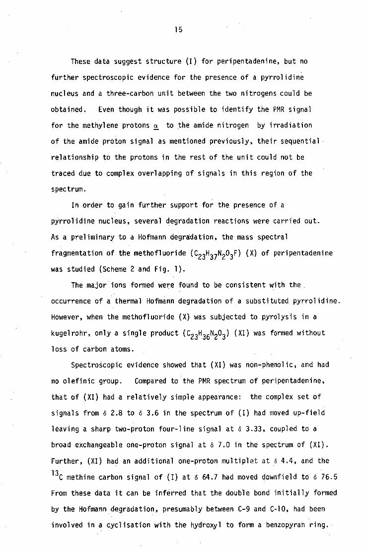
These data suggest structure (I) for peripentadenine, but no
further spectroscopic evidence for the presence of a pyrrolidine
nucleus and a three-carbon unit between the two nitrogens could be
obtained. Even though it was •possible to identify the PMR signal
for the methylene protons a to the amide nitrogen by irradiation•
of the amide proton signal as mentioned previously, their sequential
relationship to the protons in the rest of the unit could not be
traced due to complex overlapping of signals in this region of the
spectrum.
In order to gain further support for the presence of a
pyrrolidine nucleus, several degradation reactions were carried out.
As a preliminary to a Hofmann degradation, the mass spectral
fragmentation of the methofluoride (C 23H37N 203 F) (X) of peripentadenine
was studied (Scheme 2 and Fig. 1).
The major ions formed were found to be consistent with the
occurrence of a thermal Hofmann degradation of a substituted pyrrolidine.
However, when the methofluoride (X) was subjected to pyrolysis in a
kugelrohr, only a single product (C 23H 30203 ) (XI) was formed without
loss of carbon atoms.
Spectroscopic evidence showed that (XI) was non-phenolic, and had
no olefinic group. Compared to the PMR spectrum of peripentadenine,
that of (XI) had a relatively simple appearance: the complex set of
signals from 6 2.8 to 6 3.6 in the spectrum of (I) had moved up-field
leaving a sharp two-proton four-line signal at 6 3.33, coupled to a
broad exchangeable one-proton signal at 6 7.0 in the spectrum of (XI).
Further, (XI) had an additional one-proton multiplet at 6 4.4, and the
13C methine carbon signal of (I) at 6 64.7 had moved downfield to 6 76.5
From these data it can be inferred that the double bond initially formed
by the Hofmann degradation, presumably between C-9 and C-10, had been
involved in a cyclisation with the hydroxyl to form a benzopyran ring.

m/z 135 m/z 156
16
- HF
(X)
m/z 388
From a:
m/z 246 m/z 58
From b:
CH 3
M/z 201
m/z 199
From c: H2 C 0
CH ,J*L, 3 N HN
1\.) m/z 225
CEO
OH
Scheme 2

17
•
•CI-C1L1
LC)
LS)

• CH 2 j
CH2 +
CH 1 m/z 84
CH 4. CH
3 '1%1 2
CH 3
m/z 58
18
X
XI
XII
m/z 201, m/z 199 (cf. Scheme 2)
CH H 0
CH 3
From a:
From b:
m/z 156 (cf. Scheme 2) M/z 246
From c:
From a or b:
+.
m/z 160
H 3 + CEO
;
OH
m/z 135 5
0
Scheme 3

19
The absence of other products can be attributed to the directional
properties of the carbonyl group in the 0-amino keto system.
The methofluoride (XII) formed by the quaternisation of (XI)
gave a mass spectrum consistent with the benzopyran structure proposed
for (XI) containing a C3 unit between the two nitrogens (Scheme 3).
However, when the methofluoride (XII) was pyrolysed under similar
conditions to those used for (XI), a complex mixture was obtained
from which no identifiable product could be isolated, evidently
because the directional effect of the carbonyl group no longer
applied.
Presumably for the same reason, Hofmann degradation of the
methofluorides of the two isomeric lithium aluminium hydride reduction
products (XIII A and B) of (I) likewise gave complex mixtures which
could not be separated. Emde degradation of (I) also failed to give
any useful information.
In order to provide an alternative orienting effect for a two-stage
Hofmann degradation, it was decided to introduce a C8-C9 double bond
by reduction of the aromatic carbonyl function followed by dehydration.
The lithium aluminium hydride reduction products (XIII A and B) could
not be used for this purpose as the secondary amino group could lead
to several Hofmann degradation products.
Borohydride reduction of (I) gave a complex mixture from which a
pair of optically inactive epimeric alcohols (IV A and B) were
isolated. Both XIV A and B have identical spectra except for the
difference in the PMR signals for the C8 proton: in XIVA this signal
appeared as a dd (J = 11.3 and 2.5 Hz) at (S 5.53 while in B it appeared
as a dd (J = 10.9 and 2.3 Hz) at s 5.30. One of these alcohols (XIVA)
deposited white plate-like crystals from acetone. Both alcohols on
dehydrogenation under mild conditions (refluxing 10% ay. oxalic acid),
gave the same compound (XIV).
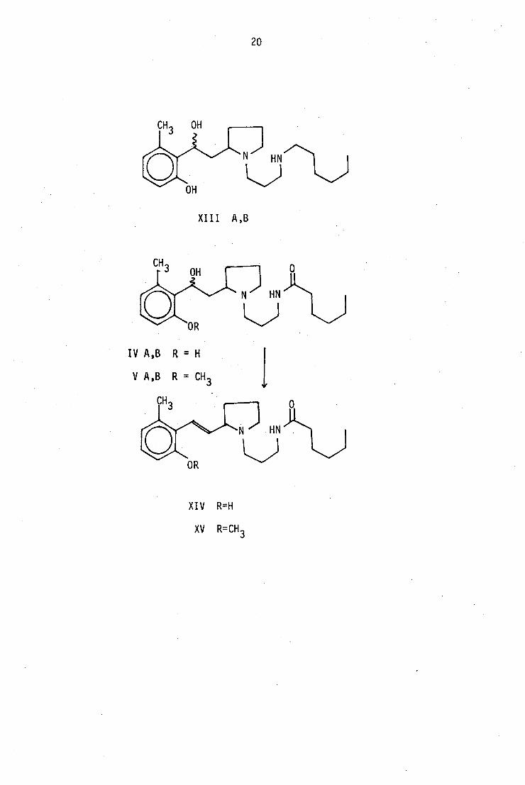
OR
IV A,B R = H
V A,B R = CH 3
CH3
OR
20
XIII A,B
XIV R=H
XV R=CH 3

1) Mel CH
3
2) F-
3) A
HN
CH3
OCH 3 1) Mel
2) F-
3) A 0
CH
3 HN)C
CH
XV
X V I
XIX
.■•■.
21
However, (IV ) was not used for the Hofmann degradation to avoid
the problem of cyclisation induced by the hydroxy group, thereby
removing the orienting effect of the double bonds in subsequent
Hofmann degradations. Instead, the corresponding 0-methylether (XV)
prepared through the alcohols (V A and B) was subjected to further
degradations (Scheme 4).
Scheme 4
On pyrolysis, the methofluoride of (XV) gave a single product
which was identified as the diene (XVI) from its UV absorption
pattern: 218 and 308 mm and PMR signals for four olefinic protons:
6 6.78 (d, J = 14), 6 6.55 (1H, dd; J = 16, 14), 6 6.25 (1H, dd,
J = 16, 7.5), 6 5.75 (1H, m). The methofluoride of (XVI) gaye
two main Hofmann degradation products: the more polar, basic,

22
nonvolatile compound was identified as N-[3(dimethylamino)propyl]
hexanamide (XVII)from its spectral data and confirmed by synthesis
(discussed in Chapter 3). The complementary fragment, the triene (XVIII),
could not be isolated as such, and it appeared to undergo cyclisation
to give the cyclohexadiene derivative (XIX) under the conditions used.
The fully aromatised analogue, 2-methoxy-6-methylbiphenyl (XX) has
been reported27
, but the small amount of (XVIII) available and the
lengthy procedure for the preparation of (XXII) did not permit the
confirmation of the structure (XIX) by comparison with a known compound.
This series of degradation reactions confirms the pyrrolidine
nature of the heterocyclic ring and demonstrates that the two nitrogens
are separated by a C3 unit: hence the structure of peripentagenine is
N{3[2(2-hydroxy-6-methoxybenzoylmethyl)pyrrolidin-l-yl]propyl}hexanamide
(I).
The structure deduced for peripentadenine has a chiral centre, but
the base isolated from the plant material, as well as the reduction
products (IV and V), have negligibly small specific rotations. The
chiral centre in hygrine (XXI), , which%presents some structural analogy
to (I), is known to be readily racemised under basic conditions such as
occur during the usual extraction procedures28
.
XX
XXI

23
2.2 Reactions of peripentadenine
2.2.1 Permanganate oxidation
The treatment of a solution of (I) in acetone with a freshly
prepared solution of permanganate followed by the usual work-up
yielded two fractions: an acidic fraction which produced white
crystals, and a neutral gum. The acid was identified as 2-hydroxy-
6-methyl benzoic acid by comparison with an authentic sample. The
gum analysed for C13H24N202 by high resolution mass spectrometry.
Its IR spectrum had a broad absorption at 3300 cm two
carbonyl absorption bands at 1650 and 1640 cm -1 . The PMR spectrum
showed the presence of the N(propyl)n-hexanamide moiety whose
signals could easily be detected by comparison with the PMR spectrum
of (I). The mass spectral fragmentation (Figure 2 and Scheme 5)
indicated the presence of an N-substituted 2-pyrrolidone, the
substituent being the N(propyl)hexanamide moiety as shown by its
PMR spectrum. This compound was thus identified as N[3(pyrrolidin-2-one)
propyl]hexanamide (XXIII).
OH
XXII
XXI I I

24
(.0 C \ r-i
+
Cv1
• 2 01
+ •••••(....)
0
r-4
00 01
C
+
1-1
C:)

-CO
0 +N)
II CH 2
m/z 98
L+I N CH
m/z 70 2
N HN
1\) m/z 240 McLafferty + H
i transfer
HO
m/z 86
m/z 197
m/z 126
1-CH3 CH2
CH2 = N=C=O
m/z 56 m/z 56
-CHO
— NH
m/z 56
25
- 1+ CEO
-NH 2
CH. 2 CH 2
L CHO Ls+ /CHO N
Scheme 5

./J
OCOCH 3 1
COCH3
26
2.2.2 Acetylation at elevated temperature
On room-temperature acetylation of (I), the above-mentioned
monoacetyl derivative (II) was formed, but when the acetylation
was carried out at 100 0 , a number of products were formed. Five
of these compounds have been identified.
The PMR spectrum of the least polar compound had two acetate
signals at (52.25 (ArCOCH 3 ) and 62.1 (NHCOCH 3 ), and an additional
signal at (56.4 for two olefinic protons. Further, the complex
set of signals for the protons on the pyrrolidine nucleus in (I)
appeared to be simplified and shifted slightly up-field. This
evidence, together with its molecular weight (460) and its mass
spectral fragmentation is consistent with a product formed by the
opening of the pyrrolidine ring (Scheme 6), and the structure (XXIV)
is assigned to this compound.
XXV R = H
XXVI R = COCH3

• COCH 3
OCOCH 3
27
The PMR and mass spectral analysis of two other products showed
that they were further acetylated derivatives of(XXIV). The
structures (XXV) and (XXVI) are assigned to these two acetates.
XX IV
XXVI
The fourth compound was identified as a transacylated product
(XXVII) by PMR and mass spectral analysis.
XXVII

28
2.2.3 Borohydride reduction
The basic fraction of the borohydride reduction products of (I)
yielded two epimeric alcohols (IV A and 13), a third compound in small
amount, and two others in extremely minute quantities. Only the first
three were available in sufficient amount to obtain satisfactory
PMR spectra, but the mass spectra of all three minor compounds had
molecular ions at m/z 376, which suggested they were isomeric with the
major alcohols. Several compounds that could be formed under the
conditions used for the borohydride reduction have been postulated
(Scheme 7), but none of them seem to fit the PMR spectrum of the
predominant minor compound.
On careful examination of the PMR spectra of the two major
alcohols (IV A and 13), apart from the differences in the PMR signals
associated with the epimeric centre, several chemical shift and
splitting pattern differences for protons remote from the epimeric
carbon were also observed. (Detailed analysis of these spectra appear
in Chapter 3). The observations suggest the presence of some
kind of conformational difference, probably caused by strong hydrogen
bonding with the phenolic hydroxy function, in addition to the
stereoisomerism around the two chiral centres. In view of this
possibility the minor isomer in question can be considered as a third
conformational isomer. The absence of such minor isomers in the
borohydride reduction products of 0-methylperipentadenine (III) is
consistent with this assumption.
The neutral fraction from the borohydride reduction of (I) was
not further investigated, but the same fraction from (III) yielded
2-methoxy-6-methyl acetophenone (XXVII). The mass spectral analysis
of a minor base isolated from the reduction of (III) revealed it to be
the complementary fragment (XXVIII) of XXVII (Scheme 8).
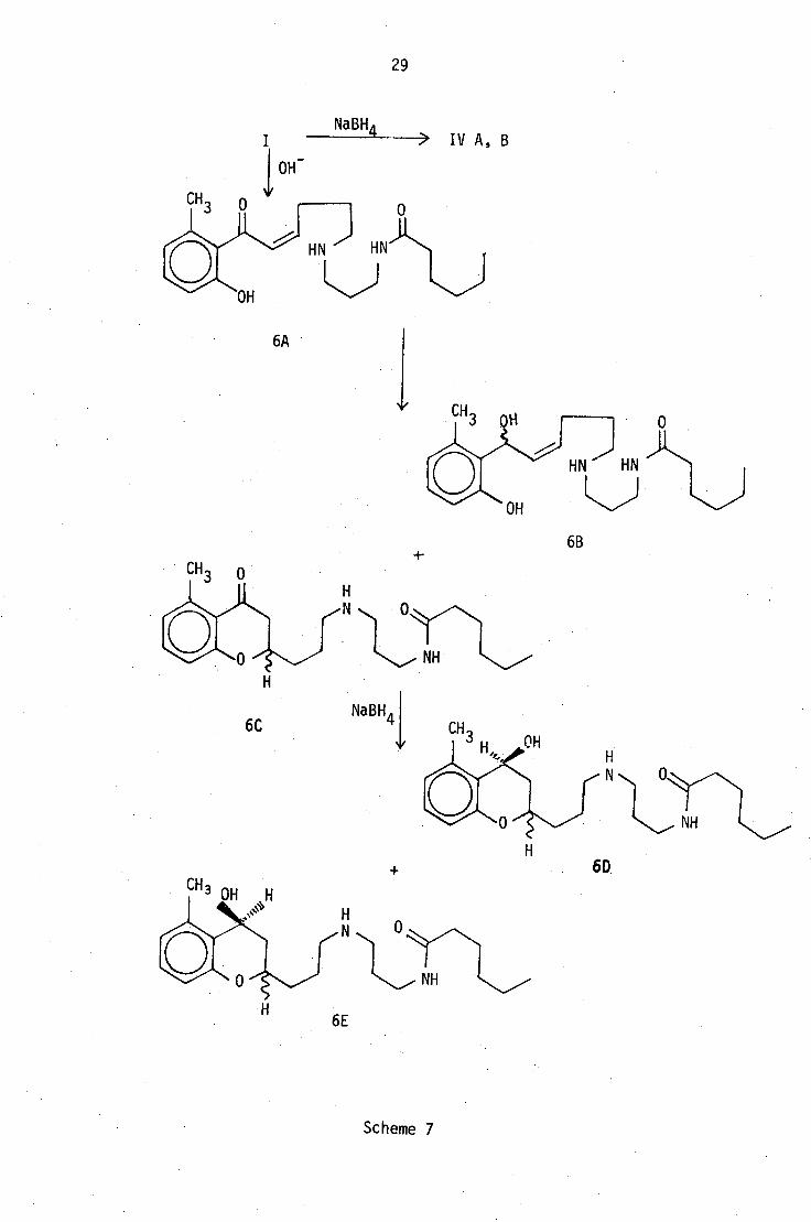
6C
N y•N
NH
NaBH41
29
NaBH4 IV A, B
OH -
6A
6B
6D
Scheme 7

30
III
CH3
XXV II
Na8H4 °I
N HN
)
OCH 3
XXVIII I NalIVIA_
XXIX
Scheme 8

31
CHAPTER 3
3.1 PMR spectral assignments of peripentadenine and its derivatives
In the preceding discussion only the salient spectral features
which were relevant to the structural elucidation of peripentadenine
were considered. It would be much more instructive if all spectral
information could be assigned, as this would facilitate the structural
elucidation of the minor bases isolated.
As mentioned earlier, the assignment of the PMR signals for the
protons attached to the (pyrrolidin-1-yl)propyl unit of
peripentadenine had not been feasible, mainly due to overcrowding
and complex coupling in the relevant regions in the 270 MHz spectra.
However, the subsequent isolation of simpler degradation products
has shed more light on this problem.
3.1.1 N-[3-(dimethylamino)propyl]hexanamide (I)
The title compound was the simplest of the basic degradation
products obtained from peripentadenine, and accordingly it has the
most easily interpreted PMR spectrum (Figure 1). The exchangeable,
one-proton signal at 57.0, and the two-proton doublet of triplets
at 63.34 have already been identified as the signals for the -CONHCH 2 -
system. Analysis of coupling constants showed that the signal at
63.34 and the two-proton , triplet at 62.45 are coupled to the two-
proton signal (triplet of triplets) at 61.69, and this confirms that
the latter two signals are due to the protons a and y to the amide
nitrogen respectively. The remaining signals at 62.16 (2H, t J = 7.3),
61.63 (2H, tt, J = 7.3), 61.31 (4H, tq, J = 7.3) and 60.87 (3H, t, J = 7.3)

32
CD
.2- •
CY) cf,
m
•N •-•1 C..)
LO •
— CD
C
cu
•r-
Cs..1
LO
C
LC) LAI
LAC) kg)
I I I I
=

33
also have been identified as the signals due to protons attached to
the C5 parafinic chain by decoupling experiments carried out earlier
on peripentadenine. It is interesting to compare these chemical
shifts with those of some of the known simple amines and amides
(Figure 2).
NH -CH2
-CH -CH2
-NH2
Ref. 29
1.6 2.6
CO-NH-CH2
-CH2
-CH2
-CH2
-NH2
3.5 1.8 1.6 2.75
Ref. 29
0 -NH-CO-NH-CH2—CH
2—CH
2-CH
3
6.53 3.23 1.4
Ref. 30
The accompanying figures indicate the chemical shifts (6) of
the protons.
Figure 2
3.1.2 ihrthydro-derivative of dihydroperipentadenine (II)
The signals for the pyrrolidine nucleus can easily be distinguished
in the PMR spectrum of the title compound as these, together with the
singlet for the aromatic methyl protons, are the only up-field signals
present, apart from the signals whose origin has already been traced
from the spectrum of the previous compound (I). The one-proton
multiplet at 63.37 is assigned to the proton at C-10, while the two-

.1■11,
-1"
LI)
—
Cr)
Lf)
vZ1"
34 LC)
CV"
N. tr>
.1•1111■■•
-
— LC)
LC)
LC)

35
proton multiplet at 63.08 and the six-proton multiplet at 61.65-1.95
are assigned to the C-13 and the C-11, 12 and 16 methylene protons
respectively (Figure 3). However, the two olefinic protons,
particularly C-9H, appeared to be shifted downfield more than
expected, and their signals overlap with those of the three aromatic
protons. By way of comparison, in the alkaloid norruspoline31
(III)
the corresponding olefinic protons appear at 66.45 and 66.07.
1.4-2.2
HO
CHO
6.45
H 6.07 \
(dd, j = 16,7)
2.85
III norruspoine31
On the other hand, when the phenolic 'hydroxyl is methylated (IV)
the olefinic protons move up-field to 66.55 (J = 16, C-8H) and 66.0
(J = 16, 7.5, C-9H) (Figure 4). The coupling between C-8H and C-9H
(16 Hz) confirms the trans-substitution, and the coupling between C-9H
and C-10H (J = 7.5 Hz) is comparable with that between the
corresponding protons in norruspoline (J = 7 Hz)31
.
3.1.3 Hofmann degradation product of (III)
The assignment of the signals is shown in Figures 5 and 6.
With the introduction of a second double bond, the signal for the
olefinic proton at C8 has moved downfield, and overlaps with the

36
aromatic proton signals. The complex set of signals between 62.0-2.4
were assigned to the methylene protons at C12 and 13, but their
splitting pattern could not be established owing to second order coupling.
3.1.4 Peripentadenine (V)
Only the methylene protons a to the aromatic carbonyl function
in peripentadenine lack a parallel in the compounds so far discussed,
and as a result the PMR signals for these protons (one-proton double
doublets at 63.47 and 62.52: Figure 7)can be identified quite
easily. The downfield shift of one of them (63.47) compared to the
chemical shifts of the corresponding protons in 0-hydroxyacetophenone
(62.55) and 2-hydroxy-6-methylacetophenone (62.4) could be ascribed to
three different factors: a) ring current effect b) hydrogen bonding
with the phenolic hydroxy and c) diamagnetic aniosotropic deshiel ding
by the amide carbonyl function. As the first two factors can operate
in acetophenone derivatives themselves, the third appears to be the
effective factor in this particular case. Complete assignment of
the PMR signals for peripentadenine, as confirmed by extensive
decoupling experiments, is given in Table 1.
2.5
./J 1.8 1 ' 3
N N 2.15 1.3
Figure 8
1.8 j
2.5 NH2
2.5 2.7
1.8
0.9

12
13 HN 19
18 2 17 1
24
23 22
16
V
37
TABLE 1
PMR signal assignment of Peripentadenine
Proton/s at Chemical shift/6
Multiplicity Coupling constants/Hz
C-4 7.2 dd J3,4
= J4,5
= 7.5
C-3 6.72 d J3,4
= 7.5
C-5 6.67 d J4,5
= 7.5
N-H 5.72 m
C-10 3.58 dddd 9A,10 = 10 J 98,10
= 5
J 10,11A = 8.3 J
10, =5
C- 9A 3.47 dd JAB = 12.75
9A,10 = 10
C-15A
3.27 ddd JAB = 10.5
J 15A'
16A
= 6.3 J15
A'16
B = 4.5
C-17A
3.23 dd JAB
= 13.5 317'1018
= 6
C-17 B 3.17 dddd J AB = 13.5
J 17B'
18 = 6.0
J 16 17 = 8.25 J A' B 16B'
17 B = 6.
C-13A 2.85 ddd JAB = 12
C-9 8 9 B dd
J 13A' 12A = 7.5 J
13A' 12B
=
JAB
= 12.75
3.
j 98,10 = 5
13 8 2.45 dd JAB =
12

38
TABLE 1 continued
15B
2.43 ddd JAB
= 10.5
J 15 B 16A = 8.5
J 15
B'16
B = 7.5
1 2.3 s
11A
2.12 ddd JAB = 12
J = 10,11A 8.3
J 11
A'12
A = 8
20 2.05 = 8.25 J 20,21
16A 1.92
168 1.72 m
11B 1.7 m
12 1.65 m
21 1.57 txt = 8.25 J21,22 22,23 1.2-1.3 m
24 0.9 t J23,24 = 8.25
Compared to the chemical shifts of the protons on simple
(pyrrolidin-l-yl)propyl compounds (Figure 8) encountered during the
synthesis of peripentadenine, a considerable degree of variation in
chemical shifts for the same protons in peripentadenine was
observed (Figure 9).
As there are no functionalities directly attached to the
pyrrolidine nucleus that can cause such changes in chemical shifts,
the changes have to be ascribed to more remote functional groups.
To satisfy this condition, even though the peripentadenine molecule
appears to be quite flexible, it must be held in a more or less rigid
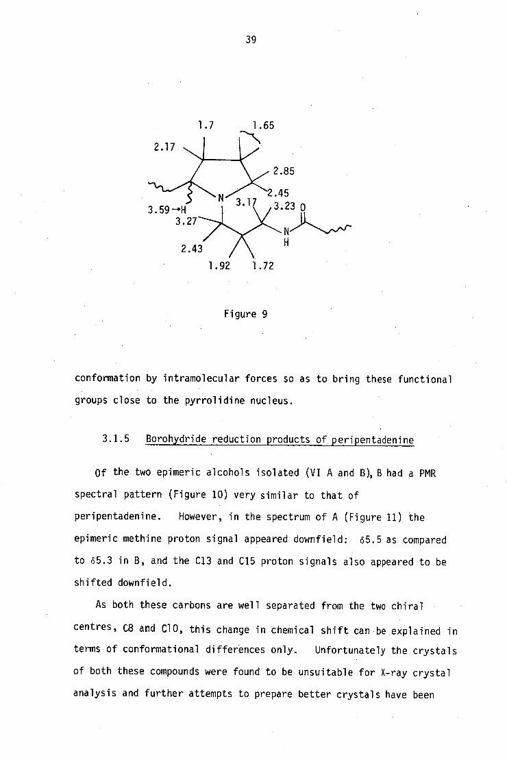
39
Figure 9
conformation by intramolecular forces so as to bring these functional
groups close to the pyrrolidine nucleus.
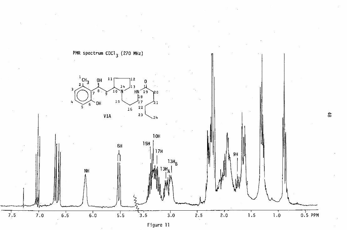
3.1.5 Borohydride reduction products of peripentadenine
Of the two epimeric alcohols isolated (VI A and B),B had a PMR
spectral pattern (Figure 10) very similar to that of
peripentadenine. However, in the spectrum of A (Figure 11) the
epimeric methine proton signal appeared downfield: 65.5 as compared
to 65.3 in B, and the C13 and C15 proton signals also appeared to be
shifted downfield.
• As both these carbons are well separated from the two chiral
centres, C8 and C10, this change in chemical shift can be explained in
terms of conformational differences only. Unfortunately the crystals
of both these compounds were found to be unsuitable for X-ray crystal
analysis and further attempts to prepare better crystals have been

40
unsuccessful. Owing to the flexibility of the aliphatic side chain,
it was difficult to decide from models which orientations could most
satisfactorily account for these differences.
3.1.6 Hofmann degradation product of Peripentadenine (VII)
Apart from the presence of a few additional signals, the
aliphatic region of the spectrum of this compound resembles that of
the amino amide (I) described earlier. Opening of the pyrrolidine
ring by Hofmann degradation has removed most of the complex coupling
present in (V), thereby making the spectrum much simpler (Figure 12).
The proton on the carbon bearing the ether oxygen, C-10H, appears as
a multiplet at 64.4, while the methylene protons a to the aromatic
carbonyl appear at 62.67 as a three line complex signal, due to
virtual coupling. Similarly the C-11, C-12 methylene protons which
appear at 61.7-1.9 also give a complex multiplet.
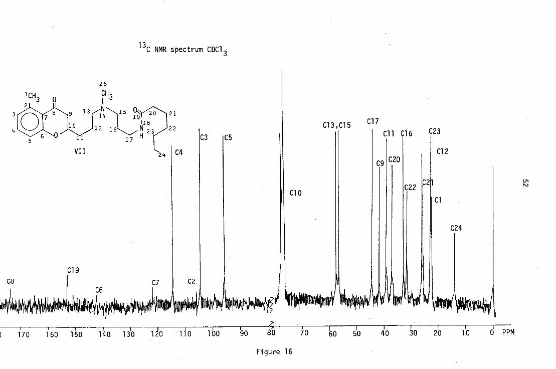
3.2 13C NMR spectral assignments
The 13C spectra of peripentadenine (V) and its two derivatives,
the dehydration product of dihydroperipentadenine (II), and
the Hofmann degradation product (VII) were recorded. The aromatic
carbon signals were assigned by comparison with calculated values32
.
(Table 2).
3.2.1 Peripentadenine (V)
The two downfield quaternary carbon signals at 6207.3 and 6173.6
were assigned to the aromatic and amide carbonyl carbons respectively.
The only up-field methine carbon signal at 664.7 was assigned
to C-10. Its chemical shift appears to be high in comparison to
those of related compounds (Figure 13), but since C-10 is the only
2
3
4
5
6
7
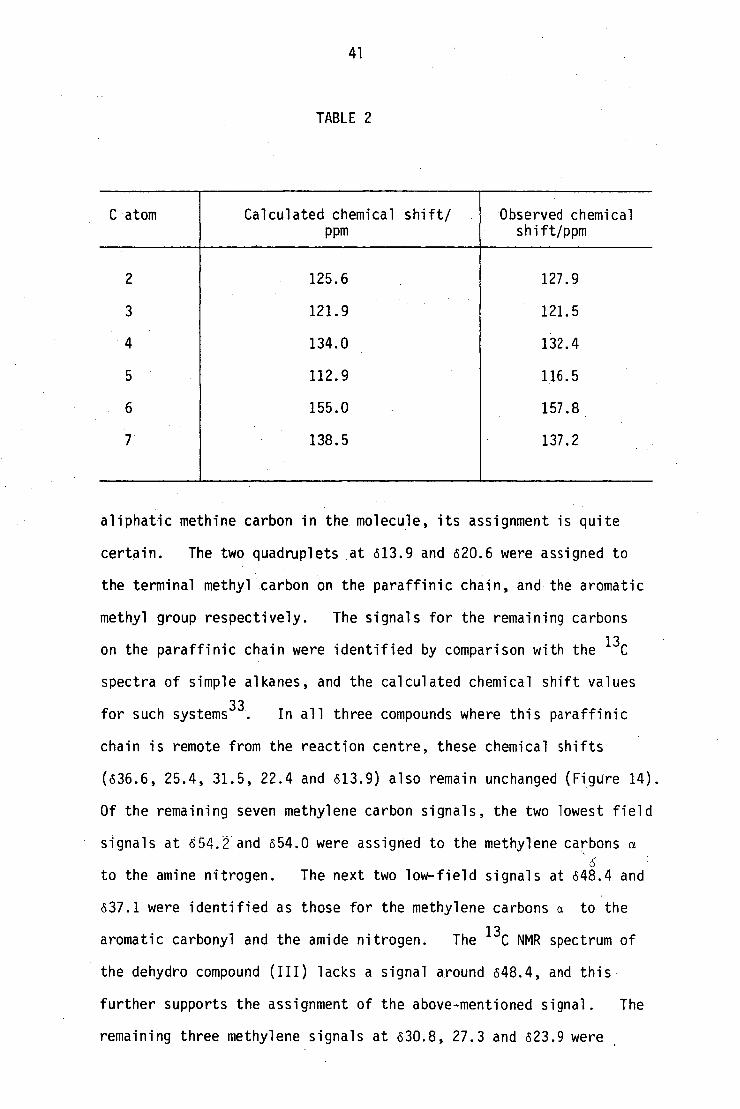
125.6
121.9
134.0
112.9
155.0
138.5
127.9
121.5
132.4
116.5
157.8
137.2
C atom Calculated chemical shift/
PPm
Observed chemical shift/ppm
41
TABLE 2
aliphatic methine carbon in the molecule, its assignment is quite
certain. The two quadruplets at 613.9 and 620.6 were assigned to
the terminal methyl carbon on the paraffinic chain, and the aromatic
methyl group respectively. The signals for the remaining carbons
on the paraffinic chain were identified by comparison with the 13
C
spectra of simple alkanes, and the calculated chemical shift values
for such systems33
. In all three compounds where this paraffinic
chain is remote from the reaction centre, these chemical shifts
(636.6, 25.4, 31.5, 22.4 and 613.9) also remain unchanged (Figure 14).
Of the remaining seven methylene carbon signals, the two lowest field
signals at 654. and 654.0 were assigned to the methylene carbons a
6 to the amine nitrogen. The next two low-field signals at 648.4 and
637.1 were identified as those for the methylene carbons a to the
aromatic carbonyl and the amide nitrogen. The 13
C NMR spectrum of
the dehydro compound (III) lacks a signal around 648.4, and this
further supports the assignment of the above-mentioned signal. The
remaining three methylene signals at 630.8, 27.3 and 623.9 were .

32.4 1
56.18.1
32.3 25.2
24.6
26.1
53.1
20.2
CH3 127.1 0
138
32.1
207.5
H\20 6
156.6 65.4
VII isoelaeocarpicine
113.4
27.7
46.6 121.1
131.4
31.8
42
assigned to the C-11, C-16 and C-12 carbons by comparison with the 13C
spectra of similar compounds.
The 13C NMR spectra of all three above-mentioned compounds with
their complete assignments appear in Figures 14-16.
13.1 39.1
norruspoline31
VI Coniine
VIII 3,4-dimethoxy w(2-piperidyl)acetophenone
The 13
C NMR spectra of VII and VIII were recorded on samples provided
by Dr. J.A. Lamberton.
Figure 13

43
LC)
'.0
IC) Ln
U,

OCH 3 I V
PMR spectrum CDC1 3 (270 MHz)
8H
CH
, 0 3 14 15
12 16 22 18 17
1
CHJ
÷ J8,9, 7 16
10H 11H
j9,10
= 9 4. J10,11
J8,9 = 9
J
= 15 J10,11
J11,12 . 7
fr
5H
9
4H
21
24
23
1 I I I I I I I I 1 1
7.1 7.0 6.9 6.8 6.7 6.5 6.4 6.3 6.2 6.1 6.0 5.9
5.8 5.7
Figure 5

45
✓-4
C'4 C
l. 0
.•
UN
.
Cr)
•= —
•all1■
11 ,
C \
Ul
LC)

0, 9HB
10Hd
))14kJJ
1;5.HB 17H 13HA
13HB
,k4
1H 41* 67)
11 HA
20H 22,23H
12H 21H
16H ,11H 16H
CDC1 3 PMR spectrum CDC1 3 (270 MHz)
23 2. 1+
7.0 6.5 6.0 5.5
3.5
3.0 2.5
2.0 1.5 1.0 PPM
Figure 7

PMR spectrum CDC1 (270 MHz)
11 CH IH
2
OH
VIB
16
17 21 221
23c 24
12 0
13
HN 19 20 18
1
8H
10H 15HB
13HB 13HA I
17H
15HA
1H
24H 20H
21H
I 9
7.5
7.0
6.5
6.0
5.5
3.0
2.5
2.0 1.5
1.0
0.5
Figure 10
01
48
r•-•
C".1
PMR spec trum C DC 1 3 ( 270 MHz )
•••■■
• ve
"
LO
Cfl
LO
—
c.)
c.) —1
4.0

■•■•••■•
(71
49
<
PMR spec trum
LC)
C7)
)■1- C•1 • C1.1
cu

50
LD
C \ I • r—
C\J
C \
CD
C \
C \ I L.)
—C
)
■i■
—
LC)
—
CD
r-
CD
-
er)
-cd-
-LO
r-
LC)
-rss
—CO
s—
-C
))
-C \ I
CO
L.)
CD
-rs..1

C■J (-)
51
re)
,••• C
C)
C.)
CN.1
CD
Li
CO
c_) ce)
CD
9-1
C21
52

53
CHAPTER 4
Synthesis of peripentadenine and N-[3-(dimethylamino)propyl]hexanamide
4.1 Synthesis of eZaeocarpus alkaloids
EZaeocarpus alkaloids have aroused considerable interest among
synthetic organic chemists, as seen for instance in the case of
elaeocarpine (XI), which has been synthesised by four independent
groups34,35,36,37
. The strategy involved in each of these syntheses
is illustrated in the following section.
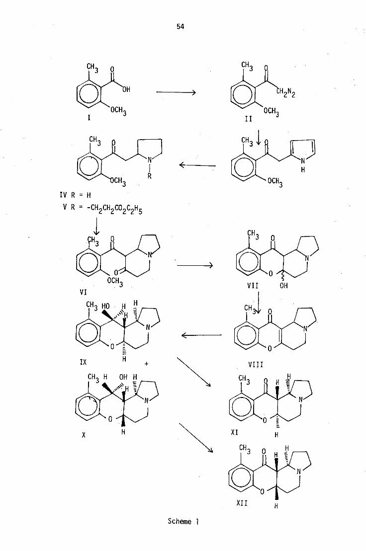
4.1.1 Synthesis of dk-elaeocarpine (XI) and dk-isoelaeocarpine
(XII) by Tanaka and Iijima 34 (Scheme 1)
The diazoketone (II) prepared by the reaction of diazomethane
on 2-methoxy-6-methyl benzoic acid (I) was condensed with excess
pyrrole in the presence of copper powder to give the pyrrolyl methyl
ketone (III), which was then hydrogenated using a platinum catalyst
to the amino ketone (IV). The hydrogenation did not affect the
aromatic carbonyl• function owing to steric hindrance. The amino
ketone (IV) was then converted to the diketone, (VI) through
addition of ethyl acrylatefollowed by a Dieckmann-type condensation.
The diketone was then demethylated to give the chromanone (VII) by
spontaneous cyclisation. The dehydrogenation of this chromanone
produced the chromone (VIII), but attempted conversion of this
compound to elaeocarpine (XI) or isoelaeocarpine (XII) by
hydrogenation was not successful. Instead, the two isomeric
alcohols (IX) and (X) obtained by borohydride reduction of (VIII)
were oxidised to (XI) and (XII).
54
IV R = H
V R = -Cl 2 CH 0 C H 5
VIII
X
Scheme •

CH3
H
)
Al H 2
XVI
55
4.1.2 Synthesis of (XI) and (XII) by Onaka 35 (Scheme 2)
In this two-step synthesis, the reaction of 2,3-dihydro-1H-
indolizinium bromide (XIV) with 2-hydroxy-6-methylbenzaldehyde
produced the alcohol (XV), which was oxidised with Jones' reagent
to give (XI) and (XII).
CH3
OH
1—N-)
OH XIII
XIV XV
XI + XII
Scheme 2
The mechanism of this condensation, which takes place in the presence
of lithium aluminium hydride, is presumed to proceed via the aromatic
chelate (XVI) as follows:

XV II I
XIX
NJ
CH3
OH
OCH3
0 -
CH3
56
4.1.3 Synthesis of (XI) and (XII) by Tufariello 36 (Scheme 3)
The amino ketone (IV) for this synthesis was obtained from the
isooxazolidine derivative (XIX), formed by addition of 1-Ryrroline-1-
oxide (XVIII) to 6-methoxy-2-methyl styrene (XVII).
OCH3
IV R = H
XXII R = CH2
CH2
CH2 OH
OCH3
XX R = H
XXI R = CH2
CH2
OH
XXIII
Scheme 3
Alkylation of the amine nitrogen could be carried out by reaction with
3-bromo-1-propanol either before or after the oxidation of (XX), but
CO2Et
LNS N CH2CO2Et
CO2 Et
57
direct treatment of (XIX) with the same reagent followed by reaction
with potassium tert-butylatend benzophenone in refluxing benzene _gave;(XXIII)
in a one-flask operation with improved yields.
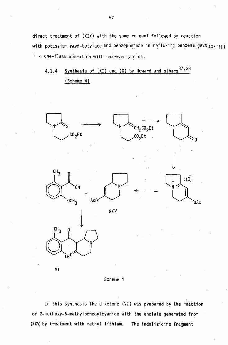
4.1.4 Synthesis of (XI) and (X) by Howard and others 37 ' 38
(Scheme 4)
XXV
Scheme 4
In this synthesis the diketone (VI) was prepared by the reaction
of 2-methoxy-6-methylbenzoylcyanide with the enolate generated from
(XXV)by treatment with methyl lithium. The indolizidine fragment

0
0
XXVIII R = -CO2C2H5
XXIX R = CH2OH
XXX R = CHO
OH
58
was synthesised through the sequence shown in Scheme 4.
Elaeokanine C was synthesised by the same workers 18,who isolated
the compound by a route similar to that described by Tanaka and
Iijima. The methods described by Tufariello and by Howard for the
synthesis of elaeocarpine and isoelaeocarpine have subsequently
been extended to the synthesis of elaeokanine A,B and C37,38,39
.
• A further synthesis of the same three compounds has been described
by Watanabe40 (Scheme 5).
2 C'H 2 5 L N ,•CO2C2H 5
XXVI XXVII
LN CHO '‘%‘H L
N >r0
0 _I XXX
XXXII
XXXI
(±) elaeokanine C
0
XXXIII
XXXXIV
XXXXV
(±) , elaeokanine B (±) elaeokanine A
Scheme 5

L
0
OH I
59
Watanabe41
went on to extend his synthesis to elaeokanine E
and the C-12 epimer of elaeokanine D. (Scheme 6).
XXX
CH3
elaeokanine E (±) 12-epielaeokanine D
Scheme 6
4.2 Synthesis of peripentadenine
The first approach to the synthesis of this compound involved
a reversal of the hydrolytic cleavage of the B-keto amino system
(Scheme 7) which gave 2-hydroxy-6-methyl acetophenone. The
isolation of this acetophenone in substantial quantities, and the
relative ease with which the corresponding iminium salt (XXXIX)
60
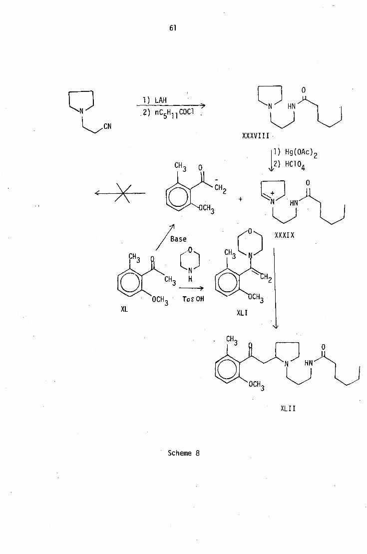
is prepared made this approach very attractive. The amino amide (XXXVIII)
was prepared by the reduction of 3(pyrrolidin-1-yl)propionitrile
followed by acylation with n-hexanoyl chloride. This was then
converted to the iminium salt (XXXIX) by oxidation with mercuric
acetate, and the perchlorate salt was then prepared as a viscous gum
which could not be obtained crystalline. An excess of this salt was
then gradually introduced into a mixture of 2-methoxy-6-methyl
acetophenone and a suitable basic catalyst. A number of solvent
systems and several bases including sodium ethoxide, potassium tert-
butoxide and sodium hydride were tried, but the intended condensation
could not be achieved, presumably due to the instability of the
iminium ion under these conditions. (Scheme 8).
Scheme 7
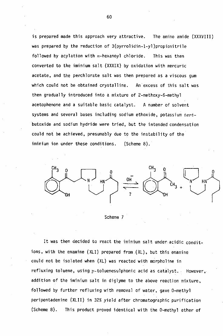
It was then decided to react the iminium salt under acidic condit-
ions, with the enamine (XLI) prepared from (XL), but this enamine
• could not be isolated when (XL) was reacted with morpholine in
refluxing toluene, using p-toluenesulphonic acid as catalyst. However,
addition of the iminium salt in diglyme to the above reaction mixture,
followed by further refluxing with removal of water, gave 0-methyl
peripentadenine (XLII) in 32% yield after chromatographic purification
(Scheme 8). This product proved identical with the 0-methyl ether of
1) LAN nC5Hvi C0C1
I j 0
HN )-( •
XXXVIII
11) Hg(0Ac) 2 2) HC104
L N)° HN
XXXI X /Base -0
OCH 3 Tos OH
CH 3
61
XL XLI
XLII
Scheme 8

62
natural alkaloid by tic, IR and PMR comparison.
After the initial failure of the above approach to the synthesis
of peripentadenine, several other routes were explored. One of the
precursors of elaeocarpine (XI) and isoelaeocarpine (XII) encountered
in two of the syntheses previously reported 34 ' 36 , 2(2-methoxy-6-methyl
benzoylmethyl)pyrrolidine (IV), could in principle be converted to
0-methyl peripentadenine by attaching the corresponding amide chain to
the amine nitrogen (Scheme 9) ]).y a process similar to the initial
reactions of Scheme 8. However, the reduction of the nitrile function
CH3 CH
3
OCH3 OCH 1
V XLIII 1 nC5H11 COC1
Scheme 9 XLII
with metal hydrides would require the protection of the aromatic carbonyl
group; an alternative procedure, catalytic hydrogenation, is known to
generate a certain amount of polymeric material. To eliminate this
inconvenience it was decided to attach the amide chain to the pyrrole
nucleus prior to its condensation with the diazoketone (II) (Scheme 10).
The amide group, being a relatively non-reactive function, was not
expected to interfere seriously with the diazoketone condensation except
for possible radical-initiated decomposition under these conditions.
The aromatic component, 2-hydroxy-6-methyl benzoic acid (I), is
a useful starting material for a number of natural products and has
been synthesised by several groups42-52
. These syntheses all

reduction
XLII
II XLIV
63
Scheme 10
involve rather long sequences, but Hauser53 has improved the procedures
42 described by Bohlmann and Piskov
43 so that large quantities of (I)
can be prepared in improved yields under routine laboratory conditions
with the minimum number of isolation steps (Scheme 11). The acid (I)
prepared according to this method was treated with thionyl chloride
to convert it to the acid chloride, which was then reacted with
ethereal diazomethane to obtain the diazoketone (II). The condensation
of (II) with N[3(pyrrol-1-yl)propyl]hexanamide (XLIV) was attempted under
34 the conditions described by Tanaka for the synthesis of (III). The
low solubility of the pyrrole derivative (XLIV) in benzene and toluene
prevented the use of these solvents, and instead dry ether was employed.
An ethereal solution of the diazoketone was slowly added to a stirred
mixture of excess of (XLIV) and freshly prepared copper powder at 00
under anhydrous conditions. The solution was then allowed to come to
room temperature and left overnight. The brown gum precipitated
resembled 0-methylperipentadenine (XLII) in being insoluble in ether,
but mass spectral analysis showed no trace of a m/z 384 species
to be present in the gum. The reaction was repeated in ether-toluene
mixture at 50-60°C in strong light but the expected product was not
found in the reaction mixture, which contained large amounts of

CH 3 0 C H
IHC1
CH3
3 Na0C
2H 5 0
H\ir CH
CH3
OCH 3
64
Synthesis of 2-methoxy-6-methylbenzoic acid (I) 53
CH
I) Br2/AcOH
2) A CO2C2H 5
OH
(CH3 ) 2SO4
OCH3
Scheme 11

65
polymeric material.
Finally the aminoketone (IV) was prepared according to Tanaka's34
method and converted to the amine (XLIII) by condensation with
acrylonitrile followed by hydrogenation in glacial acetic acid with
platinum oxide as catalyst. The amine was then acylated with
n-hexanoyl chloride to give 0-methylperipentadenine (XLII), which
was found to be identical with the natural product by tic, IR and
PMR comparison. Like the natural alkaloid, the synthetic product
could likewise not be obtained crystalline.
4.3 Synthesis of N[3-(dimethylamino)propyl]hexanamide
CH CH 100°C
3\ NH + CH2
= CH-CN ' 3\N(CH 7 7
CH3
CH 3 I
CH nC
5 H11
COC1 < CH
3'1■1(CH ) NHCOnC
5H11
3\\N(CH ) NH2
CH3
CH3
Scheme 12
The title compound, one of the degradation products of
peripentadenine, was synthesised by the route shown in Scheme 12.
Equimolar quantities of dimethylamine (25/30 w/v aq. solution) and
acrylonitrile were heated in a sealed tube with a few ml of methanol
for 3 hours. The product, 3-dimethylaminopropionitrile54
, was
isolated by distillation, and reduced to the amine 54 using lithium
aluminium hydride. The amine was acylated with n-hexanoyl chloride
to give N-[3-dimethylamino)propyT]hexanamide,which was found to be
identical with the final Hofmann degradation product by tic, IR and
PMR comparison.
CN

66
CHAPTER 5
Minor alkaloids of the bark extract
The minor alkaloids of the bark can be divided into three
groups according to their relative amounts. Three alkaloids,
peripentamine (VP, dehydroperipentamine (VIP and dinorperipentadenine
(II) were isolated in 180-75 mg quantities, and their structural
elucidation is discussed in this chapter. Three more compounds
were isolated in less than 50 mg quantities and partial structures
have been assigned. A set of six compounds was isolated in less
than 25 mg quantities; some of their spectra have been recorded,
but no structures have been assigned. The latter two groups of
compounds will be discussed in Chapter 7.
The column-chromatographic separation of the bark extract
gave three major fractions. The least polar fraction gave a mixture
of volatile compounds, peripentamine (VI) and another minor base
(PBXM2). The second fraction gave the major base, peripentadenine
(I), and the most polar fraction gave dehydroperipentanvine (VIII) and
a lower homologue (II) of peripentadenine together with seven other
compounds (PBXM3-PBXM9).
5.1 Structural elucidation of dinorperipentadenine (II)
The most polar fraction obtained from column chromatography was
subjected to preparative tic. The least polar compound on the
chromatogram gave peripentadenine (I). A slightly more polar band
gave a mixture of (II) and PBXM3; these were separated by further
ptic using a multiple development technique.
Dinorperipentadenine (II) gave a pale yellow gum which
formed a single spot on tic with several solvent systems. Like the

CH3
HN
67
major alkaloid, this compound could not be obtained crystalline.
High resolution mass spectrometry gave the formula C 20H30N20 3,and
the PMR spectrum (Figure 1) appeared to be identical with that of
peripentadenine (I) except for the absence of the multiplet between
61.15 and 1.35 which corresponded to the C22 and C23 methylene
protons on the six-carbon chain. The molecular formula, coupled with a
close examination of the PMR spectrum, suggested the structure (II)
for this compound, where the hexanamide group in peripentadenine (I)
has been replaced by a butanamide unit. The assignment of the PMR
signals and the coupling patterns were confirmed by detailed
decoupling experiments.
CH3 0
OH
II R = H
III R = CH3
This compound gave a positive Gibbs test and formed a monomethyl
ether (III) on treatment with diazomethane. Further, the mass
spectral fragmentation patterns of the compound itself and its metho-
fluoride (IV) (Figures 2 and 3) confirmed the presence of an N(propyl)
butanamide chain and a pyrrolidine nucleus.
The assignments of the PMR signals and the coupling patterns
(Figure 4) were confirmed by detailed decoupling experiments. The
assignment of the 13C values (Figure 5) was done by comparison with
those of peripentadenine (I).

PMR spectrum CDC 1 3 ( 270 MHz )
68
1--I
CS, 1-71
C‘J
LC)
CO

Z'R ■-•
1
69
NI
a) s- = cn •,—
L.I._
70
•r—
Lt—
C \Z
cs Cs.1

18
15 17
16
11
22_
OH
911A
1H
21H
8.5—
11H
11HA
12 --r
16HA 4
OH
Expansion of the PMR spectrum of dinorperipentadenine from 3.4-1.4 ppm (J values in Hz)
'
3.4
3.2
3.0
2.8
2.6
2.4 2.2 2.0 . 1.8
1 . 6
I. 4 PPm
Fi9ure 4

ca_ Ca-
CD
72
r—I
CD
cv CD
cy,
CD
C-3
CD
t•)
CD
s••••1
■/
",!\.'
a) S..- C)
•r—
U—
C—) CD
1-1
cz.) —
e--1
(.0
t--i C—)

chloride N --> H2 —
H 0
OCH3
III
CH3 n-butanoyl 0
73
5.2 Synthesis of dinorperipentadenine (II)
Structure (II) for this compound was finally confirmed by
synthesis. The amine (V), a synthetic precursor of peripentadenine,
was acylated with n-butanoyl chloride to give the 0-methyl ether of
dinorperipentadenine (III), which was found to be identical with
the 0-methyl ether of the natural product by tic, IR and PMR spectral
comparison. Like peripentadenine, this compound was also isolated as
a racemic mixture.
5.3 Structural elucidation of peripentamine (VI)
Peripentamine (VI), a pale yellow oil, was shown to have the
molecular formula C22H36N204
by chemical ionization high resolution
mass spectrometry. On electron impact this compound appeared to
lose a molecule of water quite readily, and the highest mass observed
was at m/z 374; but on low-energy electron impact and chemical
ionization mass spectroscopy the molecular ion appeared at m/z 392.
The molecular formulae show that this compound differs from
peripentadenine (I) in having the elements of a molecule of water
which is easily lost in its molecule.
Both PMR and 13C NMR spectra show the presence of a 2-hydroxy-6-
methyl benzoyl unit in the molecule: PMR signals for three ABC-coupled
aromatic proton signals at d7.17, 6.8 and 6.68 (Figure 6), 13C signals
at d205.8, 159.2, 137.9, 133.1, 125.3, 122.9 and 115.6 (Figure 7), and
a mass spectral fragment at m/z 150 (Scheme 1 and Figure 8).
Furthermore, (VI) gave a +ve Gibbs test, indicating a free p-position
1 CH3
20
14 5 ° .•:y/ ■ 21 13 9 19
10 18
OH 12 16 NH 22 24 11 17 23
HO
2
74
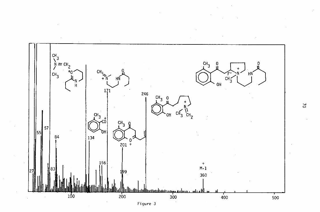
to the phenolic hydroxyl. A one-proton exchangeable PMR signal at 66.65
coupled to a methylene proton signal at 63.37, together with the IR
-1 13 absorption bands at 3300 and 1640 cm and a C signal at 6173.3,
indicated the presence of a primary amide function (-CH 2NHCO-). The
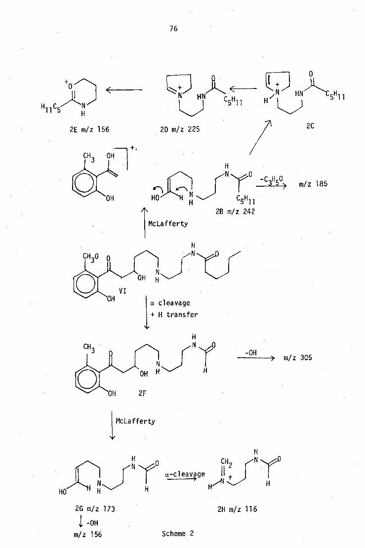
mass spectral fragments at m/z 156 and 225 (Scheme 2), which were also
present in the mass spectrum of peripentadenine, suggest the presence
of an N(3-aminopropyl)n-hexanamide unit. Further, PMR decoupling
experiments (Table 1) and 13C signals at 657.6, 25.5, 38.6, 173.3,
36.9, 25.9, 31.5, 22.4 and 13.9 also confirmed its presence.
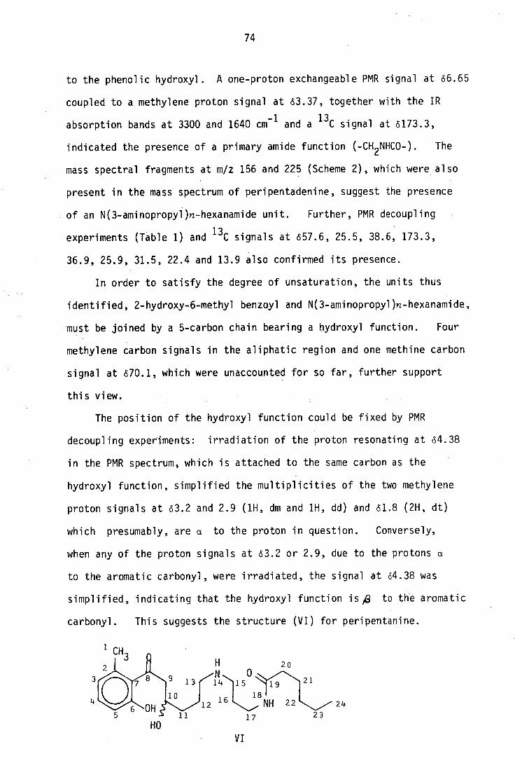
In order to satisfy the degree of unsaturation, the units thus
identified, 2-hydroxy-6-methyl benzoyl and N(3-aminopropyl)n-hexanamide,
must be joined by a 5-carbon chain bearing a hydroxyl function. Four
methylene carbon signals in the aliphatic region and one methine carbon
signal at 670.1, which were unaccounted for so far, further support
this view.
The position of the hydroxyl function could be fixed by PMR
decoupling experiments: irradiation of the proton resonating at 64.38
in the PMR spectrum, which is attached to the same carbon as the
hydroxyl function, simplified the multiplicities of the two methylene
proton signals at 63.2 and 2.9 (1H, dm and 1H, dd) and 61.8 (2H, dt)
which presumably, are a to the proton in question. Conversely,
when any of the proton signals at 63.2 or 2.9, due to the protons a
to the aromatic carbonyl, were irradiated, the signal at 64.38 was
simplified, indicating that the hydroxyl function is ie to the aromatic
carbonyl. This suggests the structure (VI) for peripentanine.
V I
OH
m/z 201
-e onium
H1‘1.Oy- /N
H
75
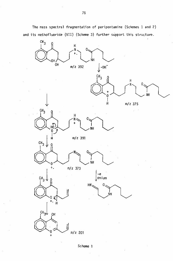
The mass spectral fragmentation of peripentamine (Schemes I and 2)
and its methofluoride (VII) (Scheme 3) further support this structure.
CH3
NH
OH m/z 392 1-0H
NH
m/z 375
Scheme 1
Scheme 2
2H m/z 116 2G m/z 1 73
1 -OH
m/z 156
76
HO Jr-17LE'7 N
McLafferty
/NN.° -C3
I150>
C5H 11 2B m/z 242
m/z 185
a cleavage
+ H transfer
McLafferty
-OH > m/z 305
CH2 • ,N
a-cleavage II t
HN
L../)
2D m/z 225
5H 11
2C 2E m/z 156
+.
H
HO
A

77
The nucleophilic displacement of the hydroxyl group of 2B, which
is formed by McLafferty rearrangement of the parent molecule, giving
rise to a stable ion at m/z 225 (2D) (Scheme 2) seems to further
support the presence of a hydroxyl function at C10.
OH
a. m/z 135 and 284
b. m/z "272" -15 > 257
c. m/z 201, "220" -18 > 202
d. m/z 156, "264" -18>
246
Scheme 3
The structure (VI) for peripentamine was confirmed by its
conversion into a known compound at a later stage, but the PMR
splitting patterns of some of the protons could not be explained
directly on the basis of this structure.
PMR decoupling experiments (Table I) showed that one of the C9
protons and the C10 proton are coupled to protons either at C22 or
C23. When the C10 proton signal at 64.38 was irradiated, the C-9
proton signals at 63.2 and 2.9 simplified into a ddd multiplet

78
TABLE 1
PMR decoupling experiments on peripentamine (10
Irradiated at/ppm
SIGNAL/S C HANGED
At/ppm From To/J Hz
4.38 3.2 dm ddd 16,5,3
2.9 dd d 16
1.8 simplified
1.3 simplified
3.37 1.75 simplified
3.2 4.38 dm 11
2.9 dd d 6
2.9 4.38 tm 8
2.6 1.7 simplified
1.8 4.38 ddd 10,8,5
1.3 4.38 simplified
1.69 tt t 8
0.9
(J = 16, 5 and 3Hz) and a doublet (J = 16) respectively. On the other
hand, when the C-11 methylene protons at 61.8 were irradiated, the C-10
proton signal at 64.38 simplified to a ddd (J = 10, 8, 5Hz) multiplet.
The irradiation at 61.3 affected both signals at 63.2 and 4.38, but
because of the overlap of the C-22 and C-23 proton signals, the
coupled protons could not be identified.
The apparent long-rangeipteraction of C-9 and C-10 protons to C22
or C23 protons suggestra somewhat rigid conformation for the
peripentamine molecule. This may be brought about by

C21H
C1 6H
C11,12H
C2OH
1 14
10 20
7N 0:..-/•\, 21 13 15
12 16 N 22 18 24
18 H 23
C24H
Cl H C22,23H
irradiated
I ' 6.5 7.0
ClOH
4.4 --;1 3.0 2.8 2.16 3.4
3 1.2
Niv
2.4 2.2 2.0 1.8 1 1 .6 1.4 1.2 1.0 0 1.8 PPM
C9HB
at d 1.75
T5H 15H I A B
r ■.•
Figure 6

80 C-)
C-)
CO
r—
r-- C1.1
C1J
r
3
01
L.) C
D
1.11
Lf)
CD
CD
C-)
CY)
CD
1•••••
CD
-• f1,1

81
I I
0
0
CO l0
I I
0
0
.:t• t\ I

82
intramolecular hydrogen bonding and this possibility is further
supported by the fact that this compound is much less polar than
other compounds with fewer -OH and -NH groups.
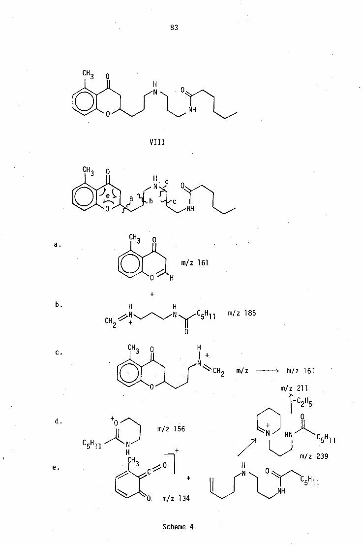
5.4 Structural elucidation of dehydroperipentamine (VIII)
Dehydroperipentamine (VIII), a white crystalline powder, was shown
to have the formula C22 H34
N2 03' isomeric with peripentadenine (I), by
both high resolution mass spectrometry and elemental analysis. Spectral
comparison with peripentadenine (I) and peripentamine (VI) showed that
both 2-oxy-6-methyl benzoyl and -N(3-aminopropyl)n-hexanamide partial
structures are present in dehydroperipentamine as well. However,
dehydroperipentamine did not give a +ve Gibbs test, and its mass
spectrum did not show a peak at m/z 150 for a 2-hydroxy-6-methyl
acetophenone fragment, which is formed by the cleavage of the bond a
to the aromatic carbonyl by a McLafferty-type rearrangement in both
peripentadenine and peripentamine. The absence of a free phenolic
hydroxyl, and the appearance of a methine carbon signal at 06.3 in the
13C NMR spectrum (Figurell), as well as a one-proton multiplet at (54.47
(Figure 10) coupled to a methylene proton signal at 62.69 in the 1 H NMR
spectrum suggested the presence of an oxygen heterocycle. The presence
of a strong peak at m/z 161 in the mass spectrum (Figure 9), which could
be formed by the cleavage of the bond a and exo to the ether oxygen in a
benzpyrano system (Scheme 4) •suggested the structure (VIII) for
dehydroperipentamine. Extensive decoupling experiments at 400 MHz led
to the confirmation of this structure, and also made possible the
unambiguous assignment of each PMR signal (Figure 10).
C .
e.
d. m/z 156
ol m/z 134
Scheme 4
83
V I I I
a.
m/z 161
b.
yiCH2 f C5Hi m/z 185
0
1 +
m/z > m/z 161
m/z 211
1-C2 H 5
0
HN C 5 H 11
m/z 239
° CO
.21H

84
The irradiation of the proton at 64.47 (C10) caused the signals
at 62.69 (C9) and 61.95 (C11) to collapse; the splitting pattern of
the C9 protons could not be distinguished due to virtual coupling.
The irradiation of the signal at 63.58 (C17) simplified the signal at
62.16 (C16), and when these protons were irradiated, signals both
63.58 (C17) and 63.03 (C15) collapsed. This enabled the signals
for the two sets of methylene protons a to the amine nitrogen, at 63.03
(C15) and 62.98 (C13), which appear very close to each other, to be
distinguished.
The assignment of the 13C signals (Figure 11) was done by
comparison with the 13C spectrum of (I),which is closely related
structurally.
TABLE 2
PMR decoupling experiments on dehydroperipentamine (VIII)
Irradiated At/ppm
SIGNAL/S CHANGED
At/ppm From To/Hz
4.47 2.69 m simplified
1.95 m simplified
3.58 2.16 tt t 6
3.03 2.16 tt t 6
2.98 1.89 m simplified
2.69 4.47 m t 7
2.27 1.64 tt t 7
1.64 2.27 t s
1.29 m simplified
1.27 1.64 ti t 7
0.89 1.29 m simplified

07._)
mass
sp
ectr
um
85
C
CNJ C\ C
LL

86
PMR sp
ectr
um C
DC13
co —
LD
Cr) J-

01
C—) 6 . 9LT
9 -1£ 6 . TE
CV CO r— C
r— C—)
C71 C
8'9Z 1-6
v... C.)
ey Z'9E 9'9C
t . £6 .1
87
u-) 6 . 9TI
L'E•TI
rtZT
617E1
17•Z9T

88
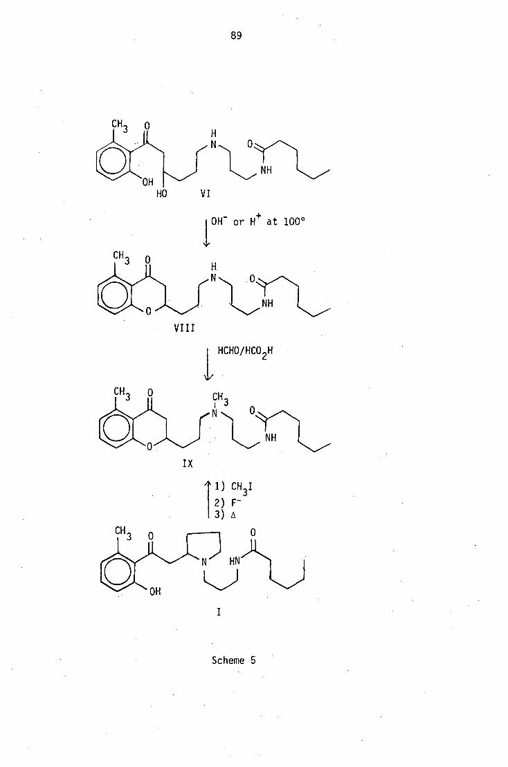
5.5 Conversion of peripentamine (VI) and dehydroperipentamine (VIII)
into a known compound: the Hofmann degradation product (IX) of
peripentadenine (I). Scheme 5).
5.5.1 'Dehydration of peripentamine (VI)
Peripentamine (I) was found to lose a molecule of water so readily
on electron impact that chemical ionization mass spectrometry had to be
utilized to assure the appearance of its molecular ion. An attempt was
made to simulate this dehydration process by pyrolysis of 150°C and
6.2 x 10⁻⁴
Hg mm. A colourless liquid distilled off, which consisted of
2-hydroxy-6-methyl acetophenone and a mixture of simple amines that could
not be separated.
No appreciable change was observed when peripentamine was left
standing in 5% methanolic sodium hydroxide solution at room temperature.
However, when refluxed at 100°C, either 5% methanolic sodium hydroxide
or 10% aqueous oxalic acid solutions converted peripentamine into
dehydroperipentamine.
5.5.2 Conversion of dehydroperipentamine (VIII) into the Hofmann
degradation product of peripentadenine
N-methylation of dehydroperipentamine using formaldehyde/formic acid
gave (IX), which was found to be identical with the Hofmann degradation
product of (I) by tic, IR and PMR comparison.
89
01/"\
NH
HO
VI
][0H- or H+ at 100 0
•
VIII
1/ HCHO/HCO 2H
I X
Scheme 5

90
CHAPTER 6
Constituents of the leaf extract
6. The column chromatographic separation of the leaf alkaloid extract
produced three main fractions. The least polar fraction contained
2-hydroxy-6-methylacetophenone and two bases: PLM2 and PLM3. The
second fraction gave peripentadenine, dinorperipentadenine and
dehydroperipentamine. The third fraction produced a series (PLM4-PLM10)
of fairly polar compounds; the structural elucidation of some of
these will be discussed in Chapter 7. Peripentamine could not be
detected in the leaf extract.
6.1 Structural elucidation of PLM2
The ptic purification of the first fraction gave PLM2 as a yellow
oil, which was further purified by sublimation. The low melting solid
thus obtained analysed for C91-1 13 N0 by high-resolution mass spectrometry
and microanalysis. It also formed a crystalline picrate, which analysed
for C15H16N408
by microanalysis.
-1 A strong IR absorption band at 1730 cm and a
13C NMR signal for
a quaternary carbon at 208.5 ppm indicated the presence of a carbonyl
function in PLM2. Further, UV absorption bands at 217, 325 and 370 nm
suggested that the carbonyl was conjugated with some unsaturated group.
In the 13
C NMR spectrum there was only one other low-field signal in
addition to that of the carbonyl carbon, due to a quaternary carbon at
173.9 ppm, and from these data it appeared that the unsaturated group
is an imine function. On treatment with methyl iodide, only one
methyl group was added to the compound, but on the other hand, after
reduction of PLM2 with borohydride, two methyl groups were added
under the same conditions. The borohydride reduction increased

91
the molecular weight by four a.m.u., and the product formed a diacetate
on treatment with pyridine/acetic anhydride at room temperature, whereas
the parent compound itself did not form an acetate under the same
conditions.
The PMR spectrum for PLM2 (Figure 3) had signals for two methyl
groups: a three-proton doublet at 1.03 ppm, and a three-proton
singlet at 2.12 ppm. The chemical shift of the latter suggested
either an acetyl function or an olefinic methyl group. From these
data the partial structures (I) or (II) can be put forward:
•-"-^-0
,,v,/iiel73.9
CH3
(2.12) i'208
4N N T H (2.12) 3
208 173.9
II
To satisfy the degree of unsaturation (four), these partial
structures would have to be incorporated into a system of two rings.
The 13C NMR spectrum (Figure 4) showed, in addition to the two low-field
quaternary carbons already described, the presence of seven aliphatic
carbons: three methine carbons, two methylene carbons and two methyl
carbons. The chemical shift of the two signals at lowest field, both
for methine carbons, at 61.5 and 55.8 ppm, suggested that they are
located a to the imine function. Furthermore, the lowest field PMR
signals at 4.5 and 3.14 ppm corresponding to one proton each
can be reasonably attributed to the protons attached to the same
methine carbons. Irradiation of the methyl proton doublet at 1.03 ppm
did not affect either of the above-mentioned PMR signals, indicating that
the methyl group is not attached to the methine carbons in question.
Therefore these two carbons can be assumed to be present at a ring
junction,and the partial structures (I) and (II) can now be extended

to (III), (IV) and (V).
92
9 loCH3 CH3 7
10
CH3
3
9 CH
3
III
IV
Significant coupling between the two methine protons at the ring
junction in structure V would be expected; however, since no such
coupling was observed, this structure can be disregarded.
Most of the spectral features of PLM2 can be explained on the
basis of structure III. The IR carbonyl absorption (1730 cm -1 ) and
the 13C chemical shift of the same carbon (6 208) are quite comparable
with those for an acetyl group. Further, both PMR and 13C NMR
values for the methyl group (C9) (6 2.15 and 6 24.4), and the prominent
loss of 43 a.m.u. (CH 3CO) from the molecular ion in the mass spectrum
(Figure 5) agree reasonably well with the presence of an acetyl
function in the molecule; formation of most of the mass•
spectral fragments can also be explained (Scheme 1).
The PMR signals for the two methyl groups (6 2.12, s, and 6 1.05, d)
can be assigned to the C9 and C10 protons respectively. The two lowest
field signals at 6 4.52 and 3.15, both one-proton signals, can be
assigned to the protons on Cl and C4 at the ring junction. The broad
signal at 6 4.52, which must be due to the Cl proton a to the
imine nitrogen, was found to be coupled to the set of four protons
resonating around 6 2.0. On the other hand, the C4 proton signal at
6 3.15 was found to be coupled to one proton only. According to
93
structure III, the C4 proton would be expected to be coupled to more than
one proton, as there would be in any case at least three such protons on
the adjoining carbon whether the methyl substituent (C10) were at C5 or C7.
The construction of a model of structure III showed that the dihedral
angle between the C4 proton and any of the protons at C5 or C7 cannot
be close enough to 90°C in each case, so as to eliminate any coupling
between them. The structure III remains unsatisfactory in this
respect.
Further evidence against structure III came from quaternisation
of PLA2 and from subsequent reduction of the salt. On quaternisation,
the three-proton singlet at 2.12 ppm moved down-field to 3.35 ppm.
The quaternisation of the imine nitrogen in structure III would not be
expected to have such a pronounced deshielding effect on the methyl
protons which are y to it. This favours the structures IV and V,
where the methyl group on the imine carbon would be expected to be
considerably affected by quaternisation.
Furthermore, when the quaternary salt was partially reduced with
sodium cyanoborohydride, the PMR spectrum of the product showed two
three-proton doublets at 1.1 and 0.85 ppm. Also, the spectrum showed
no evidence of either a hydroxy proton, or a methine proton on a carbon
bearing a hydroxy function. The PMR signal for the acetyl group in
structure III would be expected to remain more or less unaffected
during these transformations (Scheme 2), and this leaves structure IV
for PLM2. A parallel for a 2-aza-bicyclo[3,2,1]octane system could
not be found from a natural source. Further evidence, chemical or
spectroscopic, to confirm this structure could not be obtained due to
lack of sufficient material, and the difficulty in formation of
derivatives. However, the available spectroscopic data can be explained
satisfactorily with structure IV.

CH3
-COCH 3 •
94
m/z 136
c <-CH
3
N2\ CEO
m/z 94
m/z 151
'
retro-Diels-Alder
LN cH 3 11
m/z 109
CH 3
CH3
CH
- CH3 ).
CEO
m/z 57
Scheme 1
Proton decoupling experiments on PLM2 did not provide much further
evidence. They showed that the one-proton signals at 4.5, 3.15 and
1.25 ppm and the three-proton doublet at 1.03 ppm were all coupled to
the set of signals around 2 ppm, but not coupled to each other. The
close proximity of the signals around 2 ppm did not permit the
decoupling of all four protons. The irradiation of the two overlapping
one-proton signals at 1.97 ppm caused the collapse of all three signals
at 3.15, 1.25 and 1.03 ppm into singlets.
The one-proton signals at 4.5 and 3.15 ppm can be assigned to the
protons a to the imine nitrogen and the. imine carbon (Cl H and C4 H)
respectively. The fact that the former signal is a doublet (J = 3Hz)
implies that the C5 proton is coupled to one proton only, even though
there are up to four such protons present on C6 and C8. The

CH3
CH3
CH3
NaBH4
CH3
CH3
IV
CH3
V I
CH3
CH3
3
95
construction of a model of IV showed that the dihedral angles made by
the C5 proton with one of the protons attached to both C6 and C8 would
be close to 90 0 , thereby minimising the coupling between these protons.
One of the remaining two protons has to be replaced by a substituent,
the methyl function, leaving one proton that could couple with the C5
proton. Therefore the methyl group has to be positioned either at
C6 or C8.
The analysis of the 13C NMR spectrum helps the positioning of the
methyl function to a certain degree. The chemical shift values for
the signals assigned to C6, C7 and C8 in the corresponding isomeric
hydrocarbons were calculated following the Lindeman and Adams method.33
(Figure 6). However, since the effects of the heteroatoms, as well
as the ring strain, have not taken into account, these chemical
shift values cannot be accepted i with absolute certainty. Even so,
certain marked differences in the chemical shifts for the individual
carbon atoms in different isomers can be seen. The chemical shifts
for the methine carbons bearing the methyl function were calculated
to be 29.9 and 35.4 ppm for the 6-substituted (6C) and 8-substituted
(6F) isomers respectively. The observed value for this carbon in PLM2
was 28.9 ppm, which favours structure (VIII) for this compound where
the methyl function is at C6. The fact that the 6 4.5 proton (on 01)
is coupled to four other protons also favours (VIII).
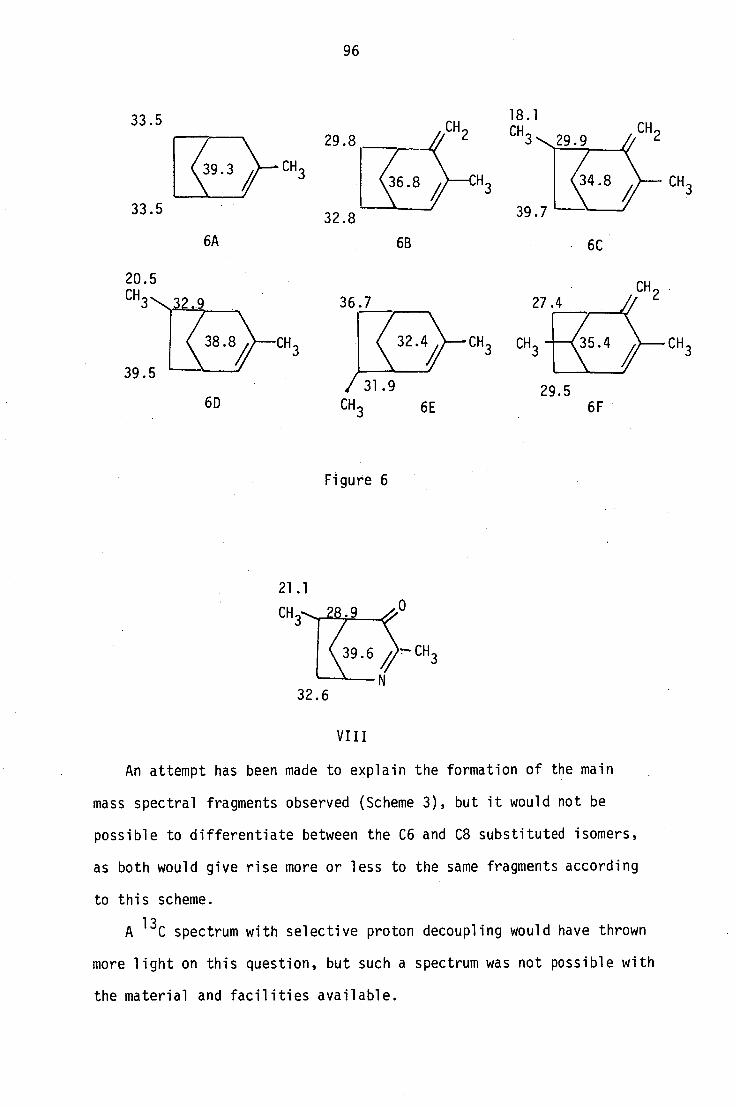
33.5 32.8
6A 6B 6C
33.5
96
36.7
CH3
6D
CH3 CH
29.5 6F
20.5 CH
3 2 9
39.5
38.8/ CH3
CH3
Figure 6
21.1
32.6
VIII
An attempt has been made to explain the formation of the main
mass spectral fragments observed (Scheme 3), but it would not be
possible to differentiate between the C6 and C8 substituted isomers,
as both would give rise more or less to the same fragments according
to this scheme.
A 13C spectrum with selective proton decoupling would have thrown
more light on this question, but such a spectrum was not possible with
the material and facilities available.

CH 8
3E m/z 82
0
1 1
3D m/z 94
CH 2
CH3
04.
3C m/z 110
C .0 NH
m/z 108
NH
3G m/z 55 m/z 70
4-
CH 3
3F m/z 69
3H m/z 41
97
Mass spectral fragmentation of PLM2
Scheme 3
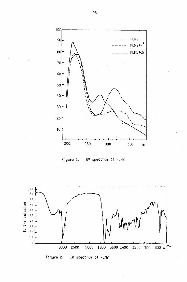
I # I __L./
3000 2500 2000 1800 1600 1400 1200 100 800 cm-1
_L
1% Transmissio
n
100
90
70
60
50
40
30
20
10
80
98
100
90
80
70
60
50
40
30
20
10
200
250
300
350 nm
Figure 1. UV spectrum of PLM2
Figure 2. IR spectrum of PLM2

LC)
LC) OM,
/••
99
•••..
LC)
PMR
spec
tru
m C
DC13
(27
0 M
Hz)
LC)
Ce)
LO

—CD
100
CD
NMR
spec
tru
m
L.)
1.0
01 C..)
3
CD - 1.f)
CD •zZ1-
CD
▪ CD
CY1 L.)
,„ CD 01 r•—•
CD

101
Mass sp
ectrum P
L M2
re)
c:1C
C-) cD r••■•
C:)
C7)
czt CO C I
LiJ CO
CO
100 80 60 I-
40 20
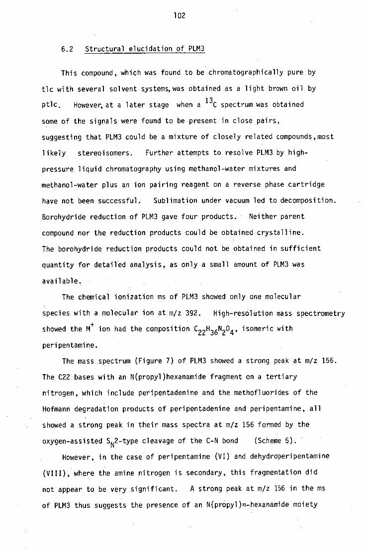
102
6.2 Structural elucidation of PLM3
This compound, which was found to be chromatographically pure by
tic with several solvent systems, was obtained as a light brown oil by
ptic. However, at a later stage when a 13C spectrum was obtained
some of the signals were found to be present in close pairs,
suggesting that PLM3 could be a mixture of closely related compounds,most
likely stereoisomers. Further attempts to resolve PLM3 by high-
pressure liquid chromatography using methanol-water mixtures and
methanol-water plus an ion pairing reagent on a reverse phase cartridge
have not been successful. Sublimation under vacuum led to decomposition.
Borohydride reduction of PLM3 gave four products. Neither parent
compound nor the reduction products could be obtained crystalline.
The borohydride reduction products could not be obtained in sufficient
quantity for detailed analysis, as only a small amount of PLM3 was
available.
The chemical ionization ms of PLM3 showed only one molecular
species with a molecular ion at m/z 392. High-resolution mass spectrometry
showed the M+
ion had the composition C22H36N2 0 4 , isomeric with
peripentamine.
The mass spectrum (Figure 7) of PLM3 showed a strong peak at m/z 156.
The C22 bases with an N(propyl)hexanamide fragment on a tertiary
nitrogen, which include peripentadenine and the methofluorides of the
Hofmann degradation products of peripentadenine and peripentamine, all
showed a strong peak in their mass spectra at m/z 156 formed by the
oxygen-assisted SN2-type cleavage of the C-N bond (Scheme 5).
However, in the case of peripentamine (VI) and dehydroperipentamine
(VIII), where the amine nitrogen is secondary, this fragmentation did
not appear to be very significant. A strong peak at m/z 156 in the ms
of PLM3 thus suggests the presence of an N(propyl)n-hexanamide moiety

103
0 SN2 type
H C 5H 11 1
m/z 156
Scheme 5
on a tertiary amine nitrogen. Its presence is further supported by
the appropriate PMR (Figure 9) and 13C NMR (Figure 8) features: an
exchangeable one-proton signal at 6 6.7 (NH) coupled to two protons
resonating at 6 3.45 and 3.2 (-CH2 -NHCO-); a quaternary carbon
signal at 6 173.3 (NHCO) and PMR and 13C NMR signals for a C5
paraffinic chain (PMR 6 2.2 (2H, 0, 1.65 (2H, txt), 1.3 (4H, m)
and 0.9 (3H, t) and 13C NMR 6 36.8, 25.6, 31.6, 22.4 and 13.9); and
a C3 unit between two nitrogens (PMR 6 3.45, 3.2 (2H) coupled to 1.8,
1.65 (2H) coupled to 2.9, 2.4 (2H) and 13
C NMR 6 55.9, 39.7 and 23.9).
However, unlike other C22 and C20 Peripentadenia alkaloids, PLM3
did not appear to contain a 2-oxy-6-methyl benzoyl unit, •as neither
its PMR or 13C NMR spectra showed any signals for an aromatic
nucleus.
As mentioned earlier, the off-resonance-decoupled 13C NMR
spectrum shows two types of signals in the aliphatic region:
a set of 10 sharp strong signals, and a set of 18 relatively less
intense signals which are present more or less in close pairs
(Figure 8). Eight of the strong aliphatic signals have been
identified already as those of the carbons on the N(propyl)n-hexanamide
unit, and a further signal in the carbonyl region can be assigned to
the amide carbon. Apart from these nine signals for this unit,

104
whose presence has been established from the mass and PMR spectra as
well, a total of 22 signals (20 in the aliphatic region: 18 of them
in pairs and 2 in the carbonyl region) could be seen in the 13
C
spectrum. Since PLM3 was shown to be chromatographically homogeneous,
these pairs can only be explained as arising from an inseparable
mixture of closely related compounds,most likely stereoisomers . .
Furthermore, the members of each pair show identical splitting in the
off-resonance proton-coupled spectrum, and the number of pairs of
carbon signals, as well as the total number of protons attached
to these carbons, as seen from their multiplicities, tally with the
molecular formula of PLM3 obtained from high-resolution mass
spectrometny, with only one oxygen unaccounted formn the formula C22
H36
N204
.
0
2 C=0 2
5 -CH 5 ru 1 m rn /N-(v"2 1 3 - "-vun `"
r 5"11
5 CH2 10
/
1 -CH3
3
C 13 H18
02
+ C9H18
N20 = C 22H36
N203
In consequence, each pair of these 18 signals will be considered
for convenience as one signal for the rest of the discussion.
An analysis of the molecular formula showed that the molecule is
tricyclic. The presence of two methine carbons at 677.32 and 76.88
indicates that the fourth oxygen is present as an ether, and this
must form a part of an oxygen heterocycle, since there are no side
chains apart from the amide chain and a methyl group attached to
another methine carbon. A further methine carbon signal at 661.37
suggested an a-substituted nitrogen heterocycle.

105
From PMR decoupling experiments, four different sequences of
coupled protons could be established:
A. Protons on the C5 paraffinic chain.
6 2.2 (2H, 0 4-4 1.6 (2H, m) 4-4 1.4-1.2 (4H, m) 4-4 0.9 (3H,
B. 6 4.5 (1H, )CH-0-) 4-4 6 3.29 (1H)
(C6) (C7)
6 2.1-1.9 (5H, m) 4-4 1.05 (3H, dd)
(C2, C3, C5) (Cl)
In the PMR spectra of the other C22 Peripentadenia alkaloids, no
parallel for this set of signals could be found. A careful
comparison of the PMR spectra of all the C22 bases showed that in the
case of PLM3, these signals have replaced the signals for the 2-oxy-6-
methylbenzoyl nucleus. The aromatic methyl proton signal at '‘, 6 2.4,
which appeared as a singlet in the spectra of the other compounds, has
been replaced by a methyl signal at 6 1.05 (dd) in the
spectrum of PLM3. An additional signal for a proton on a methine
carbon bearing an oxygen function could be seen at 6 4.55, and this
suggests that the 2-oxy-6-methylbenzoyl nucleus common to all the other
C22 alkaloids has been reduced in PLM3. Total reduction of the
aromatic nucleus should add on six protons, giving a total of nine on
this ring, but in fact, signals for seven protons only can be observed,
indicating that the reduction is partial and involves four protons
only. As no olefinic proton or carbon signals occur in the spectra
of PLM3, the remaining degree of unsaturation must be attributed to
a carbonyl function in the ring. From biosynthetic considerations,
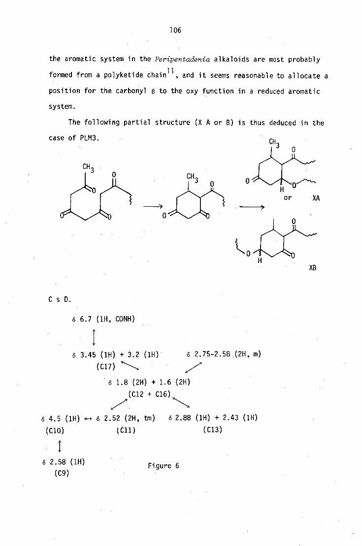
106
the aromatic system in the Peripentadenia alkaloids are most probably
formed from a polyketide chain11
, and it seems reasonable to allocate a
position for the carbonyl fi to the oxy function in a reduced aromatic
system.
The following partial structure (X A or B) is thus deduced in the
case of PLM3.
XB
(1H) o 2.75-2.58 (2H, m)
6 1.8 (2H) + 1.6 (2H)
(C12 + C16)
6 4.5 (1H) 4.-+ 0 2.52 (2H, tm) 6 2.88 (1H) + 2.43 (1H)
(C10) (C11) (C13)
C s D.
6 6.7 (1H, CONH)
6 3.45 (1H) + 3.2
(C17)
Figure 6

0
(4.5)H H H
(2.52)
107
The other two sequences of coupled protons could not be clearly
distinguished owing to overlapping of some signals, but by a careful
comparison with the PMR spectra of compounds having an N(propyl)amide
function, the PMR signals due to the protons on the propyl unit can be
identified (upper half of Figure 6). The presence of an a-substituted
nitrogen heterocycle has already been suggested from the 13C spectrum,
and the remaining sequence of signals must be due to the protons in
this system. Further analysis of their relationships and chemical
shifts suggest a 2-substituted 3-oxy piperidine nucleus (XI).
(2.58) (Ix
(2.88)
1.8, 1.6)
Chemical shift assignments are given in parenthesis in ppm.
(XI)
For the remaining oxygen heterocycle, a tetrahydro-y-pyrone
ring can be proposed on biogenetic grounds, and from analogy with the
benzypyran ring system of dehydroperipentamine. (Possible biogenetic
relationship of these alkaloids will be discussed in Chapter 8).
When all the partial units are assembled,structure (XII) for
PLM3 results.
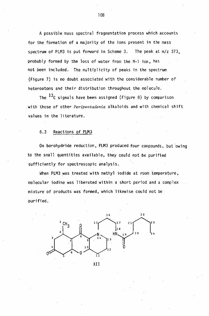
16
15 17 21
18
14 13'
20 2L.
0 12
1 CH 3
2 2
3
1 1
108
A possible mass spectral fragmentation process which accounts
for the formation of a majority of the ions present in the mass
spectrum of PLM3 is put forward in Scheme 3. The peak at m/z 373,
probably formed by the loss of water from the M-1 ion, has
not been included. The multiplicity of peaks in the spectrum
(Figure 7) is no doubt associated with the considerable number of
heteroatoms and their distribution throughout the molecule.
The 13C signals have been assigned (Figure 8) by comparison
with those of other Peripentadenia alkaloids and with chemical shift
values in the literature.
6.3 Reactions of PLM3
On borohydride reduction, PLM3 produced four compounds, but owing
to the small quantities available, they could not be purified
sufficiently for spectroscopic analysis.
When PLM3 was treated with methyl iodide at room temperature,
molecular iodine was liberated within a short period and a complex
mixture of products was formed, which likewise could not be
purified.
XII

A. 0
0
m/z 126
CH 3
0
m/z 180
C 5H 11`0
m/z 156
0
or
m/z 178 m/z 235
C8H 16 NO
( N
B.
+. HO
m/z 94
109
z rC8 H 16NO CH3
m/z 199
ii c8H,
H
0 m/z 241
1-29
irC8 H 16 NO
N
•CH 2 m/z 212
CH 3 if
Scheme 3

CH2 . 11
0
m/z 138
CH 3. CH I 3
`■./
C.
110
HO
CH 3
m/z 151 m/z 193
Scheme 3

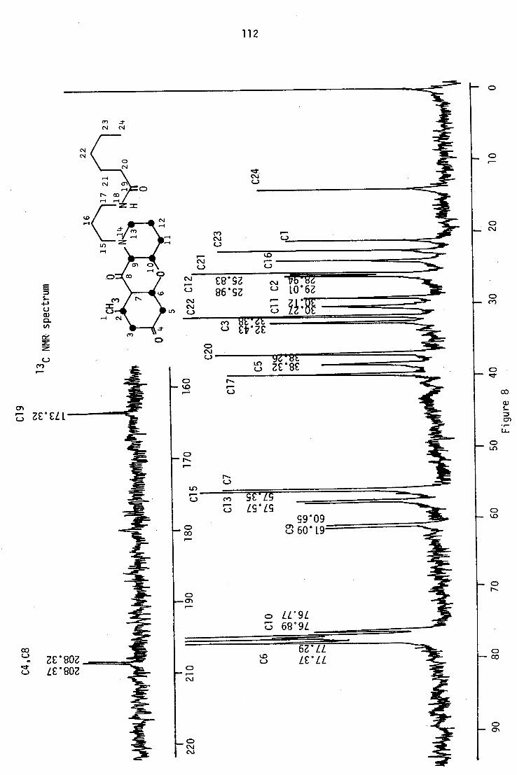
112
0
M 0.1
Q
cr.
Cr)
NMR
spec
tru
m
SZ 7It u 86'52 u 10 . 62
•-••• CY)
.-••••
CO

r-- •■■••••••1111P
113
• MMI.
,
"MIM•110
■•■■■■■■.
r--
CY)
(71
•■•

114
CHAPTER 7
Minor alkaloids of uncertain structure
7.1 Minor constituents of the bark extract
In addition to the four alkaloids described in Chapters 2 and 5,
a total of twelve other minor compounds were isolated from the three
main column chromatographic fractions of the bark extract. The very
small quantities isolated (< 25 mg) did not permit the complete
structural elucidation of these compounds.
The least polar fraction obtained from the column chromatography
of the bark extract produced three fractions after ptic separation: a
mixture of volatile compounds, peripentamine, and a second alkaloid
PBXM2.
The mixture of volatile compounds was distilled under high vacuum
in a kugelrohr and four compounds (PBVMA-PBVMD) were isolated.
7.1.1 2-Hydroxy-6-methylacetophenone (PBVMA) (I)
The compound of highest volatility, a colourless liquid, was
identified as 2-hydroxy-6-methylacetophenone l6 by spectroscopic comparison
with an authentic sample.

115
7.1.2 Dimethylsulphone (PBVMB)
The next most volatile compound deposited colourless crystals which
gave a negative Mayer's test for alkaloids. The PMR spectrum of these
crystals consisted of an isolated singlet at 2.9, and the molecular ion
appeared at m/z 94 in the mass spectrum. Comparison of the mass spectrum
89 with one reported in the literature revealed that the compound was dimethyl-
sulphone ((CH 3 ) 2S02 ), and this identification was confirmed by mixed melting
point determination with an authentic sample.
Dimethylsulphone has been reported from a plant source previously55
,
but no suggestions have been made about its biogenetic origin.
In animals, sulphones have been shown to be derived from the oxidation
of sulphoxides, which in turn are the oxidation products of thiols 56 .
If the same oxidation process is feasible in plants, dimethylsulphone may
be derived from dimethylsulphoxide which is known to be a constituent of
plant lignins.
7.1.3 PBVMC
The third compound, a liquid, which gave a positive Mayer's test for
alkaloids, could not be isolated in sufficient amounts for detailed
analysis. A 100 MHz PMR spectrum showed that it could be related to the
other C22 alkaloids.
7.1.4 Partial structural determination of PBVMD
The fourth compound was a white semi-solid soluble in chlorinated
hydrocarbons and lower alcohols. The compound gave a positive Mayer's
test for alkaloids. The tic spot of this compound appeared pink after
spraying with iodoplatinate reagent, but it turned white after a few
minutes. With Dragondroff's reagent, the tic spot remained white on an
orange background. Further attempts to improve the crystalline nature by

116
sublimation and by using various solvent systems have not been successful.
On electron impact mass spectrometry, the highest ion observed was
at m/z 183, but on chemical ionization two molecular species at m/z 455
and m/z 228 were observed. However, the former was found to be
dependent on the sample concentration and was identified as a dimer of
the m/z 228 species. High-resolution mass spectrometry showed the m/z 228
ion to have the composition C 10H 10204 , and the m/z 183 ion C 8 H 1 02 03 .
Microanalysis of the semi-solid gave C 54.74%, H 7.38% and N 9.07%;
C10H16N 2
04 requires C 53%, H 7% and N 12%.
The PMR spectrum of PBVMD (Figure 1) showed the presence of a total
of sixteen protons: 69.3 (1H, broad, exchangeable), 7.3 (1H, d), 5.7
(1H, d), 4.65 (1H, t), 3.8 (2H, d), 3.75 (2H, dq), 3.55 (2H, dq) and
1.2 (6H, t). The 13C NMR spectrum (Figure 2) consisted of eight signals
only: 6 164 (C), 150 (C), 146 (CH), 101.5 (CH), 164 (C), 150 (C), 146 (CH),
101.5 (CH), 100.3 (CH), 64.6 (CH 2 ), 51.0 (CH 2 ) and 15 (CH 3 ). However, as
the PMR spectrum showed the presence of two magnetically equivalent C 2H 5
groups, two of the 13
C signals at 664.0 and 15.0, were suspected to be
due to two carbons each. In order to clarify this possibility, the 13
C
NMR spectrum was recorded with an equimolar quantity of a shiftless
relaxation agent: chromium(III)-acetylacetonate 57 -5° . On addition of
this reagent, the intensity of the two signals •at 664.0 and 15.0 was
enhanced considerably but did not double as expected. These data appear
to favour the formula C10H16N204 for PBVMD.
The PMR spectrum had the following characteristics: an olefinic AX
system resonating at 7.3 (1H, d, J = 9 Hz) and 5.7 (1H, d, J = 9 Hz),
presumably an al3 unsaturated carbonyl system with a cis configuration,
which is further supported by the IR absorption at 1670 cm UV
absorption bands at 213 (log c 3.54) and 264 nm (3.60), and also an AX 2
system in the PMR spectrum resonating at 64.65 (1H, t, J = 5.7 Hz) and
3.8 (2H, d, J = 5.7 Hz), and two identical ABX 3 systems with signals at

117
o 3.7 (2H, dq, J = 7.1, 9.4 Hz), 3.45 (2H, dq, J = 7.1, 9.4) and 1.2 (6H,
t, J = 9.4).
On treatment with diazomethane, PBVMD formed a monomethyl derivative,
which indicates the presence of an enolic hydroxy function in the molecule.
Further, the presence of three methine and one quaternary carbon signals in
the olefinic region (o 150 (C), 146 (CH), 101.5 (CH) and 100.3 (CH))
confirmed the presence of two olefinic double bonds; one conjugated to
the carbonyl and the other bearing a hydroxy function.
The single proton triplet at 64.65 in the AX 2 system, therefore, can
be assigned to an olefinic proton, while the two-proton doublet at 63.8
can be attributed to an allylic methylene group bearing a heteroatom.
The remaining ABX3 systems can be assigned to two chemically
equivalent ethyl functions attached to hetero atoms. The geminal
coupling between the two methylene protons may be attributed to the
restricted rotation about the bond between the methylene carbon and the
heteroatom.
However, when proton decoupling experiments were carried out, the
presence of more complex second order couplings was observed, and it
became apparent that thetwo identical sets of signals at 63.7 and 3.5
were in fact not true doublets of quadruplets. Similarly, the signal at
61.2 also was shown to be not a true triplet.
When the signal at 61.2 was irradiated, the two signals at 63.7 and
3.5 were simplified to two doublets of doublets (J = 10.8, 3 Hz).
Conversely, when either of the two identical signals at 63.7 or 3.5 were
irradiated, the remaining signal and the six-proton signal at 61.2 changed
into complex multiplets. (Figure 3). This complex coupling may be
explained as second order coupling between the methylene protons with small
chemical shift differences.
On the available evidence the following partial structures can be put
forward for PBVMD. The accompanying figures indicate the observed PMR and
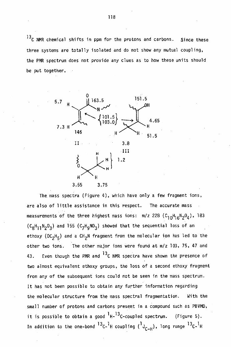
118
13C NMR chemical shifts in ppm for the protons and carbons. Since these
three systems are totally isolated and do not show any mutual coupling,
the PMR spectrum does not provide any clues as to how these units should
be put together.
0
5.7 163.5 H .,,.
151.5
FI-J'ij 1..ILL
1 'c'. 1101.51
7.3 H H
146 H 51.5
3.55 3.75
The mass spectra (Figure 4), which have only a few fragment ions,
are also of little assistance in this respect. The accurate mass
measurements of the three highest mass ions: m/z 228 (C 10H 10 204 ), 183
(y1 10203 ) and 155 (C7 H 9NO3 ) showed that the sequential loss of an
ethoxy (0C 2H 5 ) and a CH 2N fragment from the molecular ion has led to the
other two ions. The other major ions were found at m/z 103, 75, 47 and
43. Even though the PMR and 13C NMR spectra have shown the presence of
two almost equivalent ethoxy groups, the loss of a second ethoxy fragment
from any of the subsequent ions could not be seen in the mass spectrum.
It has not been possible to obtain any further information regarding
the molecular structure from the mass spectral fragmentation. With the
small number of protons and carbons present in a compound such as PBVMD,
it is possible to obtain a good 1H-
13C-coupled spectrum. (Figure 5).
In addition to the one-bond 13
C-1H coupling
1 ( J )
-C-H'' 13 1
long range C- H

119
2 coupling ( J c_H and
3J c _H ) could also be observed in this spectrum.
However, lack of sufficient information on 2J and
CC-H values for
C-H
nitrogenous compounds precluded the possibility of obtaining much useful
information from this spectrum.
The PMR and 13C chemical shifts of the two ethyl groups most strongly
suggest that they are present as ethoxy functions. However, the two
partial structures (II and III) put forward do not permit the placement
of these two equivalent ethoxy functions on carbons, and the only
remaining possibility is to locate them on the two tertiary nitrogens.
A natural product with a methoxy function on a nitrogen has been
reported61
, but much more substantial evidence would be needed before
the presence of two ethoxy functions on nitrogens in a single molecule
could be considered as established.
The lack of sufficient material did not permit any chemical work
to be carried out on this compound.
7.1.5 A tentative structure of PBXM2
The second minor alkaloid from this fraction, PBXM2, was also
isolated as a gum. Its tic spot stained purple with iodoplatinate
reagent, and the compound gave a +ve Mayer's test and a +ve Gibbs test.
The molecular ion of this compound appeared at m/z 388, and its
composition was shown to be C 22H32 N204 by high-resolution mass
spectrometry. The PMR spectrum had two low-field signals at 7.2 and
6.7 ppm corresponding to three protons, and a singlet at 2.45, also due
to three protons, which are characteristic of a 2-hydroxy-6-methylbenzoyl
-1 residue. An IR absorption at 1685 cm (ArC=0), a strong mass spectral
fragment at m/z 135 and a +ve Gibbs test further support the presence of
this unit in the molecule. The PMR and mass spectra also showed the
presence of the N(propyl)hexanamide unit which is common to most of the
Peripentadenia alkaloids. (IR absorptions at 3020 (br NH), 1670, 1645 cm-1

120
•••■•••.,
PMR
spec
trum
of PB
VM
D CD
C13
(27
0 M
Hz
)
s_ •r-LL-
•

121 0
LC)
CID
\
C \
0 CO
r•—•
C \J
CD Cv")
NMR
of P
BVM
D
Cr)
1•••••

122
H decoupling on PBVMD
4.5 4.0 3.5 3.0
2.5
2.0
1.5 PPM
Figure 3

100 1 3 200 80
60
040
20 245
80
243
40 -
47
43 75
MS OF PBVMD (CI) ammonia 113
228 35
100 200 300
MS OF PBVMD methyl ether 103
60-
170
197
20
123
103 A
MS of PBVMD (El)
75
II
100 200 300
80
60
1 70
40
20
Figure 4

vtAmil 104 102 100 98 68 66 64 62 60
124
Figure 5
54 52 50 48 18 16 14 12 PPM
H- 13C coupled spectrum of PBVMD

125
(CONHR); PMR signals at 6.5 (1H) and 3.4 ppm (2H) (-CH 2NHCO-) and a ms
fragment at m/z 156). The high relative abundance of the m/z 156 peak
also suggested that the N(propyl)hexanamide chain is on a tertiary
nitrogen. In the absence of any other side chains, the remaining
fragment of the molecule, C 5H 7 NO, which has 2 degrees of unsaturation,
must form part of a nitrogen heterocycle to satisfy this condition.
CH3
0
OH
C5H70N (CH 2 ) 3NHCOC 1
The PMR spectrum also showed the presence of a low-field one-proton
signal at 6.65 ppm, which suggested a trisubstituted olefinic double
bond, presumably conjugated (UV absorption bands at 215, 255 and 320 nm)
to the benzyl carbonyl so that the proton concerned is 13, to it. Another
PMR signal at 4.45 ppm for a non-exchangeable single proton showed that
the fourth oxygen is present as a secondary hydroxy function. From
the information it appeared that the nitrogen heterocycle is a
pyrrolidine nucleus bearing a hydroxyl function.
In view of the small sample available of this compound, no further
information could be obtained.
7.1.6 The more polar minor alkaloids of the bark extract
(PBXM3-PBXM9)
A further seven minor alkaloids were isolated in very small quantities
(<25 mg), and only their mass spectra could be satisfactorily recorded.
In certain cases, the PMR spectra also obtained, which appeared to be
very similar to those of other C 22 alkaloids; however, in the absence
of supporting data, the PMR and MS information remain insufficient for
structural elucidation.

126
The compounds isolated with their molecular ions, as determined by
electron impact (El) mass spectrometry, are listed in Figure 5.
Hosever, it must be noted that El mass spectrometry could be
unsatisfactory as a means of determining the molecular ions of some of
these compounds, as in the case of peripentamine.
compound highest mass/m/z
PBXM3 300
PBXM4 352
PBXM5 395
PBXM6 388
PBXM7 374
PBXM8 374
PBXM9 374
Figure 5
7.2 The minor constituents of the leaf extract
A total of six fractions were separated (PLM4-PLM9), and two of
these (PLM4 and PLM6) furnished crystals. Because of their high
polarity, and their low solubility in common solvents used for
spectroscopy, satisfactory physical data on the remaining four compounds
could not be obtained. The PMR spectra of the non-crystalline compounds,
when recorded in methanol, showed considerable line broadening, which
could be produced by some kind of tautomerism.
7.2.1 Methyl gallate62 (IV)
The more polar of the two crystalline compounds (PLM6), which
analysed for C8H80 5 by high-resolution mass spectrometry, was identified
as methyl gallate from its spectral data. Its identity was confirmed
by m.p., IR and PMR comparison with an authentic sample.
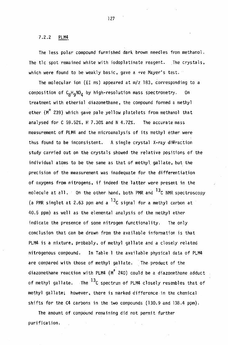
127
7.2.2 PLM4
The less polar compound furnished dark brown needles from methanol.
The tic spot remained white with iodoplatinate reagent. The crystals,
which were found to be weakly basic, gave a +ve Mayer's test.
The molecular ion (El ms) appeared at m/z 183, corresponding to a
composition of C8H9N04 by high-resolution mass spectrometry. On
treatment with etherial diazomethane, the compound formed a methyl
ether (M+ 239) which gave pale yellow platelets from methanol that
analysed for C 59.52%, H 7.30% and N 4.72%. The accurate mass
measurement of PLM4 and the microanalysis of its methyl ether were
thus found to be inconsistent. A single crystal X-ray diffraction
study carried out on the crystals showed the relative positions of the
individual atoms to be the same as that of methyl gallate, but the
precision of the measurement was inadequate for the differentiation
of oxygens from nitrogens, if indeed the latter were present in the
molecule at all. On the other hand, both PMR and 13 C NMR spectroscopy
(a PMR singlet at 2.63 ppm and a 13 C signal for a methyl carbon at
40.5 ppm) as well as the elemental analysis of the methyl ether
indicate the presence of some nitrogen functionality. The only
conclusion that can be drawn from the available information is that
PLM4 is a mixture, probably, of methyl gallate and a closely related
nitrogenous compound. In Table 1 the available physical data of PLM4
are compared with those of methyl gallate. The product of the
diazomethane reaction with PLM4 (M+ 240) could be a diazomethane adduct
of methyl gallate. The 13 C spectrum of PLM4 closely resembles that of
methyl gallate; however, there is marked difference in the chemical
shifts for the C4 carbons in the two compounds (130.9 and 138.4 ppm).
The amount of compound remaining did not permit further
purification.
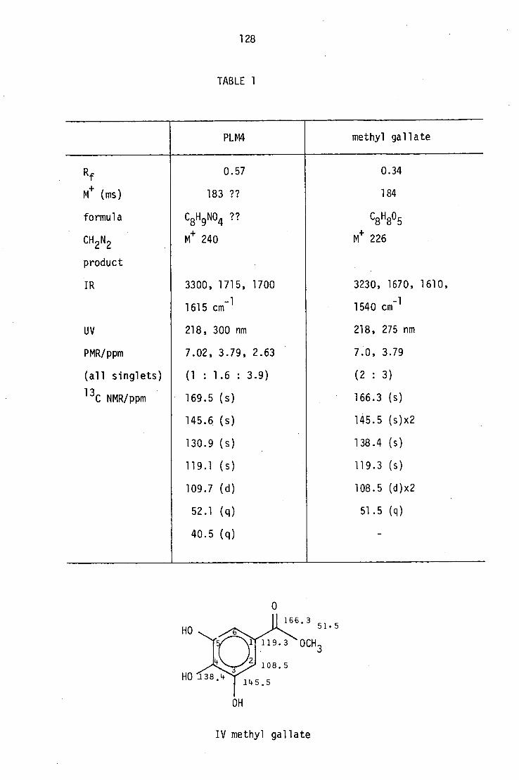
PLM4 methyl gallate
Rf
M+
(ms)
formula
CH 2 N2
product
IR
UV
PMR/ppm
(all singlets)
13C NMR/ppm
0.57
183 ??
C8 H 9
NO4 ??
M+
240
3300, 1715, 1700
1615 cm-1
218, 300 nm
7.02, 3.79, 2.63
(1 : 1.6 : 3.9)
169.5 (s)
145.6 (s)
130.9 (s)
119.1 (s)
109.7 (d)
52.1 (q)
40.5 (q)
0.34
184
C8H80 5
M+
226
3230, 1670, 1610,
1540 cm-1
218, 275 nm
7.0, 3.79
(2 : 3)
166.3 (s)
145.5 (s)x2
138.4 (s)
119.3 (s)
108.5 (d)x2
51.5 (q)
128
TABLE 1
OH
IV methyl gallate

129
CHAPTER 8
Biogenetic considerations
8.1 Introduction
A possible biogenetic pathway for both C16 and C12 EZaeocarpus
alkaloids has been put forward by Johns and Lamberton 1 " 15
according to which the elaeocarpine (I)-isoelaeocarpine ring
skeleton can be derived from the condensation of a C12 polyketide
chain and an ornithine unit, while the C12 alkaloids could be the
result of a similar process involving a C8 polyketide chain
(Scheme 1).
—4
CH /'.13 0/C° 3
Scheme 1

tryptamine CHO
IV
elaeocarpidine
130
The same authors have suggested that elaeocarpidine (IV), the only
indole alkaloid from this genus, could be derived from the condensation
of tryptamine, ornithine and a C3 unit 9 . (Scheme 2).
IV
Scheme 2
On the other hand Onaka has suggested that the nucleus of these
alkaloids could be derived through a similar process involving a C3
unit, ornithine and a polyketide chain (Scheme 3). He also has pointed
out that elaeocarpidine and 2-hydroxy-6-methyl acetophenone have both
been isolated from the same plant as (±) elaeocarpine and (±) iso-
elaeocarpine; all of these units, according to his suggestion, could
be derived from a common precursor35
.
Scheme 3

CH3
N HN
OH
VII R = C5H 11
VIII R = C3H7 Scheme 4
CH 0
OH
VI
131
8.2 Biosynthesis of Peripentadenia alkaloids
The bases isolated from this plant can be divided into two
groups: compounds with a nitrogen heterocycle, either pyrrolidine or
piperidine, in the molecule, and compounds with aliphatic amino amide
chains. The major alkaloid, peripentadenine (VII), dinorperipentadenine
(VIII), PLM3 (LX) and PBXM2 (LIX) belong to the first category, while
peripentamine (LV) and dehydroperipentamine (LVII) belong to the
latter.
The aromatic nucleus or its reduced form is most likely derived
from a polyketide unit of appropriate length by a process similar to
that of the naeocarpus alkaloids. The 2[methyl-(2-hydroxy-6-methyl
benzoyl)] pyrrolyl moiety (V) present in some of the Peripentadenia
alkaloids can be envisaged as derived from the condensation of a C10
polyketide chain with an ornithine unit, followed by decarboxylation
(Scheme 4). The C3 unit may be incorporated in a fashion similar
to that suggested by Johns and Lamberton9
for elaeocarpidine, either
before or after the condensation of the ornithine unit and the polyketide.
This could be followed by amination and acylation to give (VII) and
(VIII) (Scheme 4).

NH NH2
NH, c NH
NH2
X spermidine63
NH2 NH
XI spermine63
132
On the other hand, the open chain compounds could also be derived
• from the building up of a polyketide chain on a suitable nitrogen
substrate, but this remains biogenetically less attractive, since it
would require the formation of a polyketide chain with an odd number
of carbons (C13). It seems more likely, therefore, that the open-
chain compounds are derived from cyclic precursors.
The formation of the 2[methyl(2-hydroxy-6-methylbenzoyl)]pyrroly1
unit (V ) by the condensation of an ornithine unit and a polyketide
is biogenetically feasible, but the actual process by which the C3 unit
is introduced remains to be explained.
In order to find a parallel for this process, a search of the
literature was made for other natural products with a three-carbon
unit between two nitrogens. Some of the compounds with this
particular structural feature are listed below. Further, a number of
polypeptide alkaloids, a rapidly expanding class of natural products,
are also known to have a C3 unit joining two nitrogens. About twenty-
five of these alkaloids, belonging to either the homaline or the
lunarine type and carrying either a spermidine (X) or a homospermidine
moiety have been reported 67 .

so,H ;02H *CO 2H , CO 2 H
CO2H
133
XII a-c CH3 1
t2N R ,;1, .....'
CH 3 ,/" N NH N HN
CH 3
XIV a-b
XII - XIV acarnidin es 64 a R = CO(CH 2 ) 10CH3
R = CO(CH 2 ) 3CH=CH(CH 2 ) 5CH 3
c R = COC13H 21
XV (+) nicotianamine 65
XVII R = OH mugineic acid 66 XVIII R = H 2'-deoxymugineic acid 66
XVI (2R:3 1 R)-N-3- amino-3-carboxy propyl-azetiaine-2-carboxylic acid 65
H2
XIII a-c

H(CH0 2 ) 4
XXIII maytenine7°
134
HO
02H H CO H H CO H 2 , 2
N NOH
XIX avenic acid 66
10 11 CH3 (CH 2 ) 8CN(CH 3 )CH 2CONH(CH 2 ) 3 NCH 3 (CH2)4NCH3
68
XX
XXI 10,11-dehydro derivative 68
HO ( H ) 2CONH(CH 2 ) 3 NH(CH 2 ) 2
HO —2
. XX I I
Ph
HN H
. 1 XXIV R = (E) phCH = CHCO
Celacinine 71
XXV R = (Z) phCH = CHCO
Celallocinine 71
XXVI R = 3 furoyl
Celafurine 71
XXVII R = COph
Celabenzene 71
XXVIII R = OH
Mayfoline 71
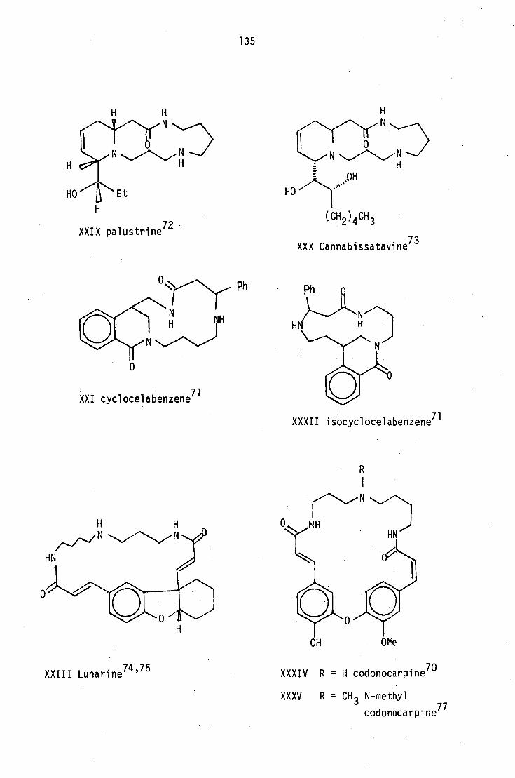
0 N
HO Et
XXIX palustrine72
N N H .
(CH 2 ) 4CH 3
XXX Cannabissatavine73
HN
135
XXI cyclocelabenzene 71
XXXII isocyclocelabenzene 71
()N H HN
OH
OMe
XXIII Lunarine74 '75
XXXIV R = H codonocarpine 70
XXXV R = CH 3 N-methyl codonocarpine 77
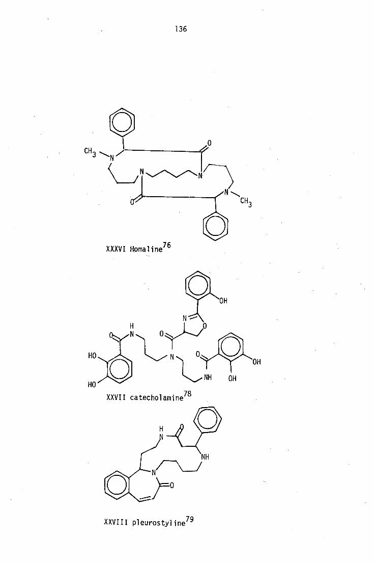
0
3 N
CH
OH
NH
XXVII catecholamine 78
OH
N
HO
HO
136
XXXVI Homaline 76
XXVIII p1eurostyline 79

HN
Ph
XLIII ephidradine 69
XLIV aphalandrine82
OH
I OH
HN OH
HN, .)
137
Ph
XXXIX periphylline 80
XL R1 = (CH2 ) 4CH3 , R2 = H Hopromine81
Hopromalino1 81 XLI R1 Ph, R2 OH
XLII R 1 = (CH2 ) 4 H , R2 = OH Hopromino1 81
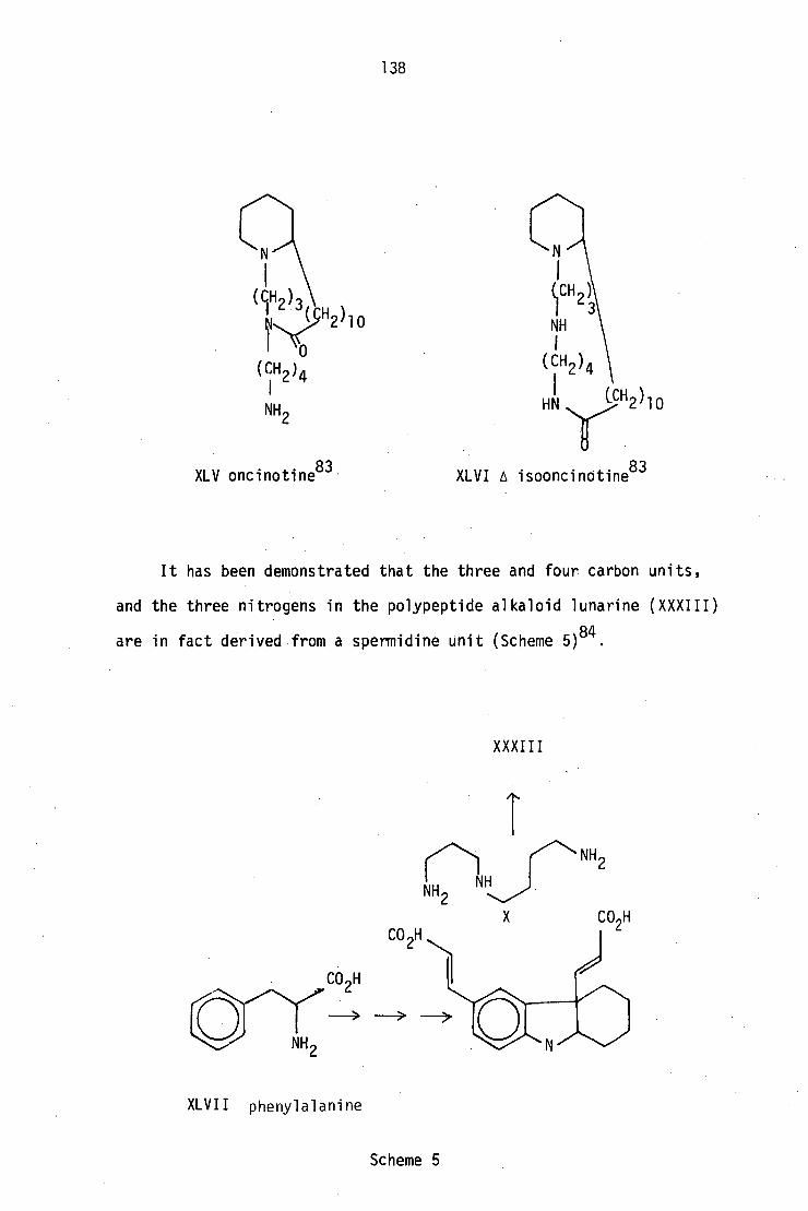
138
XLV oncinotine83
XLVI A isooncinotine83
It has been demonstrated that the three and four carbon units,
and the three nitrogens in the polypeptide alkaloid lunarine (XXXIII)
are in fact derived from a spermidine unit (Scheme 5) 84 •
XXXIII
NH2 NH
NH2
X
CO H
XLVII phenylalanine
Scheme 5

NH ■
2, CO2H CH 1
NH2
NH2 XLIX putrescine
XI spermine
139
The biosynthesis of the polyamine spermidine (X) which is
considered to be present in all animals and microorganisms and some
higher plants, has been thoroughly investigated. It has been shown
that spermidine is formed from putrescine (XLV) 88 ' 86 by the transfer
to it of a propylamine residue derived from decarboxylated S-adenosyl-
methionine (XLVI)87
(Scheme 6). Under normal conditions, S-adenosyl-
methionine acts as a methylating agent by transfer of its methyl
function to an oxygen or nitrogen.
In view of the experimental evidence in the case of spermidine89
it seems most reasonable to suggest that the C3 unit on the amine
nitrogen of Peripentadenia alkaloids could also be derived from
methionine. If this process does indeed take place in the plant,
it is quite likely that the reactions of methyl transfer and of C3
unit transfer may compete with each other. Methyl transfer should
produce the N-methylated derivative (L) (Scheme 7).
-CO2 NH 2
CO2H NH 2
XLVIII ornithine
adenosyl
'/NH2 NH2
X spermidine
Scheme 6

140
orni thine
CO 2 H CH3 transfer
V
LII R=C LIII R =C3H 7
3
OH LVI C5H 11
LX
CH 3
-00 2
0 S-adenosyl methionine
1 C3 unit transfer
- CH3
Scheme 7

CH3 oxidative
deamination
141
Isolation of an N-methyl derivative would undoubtedly lend support
to this hypothesis, but no compound of this type has so far been
detected in the plant.
The C3 unit mentioned in the proposed biosynthetic scheme for
elaeocarpidine (IV) 9 could also be derived from methionine. Further,
the other Elaeocarpus alkaloids may also be derived by this route from
the same precursors as Onaka35
has suggested (Scheme 8).
OH
elaeocarpine
isoelaeocarpine
Scheme 8
8.3 Inter-relationships between Peripentadenia alkaloids (Scheme 7)
The 1-pyrrolidinopropylamine intermediate (LI) can undergo
aromatisation followed by acylation to form peripentadenine (LII) and
dinorperipentadenine (LIII). On the other, hand the pyrrolidine nucleus

142
may undergo ring opening to form the open chain amino amide (LVI) which
may act as the precursor for both peripentamine (LV) and dehydro-
peripentamine (LVII).
The same open chain compound (LVI) could also undergo cyclisation,
perhaps through an.epoxide intermediate (LVIII) to form the piperidine
nucleus in PLM3 (LX) and PBXM2 (LIX). The hydroxy function on PBXM2,
whose position could not be fixed from the available data, can now be
located tentatively at C11, as the oxidation of the allylic position
would be preferred on biogenetic grounds.

143
CHAPTER 9
Experimental
Memorandum
The following general experimental details apply to all the
experimental sections:
Except for large-scale extractions of plant material, solvents
purified by standard methods were used for all purposes. The
evaporation of solvents was carried out under reduced pressure below
40°C.
For column chromatography, thin-layer chromatography (tic) and
preparative thin-layer chromatography (ptic), Camag silica gel DSF-5
was used. In some cases the silica gel was modified by making up
the slurry with 0.5 N potassium hydroxide solution instead of water,
which is referred to as KOH silica gel in the experimental section.
The thin-layer chromatograms were developed using a chloroform:methanol
(9:1) solvent system unless stated otherwise. On ptic plates, the
compound bands were visualised either by examination under UV light
or edge spraying with iodoplatinate reagent. The bands thus located
were separated, extracted with chloroform:methanol (1:1), evaporated,
redissolved in chloroform, dried (over Na 2SO4 ) and evaporated to give
the corresponding compounds.
As a number of ptic purification steps had to be carried out
prior to the isolation of a pure compound, in most of the cases the
percentage yields given may not represent the true composition of the
crude extract.
Melting points were determined using a Yanagimoto-Seisakusho
micro melting-point apparatus and are uncorrected.
Distillation of high-boiling substances was carried out from
144
bulb to bulb using a kugelrohr apparatus, and the oven temperature and
pressure were recorded.
Microanalyses were performed either by the Australian Microanalytical
Service or by Dr. A. Campbell, Chemistry Department, Otago University,
Dunedin, New Zealand. When the compounds could not be obtained
crystalline, high-resolution mass spectrometry (HRMS) was employed to
obtain the molecular formulae. In such cases, homogenity on tic with
several solvent systems and the consistency of their 13C NMR data were
used as the criteria for purity.
Infra red spectra (IR) were recorded on a Beckmann IR-33
spectrometer in chloroform unless otherwise stated. Ultra violet
spectra (UV) were recorded on a Hitachi-Perkin-Elmer 124 spectro-
photometer in methanol, and the logarithms of the extinction
coefficients were recorded.
Proton magnetic resonance spectra (PMR) were recorded in deutero-
chloroform at 100 MHz with .a Jeol JNM-4H-100 MHz instrument unless
otherwise specified. The 270 MHz PMR spectra and the carbon-13 NMR
spectra (13
C NMR) (67.89 Hz) were recorded with a Bruker HX 270
instrument. Tetramethylsilane was used as internal standard. Chemical
shifts (d) are given in ppm, and the coupling constants which were
confirmed by decoupling experiments are given in Hertz (Hz). The 13
C
chemical shifts recorded are for proton-decoupled spectra. The
multiplicity of the peaks in the off-resonance decoupled spectra are
given in parenthesis as singlet (s), doublet (d), triplet (0 or
quadruplet (q). Unless the full PMR spectra are given, only the salient
signals are described.
Specific optical rotations were measured in solvents specified in
the experimental section on a "PEPOL-60" spectropolarimeter.
Low resolution and high-resolution mass spectra (ms and HRMS) were
run on a Vacuum General Micromass 7070F instrument at 200° and 70 eV.

145
For chemical ionization (CI) spectra either isobutane or ammonia gas
were used. Peaks are listed in decreasing order of m/z ratios with
the intensities (parenthesis) expressed as a percentage of the base
peak.
Extraction of bark alkaloids
Plant materials for this investigation were collected at Boonjie,
north Queensland. Dried milled bark (14.5 Kg) was extracted by
percolation with ethanol at 40°C. The extract was concentrated under
reduced pressure, diluted with water and acidified with dilute sulphuric
acid, then filtered and made basic with ammonia. The crude alkaloids
were extracted with chloroform, and the chloroform solution was
repeatedly extracted with aqueous sulphuric acid (2 N) until no further
bases were removed. The crude alkaloids were then recovered by
basifying the combined acid extracts and re-extracting with chloroform.
Evaporation of the dried chloroform extract gave an oily residue of
crude alkaloids (90 g, 0.62%).
Fractionation of the bark alkaloid extract
The crude alkaloid extract (5.0 g) was chromatographed on a short
column88
of silica gel (200 g) packed in chloroform. The column was
eluted with chloroform-methanol solvent systems of increasing polarity.
The eluent was collected in 10 ml fractions, monitored by tic and
bulked accordingly. This gave five major fractions (A-E) (Table 1).
Further quantities of the crude extract (rl, 60 g) were fractionated in
similar fashion to obtain sufficient amounts of the more polar minor
alkaloids.

146
TABLE 1
Bulking summary of the fractions isolated from column chromatography
Fraction eluent CH
3:CH
3OH/volume ml
weight of recovered alkaloids/g
constituents
A 1:0 200 0.183 mainly 2-hydroxy-6-methyl
acetophenone
B 19:1 320 0.240 PBVMA-PBVMD
PBM2-peripentamine.
PBXM2
C 17:3 320 0.815 PB-peripentadenine
D 17:3 860 0.320 PB-peripentadenine
PBM3-dinorperipentadenine
PBXM3
- 3:1 380 - -
E 1:1 460 0.850 PBM4-dehydroperipentamine
PBXM4
I
PBXM9
- 1:0 500 - -
F CH3OH:H
20
9:1 200 1.050 highly polar material
containing some alkaloids

147
9.1 Experimental for Chapter 2
Peripentadenine. Fraction C gave a chromatographically pure viscous
brown oil in 16.3% yield, Rf 0.45 in chloroform:methanol (9:1) and 0.48
in chloroform:triethylamine (9:1). Subsequent separation of fraction D
furnished more of the same compound in 'v,2% yield. The physical
properties of the compound remained unchanged after further purification
by ptic and droplet countercurrent chromatography94 . The base could not
be obtained crystalline, and attempts to prepare a crystalline
derivative were likewise unsuccessful; UV Amax
228 nm (log E 3.96),
253 (3.36), 283 (3.15), 309 (3.15); UV X max (+0171) 210 nm (log c 4.08),
234 (3.99); IR vmax 3250-3020 (br NH and OH), 1690 (AT C=0), 1680 and
1650 (CONHR), 1630 cm C=0 H-bonded to ortho -OH); PMR (270 MHz)
(5 10.5 (1H, br m, Ar0H), 7.2 (1H, t, JH4/H3
= JH4/H5
= 7.5 H, H4);
6.72 (1H, d, J H5/H4
H3/H4 = 7.5 Hz, H3); 6.67 (1H, d, = 7.5 Hz, H5); -
5.72 (1H, m, NH, H18), 3.58 (1H, dddd, J1-110/
10•0, H9c J1110/H9b = 5 ' 0 '
.0 Hz); 3.47 (1H, dd, J 12.75 Hz, J1-110/H1la
= 8.3, JH10/H1lb = 5
- H9a/H9b =
= 10.0 Hz, H9a), 3.27 (1H, ddd, J J1-19a/H10 - H15a/H15b = 10 ' 5 ' '4115a/16Ha
= 6.3, = 4.5 Hz, H15a); 3.23 (1H, dd, J JH15a/H16b -H17a/H17b = 13 ' 5 '
= 6.0 Hz, H17a), 3.17 (1H, dddd, J JH17a/H18 - H17b/H17a = 13 ' 5 ' '3 1-117b/H16a
= 8.25, JH17b/H16b = H17b/H18
6.0, J = 6.0 Hz, H17b), 2.85 (1H, ddd, - -
0, J1-113a/H13b
= 12.0, JH13a/H12a
= 7.5, JH13a/H12b = 12.
J1-113a/H12b =
H13a); 2.47 (1H, dd,H9b/H9a
J = H9b/H10 =
12.75, J 5 Hz, H9b); 2.45 (1H, -
J ddd 5 Hz ., H13b); 2.43 (1H, , H,-a , dd, JH13b/H13a
= 12.0, 3H13b/H12b = 3
lb /H15b
= 7.5 Hz, H15a); 2.3 (3H, s, 3H1); = 10.5, J1-115a/H16a
= 8.5, JH15a
/H16b
2.12 (1H, ddd, J 12., J
- H11a/H1lb = 0 - 11a/H10 = 8 ' 3 ' J1-111a/H12a = 8.0 Hz,
Hlla); 2.05 (2H, t,J = 8.25 Hz); 1.92 (1H, m, H16a); 1.72 (1H, - 20/21
m, H16b); 1.7 (1H, m, H11b), 1.65 (2H, m, 2H12); 1.57 (2H, tt, J - 21/20 =
= 8.25 Hz, 2H21); 1.3-1.2 (4H, m, 2H22, 2H23); 0.9 (3H, t, J21/22
= 8.25 Hz); on addition of D20, the peaks at 10.5 and 5.75 ppm J24/23
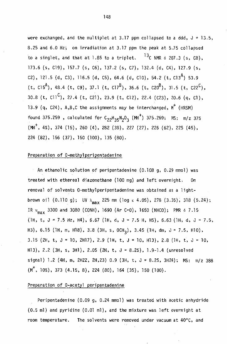
148
were exchanged, and the multiplet at 3.17 ppm collapsed to a ddd, J = 13.5,
8.25 and 6.0 Hz; on irradiation at 3.17 ppm the peak at 5.75 collapsed
to a singlet, and that at 1.85 to a triplet. 13
C NMR 6 207.3 (s, C8),
173.6 (s, C19), 157.7 (s, C6), 137.2 (s, C7), 132.4 (d, C4), 127.9 (s,
C2), 121.5 (d, C3), 116.5 (d, C5), 64.6 (d, C10), 54.2 (t, C13 A ) 53.9
(t, C15A), 48.4 (t, C9), 37.1 (t, C17
B), 36.6 (t, C20
B), 3
1.5 (t, C22
c),
30.8 (t, Cl1 C ), 27.4 (t, C21), 23.9 (t, C12), 22.4 (C23), 20.6 (q, Cl),
13.9 (q, C24), A,B,C the assignments may be interchanged, M+
(HRSM)
found 375.259 , calculated for C22H 35 N20 3 (Me) 375.269; MS: m/z 375
(Me, 4%), 374 (15), 260 (4), 282 (38), 227 (27), 226 (62), 225 (45),
224 (82), 156 (37), 150 (100), 135 (80).
Preparation of 0-methylperipentadenine
An ethanolic solution of peripentadenine (0.108 g, 0.29 mmol) was
treated with ethereal diazomethane (100 mg) and left overnight. On
removal of solvents 0-methylperipentadenine was obtained as a light-
brown oil (0.110 g); UV A max 225 mm (log E 4.05), 278 (3.35), 318 (5.24);
IR vmax 3300 and 3080 (CONK), 1690 (AT C=0), 1650 (NHCO); PMR 6 7.15
(1H, t, J = 7.5 Hz, H4), 6.67 (1H, d, J = 7.5 H, H5), 6.63 (1H, d, J = 7.5,
H3), 6.15 (1H, m, H18), 3.8 (3H, s, OCH 3 ), 3.45 (1H, dm, J = 7.5, H10),
3.15 (2H, t, J = 10, 2H17), 2.9 (1H, t, J = 10, H13), 2.8 (1H, t, J = 10,
H13), 2.2 (3H, s, 3H1), 2.05 (2H, t, J = 8.25), 1.9-1.4 (unresolved
signal) 1.2 (4H, m, 2H22, 2H,23) 0.9 (3H, t, J = 8.25, 3H24); MS: m/z 388
(Mt , 10%), 373 (4.15, 8), 224 (80), 164 (35), 150 (100).
Preparation of 0-acetyl peripentadenine
Peripentadenine (0.09 g, 0.24 mmol) was treated with acetic anhydride
(0.5 ml) and pyridine (0.01 ml), and the mixture was left overnight at
room temperature. The solvents were removed under vacuum at 40°C, and

149
the residue, purified by ptic, was obtained as a brown gum (0.068 g, 68%);
UV Amax 215 nm (log e 3.06), 305 (2.89); IR 'max 3300 (CONH), 1765
-1 (ArOCOCH 3 ), 1680, 1655 (NHC=0), 1640, 1625 cm ; PMR 6 6.8 (1H, br m,
NHCO), 2.27 (3H, s, COCH 3 ), 2.21 (3H, s, ArCH 3 ); MS: m/z 416 (M+ , 80%),
415 (75), 401 (20), 325 (13), 374 (46), 301 (56), 300 (62), 224 (10),
185 (58), 161 (43), 156 (55), 135 (100), 134 (37).
Acid hydrolysis of peripentadenine
A solution of peripentadenine (0.3 g, 0.8 mmol) in methanol (10 ml)
was treated with aqueous hydrochloric acid (10%, 90 ml) and the mixture
was heated under reflux until peripentadenine could no longer be detected
by tic. The methanol was removed under vacuum, and the aqueous phase
was extracted with chloroform (3 x 50 m1). The extract was dried and
evaporated to give a colourless gum (0.060 g, 64%); IR v max 3000, 1760,
1710 cm-1
. An ethereal solution of the gum (0.06 g) was treated with
ethereal diazomethane, left overnight, and evaporated to give an oil
(0.065 g) which was identified as methylhexanoate by glc-ms comparison
with an authentic sample. The aqueous phase was basified with ammonia
and extracted with chloroform (3 x50 ml); the residue obtained after
removal of solvents was purified by ptic to give a brown oil (0.074 g,
34%); UV Amax 225 nm (log 6 3.93), 255 (3.51), 315 (3.18); IR "max
3400-3200 (NH, OH), 1690, 1680, 1650, 1640, 1600 and 1590 cm-1; PMR 6
7.15 (1H, t, J = 7.5 Hz), 6.8 (2H, dd, J = 7.5 Hz), 3.5 (1H, m), 2.7
(2H, t, J = 6.5), 2.6 (3H, s), 2.4-2.0 (4H, m), 1.8-1.6 (6H, m), 1.2
(2H, br m, exchanged with D 20, NH 2 ); m/z 276 (M+
2%), 246 (18), 185 (30),
150 (60), 136 (100); HRMS found 276.1876, calculated for C 16H24 N202
276.1852.

150
Hofmann degradation of peripentamine
A solution of peripentamine (0.5 g, 0.14 mmole) in acetonitrile
(10 ml) was heated with methyl iodide (1 ml) to 100°C in a sealed tube
for 5 hrs. Further methyl iodide (4 x 1 ml) was added at 10 hr
intervals. The solvent was removed under vacuum, and the methiodide
salt, separated from unreacted starting material by ptic, was obtained
as a yellow gum (0.170 g, 24.5%); HRMS: found 389.2246, calculated
for C23 H37 N 203 389.2823. The methiodide was converted into the
methofluoride (0.11 g, 90%) by passing an aqueous methanol solution
(1:1, 50 ml) through an Amberlite IRA 400 (F - form, 20 g) column.
The methofluoride gave m/z 389 (M + , 4%), 388 (12), 314 (6), 247 (11),
246 (62), 201 (18), 200 (9), 199 (57), 160 (9), 156 (52), 135 (36),
98 (10), 84 (13), 70 (18), 58 (100). A sample of the methofluoride
(0.10 g) was pyrolysed in a kugelrohr at 220° and 6.3 x 10 -4 Hg mm,
and a colourless gum was obtained (0.085 g, 89%); UV X max 222 nm
(log E 2.68), 258 (2.68), 320 (2.42); IR vmax 3340 (NH), 1710, 1680,
-1 1650, 1600 cm ; PMR o 7.3 (1H, dd, J = 7.5 Hz, H4), 7.0(1H, br t,
exchanged with D 20, CONN), 6.8 (2H, dd, J = 7.5, H3, H5), 4.4 (1H, m,
H10), 3.35 (2H, dt, JH17/H18 = I-117/H16 = 6.0 Hz, 2H17), 2.67 (2H, m, J
2H9), 2.62 (3H, s, N-CH 3 ), 2.51 (2H, t, J H15/H16 = 6.5 Hz, 2H15), 2.47 -
(2H, t, H13/H12 = 6.5 Hz, 2H13), 2.28 (3H, s, 3H1), 2.15 (2H, t, j
JH20/H19 = 8.5 Hz ' 2H20), 1.8-1.75 (4H, m, 2H11, 2H12), 1.73 (2H, tt,
tt 1 5 Hz) ., .61 (2H, , J jH16/H15 = JE116/H17 = 8
H21/H20 = '31121/H22 = 8 ' 5 '
21-121), 1.3 (4H, m, 2H22, 2H23), 0.88 (3H, t, J 8.5 Hz); H24/H23 =
13C NMR IS 193.7 (s, C8), 173.1 (s, C19), 162.5 (s, C6), 142.1 (s, C7),
134.6 (d, C4), 126.2 (s, C2), 124.5 (d, C3), 115.7 (d, C5), 76.5 (d, C10),
57.5 (t, Cl3A), 56.6 (t, Cl5A ), 44.5 (q, N-CH 3 ), 41.6 (t, C10), 39.0 (t,
C17), 36.9 (t, C20), 32.6 (t, CH), 31.5 (t, C22), 25.9 (t, C16 B ), 25.5
(t, C21 B ), 22.7 (t, C23B ), 22.5 (q, Cl), 22.4 (t, C12 B ), 13.9 (q, C24),

151
A,B:assignments may be interchanged; HRMS: found 389.,2794, calculated
for C231-1 37 N 203 (Me) 389.2804.
Hofmann degradation of the first Hofmann product
The product obtained from the previous experiment (0.09 g, 0.23 mmol)
was converted to the methofluoride (0.057 g, 58%) by the same procedure
as for peripentadenine and gave m/z 403 (+ M , 15%), 388 (5), 246 (47),
225 (2), 201 (24), 199 (44), 160 (10), 157 (10), 156 (75), 135 (28),
134 (10), 105 (11), 99 (18), 84 (12), 58 (100). When subjected to
pyrolysis under similar conditions to those in the previous experiment,
a dark-brown sublimate (0.03 g, 52%) was produced, which proved to be a
complex mixture from which no pure substance could be obtained.
LAH reduction of peripentadenine
A solution of peripentadenine (0.374 g, 1 mmol) in dry dimethoxyethane
(20 ml) was added dropwise to a stirred mixture of LAH (0.300 g, 7.9 mmol)
in dimethoxyethane (30 ml) under anhydrous conditions. The mixture was
stirred for 4 hrs at room temperature, then treated successively with
water (0.3 ml), aqueous NaOH (15%, 0.3 ml) and more water (1 m1). The
white precipitate formed was filtered off and washed with methanol
(50 ml), and the combined washings and filtrate were evaporated under
vacuum to removethe.organic solvents. The remaining aqueous phase was
diluted with water, basified with ammonia, and extracted with chloroform
(3 x 30 m1). The extract was dried and evaporated to a gum (0.4 g)
which was separated by ptic into a less polar fraction (0.085 g, 22%),
UV xmax 235 nm (log e 3.44), 308 (2.90); IR vma x
3300-3200 (NH, OH),
-1 1590, 1460, 1380, 1260, 1050 cm ; PMR o 5.25 (1H, dd,J 10,
- H8/H9a =
118/H9b = 3.5 Hz, H8); m/z 362 (M + , 85%), 344 (15), 291 (3), 234 (6), J
230 (12), 225 (38), 220 (23), 217 (21), 211 (40), 210 (52), 209 (100),

152
182 (65), 135 (96); and a more polar base (0.076, 20%), UV xmax 230 nm
(log E 3.38), 310 (2.89), IR vmax 3400-3100, 1590, 1580, 1560, 1460,
1270, 1020 cm-1
; PMR 6 5.55 (1H, dm, J -H8/H9 = 12.5 Hz, H8); m/z 362
(M+ , 65%), 334 (6), 230 (15), 225 (45), 220 (30), 202 (40), 201 (65),
200 (85), 199 (100), 136 (85). Several other compounds of higher
polarity were found to be present but they could not be completely
purified.
Hofmann degradation of the LAH reduction product
The less polar of the two LAH reduction products of peripentadenine
(0.058, 0.16 mmol) was converted into its methofluoride in the usual
manner. MS: m/z 406 (6%), 392 (60), 369 (85), 242 (35), 221 (100),
199 (80), 152 (62), 150 (60), 129 (70). When the methofluoride was
subjected to pyrolysis as described earlier, a complex mixture was
obtained from which no single product could be separated pure.
Sodium borohydride reduction of peripentadenine
Peripentadenine (1.0 g, 2.7 mmol) was dissolved in aqueous methanol
(0.5 ml H 20, 30 ml methanol), and sodium borohydride (0.4 g, 10 mmol),
was added in small quantities with constant stirring. The mixture was
left overnight, then diluted with water (120 ml), acidified with dilute
sulphuric acid, and extracted with chloroform (2 x 50 mg. The extract
on evaporation gave a colourless gum (0.021 g) which was not further
examined. The aqueous phase on basification and extraction with
chloroform (3 x 50 ml) gave a yellow gum (0.723) which on ptic
separation yielded three bases. The presence of two more minor bases
was detected but their quantities did not permit isolation. The least
polar fraction formed white crystals of dihydroperipentadenine A
21 ° (0.630 g, 63%), m.p. 78° (from acetone); MD 0 (in methanol and in

153
chloroform); UV xmax 217 nm (log C 4.57), 276 (4.17); IR vmax 3300
(OH, NH), 1645 (CONH), 1600 cm-1
; PMR 6 5.95 (1H, t, Hi8/H17 = 7 Hz,
NHCO, H18), 5.53 (1H, dd,J 11.3, J 2.5 Hz, H8); - H8/H9a = H8/H9b =
MS: m/z 376 (Mt , 70%), 375 (100), 362 (10), 360 (75), 249 (8), 225
(90), 156 (47), 136 (42), 135 (50), 121 (80); found C 69.74%, H, 9.94%,
N 7.53; calculated for C 22H 30203 C 70.21%, H 9.65%, N 7.45%. The
compound of intermediate polarity gave a brown gum (0.027 g, 3%), [a] 1 =
0° (in methanol and in chloroform); UV xmax 210 nm (log e 3.57), 240 (2.93),
-1 285 (2.67); IR vmax 3300, 1670, 1645, 1630, 1600 cm ; PMR 6 8.0 (1H,
broad, exchangeable), 6.35 (1H, br m, NHCO), 5.5 (1H, dm, J = 10 Hz, WOH);
MS: m/z 376 (35%), 375 (19), 362 (42), 361 (23), 359 (45), 136 (19), 135
(100), 134 (36), 126 (16), 125 (66). The most polar fraction gave
dihydroperipentamine B, crystallised as a fine powder from acetone-water,
12, m.p. 70 0 (0.080 g, 8%); [a] 1 . 00 (inmethanol and in chloroform); UV
Amax 208 nm (log e 4.22), 218 sh (4.06), 275 (3.53); IR Vmax
3300 (NH,
-1 OH), 1645 (CONH), 1600 cm ; PMR 6 6.26 (1H, t, J H18/H17 = 6.2 Hz, H18),
-
5.30 (1H, dd,H8/H9c
J = H8/H9b =
10.9, J 2.3, CHOH, H8); MS: m/z 376 - -
(11+ , 45%), 375 (58), 361 (40), 360 (45), 359 (60), 245 (12), 226 (20),
225 (100), 121 (70).
Dehydration of dihydroperipentadenine A
A solution of dihydroperipentadenine A (0.2 g, 0.53 mmol) in 10%
aqueous oxalic acid (50 ml) was refluxed for 6 hrs, allowed to cool,
basified with ammonia, and extracted with chloroform (4 x 30 m1). The
extract on evaporation gave dehydroperipentadenine (0.182, 95%) as a
yellow gum; UV Amax 217 nm (log E 4.24), 255 (3.92), 297 (3.38), IR
-1 vmax
3300, 1650, 1600, 1590 cm ; PMR 6 7.3 (2H, m, H4, H9), 7.0 (1H,
br t, NHCO, H18), 6.8 (3H, m, H3, H5, H8), 3.37 (1H, m, H10), 3.28 (2H,
m, 2H17), 3.08 (2H, m, 2H13), 2.3 (3H, s, 3H1), 2.28 (2H, t, J H15/H16 =
7 Hz, 2H15), 2.05 (2H, t, J -H20/H21 = 8 Hz, 2H20), 1.95-1.65 (6H, m, 2H11,

154
2H12, 2H16), 1.5 (2H, tt, JH21/H20 = J
H21/H22 = 8 Hz, 2H21), 1.3-1.1
(4H, m, 2H22, 2H23), 0.88 (3H, t, JH24/H23 = 8 Hz, 3H24); 13C NMR -
173.7 (s, C19), 155.0 (s, C6), 137.4 (s, C7), 134.0 (d, C8), 127.7
(d, C4A), 127.5 (d, C9A ), 122.7 (s, C2), 121.7 (d. C3), 113.8 (d, C5),
70.5 (d, C10), 53.4 (t, Cl3 B ), 51.9 (t, Cl5 B ), 38.2 (t, C17), 36.7 (t,
C20), 31.6 (t, C22 C ), 31.4 (t, C11 C ), 37.0 (t, C16), 25.4 (t, C21),
22.3 (t, C23 p), 22.1 (t, C12 p), 20.7 (q, C1), 13.9 (q, C24); MS: m/z
359 (Me, 20%), 358 (M+ , 60), 342 (12), 315 (5), 280 (18), 231 (85),
226 (70), 202 (98), 185 (70), 156 (80), 149 (100).
A sample of dihydroperipentamine a (0.08 g, 0.21 mmol) when treated
in the same way as for dihydroperipentamine gave a product identical
with that as described above (0.068, 89%).
Sodium borohydride reduction of 0-methylperipentadenine
To a solution of 0-methylperipentadenine (2.547 g, 6.56 mmol) in
methanol (150 ml) and water (1 ml), sodium borohydride (0.8 g) was
added in small portions over a period of 6 hrs with constant stirring
at room temperature, then the mixture was left overnight. The solvent
was removed under vacuum and the residue was dissolved in chloroform
(100 ml) and extracted with dilute sulphuric acid (10%, 3 x 50 ml).
The chloroform-soluble fraction from ptic gave 2-methoxy-6-methyl
acetophenone (0.3 g, 27%): UV Amax 220 nm (log 4.55), 242 (3.63),
280 (3.56); IR vmax
1690 (AT C=0), 1600, 1590 cm -1 ; PMR 6 7.2 (1H, dd,
J = 7.5 Hz), 6.8 (1H, d, J = 7.5 Hz), 6.75 (1H, d, J = 7.5 Hz), 3.8 (3H,
s, OCH 3 ), 2.4 (3H, s, COCH 3 ), 2.25 (3H, s, ArCH 3 ); and l(2-methoxy-6-
methylphenyflethanol (0.042 g, 3.8%); UV A max 220 nm (log E 4.61), 275
(3.13), 280 (3.13); IR vmax 3300, 1600, 1590 cm -1 ; PMR 6 7.1 (1H, dd,
J = 7.5 Hz), 6.8 (1H, d, J = 7.5 Hz), 6.72 (1H, d, J = 7.5 Hz), 5.25
(1H, q, J = 7.5 Hz), 3.9 (3H, s, OCH 3 ), 2.19 (3H, s, ArCH 3 ), 1.45 (3H,
d, J =7.5 Hz, CHOHCH 3 ). The aqueous phase was basified, extracted with

155
chloroform (3 x 50 ml), and evaporated to give a brown gum (2.3 g) which
on ptic yielded 0-methyldihydroperipentadenine A (1.97 g, 79.8%); UV
Amax
208 nm (log E 3.78), 285 (3.00), 320 (2.79); IR v max 3300, 1690,
1660, 1640 and 1600 cm -1 ; .PMR 6 4.8 (1H, dd, J = 10, 3.5 Hz, CHOH); MS:
m/z 390 (M+ ); and 0-methyldihydroperipentadenine B (0.06 g, 2.3%);
UV Amax
225 nm (log 6 3.80), 280 (2.97), 335 (2.77); IR vmax 3300,
-1 1690, 1665, 1650 cm ; also some highly polar material (0.36 g, 19%)
which could not be satisfactorily purified; MS: m/z 226 (6), 225 (3),
224 (85), 156 (100), 128 (36), 98 (72), 97 (65), 84 (79), 70 (58), 58
(62).
Dehydration of 0-methyldihydroperipentadenine
A solution of 0-methyldihydroperipentadenine (1.97 g, 5.05 mmol)
in aqueous oxalic acid (10% 200 ml) was refluxed for 6 hrs, cooled,
basified, and extracted with chloroform (3 x 50 ml). Removal of
solvents yielded the dehydro compound as a yellow gum (1.49 g, 80%);
UV Amax
223 (log E 3.90), 258 (3.62), 295 (3.23); IR v 3300, 1660, MaX -1
1650, 1590 cm ; PMR cS 7.15 (1H, dd, H4/H3 = H4/H5 = 7.5 Hz, H4), -
6.8 (1H, d, J -H3/H4
= 7.5 Hz, H3), 6.78 (1H, d, J H5/H4 = 7.5 Hz, H5),
6.55 (1H, d, J - H8/H9 = 16 Hz,
H8), 6.0 (1H, dd, J H9/H8 = 16 '
7.5 Hz, H9), 3.8 (3H, s, OCH 3 ); MS: m/z 372 (M+ , 28%), 357
(30), 226 (80), 225 (100), 216 (65), 201 (18), 200 (20), 185
156 (87), 135 (50).
H9/H10 =
(s), 244
(36),
Hofmann degradation of 0-methyldehydroperipentadenine
The product from the above reaction (1.49, 4.01 mmol) was converted
to its methiodide and thence to the methofluoride (1.10 g, 92.5%) as
described for peripentadenine. Pyrolysis of the methofluoride at 160°
and 6.3 x 10⁻⁴
Hg mm, and purification of the product by ptic gave a
yellow gum (0.645 g, 69%); UV A max 2.8 nm (log E 3.30), 208 (3.00);

156
IR v max
3300, 1660, 1650, 1575 cm-1
; PMR (270 MHz) 6 7.1 (1H, dd,
H4/H3 = JH4/H5 = 7.5 Hz, H4), 6.85 (1H, d, J -H8/H9 = 16 Hz, H8), 6.8 J
(1H, d, J H3/H4 = 7.5 Hz, H5), 6.78 (1H, d, J -H5/H4 = 7.5 Hz, H4), 6.55 -
(1H, dd,J , H9), .26 (1H, dd, J - H9/H8 = 1 6 Hz, JH9/H10 = 9 Hz 6 - H10/H11 =
15 Hz, =
J 9 Hz, H9), 5.75 (1H, dt, J = 15 Hz, J H10/H9 -H11/H10 - H11/H12 =
7 Hz, H11), 3.35 (2H, dt,H17/H18 J
= JH17/H16 = 6.5 Hz, 2H17), 2.45 -
(2H, t, H15/H16 = 7 Hz, 2H15), 2.35 (3H, s, N-CH 3 ), 2.34 (2H, t, J
1113/H12 = 7 Hz, 2H13), 2.25 (3H, s, 3H1), 2.2 (2H, m, 2H12), 2.15 J
(2H, t, J , 2H20), .68 (2H, , J 7 Hz, - H20/H21 = 8 Hz 1 tt H16/H15 = H16/H17 =
2H16), 1.6 (2H, tt, J H21/H20 = H21/H22 = 8 Hz), 1.25 (4H, m, 2H22, 2H23), -
0.88 (3H, t, J H24/H23 = 8 Hz, 3H24); MS: m/z 386 (M t , 2%), 384 (8), -
372 (10), 341 (26), 244 (20), 230 (22), 216 (25), 200 (26), 199 (100),
156 (80).
Second Hofmann degradation
The above-mentioned Hofmann degradation product (0.695 g, 1.8 mmol)
was converted to the methofluoride (0.233 g, 30.8%) as before; pyrolysis
under similar conditions gave a brown oil (0.211 g), which after ptic
yielded a less polar, non-basic, semi-solid (0.065, 30%) UV x max 208 nm
(log E 4.06), 275 (3.62), 292 (3.62), 310 (3.57); IR vmax 1650, 1600,
1590 cm -1 ; PMR d 7.1 (1H, m), 6.7 (2H, m), 6.2 (2H, m), 5.6 (2H, m),
3.8 (3H, s, OCH 3 ), 2.35 (3H, s, ArCH 3 ); MS: m/z 201 (MN, 65%), 200
(25), 187 (12), 177 (36), 175 (28), 149 (60), 135 (100), 128 (80), 121
(38), 115 (95), 105 (56), 91 (72), 77 (68); and N-[3-(dimethylamino)
propyl]hexanamide as an oil (0.038 g, 18%), IR v max 3450 or 3300 (NHCO),
• 1665 (NHCO), 1450, 1430, 1420, 1180, 1050 cm -1 ; PMR (270 MHz) d 7.0
(1H, m, CONH), 3.34 (2H, dt, J = 6.5, 6.5 Hz), 2.45 (2H, t, J = 6.5 Hz),
2.28 (6H, s, N(CH 3 ) 2 ), 2.16 (2H, t, J = 7.3 Hz), 1.69 (2H, tt, J = 7.3,
7.3 Hz), 1.61 (2H, tt, J = 6.5, 6.5 Hz), 1.29 (2H, tt, J = 7.3, 7.3 Hz),
1.25 (2H, tq, J = 7.3, 7.3 Hz), 0.87 (3H, t, J = 7.3, -CH 3); MS: m/z
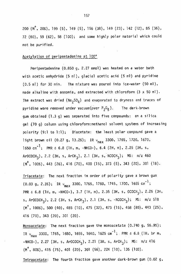
157
200 (Mt , 20%), 199 (5), 149 (5), 156 (38), 149 (23), 142 (12), 85 (36),
72 (80), 59 (62), 58 (100); and some highly polar material which could
not be purified.
Acetylation of peripentadenine at 100 0
Peripentadenine (0.850 g, 2.27 mmol) was heated on a water bath
with acetic anhydride (5 ml), glacial acetic acid (5 ml) and pyridine
(0.5 ml) for 30 min. The mixture was poured into ice-water (50 ml),
made alkaline with ammonia, and extracted with chloroform (3 x 50 m1).
The extract was dried (Na 2SO4) and evaporated to dryness and traces of
pyridine were removed under vacuum(over P 20 5 ). The dark-brown
gum obtained (1.3 g) was separated into five compounds: on a silica
gel (70 g) column using chloroform:methanol solvent systems of increasing
polarity (9:1 to 1:1); Diacetate: the least polar compound gave a
light brown oil (0.27 g, 13.2%); IR v max 3300, 1765, 1720, 1670,
1650 cm-1
; PMR 6 6.8 (1H, m, -NHCO-), 6.4 (2H, m), 2.25 (3H, s,
ArOCOCH 3 ), 2.2 (3H, s, ArCH 3 ), 2.1 (3H, s, NCOCH 3 ); MS: m/z 460
(M+ , 100%), 443 (26), 416 (70), 400 (15), 373 (5), 343 (20), 301 (18).
Triacetate: The next fraction in order of polarity gave a brown gum
(0.03 g, 2.3%); IR vmax 3300, 1765, 1760, 1745, 1700, 1655 cm-1
;
PMR 6 6.8 (1H, m, -NHCO-), 3.7 (1H, m), 2.35 (3H, s, OCOCH 3 ), 2.25 (3H,
s, ArOCOCH 3 ), 2.2 (3H, s, ArCH 3 ), 2.1 (3H, s, -NCOCH 3 ); MS: m/z 518
(Mt , 100%), 500 (40), 485 (10), 475 (32), 473 (15), 458 (80), 443 (25),
416 (70), 343 (20), 301 (20).
Monoacetate: The next fraction gave the monoacetate (0.740 g, 56.9%);
-1 IR vmax
3300, 1765, 1680, 1655, 1640, 1625 cm ; PMR 6 6.8(1H, br m,
-NHCO-), 2.27 (3H, s, ArOCOCH 3 ), 2.21 (3H, s, ArCH 3 ); MS: m/z 416
(M+ , 80%), 415 (75), 401 (20), 301 (56), 224 (10), 135 (100).
Tetraacetate: The fourth fraction gave another dark-brown gum (0.02 g,

158
1.5%); IR vmax 1770, 1765, 1745, 1700, 1650, 1640 cm -1 ; PMR 6 3.7
(1H, m), 2.35 (3H, s, OCOCH 3 ), 2.75 (3H, s, ArCOCH3 ), 2.2 (3H, s,
ArCH3 ), 2.15 (3H, s, NCOCH3 ), 2.1 (3H, s, NCOCH 3 ); MS: m/z 560
(M+ 20%), 458 (45), 416 (40), 214 (100).
Transacylated compound: the fraction of highest polarity (0.066 g, 5.1%),
IR vmax 3300 ' 1765 ' 1750, 1690, 1650 cm-1
; PMR 6 6.8 (1H, m, -NHCO-),
2.25 (3H, s, ArOCOCH 3 ), 2.2 (3H, s, ArCH 3 ), 2.1 (3H, s, NHCOCH 3 )*,
MS: m/z 360(M 30%), 318 (10), 300 (8), 258 (12), 135 (100).
Permanganate oxidation of peripentadenine
Peripentadenine (0.120 g, 0.2 mmol) was dissolved in acetone (5 ml)
that had been freshly distilled over potassium permanganate. To this
solution was added a freshly prepared solution of potassium permanganate
in acetone (1%, 14 ml) until the purple colour persisted for a few
min. The precipitate was filtered off and boiled with acetone (100 ml)
for 30 min, then the suspension was filtered and the acetone solutions
were combined and evaporated under vacuum. The gummy residue was
dissolved in chloroform (50 ml) and the solution was extracted with
aqueous sodium carbonate (10%, 3 x 25 m1). The extract was acidified
and extracted with chloroform (3 x 10 m1). The residue left after the
removal of solvents was recrystallised from benzene to give 2-hydroxy-
6-methyl benzoic acid42
, m.p. 168'; UV Xmax 220 nm (log e 3.88), 255
(3.50),315(3.17);IRvmax 3300 (OH), 1730, 1660, 1610, 1580 cm -1 ;
PMR 6 7.25 (1H, dd, J = 7.5 Hz), 6.8 (1H, d, J = 7.5 Hz), 6.7 (1H, d,
J = 7.5 Hz). The chloroform solution from above after extraction with
aqueous sodium carbonate was dried and evaporated. The residue
(0.070 g) was purified by ptic to give a brown gum (0.058 g, 70%);
IR ''max 3300 (NHCO), 1690, 1670, 1660, 1640 cm
-1; PMR 6 6.7 (1H, m,
NHCO), 3.4 (2H, 0, 3.2 (2H, td, J = 6.5, 6.5 Hz), NH-CH 2 ), 2.45 (4H, m
2.3 (2H, m), 2.1 (2H, t, J = 8.0 Hz), 1.9 (2H, m), 1.7 (411, m), 1.3

159
(4H, m), 0.8 (3H, t, J = 8 Hz); MS: m/z 241 (Me, 3%), 240 (15),
218 (3), 184 (19), 126 (18), 112 (100), 99 (48), 98 (48), 70 (42),
56 (43), 43 (63), 41 (55).

160
9.2 Experimental for Chapter 4
9.2.1 Synthesis of N-[3-(dimethylamino)propyl]hexanamide
To a mixture of dimethylamine (20 ml, 25% w/v aqueous solution,
5 g, 0.1 mol) and methanol (20 ml) at 0°, arylonitrile (5 g, 0.09 mol)
was addea dropwise. The mixture was heated at 100 0 in a sealed tube
for 3 hrs, acidified with dilute sulphuric acid, then the methanol was
removed under vacuum. The aqueous residue was basified and
extracted with chloroform, and the extract was dried and evaporated.
The residue, 3-(dimethylamino)propionitrile (7.5 g, 85%), b. PlOmm 68°
(lit.644- 10 mm 58-62°); IR 'max
(neat) 2950-2750, 2240 (CEN),
-1 1450, 1420 cm ; was dissolved in dry ether (50 ml) and added to a
stirred mixture of LAH (1.2 g) in ether (50 ml) under anhydrous
conditions. The mixture was refluxed for 1 hr, cooled, and treated
successively with water (1.2 ml), aqueous sodium hydroxide (15%, 1.2 ml)
and more water (3.5 m1). The precipitate formed was filtered off and
the filtrate was distilled to give 3-(dimethylamino)propylamine,
b.p. 133-137° (lit. 64 b.p. 133-137°) in 81% yield; IR v max (neat)
3800-3290 (NH 2 ), 1480, 1460 cm-1 . To a rapidly stirred mixture of this
base (1.02 g, 0.01 mol) in ether (10 ml) and aqueous sodium hydroxide
(10%, 20 ml) at 0°, n-hexanoyl chloride (1.34 g, 0.01 mol) was added
dropwise, and the mixture was allowed to stand overnight. The ether
phase was washed, dried, and evaporated to give N-[3-(dimethylamino)
propyl]hexanamide as .a viscous oil (1.8 g, 90%) b.p. 114'; IR v max
(neat) 3450, 3300 (CONH), 1665 (CONH), 1450, 1430 cm -1 . The compound
was found to be identical (tic, IR, PMR and ms) with the amino amide
obtained from the Hofmann degradation of peripentadenine.
N[3-(pyrrolidin-l-yl)propyljhexanamide and its iminium salt
n-Hexanoyl chloride (5 g, 3.2 mmol) in ether (20 ml) was added to

161
a magnetically-stirred mixture of 3-(pyrrolidin-l-yl)propylamine (5 g,
4 mmol) prepared by LAH reduction of 3-(pyrrolidin-l-yl)propylnitrile,
and aqueous sodium hydroxide (10%, 20 ml) at 0 0 . The mixture was left
at room temperature for 6 hrs, then the ether layer was dried and
distilled to give an oil (8 g, 96%), b.p. 10 mm 148°; IR "max (neat)
-1 3300, 3100, 1660 cm ; PMR 6 7.2 (1H, m, NHCO), 3.35 (2H, dt, J = 6.5,
6.5, CH 2NHCO), 2.7-2.4 (6H, m), 2.1 (2H, t, J -8 Hz), 1.8-1.7 (6H, m),
1.3 (4H, m), 0.9 (3H, t, J = 8 Hz). The hexanamide (1.12 g, 0.01 mmol)•
in aqueous acetic acid (5%, 50 ml), and mercuric acetate (9.56 g, 0.03
mmol) were heated on a water bath for 2 hrs. The mixture was cooled,
filtered to remove mercurous salt, and hydrogen sulphide was passed
through the filtrate until no further mercuric sulphide was precipitated.
The mixture was centrifuged, and the precipitate was washed with dilute
acetic acid. The combined washings and filtrate were evaporated to
dryness under vacuum at 45°. The residue, dried under vacuum and
dissolved in absolute ethanol (10 ml), was treated with perchloric acid
(0.5 ml) and stored overnight at 0°. The perchlorate salt separated
as a yellow gum, which was dried under vacuum for several days (1.48 g).
Attempts to crystallise the salt were not successful.
9.2.2 Synthesis of peripentadenine
Attempted condensation of 2-methoxy-6-methyl acetophenone with the
iminium salt
A. With base catalysis:
2-Methoxy-6-methyl acetophenone (0.05 g, 0.3 mmol), prepared from
2-hydroxy-6-methyl acetophenone by treatment with diazomethane, was
dissolved in absolute ethanol (20 m1). The solution was cooled in an
ice-bath under nitrogen, and a solution of sodium ethoxide (5 ml, 1%)
in absolute ethanol was added, followed by the iminium salt (0.120 g,

162
3.7 mmol) in absolute ethanol (20 ml). The mixture was stirred at room
temperature overnight, and the solvents were removed under vacuum. The
residue was analysed by tic and mass spectrometry, but no trace of
0-methylperipentadenine was found to be present.
B. With acid catalysis:
2-Methoxy-6-methylacetophenone (0.5 g, 3 mmol), morpholine (0.5 ml;
5.6 mmol) and p-toluenesulphonic acid (0.1 g) in toluene (30 ml) were
refluxed for 4 hrs in a flask fitted with a Dean and Stark separator
and dropping funnel. The iminium salt (1.0 g, 3 mmol) in diglyme
(20 ml) was added slowly, and the mixture was refluxed for a further 5 hrs,
then the solvents were removed and the residue was dissolved in dilute
sulphuric acid (10%, 50 m1). The solution was washed with chloroform,
basified with ammonia, and extracted with chloroform. The chloroform
extract was dried and evaporated, and the brown gum obtained (1.2 g)
was purified by ptic to give N{[2(2-methoxy-6-methylbenzoylmethyl)
pyrrolidin-l-yl]propyl}hexanamide (0.375 g, 32%), identical with
0-methylperipentadenine (tic, IR, PMR and ms).
Synthesis of 2-methoxy-6-methylbenzoic acid 53
Ethyl 5-methyl-3-oxocyclohexene-4-carboxylate
Ethylacetoacetate (97.5 g, 0.75 mol) was added to a magnetically-
stirred mixture of sodium (0.5 g) dissolved in absolute ethanol (150 m1).
The mixture was cooled to 0 0 , and a solution of freshly distilled
crotonaldehyde (52.5 g, 0.75 mol) in ethanol (50 ml) was added over a
period of 0.5 hrs. The mixture was stirred overnight at room temperature,
then cooled in an ice-bath, saturated with hydrogen chloride gas, and
allowed to stand for 24 hrs at room temperature. The PMR spectrum of
a sample of this mixture after removal of ethanol showed a strong signal
at 2.2 ppm for an acetyl group, indicating incomplete cyclisation. The
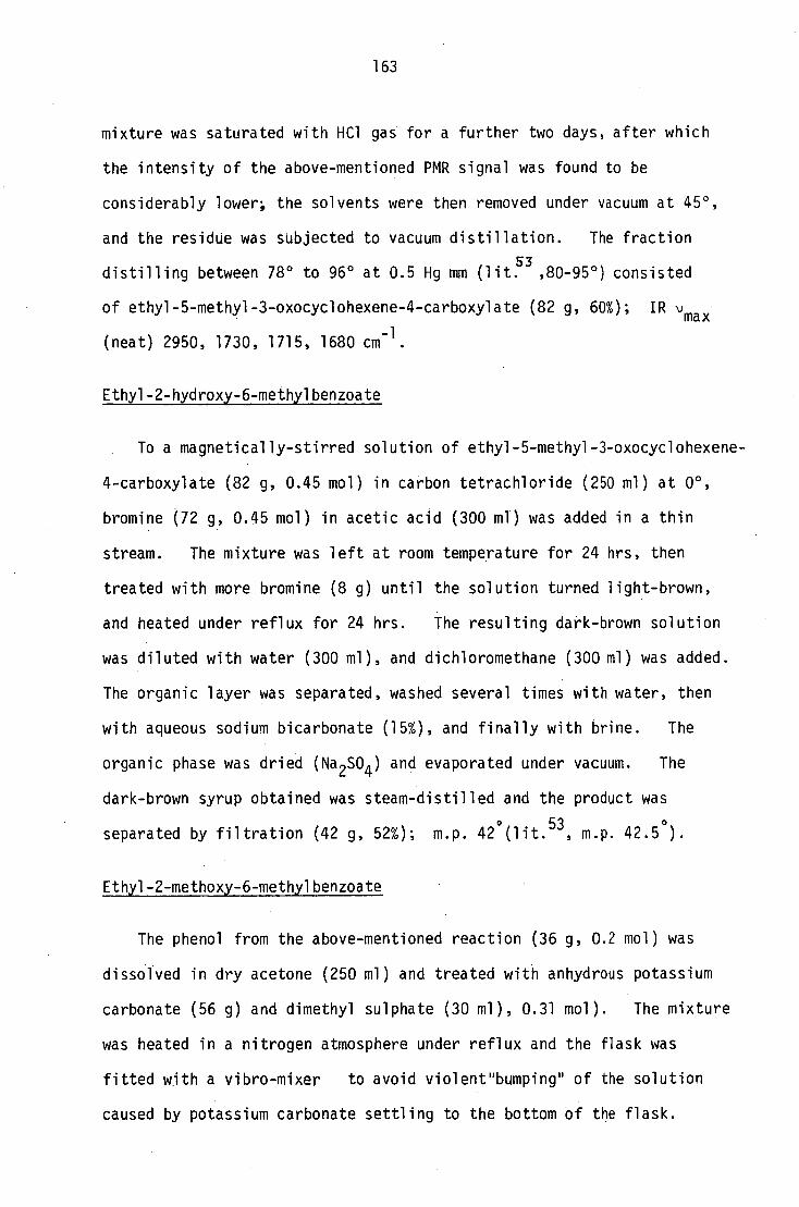
163
mixture was saturated with HC1 gas for a further two days, after which
the intensity of the above-mentioned PMR signal was found to be
considerably lower; the solvents were then removed under vacuum at 45°,
and the residue was subjected to vacuum distillation. The fraction
distilling between 78° to 96° at 0.5 Hg mm (lit.53
,80-95 ° ) consisted
of ethyl-5-methyl-3-oxocyclohexene-4-carboxylate (82 g, 600; IR vmax
(neat) 2950, 1730, 1715, 1680 cm -1 .
Ethy1-2-hydroxy-6-methylbenzoate
To a magnetically-stirred solution of ethy1-5-methy1-3-oxocyclohexene-
4-carboxylate (82 g, 0.45 mol) in carbon tetrachloride (250 ml) at 0°,
bromine (72 g, 0.45 mol) in acetic acid (300 ml) was added in a thin
stream. The mixture was left at room temperature for 24 hrs, then
treated with more bromine (8 g) until the solution turned light-brown,
and heated under reflux for 24 hrs. The resulting dark-brown solution
was diluted with water (300 ml), and dichloromethane (300 ml) was added.
The organic layer was separated, washed several times with water, then
with aqueous sodium bicarbonate (15%), and finally with brine. The
organic phase was dried (Na 2SO4 ) and evaporated under vacuum. The
dark-brown syrup obtained was steam-distilled and the product was
separated by filtration (42 g, 52%); m.p. 42 ° (lit. 53 , m.p. 42.5 ° ).
Ethy1-2-methoxy-6-methylbenzoate
The phenol from the above-mentioned reaction (36 g, 0.2 mol) was
dissolved in dry acetone (250 ml) and treated with anhydrous potassium
carbonate (56 g) and dimethyl sulphate (30 ml), 0.31 mol). The mixture
was heated in a nitrogen atmosphere under reflux and the flask was
fitted with a vibro-mixer to avoid violentnbumping" of the solution
caused by potassium carbonate settling to the bottom of the flask.

164
After the completion of the reaction,which was monitored by tic, the
acetone was removed, and the residue was dissolved in ether (400 ml),
to which an excess of triethylamine (35 g) was added. Afterl hr
the mixture was washed successively with water (2 x 50 ml), hydrochloric
acid (10%, 50 ml) and brine. The ether layer was dried (MgSO 4 ) and
evaporated to give the product (39.8 g, 82%) b.p. 80-92° (lit. 53 ,
b.p. 89-91 0 ).
2-Methoxy-6-methylbenzoic acid
The ester from the previous experiment (32 g, 0.11 mol) was refluxed
with sodium hydroxide (8 g, 0.22 mol), water (100 ml) and ethanol
(250 ml) for 12 hrs. The ethanol was removed at reduced pressure, and
the aqueous phase was repeatedly extracted with ethyl acetate (4 x 75 ml)
after acidification with dilute hydrochloric acid. Concentration of
the ethyl acetate extract gave crystals of 2-methoxy-6-methylbenzoic
acid (24 g, 91%), m.p. 140° (lit.53
, m.p. 139-141°).
2-Methoxy-6-methylbenzoyl chloride
The acid from above (3.2 g, 0.02 mol) was dissolved in chloroform
(50 ml), then dimethylformamide (0.15 g, '1 ,0.002 mol) and thionyl chloride
(2.5 ml, 0.013 mol) were added, and the mixture was heated under reflux
for 12 hrs. The solvents were removed under vacuum, and the residue
was distilled in a kugelrohr under atmospheric pressure. A yellow
viscous oil was collected at 235-240° (2.21 g, 62%); IR vmax (neat)
-1 2990, 1795, 1730, 1700 sh, 1600, 1590 cm.
2-Methoxy-6-methylphenyl diazoketone
The acid chloride from above (2.0 g, 10 mmol) in ether (100 ml)
was added dropwise under anhydrous conditions to a stirred mixture
of alcohol-free anhydrous diazomethane90
(1.5 g, 34 mmol) in ether

165
(250 ml) and dry triethylamine (1.2 g, 12 mmol) at 0 0 . The mixture
was stored overnight at 00 and the separated salts were filtered off.
The solvents were removed under vacuum and the diazoketone was obtained
as a yellow semi-solid. It was stored in the dark under nitrogen in a
refrigerator and used for the subsequent reactions within 24 hrs
without further purification91,92 .
3(pyrrol-1-yl)propylamine
To freshly distilled pyrrole (50 ml, 0.7 mol) potassium (8 g),
cut into small pieces, was added, and the mixture was heated on a water-
bath under nitrogen until the reaction was complete. Anhydrous toluene
(100 ml) was added, then the mixture was heated to reflux and a
solution of 13 chloropropionitrile (22 g) in toluene (75 ml) was added.
After refluxing for 24 hrs, the mixture was cooled and water (100 ml)
was added. The aqueous phase was separated and extracted with
toluene. The toluene extract was combined with the organic phase and
distillation at 92-97° and 0.5 Hg mm gave 3(pyrrol-1-yl)propionitrile
(29 g, 33%) (lit.93
, b.p. 90-96°, 0.4 Hg mm). The LAH reduction of
this compound followed by a standard work-up gave 3(pyrroly1)
propylamine (18.5 g, 61%), b.p. 80-82° (4 Hg mm) (lit. 93 ', 82°, 4 Hg mm).
N[3(Pyrrol-1-yl)propyl]hexanamide
To a rapidly stirred mixture of 3(pyrrol-1-yl)propylamine (5.0 g,
40 mmol) in ether (50 ml) and aqueous sodium hydroxide (10%, 100 ml),
n-hexanoyl chloride (6.0 g, 41 mmol) in ether (50 ml) was added at 0°.
The mixture was stirred for 6 hrs at room temperature, then the ether
layer was separated, washed with aqueous sodium hydroxide, dried
(Na 2SO4 ) and evaporated to give N[3(pyrrol-1-yl)propyl]hexanamide
(7.8 g, 86%); IR 'max
(neat) 3260, 1660, 1640 cm -1 ; PMR o 6.6 (2H, m) ,
6.2 (2H, m), 5.3 (1H, m, NHCO), 3.9 (2H, t, J = 6.5), 3.35 (2H, dt,

166
J = 6.5, -CH 2NHCO-), 2.0 (4H, m), 1.6 (211, m), 1.3 (4H, m), 0.9 (3H, t,
J = 8 Hz).
Attempted condensation of 2-methoxy-6-methylphenyl diazoketone and
N[3(pyrrol-1-yl)propyl]hexanamide
To a mixture of N[3(pyrrol-1-yl)propyl]hexanamide (2.0 g, 8.8 mmol)
in dry ether and freshly prepared copper powder at 00 a solution of the
above-mentioned diazoketone (0.450 g, 2.4 mmol) in dry ether was added
dropwise. The mixture was slowly heated to 50-55° and maintained at
this temperature for 5 hrs, then left at room temperature overnight.
A brown oil separated (1.6 g, but a mass spectral analysis
did not show the presence of any condensation product with a m/z value
of 384 (N13[2(2-methoxy-6-methylbenzoylmethyl)pyrrol-1-yl]propyl}
hexanamide). The reaction was repeated in an ether-toluene (1:1)
mixture as solvent, but the desired product could not be obtained.
2-(2-Methoxy-6-methylbenzoylmethyl)pyrrolidine 34
The diazoketone, prepared as described earlier from the acid
chloride (1.36 g, 8.5 mmol), was dissolved in dry chloroform (50 ml)
and added dropwise under anhydrous conditions to a magnetically-
stirred mixture of freshly distilled pyrrole . (1.7 g, 25 mmol), freshly
prepared and dried copper powder (2 g), and dry benzene (30 ml) at
room temperature. The mixture was maintained at 55-60° for 5 hrs, then
left at room temperature overnight. The solvents were removed, and the
black syrup ( 2.8 g) was chromatographed on a silica gel (100 g)
column using chloroform-methanol mixtures of increasing polarity. The
fraction eluted with a 4:1 mixture gave 2-(2-methoxy-6-methylbenzoylmethyl)
Wrole (0.52 g, 26%), m.p. 93° (lit. 34 , 93°); IR vmax 3400, 1685, 1620
-1 cm ; PMR (5 7.1-6.2 (6H, m), 4.12 (2H, s, COCH 2 ), 3.85 (3H, s, OCH3 ),
2.2 (3H, s, COCH 2 ), 3.85 (3H, s, OCH 3 ), 2.2 (3H, s, ArCH 3 ). This compound

167
was hydrogenated in glacial acetic acid (8 ml) over platinum oxide
at room temperature and atmospheric pressure for 12 hrs. The mixture
was diluted with water (100 ml), basified with ammonia, and extracted
with chloroform. The removal of solvents followed by ptic purification
gave 2-(2-methoxy-6-methylbenzoylmethyl)pyrrolidine as an oil (0.315 g,
62%); IR vmax (neat) 3400, 1685, 1620, 1590 cm -1 ; PMR 6 7.1 (1H, t,
J = 7.5), 6.8 (2H, dd, J = 7.5, 7.5), 3.85 (3H, s, OCH 3 ), 3.1 (1H, m,
-NCH-), 2.9 (2H, m, -CH 2C0-), 2.5 (2H, m), 2.2 (3H, s, ArCH 3 ), 1.8-1.6
(4H, m);
246 (4),
126 (20),
MS:
233
125
m/z 291
(4), 232
(27), 99
(M+
,
(10),
(13),
1%),
164
97
290 (M+
,
(21), 150
(14), 91
2), 289 (1), 288
(12), 149 (100),
(69), 84 (57), 70
(3), 287
127 (20),
(47).
(5),
3 2(2-Methoxy-6-methylbenzoylmethyl)pyrrolidin-l-yl]propylamine
The pyrrolidine derivative from above (0.300 g, 1.28 mmol),
acetonitrile (20 ml), acrylonitrile (0.150 g, 2.8 mmol) and a drop of
glacial acetic acid were heated under reflux for 6 hrs. The solvents
were removed and the crude nitrile thus obtained (0.305 g), IR vmax
-1 2250 cm (CEN), was hydrogenated over platinum oxide (0.3 g) in glacial
acetic acid (10 ml). The acid solution was filtered, diluted with
water, basified with ammonia and extracted with chloroform. The
chloroform extract was evaporated and purified by ptic to give a brown
oil (0.22 g, 62%); UV Amax 225 nm (log E 3.16), 245 (2.71), 280 (2.47);
IR vmax 3350 (NH 2 ), 1680, 1640, 1580, l565 cm'; PMR 6 7.1 (1H,
J = 7.5), 6.7 (1H, d, J = 7.5), 6.65 (1H, d, J = 7.5), 5.2 (2H, br m,
exchangeable -NH 2 ), 3.85 (3H, s, OCH 3 ), 3.15 (1H, m), 2.8 (2H, m), 2.2
(3H, s, ArCH 3 ). MS: m/z 291 (MH+ , 1%), 290 (e, 2), 289 (1), 288 (3),
287 (5), 246 (4), 233 (4), 232 (10), 164 (21), 150 (12), 149 (100), 127
126 (20), 125 (27), 99 (13), 97 (14), 91 (69), 84 (57), 70 (47).
(20),

168
Nf3[2(2-Methoxy-6-methy1benzoylmethyl)pyrrol1din-l-yl)propyl}hexanamide
To a rapidly stirred mixture of 3[2(2-methoxy-6-methylbenzoylmethyl)
pyrrolidin-1-61]propylamine (0.05 g, 0.17 mmol) in ether (10 ml) and
aqueous sodium hydroxide (10%, 10 ml) at 0°, n-hexanoyl chloride (0.030 g,
0.22 mmol) in ether (10 ml) was added dropwise. The solution was
stirred for another 2 hrs at room temperature, then the ether layer was
separated, and the aqueous phase was extracted with chloroform. The
chloroform extract was combined with the ether layer and evaporated,
and the residue obtained was purified by ptic to give a yellow gum,
Rf 0.45 (chloroform-methanol 9:1) (0.06 g, 89%) which was found to be
identical with 0-methylperipentadenine by tic, IR, PMR and ms comparison.

169
9.3 Experimental for Chapter 5
Separation of fraction D
On tic this gum appeared to be a mixture of two compounds:
peripentadenine (R f 0.45), and a more polar minor alkaloid (PBXM3)
(Rf 0.38) (solvent:chloroform:methanol 9:1); but when the chromatogram
was developed three times successively, using the same solvent system,
a third compound (PBM3) with an R f value slightly lower than peripenta-
denine appeared. The three compounds were separated by ptic employing
the multiple development technique.
Dinorperipentadenine
The compound between peripentadenine and PBXM3 gave dinorperipenta-
denine as a yellow gum (1.06% yield), R f 0.45 (methanol:chloroform 9:1).
The base could not be obtained crystalline, and the attempts to prepare
a crystalline derivative were also unsuccessful; UV xmax 218 nm (log E
4.01), 252 (3.15), 285 (3.15); IR vmax (neat) 3250-3020 (br, NH and OH),
1690 (AT C=0), 1680 and 1650 (CONHR), 1630 cm C=0 H-bonded to
ortho OH); PHR 6 (270 MHz) 10.5 (1H, m, Ar0H), 7.17 (1H, dd, JH4/H3 =
H5/H4 = H4/H5 = 7.5 Hz, H4), 6.72 (1H, d, J 7.5 Hz, H5), 6.65 (1H, d,
J -
H18/2H17 = 113/H4 = 7.5 Hz, H3), 6.05 (1H, br t, J 6.0 Hz, H18), 3.59
J -
(1H, dddd„ JHIO/H9a = 10.0, J H10/H9b = 5 ' 0 ' J1110/H1la = 8 ' 3 ' JH10/H1lb =
5.0 Hz, H10), 3.48 (1H, dd, JH9a/H9b = H9a/H10 =
12.75 Hz, J 10.0 Hz, H9a), -
3.27 (1H, ddd, J 10.5, J 4.5 Hz, - H15a/H15b = H15a/H16a - 6 ' 3 ' 14115a/H16b =
H15a), 3.25 (11-I, dd,J 6.0 Hz, H17a), 3.15 - H17a/H17b = 13 ' 6 ' JH17a/H18 =
(1H, dddd, LI - H17b/H17a = 13 ' 5 ' JH17b/H16a = 8.25, J - H17b/H16b = 6.0,
0 Hz = 6., H17b), 2.:7 (1H, ddd, J 12., J j1-117b/H18 - H13a/H13b = 0 -H13a/H12a
.5 Hz, H13a), 2.57 (1H, dd, J 12.75, = = 7 7.5, JH13a/H12b -H9b/H9a =
= 5 Hz, H9b), 2.52 (1H, dd, J , J JH9b/H10 - H13a/H13b = 12 - H13a/H12b 3.6 Hz '
H13b), 2.46 (1H, ddd, J - H15a/H15b =
10.5, j1115a/H16a = 8 ' 5 ' j H15a/H16b =

170
7.5 Hz, H15a), 2.3 (3H, s, 3H1), 2.2 (1H, ddd, J - 1111a/H11b = 13.0,
., Hlla), .07 (2H, , J jiilla/H10 = 8.5 ' jH11a/H12a = 120 Hz 2
t- 20/21 = 7.5 Hz,
2H20), 2.0 (1H, m, H11b), 1.95 (1H, m, H16a), 1.75 (1H, m, H16b), 1.67
(2H, m, 2H12), 1.55 (2H, tt, J - 21/20 = J21/22 = 7.5 Hz, 2H21), 0.9 (3H,
t, J 22/21 = 7.5, 3H22); 13
C NMR 6 207.1 (s, C8), 173.4 (s, C19), 157.7
(s, C6), 137.2 (s, C7), 132.5 (d, C4), 127.9 (s, C2), 121.7 (d, C3),
116.5 (d, C5), 64.6 (d, C10), 54.2 (t, C13), 53.9 (t, C15), 48.4 (t, C9),
38.6 (t, C17), 37.1 (t, C20), 30.8 (t, C11), 27.4 (t, C16), 23.9 (t, C12),
20.7 (q, Cl), 13.8 (q, C22); HRMS: M ./. found: 346.2267; calculated for
C20
H30
N203 346.2166; m/z 346 (M
+ 12%), 197 (65), 150 (82), 135 (100),
128 (36), 82 (87).
0-methyl dinorperipentadenine
A methanolic solution of dinorperipentadenine (0.03 g in 20 ml) was
treated with an excess of ethereal diazomethane and left at room
temperature overnight. Removal of the solvents gave 0-methyl
dinorperipentadenine as a light brown gum (0.03 g n. ,100%), Rf 0.45
(chloroform:methanol 9:1), PMR an additional signal at 6 3.8 (3H, s,
OCH 3 ), the broad signal at 6 10.5 disappeared; MS: m/z 360 (M 4- ; 12%),
345 (16), 197 (62), 164 (73), 135 (92), 128 (42), 82 (100).
Methofluoride of dinorperipentadenine
Dinorperipentadenine (0.045 g, 0.13 mmol) was converted to the
methofluoride, a brown gum (0.060 g), by the same procedure as for
peripentadenine; MS: m/z 360 (M±-1, 10%), 246 (63), 201 (32), 171 (71),
134 (34), 128 (98), 84 (23), 69 (40), 58 (100).
Synthesis of 0-methyldinorperipentadenine
To a rapidly-stirred mixture of an ethereal solution (20 ml) of
3[2(2-methoxy-6-methylbenzoylmethyl)pyrrolidin-l-yl]propylamine (0.05 g,

171
18.5 mmol), the synthetic precursor of peripentadenine, and a solution
(20 ml) of aqueous sodium hydroxide (10%) at 0°, n-butanoyl chloride
(0.025 g, 24 mmol) in ether (20 ml) was added dropwise, then the
solution was stirred for a further 2 hrs at room temperature. The
ether layer was separated, and the aqueous phase was extracted with
chloroform (2 x 20 ml). The chloroform layer, combined with the ether
layer, was dried and evaporated. The yellow gum obtained, after
purification by ptic (0.035 g, 53%), was found to be identical with the
methyl ether of the natural product (tic, ir and PMR).
Isolation of Peripentamine
The ptic separation of fraction B produced three sub-fractions:
a mixture of four less polar compounds (PBVM), and two bases,peripenta-
mine (PBM2) and PBXM2.
Peripentamine, a pale yellow oil (0.8%), had R f 0.75 (chloroform:
methanol 9:1). Like the majority of other Peripentadenia alkaloids,
neither peripentamine nor its derivatives could be obtained crystalline;
UV xmax
218 nm (log E 4.26), 245 (3.98), 250 (3.94), 256 (3.93), 263
(3.81), 300 (3.63); x max
(+ OH - ), 225 (4.46), 245 (4.24), 252 (4.16);
IR vmax (CHC1 3 ) 3300 (NH, OH), 1690 (AT C=0), 1680 and 1655 (RNHCO),
-1 1640 and 1630 cm ; PMR 6 (270 MHz), 6 16.4 (1H, broad m, OH), 10.4
(1H, broad m, Ar0H) both exchanged on addition of D 20, 7.15 (1H, dd,
JH4/H3 = JH4/H5= 7.5 Hz, H4), 6.8 (1H, d, H5/H4 = 7.5 Hz, H5), 6.69 J
(1H, d, J H3/H4 = 7.5 Hz, H3), 6.65 (1H, broad t, -NHCO-, H18), 4.38
(1H, m, H10), 3.37 (2H, ddd, J - H17a/H17b = 12,J..— mii/H16a = 7 ' 'H17/H18
• 6 Hz, 2H17), 3.2 (1H, dm, J 16 Hz, H9a), 2.95 (1H, dd, J H9a/H9b =
H9a/H9b =
dt 2 , H9b), .87 (1H, , J , J 16, JH9a/H10 = 5 Hz
- H13a/H13b = 12
H13/H12 = 5 Hz,
H13a), 2.6 (2H, m, 2H15), 2.44 (1H, dd, J , J / 4 Hz, - H13b/H13a = 12 -H13012 =
H13b), 2.4 (3H, s, 3H1), 2.2 (2H, t, JH20/
(8H, unresolved m, 2H11, 2H12, 2H16, 2H21), 1.3 (4H, m, 2H22, 2H23), 0.9
H21 = 7.5 Hz, 2H20), 1.85-1.5

172
(3H, t, JH24/H23 = 7.5 Hz, 3H24); 13C NMR (6 ppm) 205.8 (s, C8), 173.4
(s, C19), 159.2 (s, C6), 137.9 (s, C7), 133.1 (d, C4), 125.3 (s, C2),
122.9 (d, C3), 115.6 (d, C5), 76.0 (d, C10), 57.6 (t, C15), 56.2 (t, C13),
49.2 (t, C9), 3817 (t, C17), 36.9 (t, C20), 31.6 (t, C22), 30.11 (t, C21),
25.9 (t, C23), 25.6 (t, C16), 23.8 (t, C12), 22.5 (q, Cl), 22.5 (t, C23),
13.9 (q, C24); HRMS: M-1. found 392.2464; calculated for C22 H 30204 :
392.2587; m/z 392 (W. ', 9%), 391 (50), 375 (100), 373 (36), 305 (11),
225 (93), 224 (12), 201 (22), 173 (66), 156 (29), 151 (57), 116 (57).
Methofluoride of peripentamine
Peripentamine (0.030 g, 0.08 mmol) was converted to the methofluoride
(0.027 g, 84%) following the same procedure as for peripentadenine.
The brown semi-solid gum had MS: m/z 421 (M + , 2%), 420 (3), 419 (6), 417 (6),
405 (7), 403 (10), 490 (26), 388 (35), 387 (93), 284 (36), 257 (96),
201 (100), 156 (38).
Attempted pyrolytic dehydration of peripentamine
Peripentamine (0.017 g, 0.04 mmol) was placed in a kugelrohr and
•heated slowly under vacuum (6.3 x 10⁻⁴
Hg mm). A tic examination of
the colourless liquid collected at 110° revealed the presence of
2-hydroxy-6-methylacetophenone and a complex mixture of basic compounds
which could not be separated by tic.
Isolation of dehydroperipentamine
Fraction E (4.8 g) was separated by ptic, and the least polar
compound of the mixture, dehydroperipentamine, was isolated as a brown
solid (0.090 g, 0.6%) which gave white needles from acetone-water
mixture, m.p. 96'; microanalysis of the solid gave: C 63.37%, H 9.03%
and N 6.55%; calculated for C22 H34
N2 0320: C 62.96%, H 9.37% and
N 6.68%; Rf 0.31 (chloroform:methanol 9:1); UV Amax 214 nm (log E 4.5),

173
250 (3.64), 285 (3.42), 320 (3.44); IR v max (CHC1 3 ) 3300 (NH), 1695
(AT C=0), 1680, 1650 cm-1 ; PMR 6 (400 MHz), 7.35 (1H, dd, J - H4/H3 =
I-14/H5 = 7.5 Hz), 7.24 (1H, t, J H18/H17 = 6 Hz, H17), 6.88 (1H, d, J -
H3/H4 = I-15/H4
= 7.5 Hz, H5), 6.83 (1H, d, J 7.5 Hz, H3), 4.47 (1H, m, J -
H10), 3.58 (2H, dt, JH17/H18 = 6, JH17/H16 = Hz, 2H17), 3.03 (2H,
1-113/H14 = 6 Hz, 2H13), 2.98 (2H, t, J - H15/H16 = 6 Hz, 2H15), 2.69 J (2H, m, 2H9), 2.64 (3H, s, 3H1), 2.28 (2H, t, JH20/H21 = 7.5 Hz, 2H20),
2.16 (2H, m, 2H16), 1.95-1.89 (4H, m, 2H11, 2H12), 1.62 (2H, tt,
H21/H20 = JH21/H22 = 7.5 Hz, 2H20), 1.29 (4H, m, 2H22, 2H23), 0.89 J (3H, t, H 24/H23 = 7.5 Hz); 13 C NMR 6 (100.62 Hz), 193.4 (s, C8),
175.9 (s, C19), 162.4 (s, C6), 142.3 (s, C7), 134.9 (d, C4), 124.2
(d, C3), 119.7 (s, C2), 115.9 (d, C5), 76.27 (d, C10), 48.2 (t, C15),
45.5 (t, C13), 44.5 (t, C9), 36.5 (t, C20), 36.2 (t, C17), 31.9 (t, CH),
31.6 (t, C22), 26.8 (t, C21), 25.6 (t, C16), 22.8 (q, Cl), 22.5 (t,
C23), 22.2 (t, C12), 14.0 (q, C24); HRMS: M 374.2501,
calculated for C 22 H34 N 203 : 374.2569; MS: m/z 374 (M+, 12%), 232 (31),
225 (18), 218 (11), 211 (14), 202 (16), 185 (100), 156 (56), 135 (46).
Dehydration of Peripentamine
Peripentamine (0.052 g, 0.13 mmol) was refluxed with aqueous oxalic
acid (10%, 30 ml) for 12 hrs. The solution was cooled, basified with
ammonia, and extracted with chloroform. The gum obtained on evaporation
of the chloroform extract contained three compounds; the compound of
intermediate Rf ' when purified by ptic (0.018 g, 36%) was shown to be
identical with dehydroperipentamine by tic, UV, IR and PMR comparison.
N-Methylation of dehydroperipentamine
Dehydroperipentamine (0.035 g, 0.094 mmol) was dissolved in ice-cold
formic acid (98%, 0.5 ml, 13 mmol), and formaldehyde (37%, 1 ml, 12 mmol)
was added. The mixture was heated on a water bath for 8 hrs, then
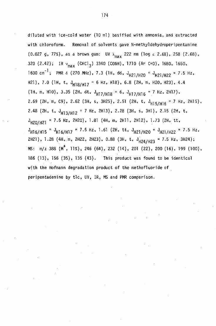
174
diluted with ice-cold water (10 ml) basified with ammonia, and extracted
with chloroform. Removal of solvents gave N-methyldehydroperipentamine
(0.027 g, 77%), as a brown gum: UV xmax 222 nm (log E 2.68), 258 (2.68),
320 (2.42); IR vmax (CHC1 3 ) 3340 (CONH), 1710 (AT C=0), 1680, 1650,
1600 cm -1 ; PMR d (270 MHz), 7.3 (1H, dd, Hzi/H20 = H21/H22 = 7.5 Hz,
H21), 7.0 (1H, t, JH1 " , 8/H17 = 6 Hz, H18), 6.8 (2H, m, H20, H23), 4.4
(1H, m, H10), 3.35 (2H, dt,J = 7 Hz, 2H17), - H17/H18 = 6 ' JH17/H16
2.69 (2H, m, C9), 2.62 (3H, s, 3H25), 2.51 (2H, t, J H15/H16 = 7 Hz, 2H15),
2.48 (2H, t, J H13/H12 = 7 Hz, 2H13), 2.28 (3H, s, 3H1), 2.15 (2H, t, -
H20/H21 7.5 Hz, 2H20), 1.81 (4H, m, 2H11, 2H12), 1.73 (2H, tt, J
tt 7.5 Hz, 1.61 (2H, , J JH16/H15 = JH16/H17 = - H21/H20 = J1121/H22 = 7.5 Hz,
2H21), 1.28 (4H, m, 2H22, 2H23), 0.88 (3H, t, JH24/H23 = 7.5 Hz, 3H24);
MS: m/z 388 (M+ , 11%), 246 (64), 232 (14), 201 (22), 200 (16), 199 (100),
186 (13), 156 (35), 135 (43). This product was found to be identical
with the Hofmann degradation product of the methofluoride of
peripentadenine by tic, UV, IR, MS and PMR comparison.

175
9.4 Experimental for Chapter 6
Extraction and fractionation of leaf alkaloids
Dried milled leaves (14 kg) were extracted with methanol at room
temperature, and the methanol was removed under vacuum at 45°. The
dark-brown residue was dissolved in glacial acetic acid (2.5 10,
poured into rapidly-stirred water (40 2) in a fine stream and left
overnight. The precipitate formed was filtered off through a bed
of celite and washed with water (10 2). The combined filtrate and
washings were evaporated to dryness, and the residue was re-dissolved
in aqueous sulphuric acid (10%, 20. The aqueous phase was
extracted with chloroform (3 x 500 ml) until no more soluble material
was removed. The aqueous phase was basified with ammonia (pH = 11)
and extracted with chloroform (12 x 250 m1). The chloroform extracts
were combined, dried (Na 2SO4 ) and evaporated to give the crude
alkaloid extract as a black viscous oil (26 g, 0.19%).
The crude extract (12 g) 'was separated on a silica gel column
(200 g) packed in dichloromethane. Elution was carried out with
dichloromethane-methanol solvent systems of increasing polarity, and
10 ml aliquots were collected. The aliquots were monitored by tic
and bulked accordingly to give three main fractions. (Table 2) (page 179).
Isolation of PLM2 and PLM3
The ptic purification of fraction A (2.3 g) gave two bases, PLM2
(0.18 g, 0.69%) and PLM3 (0.89 g, 3.4%), both pale yellow viscous oils,
and 2-hydroxy-6-methyl acetophenone (0.62 g, 2.3%).
9.4.1 PLM2
The less polar compound, R f 0.85 (chloroform:methanol 9:1), could
be further purified by sublimation, upon which it produced a low-melting

176
semi-solid, m.p. 43-44°. The tic spot of the compound appeared purple
immediately after spraying with iodoplatinate, but turned white
afterwards. PLM2 also formed a crystalline picrate, m.p. 212-213°,
when an alcoholic solution was treated with a few drops of a saturated
solution of picric acid in methanol.
PLM2; found: C 70.3%, H 8.6%, N 8.5%. C 9H13N0-1/8H 20 requires C 70.4%,
H 8.7%, N, 9.1%; picrate: found C 47.8%, H 4.3%, N 13.7%. C 9H13N0
C9 H13 NO.0
6 H3 N 3 07 (C
15 H16 N4 08 ) requires C 47.4%, H 4.3%, N 14.7%; UV Amax
217 nm (log E 3.19), 325 (2.76), 370 (2.21);Amax (+ OH- ), 218 nm (log E
3.39), 315 (3.06), 370 sh (2.82); xmax
(+ Hi), 228 (3.19), 280 (3.04),
300 (2.93); IR vmax (CHC1 3 ) 3450 (broad), 1730, 1660, 1630 cm -1 ; PMR 6
(270 MHz), 4.51 (1H, broad, H1), 3.25 (1H, d, J = 3 Hz, H5), 2.12 (3H, s,
3H9), 2.11 (1H, m), 2.05 (1H, d, J = 2.5 Hz), 1.97 (2H, m) 1.25 (1H, dd,
J = 9, 2 Hz), 1.03 (3H, d, J = 9 hz, 3H10); 13C NMR 6 208 (s, C4), 173.9
(s, C3), 61.5 (d, C1), 55.8 (d, C5), 39.6 (t, C8), 32.6 (t, C7), 28.9 (d,
C6), 24.4 (q, C9), 21.1 (q, C10); MS: m/z 152 (Mi + 1, 17%), 151 (Mi ,
100), 136 (58), 110 (20), 109 (7), 108 (8), 95 (21), 94 (31), 84 (22),
82 (11), 81 (7), 71 (10), 70 (8), 69 (48), 68 (31), 67 (14), 57 (20),
55 (22), 53 (7), 44 (13), 43 (36), 42 (41), 41 (58), 39 (34).
Borohydride reduction of PLM2
A methanolic solution of PLM2 (0.020 g, 0.13 mmol, 10 ml) was
treated with sodium borohydride (0.005 g) and stirred at room temperature
for 2 hrs, then the solvent was removed under vacuum. The residue was
dissolved in aqueous sulphuric acid (10%, 10 ml), the solution was
basified and extracted with chloroform. The chloroform layer was dried
and evaporated to give a brown gum (0.012 g, 57%); MS: m/z 156 (M i , 2%),
140 (17), 138 (38), 123 (30), 111 (25), 110 (21), 94 (23), 82 (20), 69
(100), 68 (45), 67 (30), 55 (25), 43 (72), 42 (48), 41 (85).
177
Acetylation of the reduction product of PLM2
The above-mentioned reduction product (0.006 g) was treated with
glacial acetic acid (5 drops), acetic anhydride (5 drops) and pyridine
(1 drop) and left overnight at room temperature. The mixture was
basified with ammonia and the solvents were removed under vacuum to
give a brown gum; MS: m/z 239 (M+ , 0.04), 224 (0.02), 182 (0.02),
180 (0.02), 164 (0.02), 152 (0.03), 120 (27), 105 (40), 57 (15), 43
(100).
Quaternisation of the borohydride reduction product of PLM2
The reduction product (0.006 g) in acetone (10 ml) was heated in a
sealed tube with methyl iodide (0.5 ml) on a water bath for 4 hrs.
Removal of solvents gave a brown gum; MS: m/z 185 (M+ , 10%), 184
(M+ -1, 74), 110 (57), 84 (41), 69 (50), 58 (60), 57 (53), 42 (100).
Methoidide of PLM2
PLM2 (0.047 g, 0.31 mmol) was heated in a sealed tube with acetone
(10 ml) and methyl iodide (2 x 1 ml) on a water bath. After about
4 hrs the methiodide started separating, the tube was cooled, and the
crystals were filtered off (0.086, 94%), m.p. 96°; PMR d (CD 3C0CD 3 ,
400 MHz), 4.7 (1H, broad, H1), 3.76 (1H, d, H5), 3.7 (3H, s, 3H9);
3.37 (3H, s, 3H11), 2.6 (2H, m), 2.42 (2H, m), 1.55 (1H, dd), 1.13
(3H, d); MS: m/z 166 (Mt , 10%), 165 (M 4.-1, 100), 150 (27), 136 (7),
128 (85), 127 (43), 123 (57), 122 (78), 108 (85), 95 (57), 94 (67),
82 (62), 67 (17), 55 (37), 42 (55).
Partial reduction of the methiodide of PLM2
In an attempt to selectively reduce the iminium function of the
methiodide of PLM2, ethanolic solutions of this compound were treated

178
with 1, 0.5, 0.2 and 0.1 molar equivalents of sodium cyanoborohydride
at -10 0 . Reactions with 1 and 0.5 molar equivalents of cyanoborohydride
led to the completely reduced compound which was obtained in the form
of a gum: IR: no C=0 absorption, PMR 6 4.7 (1H, m, CHOH), 2.6 (3H, s,
N-CH 3 ), 1.1 (3H, d, J = 7 Hz, CH-CH 3 ), 0.9 (3H, d, J = 7 Hz, CH-CH 3 );
MS: m/z 169 (M t , 3%), 156 (18), 155 (21), 141 (32), 126 (100), 72 (42);
but when the methiodide (0.030g. 0.1 mmol, in 10 ml ethanol) was
treated with 0.2 molar equivalent of sodium borohydride (3 ml, 0.02%
ethanolic solution) followed by the usual work up, the partially reduced
compound was obtained as a brown gum (0.008 g, 47%); IR 'max
(CHC13)
1730 cm-1
; PMR 6 2.35 (3H, s, N-CH 3 ), 1.7-1.3 (unresolved multiplet),
1.1 (3H, d, J = 7 Hz), 0.9 (3H, d, J = 7 Hz); MS: m/z 169 (M + , 0.5%),
156 (12), 155 (15), 142 (27), 126 (100), 72 (30).
9.4.2 PLM3
The more polar compound R f 0.7 (chloroform:methanol 9:1), which was
chromatographically pure, was isolated as an oil. The tic spot appeared
purple immediately after spraying with iodoplatinate reagent but turned
white after a few minutes. On attempted further purification by
sublimation under vacuum (2.5 x 10⁻³
Hg mm, 180°), the compound underwent
decomposition. On a Waters radial pack reverse phase C18 column, the
material appeared to be a single compound with methanol-water, and with
methanol-water systems containing the ion-pairing reagent PIC B8.
The oil gave a negative Gibbs test: UV xmax 212 nm (log E 3.31), 280
(2.56); Amax (+ H+ ), 215 nm (3.51), 248 sh (3.15), 310 (3.06), 370
(2.51): Amax (+ OH) 215 nm (3.29), 248 (3.37), 310 sh (2.9); IR vmax
(neat) 3300 (NH, OH), 2950, 1730, 1660, 1640 cm -1 ; PMR 6 (270 MHz),
6.7 (1H, m, H18), 4.55 (1H, broad s, H6), 4.05 (1H, m, 1-110), 3,45 (1H,
m, H17a), 3.29 (1H, broad s, H7), 3.2 (1H, m, H17b), 2.88 (1H, dm,
H13a/H13b = 10.5 Hz, H13a), 2.75-2.58 (2H, m, 2H15), 2.52 (2H, tm, J

179
2H11), 2.43 (1H, dm, H13b), 2.2 (2H, t, JH20/H21 =
7.5 Hz, 2H20), 2.1-1.9
(5H, m, H2, 2H3, 2H5), 1.8 (2H, m, 2H12), 1.6 (4H, m, 2H16, 2H21),
1.4-1.2 (4H, m, 2H22, 2H23), 0.9 (3H, t, 3H24); 13C NMR (5 208.37 (s,
C4 or C8), 208.32 (s, C4 or C8), 173.32 (s, C19), 77.37 $ 77.29 (d, C6),
61.09 A 60.65 (d. C9),
(d, C7), 39.7 (t, C17),
57.57 s
38.32 A
57.35 (t, C13),
38.26 (t, C5),
55.9 (t, C15),
36.85 (t, C20),
55.65
32.43
32.38 (t, C3), 31.59 (t, C22), 30.27 $ 30.12 (t, C11), 29.01 A 28.94
(d, C2), 25.98 /s 25.83 (t, C12), 25.59 (t, C21), 23.86 (t, C16), 22.45
(q, Cl), 13.96 (q, C24); MS: m/z 392 (M+ , 0.7%), 391 (M +-1, 0.6),
374 (1), 373 (4), 330 (1.5), 245 (25), 241 (20), 235 (12), 223 (22),
212 (12), 199 (29), 193 (52), 180 (42), 178 (19), 156 (58), 151 (50),
138 (37), 126 (100), 109 (42), 94 (25), 84 (37), 70 (52), 69 (37),
56 (22), 55 (15), 43 (50), 42 (14), 41 (42).
TABLE 2
Bulking summary of fractions isolated from column chromatography
Fraction Eluent CH2
CL 2/CH
3OH/
volume/ml
Weight/g Constituents/wt per 12 g of crude extract
1:0 260} 2.3
2-hydroxy-6-methyl-
19:1 600 acetophenone (0.620)
PLM2 (0.180) PLMS (0.890)
B . 17:3 5751 6.8
peripentadenine
3:1 650 dinorperipentadenine dehydroperipentamine PBXM4-PBXM9
C 1:1 830 1.8 PLM4 (0.120)
PLM5 (0.090)
PLM6 (0.140)
PLM7 -PLM9

180
9.5 Experimental for Chapter 7
9.5.1 Separation of the mixture of volatile compounds (PBMV)
from Fraction B
The yellow oil (1.29 g) was distilled in a kugelrohr under vacuum
(0.1 Hg, mm, 80-120'). Four different fractions were collected in
the bulbs (PBVMA-PBVMD). The compound of highest volatility gave a
colourless oil, UV Amax 252 mm (log E 3.86), 284 (3.19); IR v max
(neat) 1630 cm -1 ; PMR 6 12 (1H, exchangeable, OH), 7.22 (H, t,
J = 7.5 Hz), 6.64 (2H, dd, J = 7.5 Hz), 2.62 (3H, s, COCH 3 ), 2.56 (3H,
s, ArCH 3); MS: m/z 250 (M + , 80%), 135 (50), 136 (100), 107 (45),
79 (60), 78 (40), 77 (60). The compound was identified as 2-hydroxy-
6-methylacetophenone16
by spectral comparison with an authentic sample.
The next most volatile compound deposited white crystals (0.3%):
m.p. 108'; PMR 6 2.9 (s), MS m/z 94 (M+ , 65%), 79 (100), 63 (14), 32 (72).
It was identified as dimethylsulphone by mixed m.p. (lit. m.p. 107-108' 55 )
with an authentic sample and mass spectral comparison with a MS reported
in the literature89
. The third compound, a liquid (0.02 g, 0.03%)
(R f 0.79, chloroform:methanol 9:1), gave a +ve Mayer's test. A 100 MHz
PMR spectrum obtained appeared similar to those of other C22 alkaloids.
The fourth compound (PVMD) deposited a white semi-solid (0.064 g, 0.11%)
(R f 0.61 chloroform:methanol 9:1); microanalysis gave C 54.74%,
H 7.38%, N 9.07%; UV Amax 213 nm (log e 3.54), 264 (3.60); Ama x (4131i—)
213 (3.73), 260 (3.47); IR v max 3200 (broad), 1670 cm -1 ; PMR 6 9.5
(1H, broad, exchangeable, OH), 7.3 (1H, d, J = 9 Hz), 5.7 (1H, d,
J = 9 Hz), 4.65 (1H, t, J = 5.7 Hz), 3.8 (2H, d, J = 5.7 Hz), 3.7 (2H,
dq, J = 9.4, 7.1 Hz), 3.54 (2H, dq, J = 9.4, 7.1), 1.2 (6H, t, J = 9.4 Hz);
13 C NMR 6 163.5 (s), 151.1 (s), 146.0 (d), 101.5 (d), 100.3 (d), 64.0 (0,
51.0 (t), 15.0 (q); on addition of an equimolar quantity of chromium (III)-
acetylacetonate57-60
to the NMR solution the intensity of the peaks at

181
(5 64.0 and 15.0 was increased. 13
C NMR (1H-
13C coupled) .5 163.5
(d, J = 12 Hz), 151.1 (quintuplet, J = 4.5 Hz), 146.0 (dq, J = 180 and
4.5 Hz), 101.5 (dd, J = 177 and 3 Hz), 100.3 (d, quintuplet, J = 161
and 3 Hz), 64 (t, quintuplet, J = 142 and 3 Hz), 51.0 (td, J = 142
and 4.5), 15.0 (qt, J = 127.5 and 3 Hz); MS: m/z (El) 183 (18%), 155 (14),
112 (9), 103 (91), 82 (13), 75 (58), 55 (9), 47 (100); MS: m/z (CI,
ammonia), 246 (M+
+ NH4'
15%), 230 (M+
+ 2, 11), 229 (M+
+ 1, 100),
184 (7), 183 (67), 103 (54), 50 (37); (high resolution ms) measured
229.1384 calculated for C 10H 17 N2 04 229.1197, measured 183.0767
calculated for Ce l N2 0 3 183.0722, measured 155.0588 calculated for
C7H 9NO 3 155.0589.
Diazomethane methylation of PBVMD
The semi-solid (5 mg) was treated with excess of ethereal
diazomethane and left overnight at room temperature. The gum obtained
after the removal of solvents gave m/z (CI ammonia), 243 (M + + 1, 37%),
197 (22), 169 (7), 103 (100), 82 (18), 75 (25), 55 (8), 47 (30).
9.5.2 PBXM2
The second minor base from fraction B gave a yellow gum (R f 0.48
chloroform:methanol 9:1) UV Amax 215 nm (log E 3.48), 255 (2.98), 320
(2.49); IR vmax (CHC1 3 ) 3300, 1720, 1685, 1670, 1645 cm -1; PMR d
(1H, t, J = 7.5 Hz), 6.7 (2H, m), 6.5 (1H, broad, CONH), 4.45 (1H, m,
-CH0-), 3.4 (2H, m, -CH 2NHCO-), 3.3-2.7 (unresolved signals) 2.45 (3H,
s, ArCH3 ), 2.2 (2H, t, J = 7 Hz, -COCH 2-), 1.75-1.5 (unresolved signals),
1.3 (4H, m, -CH2 -CH2-), 0.9 (3H, t, J = 7 Hz, -CH 3 ); HRMS M+
found
388.2356 calculated for C22
H32
N204 388.2382; m/z 388 (M 374 (6),
332 (13), 285 (15), 185 (20), 184 (11), 156 (52), 135 (74), 126 (100),
113 (48), 112 (73), 100 (24), 99 (46), 98 (39), 87 (24), 84 (46), 71
182
(39), 70 (85), 56 (67).
9.6.3 The more polar minor alkaloids of the bark extract
The mixture of alkaloids left after the separation of the least
polar alkaloid, dehydroperipentamine, from fraction E was found to be
an extremely complex one. The tic analysis of this mixture on
Merck Kieselgel F254 (0.25 mm) pre-coated plates using a multiple
development technique (with 10:1, 8:1 and 6:1 CHC1 3 :CH3OH mixtures
as solvent) showed the presence of six bases. All six compounds
stained in different shades of pink with iodoplatinate reagent.
On ordinary silica plates a satisfactory separation could not be
obtained. However, ptic separation of 5 g of this mixture on KOH-
silica plates using multiple development yielded 15-30 mg quantities
of the six alkaloids. Owing to poor resolution and irreversible
adsorption onto silica, this proved to be an unsatisfactory technique
for the separation of this mixture. A preliminary investigation
showed that the mixture may be separated by HPLC on a reverse phase
(Bondapack C18) column using methanol-water systems, but the time
and equipment available did not permit further work on this mixture.
The mass spectra of seven of the minor alkaloids were recorded.
Only two compounds, PBXM7 and PBXM8, could be isolated in sufficient
amounts to record PMR spectra. (The compounds are labelled in
increasing order of polarity on KOH-silica plates).
PBXM3; MS: m/z 300 (M + , 0.6%), 299 (6), 298 (3), 269 (2), 225 (76),
156 (100), 1 x 2 (32), 124 (94), 96 (28), 84 (30), 69 (32), 55 (42),
42 (85).
PBXM4; MS: m/z 352 (M+ , 0.7%), 334 (0.4), 226 (40), 225 (33), 157
(43), 156 (49), 153 (29), 152 (23), 136 (100), 135 (72), 125 (21),
99 (25), 98 (20), 83 (48), 82 (56), 69 (29), 55 (30), 42 (80).
183
PBXM5; MS:
277 (41), 269
173 (46), 151
PBXM6; MS:
m/z 395 (M+ , 57%), 376 (14), 375 (57), 347 (37), 306 (18),
(19), 227 (21), 225 (37), 212 (26), 199 (60), 197 (100),
(98), 145 (92), 128 (15), 116 (18), 88 (21), 70 (13).
m/z 388 (M+ , 0.3%), 374 (2), 225 (12), 197 (13), 185 (28),
176 (36), 167 (38), 157 (24), 156 (25), 150 (17), 135 (53), 128 (34),
84 (70), 82 (66), 69 (38), 55 (43), 42 (100).
PBXM7; MS: m/z 375 (M+ + 1, 2%), 374 (M+ , 9%), 232 (25), 224 (15),
185 (100), 156 (51), 135 (82), 98 (21), 82 (89), 70 (44), 58 (53),
44 (92), 43 (80), 41 (67).
PBXM8; MS: m/z 374 (M+ , 20%), 232 (22), 224 (18), 186 (18), 185 (75),
156 (32), 135 (100), 82 (84), 70 (30), 58 (46), 44 (80).
PBXM9; MS: m/z 374 (M+ , 5%), 232 (12), 224 (15), 185 (35), 156 (22),
149 (28), 135 (84), 125 (17), 99 (20), 84 (28), 82 (100), 88 (25),
69 (23), 43 (39).
9.5.3 Minor constituents of the leaf extract
Repeated ptic separation of fraction C (1.8 g) on silica gel using
the multiple development technique yielded the following six fractions:
PLM4: the compound of least polarity deposited dark-brown needles from
methanol, m.p. 116 0 ; (0.120 g, 1%); Rf 0.57 (chloroform:methanol 9:1);
UV Amax
218 nm (log e 4.15), 300 (3.91); Amax
(+ OH- ) 235 nm (log E 4.12),
300 (3.76), 335 (3.69), Amax (4. H+)'
209 nm (log E 4.16), 242 (3.76),
300 (3.46); IR vmax 3600-3200 (broad), 1715, 1700, 1615 cm -1 ; PMR d
7.02 (s), 3.79 (s), 2.63 (s), (ratio 1:1.6:3.9); 13C NMR 169.5 (s),
145.6 (s), 119.1 (s), 109(d), 52.1 (q), 40.5 (q); HRMS: M4- measured
183. 0558 1 calculated for C8H9N04 183.0538 (calculated for C0 70 5
183.03001); m/z 184 (7), 183 (82), 169 (11), 152 (100), 124 (10),
96 (40); treatment of the compound with excess of ethereal diazomethane
at room temperature followed by removal of solvents gave a yellow

184
crystalline product m.p. 76°. Microanalysis gave C 59.52%, H 7.30%
and N 4.72%; PMR 6 7.3 (m), 3.9 (3 x s), 2.85 (s), (1:4:1.5); MS:
m/z 240 (18), 239 (100), 238 (28), 226 (31), 224 (40), 222 (14), 211
(36), 208 (40), 196 (17), 194 (11), 180 (12), 174 (10), 152 (9), 122
(8), 104 (12), 66 (18), 59 (15).
PLM5;
MS: m/z 420 (67), 419 (75), 402 (40), 401 (53), 371 (37), 218 (34),
205 (30), 182 (32), 167 (100), 74 (58), 57 (75).
PLM6; methyl gallate: The third compound deposited white crystals
from methanol, m.p. 200 (lit.62
, m.p. 202°, UV Amax 275 nm (log c 4.02),
218 (4.43);xmax (+ OH - ), 239 nm (log e 3.96), 278 (3.81), 318 (3.91);
IR "max
3230 (OH), 1670, 1610 cm-1; PMR 6 7.1 (2H, s), 3.8 (3H, s,
COCH3); MS: m/z 184 (M
+, 62%), 153 (100), 125 (32). On treatment
with ethereal diazomethane followed by removal of solvents, this compound
formed a trimethoxy compound, crystals from methanol, m.p. 81°, found:
C 58.69%, H. 6.22%, C 1 0140 5 requires 58.4%, H 6.24%; PMR 6 7.3 (2H, s),
3.9 (12H, s); MS: m/z 226 (M + , 100), 211 (48), 195 (22), 155 (18).
The identity of PLM6 as methylgallate was confirmed by m.p., IR and PMR
comparison with a synthetic sample.
185
REFERENCES
1. S.R. Johns, J.A. Lamberton, A.A. Sioumis and J.A. Wunderlich,
J. Chem. Soc. Chem. Commun., 290 (1968).
2. J. Hutchinson, in "The Genera of Flowering Plants", Vol. II,
pp. 495-497, Clarendon Press, Oxford, 1967.
3. I.R.C. Bick and M.A. Hai, Heterocycles, 16, 1301 (1981) and the
references therein.
4. J.W. Loder, C.S.I.R.O., Division of Applied Chemistry, Melbourne,
Australia; Personal Communication.
5. J.E. Saxton, in "The Alkaloids", Specialist Periodical Reports,
The Chemical Society, London, Vol. I, pp. 76-81 (1971).
6. S.R. Johns and J.A. Lamberton, in "The Alkaloids", ed. R.H.F. Manske,
Vol. XIV, Chap. 8, Academic Press, N.Y. 1973.
7. S.R. Johns, J.A. Lamberton, A.A. Sioumis, J. Chem. Soc. Chem. Commun.,
410 (1968).
8. S.R. Johns, J.A. Lamberton, A.A. Sioumis, H. Suares and R.I. Willing,
Aust. J. Chem., 24, 1679 (1971).
9. S.R. Johns, J.A. Lamberton and A.A. Sioumis, Aust. J. Chem., 22,
801 (1969).
10. S.R. Johns, J.A. Lamberton and A.A. Sioumis, J. Chem. Soc. Chem.
Commun., 1324 (1968).
11. S.R. Johns, J.A. Lamberton and A.A. Sioumis, Aust. J. Chem., 22,
793 (1969).
12. A.K. Barua, C. Dasgupta, S. Chacravarti, M.K. Choudry and A. Gosh,
J. Indian Chem. Soc., 53, 531 (1976).
13. A.B. Ray, L. Chand and V.B. Pandey, Phytochemistry, 18, 700 (1979).
14. N.K. Hart, S.R. Johns and J.A. Lamberton, J. Chem. Soc. Chem. Commun.,
460 (1971).
15. N.K. Hart, S.R. Johns and J.A. Lamberton, Aust. J. Chem., 25, 817
(1972).

186
16. S.R. Johns, J.A. Lamberton, A.A. Sioumis and R.I. Willing,
Aust. J. Chem., 22, 775 (1969).
17. S.R. Johns, J.A. Lamberton, A.A. Sioumis, H. Suares and
R.I. Willing, J. Chem. Soc. Chem. Commun., 804 (1970).
18. J. Harley-Mason and C.G. Taylor, J. Chem. Soc. Chem. Commun.,
281 (1969).
19. G.W. Gribble, J. Org . Chem., 35, 1944 (1970).
20. G.W. Gribble and R.M. Soll, J. Org. Chem., 46, 2433 (1981).
21. S.R. Johns, J.A. Lamberton and A.A. Sioumis, Aust. J. Chem., 24,
2399 (1971).
22. S.R. Johns, J.A. Lamberton and A.A. Sioumis, Aust. J. Chem., 25,
1600 (1972).
23. J. Smith, Chromatographic and Electrophoretic Techniques, Vol. I,
pp. 396, Interscience, N.Y. (1960).
24. H.D. Gibbs, J. Biol. Chem., 72, 664 (1927).
25. F.W. McLafferty in Interpretation of Mass Spectra, Second edition,
W.A. Benjamin Inc., Reading, Massachusetts, pp. 57-63 (1963).
26. K. Seiler and M. Hesse, Hely. Chem. Acta, 51, 1817 (1968).
27. Michinoni Oki, J. Amer. Chem. Soc., 89, 576 (1967).
28. H.G. Biot, "Ergebnisse der Alkaloid-Chemie bis 1960", Akademie-
Verlag, Berlin, 1961, P. 56.
29. J.A. Lamberton, Personal communication.
30. High Resolution NMR spectra catalog Vol. I and II, Varian
Associates, The National Press, U.S.A. (1962 and 1963).
31. F. Roessler, D. Ganzinger, S. Jone, E. Schiipp and M. Hesse
Hely. chim. Acta, 61, 1200 (1973).
32. G.C. Levy and G.R. Nelson, Carbon-13 Nuclear Magnetic Resonance
for Organic Chemists. Wiley-Interscience, N.Y., 1972.
33. L.P. Lindehan and J.Q. Adams, Anal. Chem., 43, 1245 (1971).

187
34. T. Tanaka and I. Ijima, Tetrahedron lett., 46, 3963 (1970).
35. T. Onaka, Tetrahedron Zett., 46, 4395 (1971).
36. J.J. Tufariello and S.A. Ali, J. Am. Chem. Soc., 101 (23),
7114 (1979).
37. A.S. Howard, G.C. Gerrans and C.A. Meerholz, Tetrahedron lett.,
21, 1373 (1980).
38. A.S. Howard, C.A. Meerholz and J.P. Michael, Tetrahedron lett.,
339 (1979).
39. J.J. Tufariello and S.A. Ali, Tetrahedron lett., 4445 (1979).
40. T. Watanabe, Y. Nakashita, S. Katayama and M. Yamauchi,
Heterocycles, 14, 1433 (1980).
41. T. Watanabe, Y. Nakashita, S. Katayama and M. Yamauchi,
Heterocycles, 16, 39 (1981).
42. F. Bohlmann and K. Prezewowsky, Chem. Ber., 97, 1176 (1964).
43. V. Piskov, C.A. 65, 3782 (1966).
44. T.M. Harris, T.P. Mrry, C.M. Harris and M. Gumulka, J. Chem. Soc .
Chem. Commun., 1426 (1974).
45. G. Guilford, A.I. Scott, D. Skingle, M. Yalpani, J. Chem. Soc.
Chem. Commun., 1127 (1968).
46. W.W. Kaeding and G.R. Collins, J. Org. Chem., 30, 3750 (1965).
47. K. Aghoramurthy, K.G. Sarma and T.R. Seshadri, J. Sci. and Research,
[B] 20, 437 (1961).
48. S.T. Young, J.R. Turner and D.S. Tarbell, J. Org . Chem., 28, 928
(1963).
49. S.E. Cremer and D.S. Tarbell, J. Org . Chem., 26, 3653 (1961).
50. K. Matsui and M. Ota, C.A. 53, 5257 (1959).
51. E.L. Eliel, D.E. Rivard and A.W. Burgstahler, J. Org . Chem., 18,
1679 (1953).
52. D. Peltier, C.R. Acad. Sci. Ser. C236, 1972 (1953).

188
53. F.M. Hauser and S.A. Pogany, Synthesis, 10, 814 (1980)
54. I.N. Nazarou and G.A. Shvekhgeimer, Zhur. Obschei. Khim., 24-163-9
(1954); C.A. 49, 3034i (1955).
55. J. Bruun and N.A. Sorensen, Acta chemica Scandinavia 8, 703 (1954).
56. P.C. Jocelyn in "Biochemistry of the SH group", p. 110, Academic
Press, London, 1972.
57. G.N. La Mar, J. Amer. Chem. Soc., 93, 1040 (1971).
58. R. Freeman, K.G.R. Pachler and G.N. La Mar, J. Chem. Phys., 55, 4586
(1971).
59. S. Barcza and N. Engstorm, J. Amer. Chem. Soc., 94, 1762 (1972).
60. G.C. Levy, W.J.D. Cargioli, J. Magn. Res., 10, 231 (1973).
61. D.W. Nagel, K.G.R. Pachler, P.S. Steyn, R. Vleggaar and P.L. Wessels,
Tetrahedron, 32, 2625 (1976).
62. Atlas of spectral data and physical constants for organic compounds,
2nd edition, ed. J.G. Grasselli and W.M. Ritchey, CRC Press, Ohio,
1975.
63. G.A. Cordell in "Introduction to Alkaloids", p. 936, Wiley-
Interscience, N.Y., (1981).
64. G.T. Carter and K.L. Rinehart, J. Amer. Chem. Soc., 100, 4302 (1978).
65. H. Ripperger, J. Faust and G. Scholz, Phytochemistry, 21, 1285
(1982).
66. Y. Ohfuune, M. Tomita and K. Nomoto, J. Amer. Chem. Soc., 103,
2409 (1981).
67. E.W. Warnhoff in "Progress in Organic Natural Products Chemistry",
28, 162 (1970).
68. F.J. Schmitz, K.H. Hollenbeak and A.S. Prasad, Tetrahedron lett.,
3387 (1979).
69. M. Tamada, K. Endo, and. H. Hikino, Heterocycles, 12, 783 (1979).
70. R.W. Doskotch, A.B. Ray and J.L. Beal, J. Chem. Soc. Chem. Commun.,
300 (1971).

189
71. J. Burghart and W.E. Hull, Tetrahedron lett., 3893 (1978).
72. P. Ruedi and C.H. Eugster, Hely. Chim. Acta, 61, 899 (1978).
73. A.G. McInnes, D.G. Smith, C.K. Wat, L.C. Wining and J.L.C. Wright,
J. Chem. Soc. Chem. Commun., 281 (1974).
74. C. Tamura, A.G. Sim, J.A.D. Jeffryes, P. Bladon and G. Ferguson,
J. Chem. Soc. Chem. Commun., 485 (1965).
75. C. Tamura and G.A. Sim, J. Chem. Soc. (B), 991 (1970).
76. M. Pais, G. Ratle, R. Sarfati and F.-X. Jarreau,
Compt. Rend., C., 266, 37 (1968).
77. R.W. Doskotch, A.B. Ray, W. Kubelka, E.H. Fairchild, C.D. Hufford
and J.L. Beal, Tetrahedron, 30, 3229 (1974).
78. T. Peterson and J.B. Neilands, Tetrahedron lett., 4805 (1979).
79. H. Wagner, T. Burghart and S. Bladt, Tetrahedron lett., 781 (1978).
80. R. Hocquemiller, M. Leboeuf, B.C. Das, H.P. Husson, P. Potier and
A. Cave, Compt. Rend., C., 278, 525 (1974).
81. M. Pais, R. Sarfati, F.-X. Jarreau and R. Goutarel, Tetrahedron, 29,
1001 (1973).
82. P. Daetwyler, H. Bosshardt, H.O. Bernard, M. Hesse and S. Johne,
Hely. Chim. Acta, 61, 2646 (1978).
83. M.M. Badavi, A. Guggisberg, P. van den Broek, M. Hene and H. Schmid,
Rely. Chim. Acta, 51, 1813 (1968).
84. C. Poupat and G. Kunesh, Compt. Rend., C., 273, 433 (1971).
85. H. Tabor, C.W. Tabor and S.M. Rosenthal, Ann. Rev. Biochem., 30,
579 (1961).
86. T. Tabor, and C. Tabor, Pharmacol. Rev., 16, 245 (1964).
87. R.G. Greene, J. Amer. Chem. Soc., 79, 3929 (1957).
88. B.J. Hunt and W. Rigby, Chemistry and Industry, 1868 (1967).
89. EPA/NIH Mass spectral data base, Vol. I, p. 58, ed. S.R. Heller
and G.W.A. Milne, U.S. Government Printing Office, Washington,
1978.

190
90. T.J. de Boer and H.J. Backer, Org. Syn., Coll. Vol., 4, 250
(1963).
91. W. Bradley and R. Robinson, J. Chem. Soc., 1310 (1928).
92. W.E. Bachman, Organic Reactions, Vol. 1, p. 38 (1942).
93. W. Herz, J. Org . Chem., 21, 896 (1956).
94. K. Nakanishi, J. Chromatography, 170, 355 (1979).
191
CHAPTER 10
Alkaloids of Hedycarya angustifblia
10.1 Introduction
Plants belonging to the family Monimiaceae are found mainly in
countries bordering the South Pacific. The family consists of about
35 genera and 350 species, of which 9 genera and 20 species have been
found to contain alkaloids, mostly derived from isoquinolines i . In
order of frequency, the types of alkaloids so far isolated are:
bisbenzylisoquinoline, aporphine, oxaporphine, benzylisoquinoline,
isoquinoline, pheanthrene, sparteine, aristolactam and morphine.
The only monimiaceous species found in Tasmania are Atherosperma
moschatum (Sassafras) and Hedycarya angustifblia. Both plants grow on
the mainland of Australia as well, but H. angustifbZia does not reach
further south than King Island, off the north-west coast of Tasmania.
The genus Daphnandra, which is confined to Queensland and eastern
New South Wales, was found to be a rather rich source of
bisbenzylisoquinoline alka1oids2-14 . Atherosperma moschatum, in
fact the first Australian plant from which an alkaloid was
isolated15, was shown to dontain two bisbenzylisoquinolines in large
quantities, two oxoaporphines, two phenanthrenes and an aporphine16,17
On the other hand, no bisbenzylisoquinolines were detected 18 in a
preliminary phytochemical investigation on H. angustifblia.
In the present investigation structural studies on the Hedycarya
alkaloids were continued, and four new alkaloids were isolated, one
each of the benzylisoquinoline, tetrahydrobenzylisoquinoline,
phenanthrene and dehydroaporphine types,together with five known
aporphines.

192
Literature on aporphine, oxoaporphine and phenanthrene alkaloids has
been reviewed by Cave et aZ. 19'20
, and their physical data have been
presented in a very useful manner so that the structural elucidation
of alkaloids isolated can be carried out quite swiftly. The main
techniques employed in structural elucidation were PMR and mass
spectroscopy. The use of PMR techniques has been described by Bick 21 ,
Baarschers22
and Pachler23
, and the application of mass spectroscopy
by Ohashi24
and Jackson25
. An account concerning the use of 13
C NMR
spectroscopy has also appeared26
. The two monographs by Shamma27
afford a general cover of all aspects of isoquinoline alkaloids.
Kametani28 has compiled data on all isoquinolines known up to 1969, and
thereafter the Specialist Periodical Reports of the Chemical Society
provides an annual summary of the literature.
Compared to the vast number of compounds reported for other groups
of isoquinoline alkaloids, the benzylisoquinolines, which have been
. listed in Table 1, appear to be a minority.
II
10.2 Results and Discussion
10.2.1 Extraction and separation of alkaloid and non-alkaloid
The plant material was collected from Little Grassy Creek
King Island (Map. Ref. BR514678), in March 1978 and March 1980.
The alkaloid content of the plant is quite low (2 x 10 -3%)
and variable compared to that of certain other members of the family,

193
TABLE 1
Naturally occurring benzylisoquinolines
R 1 R2 R3 R4 R6
1. Papaverine (1) 29 H OCH 3 OCH3 OCH3 OCH 3 H 2
2. Escholamine (11) 30 H 0-CH 2 -0 0-C H 2-0 H 2
3. Takatonine (11) 31 OCH 3 OCH 3 OCH 3 H OCH 3 H 2
4. Xanthaline (1) 32 H OCH 3 OCH 3 OCH 3 OCH 3 0
5. N-Methylxanthaline (ii) OCH3 OCH
3 OCH
3 0
6. Arenine (1)34
X1 OCH 3 H O-C 2 -0 H 2
7. Yuzirine (1) 35 H OCH 3 H O-C 2 -0 H 2
8. Macrostomine (1) 36 X 1 OCH 3 OCH 3 O-C 2 -0 H 2
9. Dehydromacrostomine (1) 36 X2 OCH 3 OCH 3 0-C 2 -O
10. Sevanine (1) 37 H OH OCH 3 0-C 2 -0 H 2
11. Palaudine (1) 38 H OCH3
OCH3
OH OCH 3 H 2
x i X2 =

194
and is apparently subject to seasonal variations, since a third lot
collected in July of 1979 from the same location failed to yield any
alkaloids.
During the extraction procedure, two crystalline non-alkaloid
substances were separated. They were, however, contaminated with
alkaloidal material which proved very difficult to remove completely,
and in the preliminary investigation 18 they were suspected of bearing
some structural resemblance to the non-phenolic alkaloids.
One of the compounds analysed for C10H1802'
No double bonds
are present in the molecule from its 13
C NMR (Figure 1) and IR
spectra l which suggested that both oxygens are present as hydroxy
functions. Since the degree of unsaturation is two, the molecule has
to be a bicyclic one. The 13
C NMR also showed the presence of three
methyl carbons, two methylene carbons, three methine carbons, two of
which bearing hydroxy functions, and one quaternary carbon. A number
of bicyclic systems could be suggested for this compound but a camphane
skeleton appeared to be the best fit. The compound was finally
identified as 2,3-camphanediol (III) comparison •of its melting
point and specific rotation with those of known camphanediols.
cb0H
III 2,3-camphanediol
The other crystalline compound was identified as a mixture of a and
eudesmol (IV C s D)40a
by PMR and mass spectral comparison with
authentic samples.
In two previous examinations of the leaf oil of Hedycarya
OH
40b angistifolia, the presence of two sesquiterpene alcohols, elemol (IVB)

IVA hedycaroyl
CH3
IVB elemol
CH s 3
OH
195
and hedycaryol (IVC)40c
have been detected. It also had been shown
that hedycaryol is the biogenetic precursor of elemo1 4°c . The
presence of a and a eudesmols is also of considerable biogenetic
significance as these two compounds also could be derived from the
same precursor hedycaryol.
IVC a-eudesmol
IVD a-eudesmol
The alkaloids were initially separated into phenolic and non-phenolic
fractions, but analytical tic showed no significant qualitative
difference between the two. PMR spectroscopy of a crude alkaloid
fraction indicated the presence of a considerable amount of 2,3-
camphanediol in both alkaloid fractions. Even continuous extraction
with diethylether of an aqueous acidic solution of the alkaloids failed
to remove this completely. The complexity of the mixture and the
presence of 2,3-camphanediol did not permit the purification of the
alkaloids by ptic, but the size of the sample (0.8 g) made it ideal
for separation by droplet countercurrent chromatography41
. This

196
produced 9 fractions consisting of comparatively simple mixtures free
from 2,3-camphanediol; the individual alkaloids could be purified
subsequently by ptic.
10.2.2 Structural determination of the aporphine bases:
corydine (V), laurotetanine boldine (X),
laureline (XI) and cilaucine (IX)
UV spectroscopy has played a major role in identifying aporphines
and to a certain degree in establishing their substitution patterns.
Aporphines with the 1,2,9-substitution pattern tend to have peaks
around 233 sh, 280 and 310 sh nm, while those with the 1,2,10-
substitution pattern have peaks around 226, 266, 275 and 305 nm42,43
Tetraoxygenated aporphines with the substitution pattern 1,2,9,10-
exhibit maxima near 220, 282 and 305 nm while the 1,2,10,11-substitution
pattern is characterised by maxima around 220, 270 and 305 nm44.
Mass spectroscopy also provides some information about the
substitution pattern. In the 1,2,10,11 series, the base peak is the
molecular ion (e), while in the 1,2,9,10 series, the (M-1) which
is formed by loss of the 6a proton, forms the base peak. The major
fragmentation pathways of aporphines are illustrated in Scheme 1. The
1,2,10,11 series seem to have more intense (M-15)+, (M-17)
+ and (M-31)
+
ions23
.
PMR appears to be the most informative spectroscopic technique.
Since all known aporphines are substituted at 1 and 2,the position of
a methylenedioxy function in a trisubstituted aporphine is virtually
fixed. In a tetrasubstituted base on the other hand, when the methylene-
dioxy group is at positions 1,2 or 10,11, its PMR signal appears as a
doublet (J = 1.2 Hz) due to the asymmetry of the biphenyl residue,
while 9,10-substitution results in a singlet. The shielding of the
methoxyl protons on ring D increases in the following order: C-9 (4.07),

+.
R = H (M-29) +.
R = CH3
(M-43)+.
Scheme 1
C-11 (3.9), C-8 (3.84) and C-10 (3.82 ppm) in known trisubstituted
aporphines. In tetrasubstituted aporphines the C-1 methoxy protons
appear at highest field (3.4-3.7 ppm) followed by C-11 (3.6-3.8 ppm)
and at the remaining positions C-2, C-9 and C-10 (3.8-3.9 ppm).
Among the aromatic protons, the C-3 proton appears at highest
197

198
field (6.5-6.7 ppm) while the C-11 proton appears at lowest field (7.6-
8.2 ppm). The C-8 and C-9 ortho protons form an AB quartet only when
ring D is either substituted at C-10, or bears a methoxy group at
both C-10 and C-11.
The first fraction gave an alkaloid with a molecular ion at m/z 341,
also PMR signals for three methoxy groups (3.88 and 3.73 ppm). This
information, together with the UV spectrum (Amax 218, 262, 270 and
305 nm) suggested that the alkaloid was a 1,2,10,11-tetrasubstituted
aporphine. The AB quartet for the C-8 and C-9 aromatic protons (7.07
and 6.83 ppm J = 8 Hz) indicated that both the 10 and 11 positions are
methoxylated, while the comparative deshielding of the methoxyl protons
(all above 3.73 ppm) suggested that the third methoxyl was located at
position 2. This resulted in structure (V), and the alkaloid was
identified as corydine 19 by comparison with an authentic sample.
CH 30
HO
CH 30
CH 30
V corydine
The second alkaloid from fraction 1 turned out to be a 1,2,9,10-
tetrasubstituted noraporphine (Amax 220, 280 and 308 nm; no PMR signal
for a methylamino group). The methoxyl signal at 3.52 ppm suggested
a methoxyl at C-1. Three of the four possible isomeric 1,2,9,10-
substituted hydroxy trimethoxy noraporphines are known: norpredicentrine
(VI)19
, wilsonirine (VII)19
and laurotetatine (VIII) 19. The UV spectrum
of this alkaloid showed a pronounced bathochromic shift, associated with
a hyperchromic effect, between 300 and 330 nm, indicating a phenolic

199
group at C-944
. This suggests the structure (VIII)19
, and the compound
was finally identified as laurotetanine by comparison with an authentic
sample.
OR4
(VI) norpredicentrine (R 2 = R = H, R = R4
= R5
= CH3
)
(VII) wilsoninine (R1 = R
3 = H, R
2 = R
4 = R
5 = CH
3 )
(VIII) Laurotetanine (R3 = R
4 = H, R
1 = R
2 = R
5 = CH
3 )
(IX) glaucine (R1
= R2
= R3
= R4
= R5
= CH3
)
(X) Boldine (R2 = R
4 = H, R
1 = R
3 = R
5 = CH
3 )
The second fraction produced corydine19
(V) and another homologue
of laurotetanine which was shown to have a methylamino group and two
methoxy groups by PMR spectroscopy. The chemical shifts of the
methoxy protons indicated that one methoxyl is located at C-1 and this
assignment was further supported by the presence of a strong (M-31) +
22,23 peak . The spectroscopic evidence indicated that the compound was
19 isoboldine (X) , and its identity was confirmed by comparison with an
authentic sample.
The seventh fraction gave another 1,2,9,10-tetrasubstituted
aporphine whose PMR spectrum indicated the presence of a methylamino
group and four methoxyl groups, which led to the structure (IX).
The alkaloid was confirmed as glaucine (IX) 19 by comparison with an
authentic sample.
Fraction four produced two alkaloids, the more polar of which had
a UV absorption pattern analogous to that of a 1,2,10-trisubstituted

200
aporphine. Its PMR spectrum indicated the presence of methylenedioxy,
methylamino and methoxy functions, and the molecular ion at m/z 309
corresponded to an aporphine bearing these substituents. The
aromatic proton pattern in the PMR spectrum showed a singlet at 7.66 ppm
(11-H), an AB double doublet at 7.15 and 6.75 ppm (J = 10 Hz), and a
singlet at 6.52 ppm (3-H). These observations further supported the
1,2,10-substitution pattern, leading to the structure (XI). Comparison
with an authentic sample of laureline (XI)19
prepared by 0-methylation
of mecambroline (XIII)19
, confirmed the structure of this alkaloid.
(XI) laureline (R = R 2 = CH 3 )
(XII) norlaureline (R 1 = H, R 2 = CH 3 )
(III) mecambroline (R 1 = CH3 , R2 = H)
10.2.3 Structural elucidation of 6,6a-dehydronorlaureline (XIV)
The less polar base from fraction 4 had a PMR pattern very
similar to that of laureline except for the absence of a methylamino
signal. The UV absorption spectrum differed from that of laureline
in having an additional band at longer wavelength (330 nm) indicating
some kind of extended conjugation. Several attempts •to N-methylate the
compound using formaldehyde and borohydride failed to give laureline.
High-resolution mass spectroscopy gave the formula C 10 1003 ; this
together with the other data suggested that this compound is a dehydro-
noraporphine. The PMR signal for the 1,2-methylenedioxy protons

201
consists of a doublet unless ring B is aromatised, as in the case of
oxoaporphines 19 . The methylenedioxy signal in the alkaloid in question
appeared as a singlet at 6.05 ppm indicating unsaturation in ring B,
probably between N and C-6a as indicated by the UV spectrum and the
failure of the attempted N-methylation. Further evidence came from a
quaternisation experiment which introduced only one N-methyl group.
This quaternary salt on reduction with borohydride gave laureline; on
this evidence the compound was identified as 6,6a-dehydronorlaureline
(XIV).
CH
(XIV) 6,6a-dehydronorlaureline
(xV)
The quaternary salt appeared to undergo further oxidation
on exposure to air. The product showed an additional downfield AB
double doublet in its PMR spectrum at 8.75 and 8.35 ppm (2H, A8q,
J = 8 Hz, 4H and 5H), and its UV spectrum appeared to bear a close
resemblance to that of an oxoaporphine18
; structure (XV) is suggested
for this compound.
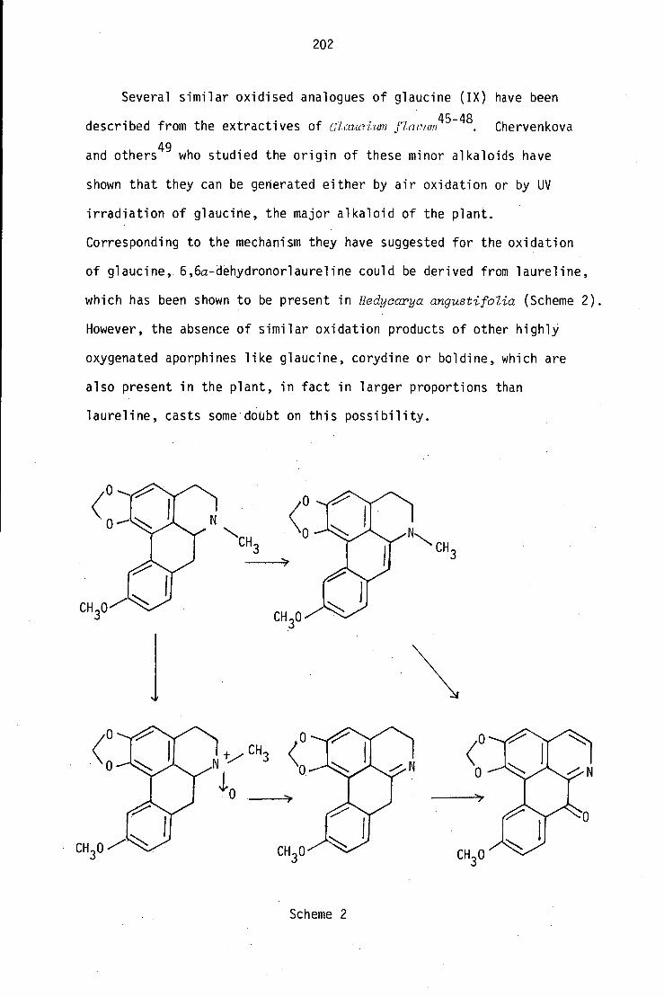
202
Several similar oxidised analogues of glaucine (IX) have been
described from the extractives of Clauoium Paolim45-48
. Chervenkova
and others49
who studied the origin of these minor alkaloids have
shown that they can be generated either by air oxidation or by UV
irradiation of glaucine, the major alkaloid of the plant.
Corresponding to the mechanism they have suggested for the oxidation
of glaucine, 6,6a-dehydronorlaureline could be derived from laureline,
which has been shown to be present in Hedyearya angustifblia (Scheme 2).
However, the absence of similar oxidation products of other highly
oxygenated aporphines like glaucine, corydine or boldine, which are
also present in the plant, in fact in larger proportions than
laureline, casts some doubt on this possibility.
Scheme 2

203
10.2.4 Structural elucidation of isosevanine (XVIA)
The third fraction gave a base which was isolated as a gum.
The alkaloid had the formula C18H15N04
by high-resolution MS and its PMR
spectrum had a down-field aromatic AB double doublet at 8.25 and 2.36 ppm
(J = 4.5 Hz) indicative of an aromatic heterocyclic system; also signals
for a methylenedioxy and a methoxyl group. The UV spectrum suggested
a benzylisoquinoline structure.
This compound, which has been named isosevanine,deposited yellow
needle-like crystals on cold storage, and a crystal X-ray diffraction
study carried out by Dr. A.H. White, University of Western Australia,
revealed its structure as the benzylisoquinoline (XVIA)isomeric with the
known alkaloid sevanine (XVIB).
XVIA isosevanine XVIB seyanine
Its PMR spectrum showed, in addition to the AB double doublet,
two one-proton singlets at 7.35 and 7.04 ppm (C5-H, C8-H), a three-
proton multiplet at 6.73 ppm (C2'-H, C5'-H, C6'-H), a two-proton
singlet at 6.06 ppm for the methylenedioxy protons, a two-proton
singlet at 4.4 ppm for the benzylic protons and a three-proton singlet
at 3.8 ppm for the methoxy protons.
10.2.5 Structural elucidation of isouvariopsine (XVII)
The sixth fraction gave two compounds, glaucine and a less polar
compound with the formula C 20 1-1 2 003 . The UV spectrum with six
absorption bands suggested a phenanthrene nucleus. The PMR spectrum
showed the presence of a methylenedioxy (6.25 ppm), a methoxy (4.0 ppm)

204
and methylamino (1.43 ppm) groups and a total of six aromatic protons
(8.6, 1H and 7.8-7.15, 5H) in the molecule. On biogenetic grounds the
methylenedioxy group is preferred on ring A,and the down-field aromatic
proton singlet at 8.6 ppm represents a bay proton (C-5H) in the
phenanthrene system, indicating the position of the methoxy group as
at C-6. This suggested the structure (XVII) for this alkaloid, which
has been named isouvariopsine as it is isomeric with the known alkaloid
uvariopsine (XVIII) 19 .
The structure was confirmed by a comparison of its spectra with
those of the Hofmann degradation product of the quaternary ammonium
salt of laureline. 2
CH3
OCH3
XVII isouvariopsine
XVIII uvariopsine
10.2.6 Structural elucidation of 0-methylcinnamolaurine (XX)
The fifth fraction gave laureline and a yellow gum which was shown
to be chromatographically pure, with the formula C 19H200 3 from high-
resolution mass spectrometry. The MS fragmentation (Scheme 3) and
the UV spectrum suggested a tetrahydrobenzylisoquinoline alkaloid.
The prominent MS fragments at m/z 190 and 121 indicated an N-methylated
benzylisoquinoline with a methylenedioxy function on ring A and a
methoxy function on ring C. From biogenetic considerations, the
methylenedioxy function is most likely at the C-6, C-7 positions, and
this is further supported by the presence of two one-proton singlets
at 6.55 and 6.2 ppm, corresponding to the C-5 and C-8 protons
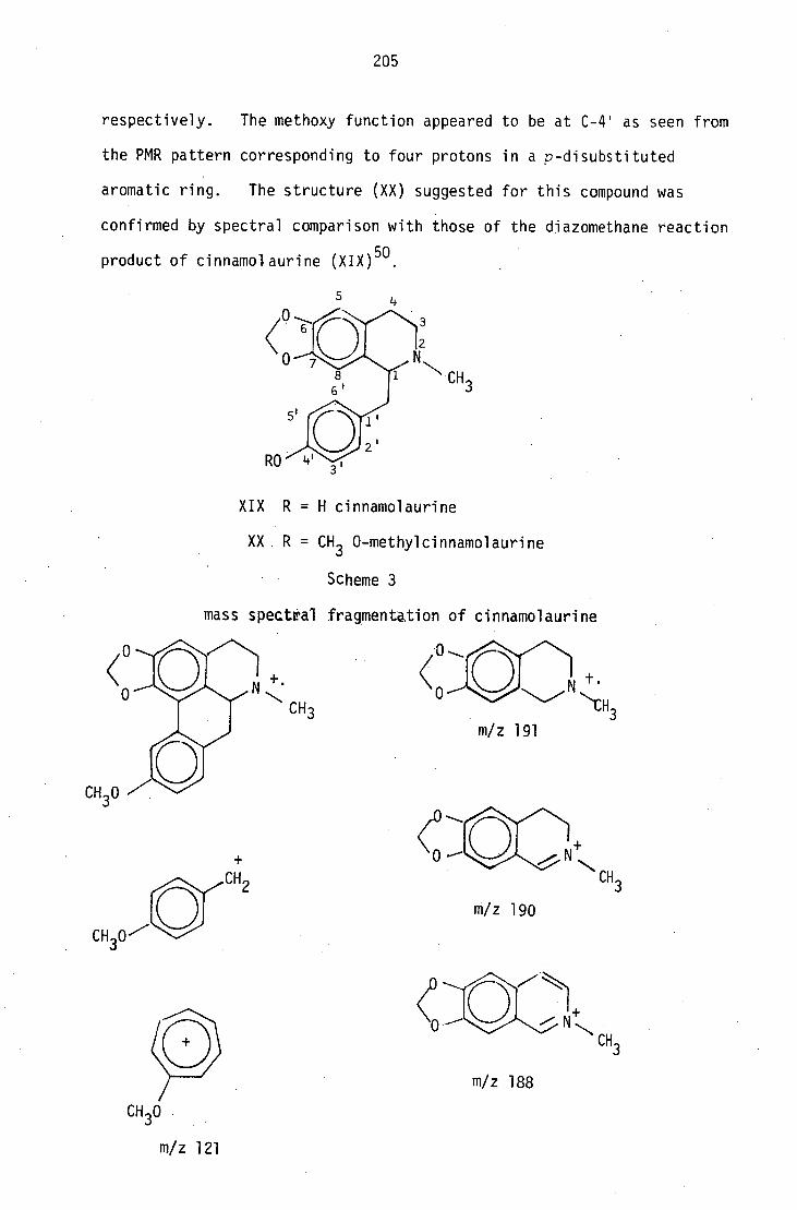
3
0
0
m/z 191
m/z 190
m/z 188
m/z 121
205
respectively. The methoxy function appeared to be at C-4' as seen from
the PMR pattern corresponding to four protons in a p-disubstituted
aromatic ring. The structure (XX) suggested for this compound was
confirmed by spectral comparison with those of the diazomethane reaction
product of cinnamolaurine (XIX) 50 .
XIX R = H cinnamolaurine
XX R = CH3 0-methylcinnamolaurine
Scheme 3
mass spectral fragmentation of cinnamolaurine

206
10.3 Experimental
Extraction of Hedycarya angustifolia
Leaves, twigs and bark of Hedycarya angustifolia collected from King Island
in March 1978 were air-dried, milled to a fine powder (22.4 Kg) and
exhaustively extracted with methanol until a test sample gave a negative
alkaloid test with Mayer's reagent. The methanol extract was
concentrated (2 0 in vacuo and dissolved in glacial acetic acid (1.5 z).
The solution was poured in a fine stream into rapidly-stirred water (20 z)
and left overnight, then the non-alkaloid precipitate was filtered off
through celite. The filtrate was evaporated to dryness in vacuo below
45°C and the residue was re-dissolved in 5% aqueous sulphuric acid.
The acid solution was extracted with ether (100 x 5 ml), basified with
ammonia and extracted with chloroform (100 x 10 ml). The chloroform
layer was dried (Na 2SO4 ) and evaporated to dryness in vacuo to give the
crude alkaloid extract as a yellow amorphous powder (12.5 g, 0.056%).
The ethereal extract on evaporation gave a pale yellow crystalline,
non-alkaloidal material (6 g). The methanol recovered from the initial
extract was found to deposit a white crystalline somewhat volatile non-
alkaloidal material (0.7 g).
The crude alkaloids were dissolved in chloroform (100 ml) and the
chloroform solution was extracted with sodium hydroxide (10 x 5 ml).
The aqueous alkaline solution was treated with ammonium chloride and then
extracted with chloroform (10 x 10 ml), dried (Na 2SO4 ) and evaporated to
give the phenolic fraction (2.7 g). The chloroform solution remaining
after extraction with alkali, upon concentration deposited a white
crystalline solid which was later found to be identical with the ether-
soluble non-alkaloidal material. The crystals were filtered off, and the
mother liquor was evaporated to give the non-phenolic alkaloid fraction
(12.5 g, 0.056%). The two fractions did not show any significant
qualitative difference on tic with several different solvent systems,

207
and both were found to contain considerable quantities of non-alkaloidal
material.
A second batch of plant material (25 Kg) collected from the same
locality in July 1979 failed to give any alkaloids when extracted in
the identical way. However, a third batch (23 Kg) collected in March
the following year gave a further 5.9 g crude alkaloid extract.
All extracts (20.1 g) were dissolved in CHC1 3 and extracted with
dilute hydrochloric acid (0.05%, 25 x 4 m1). The aqueous acid extract
was exhaustively extracted with ether in a liquid/liquid extractor for
30 hrs, basified with ammonia (pH = 13) and extracted with chloroform
(25 x 6 m1). The combined extracts were dried and evaporated to give
the crude alkaloid extract (0.8 g, 1.76 x 10 -3%).
2.3 Camphanediol
The ether-soluble non-alkaloidal material on recrystallisation from
methanol gave 2,3-camphanedio1 as white needles, m.p. 253-4° (lit.
27 250-3°
39); [a] D
- 8° (methanol); vmax ( KBr): 3300 (OH), 2950 (CH),
-1 1450 cm ; PMR (6 ppm): 3.9 (1H, m, H-C 3
), 3.8 (1H, m, H-C 2), 2.3 (2H,
H-C6 , H-C 6 ), 1.7 (1H, d x m, J = 12.5 Hz, H-C4 ), 1.35 (2H, m, H-C 6 , H-C 6 ),
1.09 (3H, s, 3H-C10 ), 0.88 (3H, s, 3H-C8 ), 0.81 (3H, s, 3H-C9); 13
C PMR
(6 ppm): 76.3 (d, C 2 ), 75.8 (dm, C 3 ), 53.7 (d, C 4 ), 51.32 (s, C7 ), 48.48
(s, C 1 ), 39.1 (tm, CO, 36.69 (tt, C 6 ), 21.35 (qm, C8 ), 20.12 (qm, C 9 ),
13.14 (q, C 10 ); m/z 170 (M+, 7%), 169 (M
+-1, 15), 154 (98), 153 (M-OH, 100),
146 (50), 137 (60), 136 (78), 126 (88), 125 (86), 124 (48), 123 (70), 112 (25),
110 (80), 93 (89); found: C 70.73, H, 10.71, C 10H 1802 requires C, 70.58,
H, 10.58.
Volatile non-alkaloid fraction
The methanol recovered from the extraction deposited white crystals
(0.7 g, 3.15 x 10 -5%), recrystallised from methanol as white flakes, m.p. 62°,

208
vmax
(KBr) 2900 (CH), 1430 cm; MS: m/z, 204 (10%, M+ *), 189 (9), 164 (7),
161 (12), 149 (22), 135 (5), 122 (8), 109 (14), 108 (12), 81 (12), 79 (8).
The compound was identified as a mixture of a- and a-eudesmol40c
by PMR
and mass spectral comparison with authentic samples.
Separation of the alkaloid mixture
On tic the presence of seven alkaloids could be detected, but a
considerable amount of non-alkaloidal material was still present, and this
together with the small amount of sample available, made the purification
of these compounds by ptic alone a difficult task. However when the
mixture was subjected to droplet countercurrent chromatography41
,
relatively simple mixtures free from non-alkaloidal material could be
obtained. The instrument used consisted of a hundred tubes each of 4 mm
in diameter and 1 m in length. The solvents used were obtained by
equilibrating methanol:chloroform:water (5:5:3); the lower phase was used
as the stationary phase while the upper phase, containing sufficient
sulphuric acid to give a concentration of N/1000, was used as the mobile
phase. The latter as it emerged from the apparatus was monitored by a
UV detector (254 mm), and 3 ml fractions were collected. Every fifth
fraction was tested by tic after basification with ammonia and extraction
with chloroform. The fractions were bulked accordingly and the bulking
summary is shown in Table 1. Methanol and chloroform were removed from
each of the fractions by careful evaporation under vacuum below 30 ° C,
and the aqueous residues were basified, extracted with chloroform, dried,
and evaporated to give the crude alkaloid fractions which were further
purified by ptic to give the individual alkaloids.
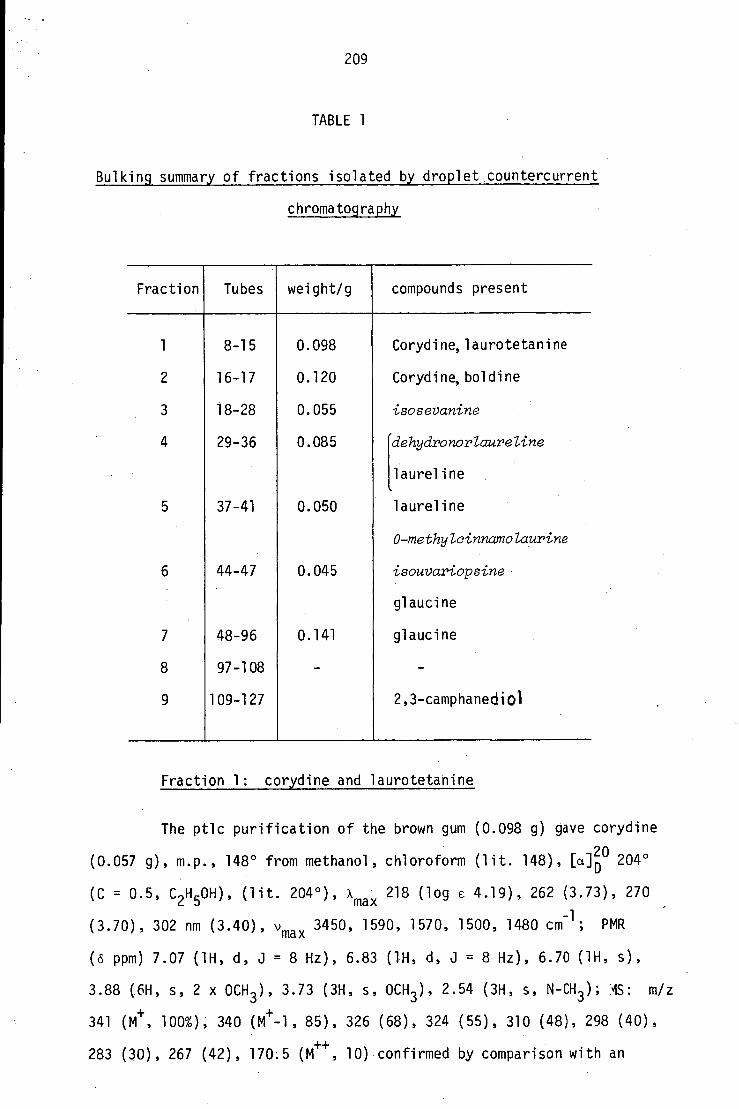
209
TABLE 1
Bulking summary of fractions isolated by droplet countercurrent
chromatography
Fraction Tubes weight/g compounds present
1 8-15 0.098 Corydine,laurotetanine
2 16-17 0.120 Corydine,boldine
3 18-28 0.055 isosevanine
4 29-36 0.085 'dehydronorlaureline
,laureline
5 37-41 0.050 laureline
0-methylcinnamolaurine
6 44-47 0.045 isouvariopsine
glaucine
7 48-96 0.141 glaucine
8 97-108 - -
9 109-127 2,3-camphanedi01
Fraction 1: corydine and laurotetanine
The ptic purification of the brown gum (0.098 g) gave corydine
a (0.057 g), m.p., 148° from methanol, chloroform (lit. 148), [] 2D0 204°
(C = 0.5, C 2 H 5OH), (lit. 204°), xmax 218 (log 6 4.19), 262 (3.73), 270
(3.70), 302 nm (3.40), vmax 3450, 1590, 1570, 1500, 1480 cm -1 ; PMR
(6 ppm) 7.07 (1H, d, J = 8 Hz), 6.83 (1H, d, J = 8 Hz), 6.70 (1H, s),
3.88 (6H, s, 2 x OCH 3 ), 3.73 (3H, s, OCH 3 ), 2.54 (3H, s, N-CH 3 ); IS: m/z
341 (Mt , 100%), 340 (M+-1, 85), 326 (68), 324 (55), 310 (48), 298 (40),
283 (30), 267 (42), 170.5 (M++ , 10) confirmed by comparison with an

210
authentic sample of corydine 19 ; and laurotetanine19 (0.026 g) which
could not be obtained crystalline, vmax 220 (log e 4.25), 280 (3.22),
308 nm (3.15), +OH - 325 (3.35); vmax 3350' 1580, 1500 cm
-1; PMR (6 ppm)
7.88 (1H, s), 6.55 (2H, m), 3.83 (6H, s, 2 x 0CH 3 ), 3.52 (3H, s, OCH 3 ); MS:
m/z 327 (M+ , 68), 326 (M+-1, 100), 312 (26), 310 (18), 298 (12), 296 (21),
283 (8), 267 (10), 163.5 (M++ , 8). The identity of this compound was
confirmed by comparison with an authentic sample of laurotetanine.
Fraction 2: corydine and boldine
The ptic purification of this fraction (0.120 g) gave cz o d2Din:19108.
(0.035 g) and boldine19 (0.072 g) m.p., 161 0 (lit. 19 , 161
. ),
(C = 1, C2H 5OH), (lit. 19 + 111°), xmax 220 (log c 4.6), 283 (4.21, 304 nm
(4.23); PMR (6 ppm, CD30D), 7.99 (1H, s), 6.90 (1H, s), 6.60 (1H, s),
3.92 (3H, s, OCH 3 ), 3.61 (3H, s, OCH 3 ), 2.58 (3H, s, N-CH 3 ); MS: m/z 327
(M+ , 85%), 326 (M+-1, 100), 312 (32), 310(38), 296 (29), 284 (68), 269 (72),
253 (52), 163.5 (5), which was confirmed by comparison with an authentic
sample of boldine 19 .
Fraction 3: isosevanine
Ptic of the gum (0.055 6) gave isosevanine (0.016 g) l colourless
needles from methanol chloroform, m.p. 148°; 'max 236 (log E 4.12), 270
(3.71), 314 (3.41), 330 nm (3.32); v max 3400, 1640, 1610, 1580 cm -1 ;
PMR (6 ppm), 8.28 (1H, d, J = 4.5 H 2 ), 7.36 (1H, d, J = 4.5 Hz), 7.35
(1H, s), 7.05 (1H, s), 6.7 (3H, m), 6.05 (2H, s), 4.45 (2H, s), 3.83
(3H, s, OCH3 ); MS: m/z 309 (MI- , 60%), 308 (M+-1, 100), 294 (23), 278 (8),
137 (11), 83 (56), 77 (22); measured 309.0987,calculated for C 10 1 004
309.1017.

211
Fraction 4: dehydronorlaureline and laureline
Ptic purification of this fraction gave laureline (0.020 g) as
a brown gum l \max 218 (log e 4.20), 264 (3.95), 273 (4.1), 315 (3.57),
325 nm (3.25); PMR ((5 ppm) 7.66 (1H, d, J = 2.5 H4), 7.15 (1H, d,
J = 10 Hz), 6.75 (1H, d, J = 10 Hz), 6.53 (1H, s), 6.03 (1H, d, J = 1.5 Hz),
5.88 (1H, d, J = 1.5); MS: m/z 309 (M+, 65%), 308 (M
+-1, 100%), 294 (53),
266 (48), which was identified by comparison with an authentic sample,
and a brown gum (0.043 g) which was identified as 6,6a-dehydronor1aure1ine,
\max 248 (log E 4.32), 278 (4.08), 317 (3.83) and 330 nm (3.85); v max
-1 1690, 1640 and 1600 cm ; PMR ( ppm) 7.35 (1H, s, 11-H), 7.2 (1H, s, 3-H),
7.1 (1H, d, J = 10 Hz, 9-H), 6.75 (1H, d, J = 10 Hz, 8-H), 6.1 (2H, s,
0-CH2-0), 4.49 (2H, m, 7-H), 3.7 (3H, s, OCH 3 ), MS: m/z 293 (le, 55), 292 (100),
288 (30), 262 (18), 249 (2), 149 (20), 146.5 (M ++ , 2); measured 293.1043
calculated for C 10 1 003 293.1052.
Attempted N-methylation of 6,6a-dehydronorlaureline
The compound (0.010 g) in methanol (3 ml) was stirred with formaldehyde
(37%, 0.2 ml) at room temperature for 5 hrs, then sodium borohydride
(0.050 g) was added in small portions. The solvents were removed, the
residue dissolved in dilute aqueous hydrochloric acid (10 ml), and the
solution was basified with ammonia and extracted with chloroform. The
product obtained did not show the presence of an N-CH 3 group (PMR and MS)
but the molecular weight was found to have increased by 2 a.m.u. The
compound was later identified as norlaureline.
Conversion of 6,6'-dehydronorlaureline to laureline
The compound (0.020 g) dissolved in acetone (5 ml) was treated with
methyl iodide (0.05 ml) in a sealed tube and left overnight at room
temperature. Removal of the solvent gave a brown solid, m/z 308, which

212
was dissolved in methanol (5 ml) and treated with sodium borohydride
(0.020 g). The usual work-up of the reaction mixture gave laureline
(0.018 g) as a brown gum; A max 218 (log c 4.20), 264 (3.95), 273 (4.1),
315 (3.57), 325 nm (3.25); PMR (6 ppm) 7.66 (1H, d, J = 2.5), 7.15 (1H,
d, J = 10 Hz), 6.75 (1H, d, J = 10 Hz), 6.53 (1H, s), 6.03 (1H, d,
J = 1.5 Hz), 5.88 (1H, d, J = 1.5 Hz); MS: m/z 309 (M+, 65%), 308 (M
+-1,
100%), 294 (53), 266 (48), which was identified by comparison with an
authentic sample of laureline.
Fraction 5: laureline and 0-methylcinnamolaurine
Ptic purification of this fraction gave laurel ine,and a more polar
compound as a brown gum which could not be crystallised. Amax 225 (log E
4.25), 270 (4.12), 275 nm (4.06); vmax 1680, 1650, 1600, 1510 cm -1 ; PMR
(6 ppm) 7.05 (2H, d, J = 8 Hz, 2'-H, 6'-H), 6.81 (2H, d, J = 8 Hz, 3'-H,
5'-H), 6.55 (1H, s, 5-H), 6.2 (1H, s, 8H), 5.86 (2H, dd, J = 8, 1.5 Hz),
0-CH 2-0), 5.1 (1H, m, 1H), 3.78(3H, s, OCH 3 ), 2.5 (3H, s, NCH 3 ); MS: m/z
311 (MI- , 0.2%), 310 (0.2), 296 (0.2), 191 (12), 190 (100), 188 (7), 174
(13), 160 (7), 144 (12), 132 (8), 121 (80), 77 (15), 59(21), 43 (36).
HRMS 411.1515 calculated for C19H21NO3
311.1521. The compound was
identified as 0-methylcinnamolaurine by comparison with an authentic
sample prepared by diazomethane,methylation of cinnamolaurine 48 .
Fraction 6
This fraction yielded two compounds on ptic. The less polar
compound, isouvariopsine,a yellow solid, m.p. 155-157°, gave xmax 218
(log c 3.93), 250 (4.35), 260 (4.35), 313 (3.88), 325 (3.91), 360 (3.62),
378 nm (3.65); vmax 1610, 1590, 1500, 1450 cm -1 , PMR Co ppm) 8.6 (1H, m),
7.8-7.15 (5H, m), 6.25 (2H, s, 0-CH 2-0), 4.0 (3H, s, 0CH3 ), 3.27 (2H, m),
2.65 (2H, m), 2.43 (6H, N-CH 3 x 2); MS: m/z 323 (M+ , 60%), 308 (12), 292 (16),
278 (30), 265 (42), 247 (8), 222 (18), 205 (7), 176 (27), 163 (36), 58 (TOO).
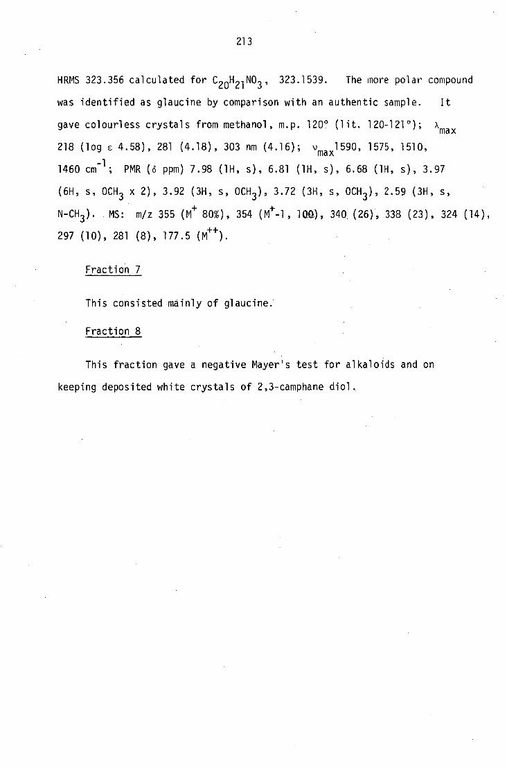
213
HRMS 323.356 calculated for C 20 2003 , 323.1539. The more polar compound
was identified as glaucine by comparison with an authentic sample. It
gave colourless crystals from methanol, m.p. 120 0 (lit. 120-1210); Amax
218 (log E 4.58), 281 (4.18), 303 nm (4.16); vmaxi"°
1575, 1510,
-1 1460 cm ; PMR (6 ppm) 7.98 (1H, s), 6.81 (1H, s), 6.68 (1H, s), 3.97
(6H, s, OCH 3 x 2), 3.92 (3H, s, OCH 3 ), 3.72 (3H, s, OCH 3 ), 2.59 (3H, s,
N-CH3 ). MS: m/z 355 (M+ 80%), 354 (M
+-1, 1011), 340 (26), 338 (23), 324 (14),
297 (10), 281 (8), 177.5 (M++ ).
Fraction 7
This consisted mainly of glaucine.
Fraction 8
This fraction gave a negative Mayer's test for alkaloids and on
keeping deposited white crystals of 2,3-camphane diol.

214
REFERENCES
1. A. Urzua and B.K. Cassels, J. Natural Products, 41, 98 (1978).
2. I.R.C. Bick and T.G. Whalley, Univ. Queensland Papers, Dept. of
Chem., 1 ., No. 28 (1946); /, No. 30 (1947); /, No. 33 (1948).
3. I.R.C. Bick and A.R. Todd, J. Chem. Soc., 2170 (1948).
4. I.R.C. Bick, E.S.'Ewen and A.R. Todd, J. Chem. Soc., 2767
(1949).
5. I.R.C. Bick and A.R. Todd, J. Chem. Soc., 1606 (1950).
6. I.R.C. Bick, K. Doebel, W.I. Taylor and A.R. Todd, J. Chem. Soc.,
692 (1953).
7. I.R.C. Bick, W.I. Taylor and A.R. Todd, J. Chem. Soc., 695
(1953).
8. I.R.C. Bick, P.S. Clezy and M.J. Vernengo, J. Chem. Soc., 4928
(1960).
9. J. Harley-Mason, A.S. Howard, W.I. Taylor, M.J. Vernengo,
I.R.C. Bick and P.S. Clezy, J. Chem. Soc., 1948 (1967).
10. I.R.C. Bick and W.I. Taylor, J. Chem. Soc.(C), 3779 (1971).
11. I.R.C. Bick and S. Sotheeswaran, Aust. J. Chem., 31, 2077 (1978).
12. I.R.C. Bick, H.M. Leow and S. Sotheeswaran, Tetrahedron Lett.,
2219 (1975).
13. I.R.C. Bick and H.M. Leow, Aust. J. Chem., 31, 2539 (1978).
14. I.R.C. Bick, H.M. Leow and M.J. Richards, Aust. J. Chem., 33, 225
(1980).
15. N. Zeyer, Vjschr. Prakt. Pharm., 10, 504. (Quoted by T.A. Henry,
"The Plant Alkaloids", 4th ed., J. and A. Churchill, London, 1949).
16. I.R.C. Bick, P.S. Clezy and W.D. Crow, Aust. J. Chem., 9, 111
(1956).
17. I.R.C. Bick and G.K. Douglas, Aust. J. Chem., 18, 1997 (1965).
18. H.M. Leow, Ph.D. Thesis, University of Tasmania, (1979).
215
19. H. Guinaudeau, M. Leboeuf and A. Cave, J. Natural Products, 38,
275 (1975).
20. H. Guinaudeau, M. Leboeuf and A. Cave, J. Natural Products, 42,
325 (1979).
21. I.R.C. Bick, J. Harley-Mason, N. Sheppard and M.J. Vernengo,
J. Chem. Soc., 1896 (1961).
22. W.H. Barschers, R.R. Arndt, K. Pachler, J.A. Weisbach and
B. Douglas, J. Chem. Soc., 4778 (1974).
23. K.G.R. Pachler, R.R. Arndt and W.H. Barschers, Tetrahedron, 21,
2159 (1965).
24. M. Ohashi, J.M. Wilson, H. Budzikieweiz,M. Shamma, W.A. Slusarchyk
and C. Djerassi, J. Amer. Chem. Soc., 85, 2807 (1963).
25. A.H. Jackson and J.A. Martin, J. Chem. Soc. (C), 2181 (1966).
26. L.M. Jackman, J.C. Trewella, J.L. Moniot, M. Shama, R.L. Stephens,
E. Wenkert, M. Leboeuf and A. Cave, J. Natural Products, 42, 438
(1979).
27. M. Shamma, in "The Isoquinoline Alkaloids", 1972, Academic Press
•and "Isoquinoline Alkaloid Research 1972-1977", 1978 Plenum Press,
•N.Y.
28. T. Kametani, in "The Isoquinoline Alkaloids", Hirokawa Publishing
Company, Tokyo, 1969.
29. G. Merk, Ann., 66, 125 (1848); 73, 50 (1856).
30. H. Furukawa, T.H. Yang, T.J. Lin, J. Pharm. Soc. Japan, 85, 472
(1965).
31. E. Fujita, T. Tomimatsu, J. Pharm. Soc. Japan, 79, 1082 (1959).
32. A. Galart, J. Amer. Chem. Soc., 72, 4436 (1950); 73, 3654 (1951).
33. J. Kunitomo, E. Yuge, Y. Nagai, J. Pharm. Soc. Japan, 86, 456
(1966).
34. I.A. Israilov, M.A. Manushakyan and M.S. Yunusov, Khim. Prir.
Soedin. 1 412 (1978).

216
35. R. Ziyaey, T. Irgashev, I.A. Israilov, N.D. Abdullaev,
M.S. Yunusov and S.Y. Yunusov, Khim. Prir. Soedin, 239 (1979).
36. V.A. Mnatsakanayan, V. Preininger, V. Simanek, J. Juriana,
A. Klasek, L. Dolejs and F. Santavy, Collect. Czech, Chem. Commun.,
42, 1421.
37. V.A. Mnatsakanayan, V. Preininger, V. Simanek, A. Klasek,
L. Dolejs and F. Santavy, Tetrahedron Lett., 851 (1974).
38. E. Brochmann-Hanssen, K. Hirai, J. Pharm. Sci., 57, 940 (1968).
39. H. Rupe and F. Muller, Hely. chim. Acta, 24, 1093 (1941).
40. (a) L. Ruzicka, J. Meyer, U.M. Mingazzini, Hely. chim. Acta, 5,
345 (1972).
(b) R.O. Hellyer, Aust. J. Chem., 15, 157 (1962).
(c) R.V.H. Jones and M.D. Sutherland, Chem. Commun., 1229 (1968).
41. K. Nakanishi, J. Chromatography, 170, 355 (1979) and the references
therein.
42. M. Shamma, Experientia, 16, 484 (1960).
43. M. Shamma and S.S. Salgar, Phytochemistry, 12, 1505 (1973).
44. M. Shamma, S.Y. Yao, J. Org . Chem., 36, 3253 (1971).
45. I. Ribas, J. Sueres and L. Castedo, Tetrahedron Lett., 3093 (1971).
46. I. Ribas, J. Sueres and L. Castedo, Tetrahedron Lett., 2033 (1972).
47. H.B. Dutschewska, A.S. Orahovats and N.M. Molloy, C.R. Acad. Bulg.
Sci., 26, 899 (1973).
48. H.G. Kiriyakov and P. Panov, C.R. Acad. Bulg. Sci., 22, 1019 (1969).
49. V.B. Shervenkova, N.M. Molloy and S. Paszyc, Phytochamistry, 20,
2285 (1981).
50. E. Gellert and R.E. Summons, Aust. J. Chem., 23, 2095 (1970).

217
Alkaloids of Macadamia integrifaia
(Queensland nuts( Proteaceae)) .
In a preliminary screening of plants belonging to the family
PPoteaceae, the kernels were found to give a +ve test for alkaloids'.
The presence of alkaloids in the edible portion of these nuts had not
been recorded in previous surveys of Queensland plants .
The presence of large amounts of fats had seriously interfered
with the preliminary experiments in extraction of alkaloids.
Complete removal of the fats and oils by extraction with petroleum
ether prior to the extraction of alkaloids was difficult, and serious
problems with emulsions were encountered during this process, and
subsequently with the extraction of the fine powdery residue to remove
the alkaloids1
.
In the present investigation, a number of extraction procedures
were explored.
The kernels (500 g) were homogenised in a blender with a mixture
of methanol:chloroform:aqueous sulphuric acid (5:5:3, I t). The
milky suspension obtained was left in a separatory funnel overnight.
Three layers were formed. All cellular material and possibly some
fats and oils were incorporated into the heaviest chloroform phase.
The next aqueous acidic phase, which remained clear, contained the
alkaloids, while the top layer consisted of a clear oil (260 g).
The three layers were separated, and the chloroform layer was washed
with further quantities of aqueous sulphuric acid until a sample gave
a negative alkaloid test. The washings were combined with the
initial aqueous phase and extracted exhaustively with ethyl acetate
until most of the non-basic material was removed. The remaining
aqueous solution gave a very strong +ve test for alkaloids with
Mayer's reagent and for sugars with Molisch test.
218
On neutralisation of the acidic solution with ammonia, a
gelatinous precipitate was formed (0.820 g). The precipitate was
recovered but when it was re-dissolved in aqueous sulphuric acid it
gave only a weak +ve test for alkaloids, and furthermore, it was
insoluble in common organic solvents.
The remaining aqueous phase was basified (pH = 9) with ammonia
and extracted with chloroform and with ethyl acetate, but none of the
extracts were found to contain any alkaloid material.
At this stage, it was presumed that the alkaloids could be either
quaternary ammonium salts or N-oxides. A sample of the aqueous
solution was treated with zinc powder and acetic acid and extracted
as previously after basification. However, none of the solvents
carried any alkaloid material, and this eliminated the possibility of
the alkaloids being N-oxides.
Another sample was acidified to pH 4 and treated with a saturated
aqueous solution of ammonium reineckate. A heavy pink precipitate of
reineckate complexes was formed, suggesting that the alkaloids could
be quaternary salts.
The aqueous phase was acidified (pH = 4) and treated with
saturated ammonium reineckate solution until precipitation was complete,
then the precipitate was centrifuged and dried (11.5 g). The reineckate
complex was then treated in the standard way: it was dissolved in
absolute acetone, filtered, and the filtrate was diluted with water
(1:1) and treated with aqueous silver sulphate solution until the
precipitation of silver reineckate was complete. The solution was
filtered, and the filtrate was treated with an aqueous solution of
barium chloride to precipitate silver as the chloride, then the
solution was again filtered2
. The final filtrate did not give a
+ve alkaloid test, but it was found to contain ammonium magnesium
phosphate (0.8 g).

219
It was discovered later that the reineckate complex had decomposed
when dissolved in dry acetone. The gelatinous precipitate filtered
off at this stage was found to give a strong +ve test for alkaloids.
In a separate experiment, the kernels were extracted using a
methanol:chloroform:water (5:5:3) system, and the aqueous phase, after
extraction with ethyl acetate, was freeze-dried. The glassy substance
obtained (9.8 g) gave a strong +ve test for alkaloids. This was
found to be insoluble in most of the organic solvents except pyridine
and dimethylsulphoxide. This material when chromatographed on
cellulose plates using n-butyl acetate, n-butanol, acetic acid, water
(85:15:40:22) was found to contain substances that stained with both
iodoplatinate and Dragendroff reagents, but did not form distinct
spots as such. Solvent systems of different composition were tried
but a satisfactory separation could not be obtained.
An attempt was made to separate this mixture by gel filtration
on Sephadex G20, but no soluble fraction could be obtained.
When refluxed with mineral acids, the freeze-dried substance was
found to release some chloroform-soluble material which gave an
alkaloid test. However, neither the concentration of the acid nor
the hydrolysis time had any effect on the final yield of the alkaloids.
Different acid concentrations (1 to 15%) and different reaction times
were employed, but under all reaction conditions, using 0.5 g of
the material, only 3-5 mg of the supposed alkaloid mixture was obtained;
when a single 0.5 g sample was subjected to successive hydrolyses
followed by work-up at every stage it yielded the same amount of
alkaloids. Finally, when a larger sample (5 g) of the substance was
subjected to hydrolysis using 7% aqueous hydrochloric acid for 0.5 hours,
the yield was still only 4 mg of the alkaloid mixture.
This observation suggests that the alkaloids released by hydrolysis
are unstable under these reaction conditions.

220
An attempt to hydrolyse the freeze-dried material using chymotrypsin
has so far been unsuccessful.
REFERENCES
1. H.M. Leow, Ph.D. Thesis, University of Tasmania, 1979.
2. Z.M. Khan, M. Hesse and H. Schmid, Hav. Chim. Acta., 48, 1957
(1965).