Embed Size (px)
Citation preview

University of Calgary
PRISM: University of Calgary's Digital Repository
Graduate Studies The Vault: Electronic Theses and Dissertations
2015-08-24
Structural Studies of Human Norovirus Protease
Complexes with RNA and Peptides
Sikandar, Asfandyar
Sikandar, A. (2015). Structural Studies of Human Norovirus Protease Complexes with RNA and
Peptides (Unpublished master's thesis). University of Calgary, Calgary, AB.
doi:10.11575/PRISM/27903
http://hdl.handle.net/11023/2403
master thesis
University of Calgary graduate students retain copyright ownership and moral rights for their
thesis. You may use this material in any way that is permitted by the Copyright Act or through
licensing that has been assigned to the document. For uses that are not allowable under
copyright legislation or licensing, you are required to seek permission.
Downloaded from PRISM: https://prism.ucalgary.ca

UNIVERSITY OF CALGARY
Structural Studies of Human Norovirus Protease Complexes with RNA and Peptides
by
Asfandyar Sikandar
A THESIS
SUBMITTED TO THE FACULTY OF GRADUATE STUDIES
IN PARTIAL FULFILMENT OF THE REQUIREMENTS FOR THE
DEGREE OF MASTERS OF SCIENCE
GRADUATE PROGRAM IN BIOLOGICAL SCIENCES
CALGARY, ALBERTA
August, 2015
© Asfandyar Sikandar 2015

ii
Abstract
Noroviruses are single-stranded RNA viruses. They encode a protease that cleaves a viral
polyprotein at specific sites to produce mature viral proteins. In addition, the protease also
binds to viral RNA, and thus is thought to regulate viral replication. However, to date no
structural information is available for protease-substrate complexes that might explain the
interactions made by peptide residues P’-side of cleavage junctions or RNA. Here I report the
work carried out to characterize these interactions in human norovirus protease using X-ray
crystallography. The protease was successfully expressed, purified and the crystallization
conditions were optimized to grow crystals for structure determination. Unfortunately, RNA
and peptide electron density were not observed in co-crystal structures. The packing of
protease molecules in one of the crystal forms shows the interaction of protease C-terminal
residues with the peptide-binding groove of a neighboring molecule in the crystal, thereby
providing the view of a protease-product complex.
iii
Acknowledgments
It would not have been possible to work on this project without the support and help of people
around me.
First, I would like to thank my family for their unconditional support. Then there are my friends
Saad Rajput, Saad Amjad, Mujeeb Mufti and Alexander Carleton, who made graduate life fun
and were there for me whenever I needed them.
This thesis would not have been possible without the help of our collaborators: Jared May,
Prasanth Viswanathan and Brent Korba from the University of Georgetown, USA. They
expressed both the wild type and mutant protease for this project. I would like to thank all the
current members of Dr. Ng’s lab- especially Dr. Md Munan Shaik for his help in refinement and
analysis of X-ray diffraction data.
I would also like to extend my gratitude to my committee members, Dr. Zimmerly and Dr.
Tieleman, for their suggestions and sound advice.
Last, but certainly not the least, my supervisor Dr. Kenneth Ng for giving me the opportunity to
work in his lab. I cannot thank him enough for his patience and trust in my abilities.
iv
Table of Contents
Abstract .......................................................................................................................................... ii Acknowledgments ........................................................................................................................ iii Table of Contents ......................................................................................................................... iv List of Tables ................................................................................................................................ vi List of Figures .............................................................................................................................. vii List of Abbreviations ................................................................................................................... ix Physical Units Abbreviation ....................................................................................................... xi Amino Acid Abbreviations ......................................................................................................... xii Chapter 1: Introduction ............................................................................................................... 1
a. Viral taxonomy ................................................................................................................................ 1 b. Epidemiology and pathophysiology ............................................................................................... 2 c. Virus genome organization ............................................................................................................. 3 d. NV life cycle ..................................................................................................................................... 4 e. NV Pro .............................................................................................................................................. 7
I. Structural features of NV Pro ......................................................................................................... 7 II. Catalytic site and mechanism of proteolysis ................................................................................ 9 III. Peptide substrate recognition and proteolytic processing order ................................................ 11
f. NV Pro-RNA interaction ............................................................................................................... 16 Chapter 2: Objectives of the Study ........................................................................................... 18
a. Interaction of HuNV Pro with RNA oligonucleotides ................................................................ 18 b. Interaction of HuNV Pro with P’ residues .................................................................................. 20
Chapter 3: Materials and Methods ........................................................................................... 21 a. Protein expression ......................................................................................................................... 21 b. Ion exchange chromatography ..................................................................................................... 21 c. Gel-filtration chromatography ..................................................................................................... 21 d. Dialysis and concentration ........................................................................................................... 22 e. RNA and peptide preparation ...................................................................................................... 22 f. Crystallization methodology .......................................................................................................... 22
I. Sparse matrix screening ............................................................................................................... 23 II. Crystal optimization .................................................................................................................... 24 III. Soaking ...................................................................................................................................... 25 IV. Crystal harvesting ...................................................................................................................... 25
g. X-ray data collection and structure determination .................................................................... 26 Chapter 4: Results and Discussion ............................................................................................ 27
a. HuNV Pro-RNA interaction ......................................................................................................... 27 I. Expression and purification ......................................................................................................... 27 II. Screening and hit optimization ................................................................................................... 29 III. Optimization of purification protocol ........................................................................................ 32 IV. Optimization of Thiocyanate condition ..................................................................................... 34

v
V. Soaking ....................................................................................................................................... 36 VI. X-ray data collection and structure determination ..................................................................... 37 VII. Structural analysis and discussion of AS59 ............................................................................. 40
b. HuNV Pro-peptide interaction ..................................................................................................... 44 I. Expression and purification ......................................................................................................... 44 II. Screening and optimization ........................................................................................................ 45 III. X-ray data collection and structure determination .................................................................... 47 IV. Structural analysis and discussion of AS106 ............................................................................. 51
Chapter 4: Conclusion and Future Work ................................................................................. 56 Bibliography ................................................................................................................................ 59 Appendix ...................................................................................................................................... 65

vi
List of Tables
TABLE 1: EFFECT OF RNA ON HUNV PRO ACTIVITY.. ............................................................................... 17
TABLE 2: RNA OLIGONUCLEOTIDE INFORMATION AND SEQUENCE.. ......................................................... 22
TABLE 3: SYNTHETIC PEPTIDE INFORMATION AND SEQUENCE.. ................................................................. 22
TABLE 4: CRYSTALLIZATION HITS IDENTIFIED FROM SCREENS PERFORMED IN THE PRESENCE OF HUNV
PRO AND RNA OLIGONUCLEOTIDES.. ................................................................................................. 29
TABLE 5: SUMMARY OF CHANGES MADE TO THE ORIGINAL CRYSTALLIZATION CONDITION IDENTIFIED TO
SLOW DOWN THE RATE OF CRYSTALLIZATION. .................................................................................. 32
TABLE 6: SUMMARY OF SALT CONDITIONS THAT YIELDED CRYSTALS. ...................................................... 35
TABLE 7: SUMMARY OF CONDITIONS USED FOR SOAKING EXPERIMENTS. .................................................. 36
TABLE 8: X-RAY DIFFRACTION DATA SETS COLLECTED FOR CO-CRYSTALS GROWN AND/OR SOAKED IN
DIFFERENT CONDITIONS.. .................................................................................................................... 38
TABLE 9: CRYSTALLOGRAPHIC DATA COLLECTION AND MODEL REFINEMENT STATISTS FOR AS59.. ....... 39
TABLE 10: CRYSTALLIZATION HITS IDENTIFIED FROM SCREENS PERFORMED IN THE PRESENCE OF MUTANT
HUNV PRO AND DIFFERENT PEPTIDES. .............................................................................................. 46
TABLE 11: SUMMARY OF THE CHANGES MADE TO THE CRYSTALLIZATION HITS TO GROW DIFFRACTION
QUALITY CRYSTALS. ........................................................................................................................... 47
TABLE 12: X-RAY DATA SETS COLLECTED FOR HUNV PRO-PEPTIDE CRYSTALS GROWN AND/OR SOAKED
IN DIFFERENT CONDITIONS. ................................................................................................................ 49
TABLE 13: CRYSTALLOGRAPHIC DATA COLLECTION AND MODEL REFINEMENT STATISTS FOR AS106.. ... 50

vii
List of Figures
FIGURE 1: CLASSIFICATION OF NOROVIRUSES INTO 5 GENOGROUPS (GI-V) AND 35 GENOTYPES BASED ON
SEQUENCE DIVERSITY IN THE COMPLETE CAPSID PROTEIN ................................................................. 2
FIGURE 2: STRUCTURE OF NOROVIRUS CAPSID AND NOROVIRUS GENOME ORGANIZTION. .......................... 4
FIGURE 3: LIFE CYCLE OF NOROVIRUS. ......................................................................................................... 6
FIGURE 4: STRUCTURE OF NV PRO. ............................................................................................................. 8
FIGURE 5: STRUCTURAL COMPARISON OF THE NV PRO (PDB-ID: 1QWS) WITH PV (PDB-ID: 1L1N) AND
HRV PROTEASES (PDB-ID: 1CQQ) ..................................................................................................... 9
FIGURE 6: NV PRO CATALYTIC MECHANISM .............................................................................................. 10
FIGURE 7: THE PROCESSING OF ORF1 POLYPROTEIN ................................................................................. 14
FIGURE 8: RIBBON REPRESENTATION OF HUNV PRO-PRODUCT COMPLEX STRUCTURE ............................ 14
FIGURE 9: SURFACE REPRESENTATION OF NV PRO (PDB ID: 4IN2) WITH COLOR-CODED ACTIVE SITE
AND SUBSTRATE BINDING POCKETS ................................................................................................... 15
FIGURE 10: SURFACE REPRESENTATION OF PEPTIDE-INDUCED CONFORMATIONAL CHANGES IN S2 AND S4
POCKETS OF HUNV PRO.. ................................................................................................................... 15
FIGURE 11: RNA BINDING BY HUNV PRO AND ITS AFFECT ON ENZYME KINETICS .................................... 17
FIGURE 12: PUTATIVE RNA BINDING SITES ON NV PRO BOUND TO PEPTIDE SUBSTRATE ........................ 19
FIGURE 13: ION EXCHANGE CHROMATOGRAM AND SDS-PAGE PROFILE OF HUNV PRO . ....................... 28
FIGURE 14: OPTIMIZATION OF THIOCYANATE CONDITION. ........................................................................ 31
FIGURE 15: X-RAY DIFFRACTION PATTERN OF CRYSTAL WITH AND WITHOUT DEFECTS ........................... 31
FIGURE 16: HUNV PRO GEL-FILTRATION ELUTION AND SDS-PAGE PROFILE.. ......................................... 33
FIGURE 17:THE CHANGE IN SIZE OF SEED CRYSTAL TRANSFERRED TO KCL CONDITION. .......................... 35
FIGURE 18: THE DIMERIC ARRANGEMENT OBSERVED IN AS59 . ................................................................ 41
FIGURE 19: COMPARISON OF DIMER STRUCTURES AND INTERFACE OF NV PRO DIMERS WITH AS59. ...... 42
FIGURE 20: OVERLAY OF AS59 MONOMER A (RED) AND B (GREEN) SHOWN AS CARTOONS ..................... 42
FIGURE 21: STRUCTURAL COMPARISON OF AS59 WITH OTHER HUNV PROTEASES. .................................. 43
FIGURE 22:STRUCTURAL COMPARISON OF HUNV PRO CRYSTAL GROWN IN THE PRESENCE (GREEN) AND
ABSENCE OF RNA OLIGONUCLEOTIDES (RED). .................................................................................. 43
FIGURE 23: MUTANT NV PRO GEL-FILTRATION ELUTION AND SDS-PAGE PROFILE (TOP). ..................... 44
FIGURE 24: HUNV PRO-PEPTIDE CRYSTAL BEFORE (LEFT) AND AFTER OPTIMIZATION (RIGHT).. .............. 46
FIGURE 25: CRYSTAL PACKING OF AS106 IN THE P21 SPACE GROUP, WITH FOUR MOLECULES IN THE
ASYMMETRIC UNIT .. ........................................................................................................................... 52
FIGURE 26:OVERLAY OF AS106 MONOMERS REPRESENTED AS RIBBON DIAGRAM.. .................................. 53

viii
FIGURE 27: STRUCTURAL COMPARISON OF AS106 MONOMER WITH OTHER NV PROTEASES REPRESENTED
AS RIBBON DIAGRAM. ......................................................................................................................... 53
FIGURE 28: STRUCTURAL COMPARISON OF C-TERMINAL TAIL INTERACTIONS OBSERVED IN HUNV PRO-
PEPTIDE COMPLEXES ........................................................................................................................... 54
FIGURE 29:INTERACTION OF MONONER C AND D ....................................................................................... 55
FIGURE 30: SCHEMATIC REPRESENTATION OF SCHECHTER AND BERGER NOMENCLATURE. ..................... 65
FIGURE 31: THE PREDICTED BINDING ROUTES FOR P’ RESIDUES IN NOROVIRUS PROTEASE. ..................... 66
FIGURE 32: STRUCTURAL COMPARISON OF HUNV PRO DIMERS OBTAINED IN THE PRESENCE OF RNA
(RED) AND RNA/PEPTIDE (GREEN). ................................................................................................... 68

ix
List of Abbreviations
CHAPS 3-[(3-Cholamidopropyl) dimethylammonio]-1- propanesulfonate CDC Center for disease control and prevention DTT Dithiothreitol EDTA Ethylenediaminetetraacetic acid FCV Feline calicivirus HCA Hepatitis A virus HEPES 2-[4-(2-hydroxyethyl)piperazin-1-yl]ethanesulfonic acid HRV Human rhinovirus HuNV Human norovirus HBGAs Histo-blood group antigens MOPS 3-morpholinopropane-1-sulfonic acid MNV Murine norovirus mRNA Messenger ribonucleic acid NV Norovirus ORF Open reading frame PV Poliovirus PEG Polyethylene glycol PEG MME Polyethylene glycol monomethyl ether PDB Protein data bank PDB ID Protein data bank identification code Pro Protease Pol Polymerase

x
ProPol Protease-Polymerase RNA Ribonucleic acid RdRp RNA dependent RNA polymerase RMSD Root mean square deviation SaV Sapovirus SDS-PAGE Sodium dodecyl supfate-polyacrylamide gel electrophoresis Tris 2-Amino-2-hydroxymethyl-propane-1,3-diol VPg Viral Protein genome-linked VP1/2 Viral capsid protein

xi
Physical Units Abbreviation
° Degree °C Degree centigrade Å Angstrom g Gram K Kelvin kDa kilo Dalton M Molar mg Milligram min Minute ml Milliliter mM Millimolar μL Microliter
xii
Amino Acid Abbreviations
Ala /A Alanine Arg /R Arganine Asn /N Asparagine Asp /D Aspartate Cys /C Cysteine Glu /E Glutamine Gln /Q Glycine His /H Histidine Ile /I Isoleucine Leu /L Leucine Lys /K Lysine Met /M Methionine Phe /F Phenylalanine Pro /P Proline Ser /S Serine Thr /T Threonine Trp /W Tryptophan Tyr /Y Tyrosine Val /V Valine

1
Chapter 1: Introduction
a. Viral taxonomy Noroviruses, previously referred to as Norwalk-like viruses [1], are a group of small, non-
enveloped, positive-sense RNA viruses that belong to the Caliciviridae family [2]. The
Caliciviridae family is divided into five genera: Vesivirus, Lagovirus, Nebovirus, Sapovirus and
Norovirus, of which only Norovirus and Sapovirus are able to infect humans causing
gastroenteritis (more commonly known as the “stomach flu”) [3]. The genus Norovirus (NV)
based on the gene sequence of the capsid protein is subdivided into at least five genogroups (GI-
V), consisting of more than 30 strains (Fig. 1) [2]. Strains from genogroups I, II and IV primarily
infect humans and are collectively called human noroviruses (HuNVs). NV strains that infect
cattle and mice are found in GIII and GV (Fig. 1). The presence of GII and GIV strains in pigs
and dogs has raised questions about zoonotic potential (cross-species transmission) and whether
animals represent a reservoir from which more virulent strains may emerge. To date no zoonotic
infection has been reported, but additional studies are needed to allay these concerns [3, 4, 5].
The inability of clinically important HuNVs to be cultured in vitro has hampered the
understanding of the viral life cycle. Currently, murine norovirus (MNV), belonging to GV, is
the only NV that replicates in cell cultures and in immunocompromised mice [6, 3, 7]. As a
result MNV has become a model of choice to study HuNVs. However, MNV infection in
immunocompromised mice does not result in overt signs of gastroenteritis. This contrasts with
HuNVs, where healthy individuals are clearly susceptible to symptomatic infections [8, 9].
Therefore, there are limitations to the usefulness of this model system to study HuNVs, which
have hampered the understanding of viral attachment to, entry into, and replication within the
cells of the human gastrointestinal tract, and consequently the development of much needed anti-
HuNV therapeutics and vaccines [10].

2
Figure 1: Classification of noroviruses into 5 genogroups (GI-V) and 35 genotypes based on sequence diversity in
the complete capsid protein (VP1). Human strains are cluster within GI, II and III [11].
b. Epidemiology and pathophysiology HuNVs infect people of all ages and are a leading cause of acute non-bacterial gastroenteritis
worldwide – responsible for an estimated 20 million cases, 56,000-71,000 hospitalizations and
500-700 deaths per year in the United States alone [12]. The situation in developing countries is
much worse, where it is estimated to be responsible for 1.1 million hospitalization and
approximately 200,000 deaths annually [10, 12]. HuNV outbreaks are reported throughout the
year, but in temperate climates cold and dry weather has been associated with a short-term
increase in NV cases. Summer peaks have also been reported [13, 14] and due to the complex
ways in which the environmental factors interact with host factors, it is difficult to fully
understand the reasons behind the seasonal variation [15]. However, it has been postulated that
the prevalence of HuNV during winter is due to better viral survival, host crowding and
reduction in vitamin D levels in humans, which impairs immune responses [16].

3
Humans are believed to be the only host of human norovirus infection, as to date no cross-
species transmission has been reported. Transmission occurs by fecal-oral route with person-to-
person and food- or waterborne spread being the most common. Exposure to minute amounts of
virus (less than 10 virions) carries a high risk of infection [17]. In addition, NVs have a very
short incubation time (1-3 days) and viral shedding by patients can continue up to 1-2 months
after the illness. To make matters worse, the virus has been found to be resistant to most
disinfectants [18]. These characteristics of the virus along with the unavailability of NV vaccine
and anti-NV drugs make it impossible to control or prevent NV infections and outbreaks, leading
the Centers for Diseases Control and Prevention (CDC) to classify NVs as a Category B threat
[10].
In otherwise healthy individuals, NV infection is rarely fatal and is characterized by acute onset
of nausea, vomiting, abdominal cramps, fever and muscle pain that is followed by non-bloody
diarrhea that lasts no more than 4 days [18, 19]. However, in immunocompromised patients
diarrhea lasting up to 6 months has been reported with a mortality rate as high as 25% [10].
Recently, HuNVs have also been linked to more serious conditions such as necrotizing
enterocolitis (inflammation and death of intestinal tissue) [20] and seizures in infants [21].
The norovirus infection is usually self-limiting with no need for hospitalization. The treatment
for NV gastroenteritis, like other diarrheal illnesses, requires oral rehydration therapy with fluids
and electrolytes. In severe cases, parenteral nutrition (also known as IV nutrition therapy), along
with anti-motility and anti-secretory agents are prescribed to prevent dehydration [19, 10].
However, to date there are still no vaccines and/or anti-NV drugs available in the market that
might reduce the severity of the infection or prevent it altogether.
c. Virus genome organization NVs have a non-enveloped icosahedral capsid that encapsulates the viral genome, which is
positive-sense RNA approximately 7.7 kb in length [22, 2]. The genome is covalently attached to
viral genome-linked protein (VPg) at the 5’ end and polyadenylated at the 3’ end. It is organized
into three open reading frames (ORF1-3; Fig. 2), with the exception of MNV, which has a fourth

4
alternative open reading frame [23]. ORF1 encodes a 200 kDa precursor polyprotein, which is
co- and post-translationally cleaved by the viral protease into six non-structural proteins in their
mature or intermediate forms that are essential for viral replication [3]. The mature non-structural
proteins include p48, NTPase, p22, VPg, protease (Pro) and RNA-dependent RNA polymerase
(RdRp/Pol) (Fig. 2), whereas the stable intermediate forms include p22VPg and ProPol [24]. The
functional importance of these intermediates is not fully understood. On the other hand,
structural proteins, which include major capsid (VP1) and minor capsid (VP2) proteins, are
encoded by ORF2 and ORF3 [22, 3]. ORF4, which is unique to MNV in Caliciviridae family,
overlaps with ORF2 and encodes a protein called virulence factor 1 (VPF1) [22, 3].
Figure 2: a) Structure of norovirus capsid. The viral genome is encapsulated in a non-enveloped capsid composed of
180 VP1 proteins. The virion is roughly 38nm is diameter (Image adapted from ViralZone, http://viralzone.expasy.org/all_by_species/194.html). b) Norovirus genome organization. The viral genome is
organized into three open reading frames (ORF 1-3), with the exception of MNV, which has a fourth open reading frame (not shown). The genome is covalently attached to VPg at its 5’ end, whereas the 3’ end is polyadenylated.
d. NV life cycle The life cycle of NV can be divided into 6 major steps: entry, uncoating, translation, RNA
replication, virion assembly, and release. The attachment of NV to cells is mediated by
carbohydrate structures present on the cell surface, which in the case of HuNVs include
interaction of viral capsid protein (VP1) with histo-blood group antigens (HGBAs) in the
gastrointestinal tract [25, 2]. However, this interaction is not sufficient to mediate entry and
5
binding to an unidentified protein receptor is thought to be required (Fig. 3; steps 1-3) [3].
Following entry, the virus is uncoated (Fig. 3; step 4) and the viral positive-sense RNA is
released into the cytoplasm through as-yet-undefined pathways, where it acts as a messenger
RNA (mRNA) template for the viral protein synthesis. The 5’ end covalently attached VPg
interacts with the host cell translation initiation factors to initiate viral genome translation via
recruitment of 43S ribosomal pre-imitation complex (Fig. 3; step 5) [26, 27]. The translation of
the viral genome, followed by co- and post-translational processing by the viral protease, results
in the release of viral non-structural proteins including RdRp, which carries out RNA replication
(Fig. 3; step 8) [3].
The first step of RNA replication involves the synthesis of negative strand RNA (Fig. 3; blue
colored RNA), which is then used as template for the transcription of genomic and sub-genomic
RNA by the viral polymerase (Fig. 3; steps 8-9). The viral genomic and sub-genomic RNAs can
then either be used for more rounds of translation (Fig. 3; step 5) or are directed towards virion
assembly and exit pathways (Fig. 3; Steps 9-10) [3].

6
Figure 3: Life cycle of norovirus [3].

7
e. NV Pro NVs encode a single Pro, which is roughly 20 kDa in size. NV Pro plays a pivotal role in the
viral life cycle through proteolytic cleavage of ORF1 encoded non-structural polyprotein into
their mature or intermediate forms [3]. The intermediate forms of NV Pro include NV Pro
precursors protein, ProPol, which possesses both protease and polymerase activity [24]. In
related human sapovirus (SaV) and feline calicivirus (FCV), it has been shown that the virus
does not produce either mature Pro or Pol, but instead relies on ProPol precursor for both
polyprotein processing and RNA replication [28]. Therefore, it is possible that the NV ProPol
precursor, like FCV and SaV, is important for viral replication strategies. This is supported by
previous studies, which have shown that NV ProPol precursor is an active part of the viral
replicase, but the role of NV Pro in this complex is still not clear [29]. Recently, direct
ribonucleic acid (RNA) binding was shown to inhibit NV Pro activity in a non-competitive
manner [28]. These results provide support for the participation of NV Pro or its precursor
ProPol in regulation of viral replication, something that has been demonstrated but not yet fully
understood in a wide range of RNA viruses. Although viral protease polyprotein processing
activity and the interaction with the viral RNA has been studied in some detail for various
positive-sense RNA viruses, including NVs, the underlying mechanism(s) regulating both these
activities is not clear. Moreover, it is also not well-defined if either these functions are mutually
exclusive or Pro interaction with peptide substrate regulates RNA binding.
I. Structural features of NV Pro The first crystal structure of NV Pro was solved in 2005 [30]. Since then numerous other
structures of Pro have been solved (Appendix 1), all confirming that it adopts a chymotrypsin-
like fold comprised of two domains: N-terminal anti-parallel β-sheet and C-terminal β-barrel
domain, separated by a groove where the active site is located. The N-terminal domain is
composed of two α-helices (not labeled) and five well defined β-strands (aI, bI, cI, fI, and gI)-
which form a twisted anti-parallel β-sheet (Fig. 4). The N-terminal is connected, via a large loop
(amino acids 61-79), to a much lager C-terminal domain that is defined by a six-stranded anti-
parallel β-barrel, formed by aII, bII, cII, dII, eII and fII [31, 30, 32].

8
NV Pro although generally similar to other viral cysteine protease e.g. poliovirus (PV), hepatitis
A virus (HAV) and human rhinovirus (HRV), has one distinct structural difference i.e. the
presence of anti-parallel β-sheet instead of a β-barrel domain in N-terminal (Fig. 5) [30, 31].
Figure 4: Structure of NV Pro. (A) Cartoon representation of NV Pro (PDB ID: 1QWS) with active site catalytic
residues (His30, Glu54 and Cys139) shown as sticks inside a dotted circle. (B) NV Pro secondary structure topology. The location of active site catalytic residues is indicated by different colored stars- His30 (Red star),
Glu54 (Blue star) and Cys139 (Green star) [30].

9
Figure 5: Structural comparison of the NV Pro (PDB-ID: 1QWS) with PV (PDB-ID: 1L1N) and HRV proteases
(PDB-ID: 1CQQ). (A) Overlay of PV and HRV proteases. β-barrel domain that is present in both C- and N-terminal domain is labeled (B) Overlay of PV and NV proteases showing their similar overall structure. The cartoon
representations of proteases were prepared using PyMOL (Schrödinger, LLC).
II. Catalytic site and mechanism of proteolysis The active site of NVs Pro consists of a conserved catalytic triad made up with cysteine
(Cys139) as the nucleophile, histidine (His30) as the general base catalyst and the glutamic acid
(Glu54) as the anion to orient the imidazole ring of His30. These residues are located deep
within the cleft between N- and C-terminal domains (Fig. 4). His30 and Glu54 are part of the N-
terminal domain and are located on two separate loops that connect cI-dI and fI-gI strands (Fig.
4). Cys139 on the other hand is part of C-terminal domain and is present on a loop that connects
strands cII to dII (Fig. 4) [32, 30]. The peptide substrate binds to the region between the two
10
domains, mainly through interactions with the β-barrel eII strand, which is part of C-terminal
domain (Fig. 4). On binding it adapts a β-strand conformation, which is stabilized primarily by
the hydrogen bond interactions with Ala160, Ala158, His157, Gln110 and Arg108. These
interactions allow for the correct positioning of the substrate in the active site for proteolysis
(breakdown of protein into smaller polypeptides or amino acids) [31].
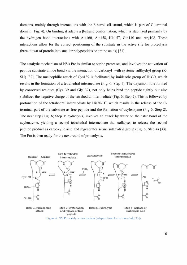
The catalytic mechanism of NVs Pro is similar to serine proteases, and involves the activation of
peptide substrate amide bond via the interaction of carbonyl with cysteine sulfhydryl group (R-
SH) [32]. The nucleophilic attack of Cys139 is facilitated by imidazole group of His30, which
results in the formation of a tetrahedral intermediate (Fig. 6: Step 1). The oxyanion hole formed
by conserved residues (Cys139 and Gly137), not only helps bind the peptide tightly but also
stabilizes the negative charge of the tetrahedral intermediate (Fig. 6; Step 2). This is followed by
protonation of the tetrahedral intermediate by His30-H+, which results in the release of the C-
terminal part of the substrate as free peptide and the formation of acylenzyme (Fig 6; Step 2).
The next step (Fig. 6; Step 3: hydrolysis) involves an attack by water on the ester bond of the
acylenzyme, yielding a second tetrahedral intermediate that collapses to release the second
peptide product as carboxylic acid and regenerates serine sulfhydryl group (Fig. 6; Step 4) [33].
The Pro is then ready for the next round of proteolysis.
Figure 6: NV Pro catalytic mechanism (adapted from Hedstrom et al. [33])

11
III. Peptide substrate recognition and proteolytic processing order The processing of ORF1 by NV Pro occurs at five sites, with either glumatime-glycine (Q-G),
glutamic acid-glycine (E-G) or glutamic acid-alanine (E-A) as the cleavage junction. These
cleavage sites, based on the processing order in vivo and in vitro studies, can be grouped into
“early and “late” cleavage sites- with Q-G sites cleaved first, followed by E-A and Q-G sites
(Fig. 7A) [34, 35, 36]. Mutational studies have shown that substitution of early cleavage site with
the late one (Q-G > E-G) is not enough to alter the proteolytic processing order, suggesting that
cleavage sites are processed independently of one another and are regulated by the residues
flanking the cleavage sites (Fig. 7B) [27, 37]. Recently, kinetic studies have confirmed that the
flanking residues, especially P4-P2’ (Appendix 2: Schechter and Borger nomenclature),
determines the processing order primarily via regulation of NV Pro rate of catalytic efficiency
(Kcat/Km) [34]. It is primarily the rate of reaction (Kcat) and not the binding affinity (Km) that
determines the preference of Q-G sites over E-G and E-A (Fig. 7C). Moreover, it has been
shown that by replacing the P4-P2’ residues of early cleavage site with late cleavage site, the
processing order can also be altered. All these results suggest that P4-P2’ forms a core
sequences that contain all the information necessary for the regulation of ORF1 polyprotein
processing [35].
The structures of NV Pro-product complexes have been solved by adventitious crystallization.
Crystal packing in these complexes resulted in the interaction of C-terminal tail of one molecule,
representing the P4-P1 residues, into the active site of another (Fig. 8) [38, 36]. Comparative
studies of these Pro-product complexes with unbound (native) NV Pro structures have provided
insight into how different amino acid residues are accommodated by the Pro and how substrate-
induced conformational changes might regulate its activity. The majority of the interactions with
the substrate involve hydrophobic S1 and S2 pockets (Appendix 2: Schechter and Berger
nomenclature) located in the N-terminal domain. S1 pocket is formed by His157, Ala160 and
Thr134 [32],whereas S2 pocket is defined by the hydrophobic residues Ile109, Glu110, Arg111,
and Val114, which are part of a loop that connect bII and cII β-strands (Fig. 9 and 10). S2 pocket
is not only larger than S1 pocket but also undergoes considerable conformation change upon

12
substrate binding (Fig. 9 A, B and C), allowing it to accommodate variation in the P2 position of
the substrate (Fig. 7) [36]. Depending on the absence or the presence of particular residues, S2
pocket depicts closed, semi-open or open conformations (Fig. 9). Moreover, the hydrophobic
nature of S2 also explains its selectivity for hydrophobic amino acids at P2 position. On the other
hand, the S1 pocket does not show significant difference upon substrate binding. This makes
sense as S1 pocket binds only to two similar residues i.e. either Gln (Q) or Glu (E) residue (Fig.
7A).
S3 pocket is not well defined and the lack of conserved interactions observed in crystal structures
with P3 residues explains the diversity of peptide residues at this position in NVs (Fig. 7 B) [36,
38]. In contrast, kinetic studies have implicated P3 residue to play a role in regulation of
polyprotein processing by modulating HuNV Pro efficiency [39]. This residue had been
overlooked because most of the earlier studies utilized a NV strain (Norwalk) as a standard
peptide, which contains His at P3 site. The peptide containing Glu at P3 instead of His was
observed to increase the NV Pro enzyme efficiency 4-fold [35]. Based on the modeling studies, it
has been proposed that P3-Glu carboxylate group (negatively charged residue) forms favorable
electrostatic and hydrogen-bonding interactions with residue-162 (Lys or Arg in GI/GII HuNV)
and P1-Glutamine (Gln), thereby increasing the HuNV Pro efficiency by promoting
“enzyme:transition-state” complex formation. In contrast, these interactions were not observed in
P3-His (positively charged residue) models, supporting a potential role of P3 residues in
polyprotein processing [35]. The preference for positively charged residues over negatively
charged ones at P3 position, with a 5-14 fold increase in activity, has been previously reported
for Coronavirus protease [40]. Therefore, it is possible that viral proteases have evolved to
utilize charged residues at P3 position as a common strategy to regulate polyprotein processing.
However, given its minimal affect in NVs i.e. only 4-fold increase in enzyme efficiency, it is
likely that P3 residue does not play a major role in HuNV Pro substrate specificity and
polyprotein processing.
S4 pocket (Fig. 9) is formed by hydrophobic residues (Met107, Arg108, Ile109, Thr166 and Val
168), which explain its preference for hydrophobic residues at P4 (Phe, Ile, Leu and Ala). S4 also
exhibits significant conformational change upon peptide binding (Fig. 10 C, D and E). The

13
substrate-induced conformational changes are coordinated between S2 and S4, such that the
constriction of S2 results in the widening of S4 pocket and vice versa (Fig. 10). The coordinated
changes observed are due to bII-cII loop that is shared by both S2 and S4 pocket (Fig. 9) [36].
Beyond S4 there appears to be little specific interactions between the peptide and NV Pro [36,
38]. This observation supports the results of kinetic studies, mentioned earlier, that residues
beyond P4 are not important for polyprotein processing.
However, as far as the P’ residues are concerned, there are no structural data available which
might help explain how they interact with NV Pro. Surprisingly, in kinetic studies the
substitution of P2’ glycine of the ProPol late cleavage site for early cleavage sites P2’ proline,
resulted in a 3-fold increase in HuNV efficiency [39]. Modeling studies indicate that the peptide
containing glycine at P2’ position can be stabilized by additional interactions between glycine
and other P’ residues [39]. In addition, modeling studies have also indicated that the NV Pro can
accommodate P’ residues up to P3’ [30] or P4’ [35]. However, based on the visual inspection of
the available NV Pro crystal structures in the presence and absence of peptide substrate or
peptide-based inhibitors, there appears to be no major binding groove beyond S1’ pocket [41],
and P’ residues beyond P2’ can take two different routes (Appendix 3: Peptide Route 1 and 2)
[31]. Based on these results it can be concluded that there is a potential for NV Pro-P’
interactions to occur, which may lack specificity beyond P1’. Therefore, further structural studies
are needed to help understand how P’ residues interact with the enzyme and bring about any
substrate-induced conformational changes that might explain their role in binding specificity.

14
Figure 7: (A) The processing of ORF1 polyprotein occurs at five sites, which can be grouped into “early” and “late” cleavage sites (1-5) [36]. (B) The most common HuNV amino acid sequence (P7-P7’) along each cleavage site (P1-
P1’) [34]. (C) Kinetics of HuNV Pro with ORF1 substrates [34].
Figure 8: Ribbon representation of HuNV Pro-product complex structure (PDB ID: 4IN1). The two monomers (A
and B) interact such that the C–terminal residues i.e. 178-181 of one monomer, represented by sticks in dashed boxes, corresponding to positions P4 to P1 of the native substrate are inserted into the active sites (blue sticks) of the
second monomer.

15
Figure 9: Surface representation of NV Pro (PDB ID: 4IN2) with color-coded active site (red) and substrate binding
pockets. The dashed line represents the peptide-binding groove where the peptide substrate binds.
Figure 10: Surface representation of peptide-induced conformational changes in S2 and S4 pockets of HuNV Pro.
Pockets exhibit three different conformational states: closed/narrower, semi-open/narrow and open/wider, depending on the absence or presence of particular residue. The peptide sequences (TALE and INFE) are shown along the
length of the substrate and represent P4-P1 positions of the natural substrate. The active site is depicted in red, the S1 pocket in green, the S2 pocket in teal, and the S4 pocket in magenta. (A/D) The S2 and S4 pockets in the absence of substrate. (B/E) The S2 and S4 showing semi-open and narrow conformations in the presence of TALE substrate.
(C/F) The conformational changes in S2 and S4 pockets to accommodate bulkier substrate residues (F) at P2 position in INFE substrate. S2 adopts an open conformation to accommodate bulkier F (Phe) residue, which is
facilitated by the movement of bII-cII loop. This loop is shared by both S2 and S4 such that widening of S2 results in constriction of S4 pocket and vice versa [36].

16
f. NV Pro-RNA interaction Previous studies have indicated that the HuNV ProPol precursor is an active part of the viral
replicase. However, no precise role for Pro in this complex was observed [29, 42]. Recently,
direct RNA binding by HuNV Pro was shown to inhibit Pro activity in a non-competitive manner
(Fig. 11) [28]. These results provide support for the participation of NV Pro in regulating of viral
replication. In related PV and HRV proteases, the RNA-binding motif has been mapped to the
highly conserved amino acid motif KFRDI, which is absent in NV Pro [43, 44, 45]. Therefore, it
is probable that the binding of RNA involves a novel, as-yet-unidentified mechanism and
RNA/protein interactions. Moreover, based on the initial enzyme inhibition studies there appears
to be little evidence for RNA sequence specificity for the NV Pro, although length was observed
to have an effect on inhibition, with longer RNA oligonucleotides being responsible for more
potent inhibition (Table 1) [28].
RNA binding by other related cysteine proteases, such as the PV and HRV proteases, is well
established [46]. Studies have indicated that the binding of PV and HRV Pro and/or ProPol
precursor to the RNA secondary structures, termed cis-acting replication elements (CREs)-
located in the untranslated regions (UTRs) and at an internal position within the viral RNA- is
important for genome replication [47]. It has been proposed that the binding of CREs to Pro
leads to unwinding of these secondary structures, which then serve as the template for
uridylylation of primer protein VPg by the viral polymerase to initiate viral replication [48].
Therefore, NV Pro might be involved in the regulation of viral replication, perhaps through a
similar mechanism observed in related PV and HRV proteases [48, 46, 28, 49]. However, further
studies are warranted to help understand the role of CREs, RNA binding mechanism, interplay
between NV Pro peptide and RNA binding sites, and different roles of Pro and ProPol in HuNV
life cycle.

17
Figure 11: RNA binding by HuNV Pro and its effect on enzyme kinetics. A) RNA binding by HuNV Pro was confirmed by mobility shift electrophoresis. B) Lineweaver-Burk analysis of the effect of RNA on HuNV Pro
activity [28].
Table 1: Effect of RNA on HuNV Pro activity. IC50 (2-fold reduction in activity) and IC90 (10-fold reduction in
activity) were calculated relative to an inhibited proteolysis reaction. pET32 represent a non-NV RNA [28].
Oligonucleotides (14-mer)
IC50 (μM) IC90 (μM)
5’-End, sense 5.5 ± 0.1 23.3 ± 1.1 5’-End antisense 4.8 ± 0.5 18.6 ± 1.2
3’-End, sense 5.4 ± 0.1 21.5 ± 0.2
3’-End antisense 3.5 ± 0.2 10.7 ± 0.9 RNAs
NV Pro (642nt) 0.013 ± 0.001 0.46 ± 0.003
pET32 (589nt) 0.017 ± 0.002 0.62 ± 0.002

18
Chapter 2: Objectives of the Study
The goals of this study were to determine the three-dimensional structure of HuNV Pro in
complex with peptides and RNA substrates by X-ray crystallography. Structural data of HuNV
Pro-substrate complexes will uncover details of the interactions made by the NV Pro with RNA
oligonucleotides and the peptide residues, prime side of the peptide cleavage junctions. This will
help identity the mechanism of RNA binding in viral proteases and explain how variation in
peptide cleavage sequences regulate HuNV Pro-Peptide binding and activity.
a. Interaction of HuNV Pro with RNA oligonucleotides Even though HuNV Pro has been observed to bind viral RNA, the amino acids involved in RNA
binding are yet to be determined, which based on the non-competitive nature of inhibition
supports the presence of a distinct RNA binding motif on the NV Pro. Since no structures are
available for any viral protease in complex with RNA, we have undertaken X-ray
crystallographic studies of HuNV Pro in complex with RNA oligonucleotides, which were
identified in already published NV Pro-RNA binding studies [28]. These Pro-RNA complexes
would allow us to identify the mechanism of RNA binding and to elucidate how RNA can inhibit
proteolytic activity in a non-competitive manner. Since the NV Pro residues involved in RNA
binding are unknown [28], we propose two putative RNA binding sites on NV Pro (Fig. 12),
which due to their distinct positions from peptide-binding site are consistent with the observed
non-competitive inhibition. RNA binding proteins are known to bind RNA mostly via positively
charged residues that compensates the negative charge of the RNA [50]. Keeping this in mind
the two proposed RNA-binding sites on NV Pro-based on the visual inspecting of the surface
potential-represent the two largest positive potential patches on the enzyme (Fig. 12). In a
distantly related viral cysteine protease, HAV 3C, it has been reported that dimerization results in
the formation of an extended RNA-binding site, which improves RNA binding affinity [43].
Dimerization of NV Pro has also been reported in crystallographic studies [51, 31, 52, 36], but
the dimer interfaces observed in NV Pro crystallographic studies are quite different from each
other [51]. Considering it is unlikely that biologically relevant dimer would have different
contacts as observed for NVs Pro, the role of NV Pro dimerization in RNA binding warrants

19
further studies. The structures of HuNV Pro in complex with different RNA oligonucleotides
would also shed light on the possible role of NV Pro dimerization in RNA binding.
To help solve the structure of HuNV Pro in complex with RNA oligonucleotides, the project was
divided into the following specific objectives:
I. To develop efficient expression and purification protocols for wild type HuNV Pro in
order to meet the high sample quantity and purity requirements of crystallization.
II. To identify crystallization conditions that promotes the formation of HuNV-RNA
complexes.
III. To collect and process X-ray data to understand the nature of HuNV Pro-RNA
interaction.
Figure 12: Putative RNA binding sites on NV Pro (PDB ID: 4ASH) bound to peptide substrate (yellow sticks). The
electrostatic potential distribution on the protein surface was calculated using PyMOL. The positive potential is colored blue and negative potential red.

20
b. Interaction of HuNV Pro with P’ residues As mentioned earlier, there is no structural information available on NV Pro-peptide complexes
which might shed light on the interaction made by the Pro with P’ residues. It is also not clear
how many P’ residues can be accommodated by the enzyme. Keeping this in mind, synthetic
peptides with a different number of P’ residues were used in this study to prevent overhanging
residues that might interfere with the crystallization of HuNV Pro-peptide complex. Moreover, to
promote substrate binding the sequence of these peptides were based on the early cleavage site
(N-term/NTP-ase), as it has the strongest binding affinity to HuNV Pro (Fig. 7 A and C). These
peptide sequences also contain a glutamic acid (E) at P3 position. Therefore, this project has the
potential to help understand how P’ and P3 residues interact with HuNV Pro, thereby providing
insight into polyprotein processing. Ultimately, this work would aid in the development of much
needed anti-viral treatments for HuNV infection.
To achieve this goal, several specific objectives were developed in an attempt to solve the
structure of HuNV Pro-peptide complexes.
I. To develop an efficient expression system for inactive mutant HuNV Pro (C139A). This
is needed to prevent autolysis of synthetic peptides used in co-crystallization studies.
II. To develop efficient purification protocol in order to meet the high sample quantity and
purity requirements required for crystallographic studies.
III. To identify crystallization conditions that promotes the formation of HuNV Pro-peptide
complexes.
IV. To collect and process X-ray data to determine the structures of mutant HuNV Pro in
complex with peptide substrates.

21
Chapter 3: Materials and Methods
a. Protein expression Our collaborators at the University of Georgetown, US, expressed both the wild type and mutant
(C139A) HuNV Pro (strain Hu/G1.1/8FlIa/1968/USA, accession number JX023285) with His6-
tag at N-terminal following previously described methods [28].
b. Ion exchange chromatography The HuNV Pro sample received from the collaborators was purified with ion exchange
chromatography. Protein was loaded onto a 5ml SP Sepharose High Performance column
(HiTrap SP Hp, GE Healthcare Life Sciences) already equilibrated with 5 column volumes of
binding buffer, Buffer A (20 mM MOPS, pH 7.0, 0.5 mM EDTA, pH 8.0, 2 mM DTT and 10%
glycerol). The bound protein was eluted in 1 ml fractions with a 30 mL gradient from 0 to 1 M
NaCl using elution buffer, Buffer B (20 mM Tris-Cl, pH 7.0, 1 M NaCl, 0.5 mM EDTA, pH 8.0,
2 mM DTT and 10% glycerol). The concentration and the purity of the protein in the fractions
were measured with NanoDrop (Thermo Scientific) and SDS-PAGE gel.
c. Gel-filtration chromatography Ion exchange fractions of highest purity and concentration were pooled together and
concentrated to 4.5 ml by centrifugation (6000-8000 RPM). The concentrated protease sample
was then applied to a gel filtration column (HiLoad 16/60 Superdex 200 pg, GE Healthcare),
which had been equilibrated with buffer, Buffer C (20 mM MOPS, pH 7.0, 100 mM NaCl, 0.5
mM EDTA, pH 8.0, 5% glycerol, 2 mM DTT). The 1 ml fractions were eluted at a flow rate of
0.2 ml/min and collected in a 48 spot collector tray by auto-fraction collector. The presence of
HuNV Pro oligomers were confirmed by running the sample against Gel filtration Markers kit
for Protein Molecular Weight 12-200 kDa (Sigma Aldrich).

22
d. Dialysis and concentration Fractions of highest concentration and purity were pooled together and dialyzed against low salt
storage buffer (20 mM Tris-Cl, pH 7.0, 20 mM NaCl, 0.5 mM EDTA, pH 8.0, 2 mM DTT and
10% glycerol) overnight. The dialyzed protein was then concentrated by ultrafiltration until a
concentration of approximately 0.12-0.20 mM (A280 = 3.0-5.0 mg/ml) was achieved. The purified
wild type and mutant HuNV Pro samples were flash frozen as 50 μL aliquots in thin-walled PCR
tubes and stored at -80°C.
e. RNA and peptide preparation RNA oligonucleotides of different sequences (Table 2) were ordered from the University of
Calgary CORE DNA services, whereas, the synthetic peptides (Table 3) were purchased from
CanPeptide Inc. RNA and peptides were neutralized to pH 7.0-7.5, and dissolved in UltraPureTM
DNase/RNAse-Free Distilled Water (Life Technologies) at different concentrations ranging from
10-30 mM.
Table 2: RNA oligonucleotide information and sequence. These sequences are based on the published HuNV Pro-
RNA binding studies [28].
RNA Oligonucleotide Sequence 1. NVpla (10-mer; 5’-end of NV viral genome) 5’-GUGAAUGAUG-3’ 2. NVp2 (14-mer; 3’-end of NV viral genome) 5’-UUUAAUUUGAUGUU-3’
Table 3: Synthetic peptide information and sequence. These sequences represent the residues surrounding the early NS2-3 cleavage site [34, 39].
Synthetic Peptide Sequence 1. Long Peptide (9 amino acids; P5-P4’) Ac-DYELQGPED-NH2 2. Short Peptide (5 amino acids; P4-P1’) Ac-YELQG-NH2
f. Crystallization methodology The vapor diffusion method was used for the crystallization studies. This is the most widely used
method due to the relative ease with which it can be set up compared to other methods. In this
method, the protein is mixed with the reservoir solution is equilibrated in a closed system against

23
the reservoir solution containing different crystallization precipitants e.g. salts, organic solvents
and polymers etc., at a much higher concentration. Initially, the protein solution contains
precipitants at lower concentration than required for crystallization but, due to the net movement
of water vapors from the protein to the reservoir solution, the concentration of precipitants in
protein solution increases gradually to a level optimal for crystallization. Whether the protein
forms crystals or amorphous solid, depends on many properties of the protein and reservoir
solution, including protein purity, protein concentration, temperature, pH, precipitant and its
concentration, and ionic strength [53]. Due to its multi-parametric nature, it is difficult to predict
crystallization conditions a prior. As a result, several thousand crystallization conditions have to
be tested, which, with the advent of crystallization robots and commercially available
crystallization screens, has become easier. First step towards growing protein crystals is
automated screening, using commercially available crystallization screens, to cover a large part
of chemical space to determine initial crystallization conditions (called “hits”) under which
protein has the propensity to crystallize. These hits are then fine-tuned manually during crystal
optimization step to grow diffraction quality crystals for X-ray data collection.
I. Sparse matrix screening In an attempt to co-crystallize the wild type HuNV Pro-RNA and mutant HuNV Pro-peptide
complexes, the protein (0.12-0.20 mM) was mixed with varying concentration of synthetic
peptides (1.2-5.4 mM) and RNA oligonucleotide (0.60 mM). These protein-substrate mixes were
equilibrated for 2 hours to ensure binding of substrates to HuNV Pro, before setting up the
crystallization screens. Since there is no way to predict the set of conditions that will give rise to
a single, well diffracting crystal, automated setup was used to screen hundreds of crystallization
conditions using commercially available sparse matrix crystallization screens (Appendix 4:
Sparse Matrix Screening). These sparse matrix screens involve an intentional bias towards
combination of crystallization conditions that have worked previously and cover a large chemical
space by using many different crystallization reagents [54]. Each screen used contained 96
different conditions, varying in pH, buffer, precipitant and salt concentrations. Using the Matrix
Hydra II eDrop pipetting robot (Thermo Scientific) crystallization trials were set up at room

24
temperature (25 °C) and monitored periodically under a microscope for over a course of ~12
weeks to identify crystallization hits.
The robotic setup involved placing 40 µl of crystallization solution into the base (reservoir) of a
96 well sitting drop vapor diffusion plate (Hampton Research). This was followed by mixing 0.4
µl of crystallization solution with 0.4 µl of HuNV Pro-substrate (RNA or peptide) solution into
the top sample container. These plates were then sealed by hand to prevent drying out of the
reservoir and sample container using Crystal Clear Sealing Tape (Hampton Research).
II. Crystal optimization The hits identified from sparse matrix screening were repeated manually and optimized by
systematically varying each component of the crystallization conditions e.g. protein
concentration, substrate concentration, pH, salt and its concentration, and precipitant
concentration etc. Crystals were grown using hanging drop vapor diffusion method in 24 well
VDXm plates (Hampton Research). HuNV Pro (0.12-0.20 mM) was mixed with either RNA
(0.60-2.0 mM) or peptide (1.2-6.0 mM) and equilibrated on ice for 2 hours. The protein-substrate
solution (1-3 µl) was then mixed with crystallization solution (1-3 µl) in different ratios e.g. 1:1,
1:2, 2:1 and 3:1, and equilibrated against 0.5 mL crystallization solution (i.e. crystallization
condition identified from the screens). These plates were then either placed at room temperature
or 4° C. Optimization was also carried out for crystals grown in the absence of substrates. These
native crystals were used for soaking experiments (explained later; page 25).
As an additional optimization strategy, seeding technique [55] was also used to improve the
quality of co- and native crystals. Small seed crystals were transferred by pipette tip or 18 mm
mounted CryoloopTM (Hampton Research) of different diameters (25-150 µm) to a pre-
equilibrated drop containing modified crystallization condition.

25
III. Soaking In addition to co-crystallization of HuNV Pro with either RNA or peptide, the soaking technique
was also used [56]. Since this technique relies on pre-formed crystal, it allows one to easily
change the crystal conditions, e.g. pH, salt, ligand concentration etc., in order to promote ligand
binding. After native and/or co-crystals had grown to the maximum size, they were transferred
using a mounted cryloop (Hampton Research) to a drop containing the soaking solution (1-2 µl)
containing varying concentrations of RNA (0.6–7.5 mM) and/or peptide (2 mM-20 mM) for 30
min to couple of days. The drop was setup using the 24 well VDXm plates (Hampton Research).
Moreover, in order to avoid damage to the crystal either due to the sudden change in crystal
environment or physical manipulation of crystal, a modified soaking methodology was also tried
to gently alter the condition. The drop (1-2 µl) containing the crystal was diluted with soaking
solution (1-2 µl) and left to equilibrate for 30 min. The next step involved the removal of 1-2 µl
of solution, followed by addition of soaking solution (1-2 µl) and equilibration. This protocol
was followed until the drop solution was completely replaced with the soaking solution.
Depending on the stability of the crystal in the soaking solution, varying soaking times were tried
(30 min – 2 days).
IV. Crystal harvesting Due to limited time and resources, only a few crystals without any visible defects were selected
for each synchrotron trip. Crystal harvesting involved the transfer of a protein crystal from its
growth solution into a suitable 18 mm mounting CryoloopTM (Hampton Research). Depending on
the size of the crystal, cryoloop with a suitable diameter (50-200 µm) was selected beforehand.
Then the coverslip (18 x 18 mm) containing the crystal was gently flipped and placed under the
microscope. While looking under the microscope the selected crystal was quickly picked and
flash-frozen in liquid nitrogen and transferred into a CrystalCapTM vial (Hampton Research),
submerged in liquid nitrogen. Finally, the crystals are transferred into a 96-sample ports storage
cassette (MiTeGen) and shipped in storage dewar to synchrotron for X-ray data collection.

26
During harvesting crystal exposure to air is minimized. This prevents and/or reduces damage to
the crystal due to dehydration. The average solvent content in protein crystal is around 43% [57]
and plays an important role in its structural stability [58]. Therefore, excess dehydration can lead
to crystal damage by structural transformation. Moreover, all the crystals were grown in the
presence of cryoprotectant e.g. glycerol and polyethylene glycol, and flash frozen quickly to
prevent the formation of ice crystals. These ice crystals not only damage the crystal due to
sudden expansion of water upon freezing but, as ice also diffracts X-ray very strongly, it can also
interfere with the accuracy of measuring protein diffraction [59].
g. X-ray data collection and structure determination X-ray diffraction data was collected either at the Stanford Synchrotron Radiation Lightsource
(SSRL) or at the Canadian Light Source (CLS). The cryoloop containing the crystal was
mounted onto the goniometer, where it is held in a stream of nitrogen gas (100 K). Each crystal
was first screened by measuring two X-ray diffraction patterns at two orientations separated by
90° with X-ray beam normal to the plane of the crystal. If the crystal was found to be relatively
defect-free then complete X-ray data set was collected, which involved collecting a total of 400-
500 frames with 0.5° oscillations and 2s of exposure per frame. Data were processed and scaled
using XDS [60]. Molecular replacement (MR) analysis was performed using Phaser [90] with the
structure of HuNV Pro (PDB ID: 3UR6; 99% sequence similarity) as the search model. The
model building and refinement was carried out using COOT [61] and Refmac [62]. Residues
were added and adjusted to fit the electron density map (called model building), followed by
refinement to obtain a model which best explains the experimental X-ray data. Progress was
monitored by R-factor and R-free values, both of which improve progressively with the
improvement in model quality [63]. All the representations of HuNV Pro were generated with
PyMol (The PyMol Graphic System, Version 1.7.4 Schrödinger, LLC).

27
Chapter 4: Results and Discussion
a. HuNV Pro-RNA interaction
I. Expression and purification Protein expression and purification are often rate limiting steps in crystallography, as it requires
milligram amounts of highly purified protein. Our collaborators successfully expressed wild type
HuNV Pro as His6-Pro fusion protein (approx. 20 kDa), using previously established protocols
[35, 28]. The protein was purified using ion-exchange chromatography. The pattern of elution is
presented below (Fig. 12), where the peak 1 and peak 2 correspond to elution of the protein at
around 20-30% buffer B (200-300 mM NaCl). Since the SDS-PAGE profile for both peak 1 and
2 is identical, it is possible that peak 1 represents the HuNV Pro dimeric form. The charged
residues probably buried in the dimer interface, weakens dimer interactions, compared to the
monomer, with the ion-exchange column, thereby resulting in the elution of protein dimer
followed by the monomer. Peak fractions were analyzed for purity by SDS-PAGE, and the
fractions of the highest purity (Fig. 13) were pooled together and dialyzed against low salt
storage buffer. The protein solution was then concentrated by ultrafiltration to 0.12-0.20 mM and
SDS-PAGE analysis was performed again to determine sample purity (Fig. 13b). Impurities were
still present in the sample after ion exchange, ultrafiltration and dialysis; however, based on the
limited amount of protein available and the work of previous project students the quality was
deemed to be good enough for crystallization.

28
Figure 13: A) Ion exchange chromatogram of HuNV Pro (approx. 20 kDa). The elution profile of protein is shown
in green and the arrow indicates fractions that were analyzed by SDS-PAGE. B) Fractions tested for purity by SDS-PAGE. The fractions (9-14) were pooled together, dialyzed and concentrated for crystallization experiments. HuNV
Pro received from the collaborators was also loaded (sample next to MW ladder) to observe the improvement in sample purity. C) SDS-PAGE analysis of concentrated (3.0 mg/ml) HuNV Pro sample after purification.

29
II. Screening and hit optimization Since the conditions for crystallization of HuNV Pro in complex with RNA were unknown, a
number of commercially available crystallization screens were tested. These screens were carried
out in the presence of NVP1a or NVP2 RNA oligonucleotide. NVP1a screens were prepared and
analyzed by the project students before I joined the lab, whereas I was responsible for NVP2
screening. Initial screening carried out using Index screen (Hampton Research) was unsuccessful
as most drops were clear even after several weeks. The lack of crystal growth or precipitation
suggested that either this particular sequence and length of RNA was not suitable for
crystallization or the protein sample was under-saturated [64]. Therefore, for the next two
screens, the concentration of protein was increased to avoid under-saturation. This increase in
protein concentration resulted in the identification of a hit, which is mentioned in Table 4. The
thiocyanate condition represented a weak hit characterized by very small, clumpy and extremely
layered crystals, which are unsuitable for X-ray data measurement. As a result, the components
of the thiocyanate condition were systematically varied and optimized to grow larger, unlayered
individual crystals for high-resolution analysis by X-ray diffraction. Ideally further screening
should have been conducted to identify additional hits. However, due to the limited amount of
protein and RNA oligonucleotides available, the strategy was to focus on the optimization of the
single hit identified.
Table 4: Crystallization hits identified from screens performed in the presence of HuNV Pro and RNA
oligonucleotides. Previous project students were responsible for the identified of crystallization hit for NVP1a.
Screen (Well No.)
[Protein] mM RNA Hit condition
1. Index (H11)
0.12 NVP1a 0.1 M KSCN, 30% PEG MME 2000
2. Index
0.12 NVP2 No hit
3. Index (H11)
0.20 NVP2 0.1 M KSCN, 30% PEG MME 2000
4. Crystal
0.20 NVP2 No hit
Thiocyanate condition was successfully optimized and the crystals of HuNV Pro in the presence
of either NVP1a or NVP2, were obtained overnight in 0.10-0.30 M Potassium thiocyanate, 0.10
M Tris-Cl, pH 7.5, and 32-36% PEG MME 2000 (Fig. 12). However, the X-ray diffraction data

30
collected for these crystals had irregular (smeary) reflections, which is an indication of crystal
defects (Fig. 14). A high level of smearing makes it impossible to distinguish between individual
diffraction reflections and since each reflection recorded contains information about the position
of the atoms in the structure, it was impossible to generate electron-density maps from
diffraction patterns showing high degree of smearing.
Defects in crystals are mainly due to the presence of impurities in protein sample and/or fast rate
of crystallization [65, 66]. Rapid growth of crystal is frequently associated with the occurrence of
packing disorder, lattice distortion, and the incorporation of impurities in the crystal [67].
Impurities can also create and/or enhance crystal disorder in several ways [66, 68, 65]. Since the
amount of disorder and defects incorporated into a crystal is at least partially dependent on the
rate of crystallization [65], one can typically reduce the amount of defects in a crystal by slowing
down the rate of crystallization. The crystals grown in the presence of the original thiocyanate
conditions had a very fast rate of crystallization (crystals appeared overnight), therefore, efforts
were made to adjust the crystallization conditions in a manner that would slow down the rate of
crystallization and reduce the amount of defects incorporated into growing crystals.
Several different approaches were tried to slow down the rate of crystallization including
lowering of precipitant concentration, use of paraffin and silicon oil [69], seeding [70], lowering
of precipitant concentration after nucleation has occurred [55], temperature variation [71],
increase in drop size [72], variation in pH, and the use of additives to increase the solubility of
protein [73]. Out of all the methods tried addition of additive (glycerol: 5-10%), use of oils (70
μl silicon and 30 μl paraffin oil placed on top of the reservoir solution) and lowering of
precipitant concentration (28-32% PEG MME 2000) had the most profound affect (Table 5). The
slowest rate of crystallization was observed for HuNV Pro-RNA crystals grown in the presence
of 0.10 KSCN, 28% PEG MME 2000, 8% (w/v) glycerol, 0.10 M Tris-Cl, pH 7.5-8.5. These
crystals appeared in the protein drop after 3-4 days, rather than overnight, and then continued to
grow slowly for another 4 days. However, this decrease in the rate of crystallization did not
improve crystal quality as X-ray diffraction data collected for all these crystals (Table 5) still had
irregular (smeary) reflections (Fig. 15). Consequently, focus was shifted towards the other
possible source of crystal disorder i.e. HuNV Pro sample purity.

31
Figure 14: Optimization of thiocyanate condition. A) Small, clumpy and layered crystals obtained in Index screen. B) Several rounds of optimization by systematically varying crystallization parameters led to the growth of larger
and individual crystals.
Figure 15: X-ray diffraction pattern of crystal with (left) and without defects (right). A small section of X-ray image
is magnified by x4 to clearly represent the irregular (smeared) X-ray reflections associated with crystal defects.
32
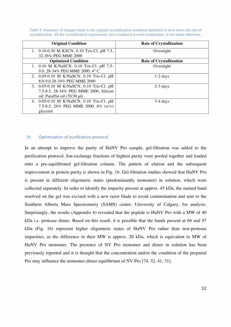
Table 5: Summary of changes made to the original crystallization condition identified to slow down the rate of crystallization. All the crystallization experiments were conduced at room temperature, if not stated otherwise.
Original Condition Rate of Crystallization
1. 0.10-0.30 M KSCN, 0.10 Tris-Cl, pH 7.5, 32-36% PEG MME 2000
Overnight
Optimized Condition Rate of Crystallization 1. 0.10 M K/NaSCN, 0.10 Tris-Cl, pH 7.5-
9.0, 28-34% PEG MME 2000, 4° C Overnight
2. 0.05-0.10 M K/NaSCN, 0.10 Tris-Cl, pH 8.0-9.0 28-34% PEG MME 2000
1-2 days
3. 0.05-0.10 M K/NaSCN, 0.10 Tris-Cl, pH 7.5-8.5, 28-34% PEG MME 2000, Silicon oil: Paraffin oil (70/30 μl)
2-3 days
4. 0.05-0.10 M K/NaSCN, 0.10 Tris-Cl, pH 7.5-8.5, 26% PEG MME 2000, 8% (w/v) glycerol
3-4 days
III. Optimization of purification protocol In an attempt to improve the purity of HuNV Pro sample, gel-filtration was added to the
purification protocol. Ion-exchange fractions of highest purity were pooled together and loaded
onto a pre-equilibrated gel-filtration column. The pattern of elution and the subsequent
improvement in protein purity is shown in Fig. 16. Gel-filtration studies showed that HuNV Pro
is present in different oligomeric states (predominantly monomer) in solution, which were
collected separately. In order to identify the impurity present at approx. 45 kDa, the stained band
resolved on the gel was excised with a new razor blade to avoid contamination and sent to the
Southern Alberta Mass Spectrometry (SAMS) center, University of Calgary, for analysis.
Surprisingly, the results (Appendix 6) revealed that the peptide is HuNV Pro with a MW of 40
kDa i.e. protease dimer. Based on this result, it is possible that the bands present at 66 and 97
kDa (Fig. 16) represent higher oligomeric states of HuNV Pro rather than non-protease
impurities, as the difference in their MW is approx. 20 kDa, which is equivalent to MW of
HuNV Pro monomer. The presence of NV Pro monomer and dimer in solution has been
previously reported and it is thought that the concentration and/or the condition of the prepared
Pro may influence the monomer-dimer equilibrium of NV Pro [74, 32, 41, 31].

33
Figure 16: A) HuNV Pro gel-filtration elution profile. Peak 1 and 2 corresponds to HuNV Pro dimeric and
monomeric forms. B and C) Fractions were tested for purity by SDS-PAGE and the fractions of highest purity i.e. fractions 13-17 (peak 1; B) and 28-33 (peak 2; C) were pooled separately and concentrated for crystallization
experiments. D) SDS-PAGE analysis of concentrated protein samples after each purification step. (Sample: protein sample from our collaborators; IEX: sample after ion-exchange chromatography, and GF P1 and GF P2:
concentrated peak 1 and peak 2 samples after gel- filtration.

34
IV. Optimization of Thiocyanate condition HuNV Pro-RNA crystals were grown in the original thiocyanate condition (Table 5) to test if the
improvement in purity had an effect on crystal quality. X-ray diffraction data collected for most
of these crystals did not suffer from irregular reflections, confirming that the improvement in
purity and homogeneity of the protein sample was responsible for the reduction in crystal
defects. Protein-ligand complexes are usually sensitive to high salt concentration, since
increasing ionic strength can interfere with the interactions needed to stabilize the complex [75].
Keeping this in mind, crystallization experiments were also conducted in the presence of low
thiocyanate concentration. In addition, crystallization experiments were also carried out in the
presence of non-thiocyanate salts (Table 6). Efforts to completely replace the potassium
thiocyanate with other salts failed as the replacement of thiocyanate inhibited spontaneous
nucleation and crystallization. Based on these results it was hypothesized that thiocyanate in
crystallization conditions is necessary for spontaneous nucleation, but not for crystal growth.
Spontaneous nucleation is kinetically demanding and conditions optimal for nucleation are not
ideal to support crystal growth. However, nucleation and growth can be uncoupled by using a
technique called seeding [55], which was used as an additional optimization strategy. Seeding
involves transfer of crystals or seeds that act as sites for nucleation, to the drops that cannot
support spontaneous nucleation. Seed crystals (20-50 μm) grown in conditions containing
thiocyanate and/or thiocyanate in combination with other salts, were transferred to a pre-
equilibrated drop containing different precipitant (PEGs), additives, salt and pH etc.
Unfortunately, seeding experiments did not yield any new crystals, but seeds in a few conditions
did continue to grow. This observation supported our hypothesis that thiocyanate is necessary for
spontaneous nucleation, but not for crystal growth. The best quality crystals were obtained when
seeds were transferred to a drop containing 0.050-0.10 KCl, 22-26% PEG MME 2000, 0.10 M
Tris-Cl, pH 7.5. These crystals grew to full size within 10 days after seeding (Fig. 17).

35
Table 6: Summary of salt combinations that yielded crystals.
Initial crystallization Condition
Precipitant Salt Buffer pH PEG MME 2000
(22-28%) Potassium or Sodium thiocyanate
(0.050-0.10 M) Tris-Cl (pH 7.5-9.0)
Salt Optimization
Precipitant Salt Combination Buffer pH PEG MME 2000
(22-28%) PEG 3000 (22-28%)
Crystals were grown in the presence of Sodium/Potassium thiocyanate (0.010-0.10 M) and:
1. Sodium Chloride/Bromide/Flouride/Iodide
(0.020-0.10 M) 2. Potassium Chloride/Bromide/Iodide
(0.050-0.10 M) 3. Cesium Chloride
(0.050 M) 4. Calcium Chloride
(0.025-0.050 M) 5. Ammonium Sulfate/ Acetate
(0.020-0.050 M) 6. Potassium Acetate
(0.020-0.050 M)
Tris-Cl (pH 7.0-8.0)
Figure 17:The change in size of seed crystal transferred to potassium chloride (KCl) condition. The crystal on the left shows the crystal seed after 2 days, whereas, the one on the right represents the full size achieved after a few
more days.

36
V. Soaking HuNV Pro-RNA binding is weak (ki: μM range) [28], therefore co-crystallization experiments
were also performed in the presence of very high RNA concentration in order to saturate the
RNA binding site on the protein. However, these experiments were not successful and RNA
concentration above 2.5 mM was found to inhibit nucleation and growth. As a result crystals
grown in the presence of 0.60-2.5 mM RNA were soaked to further increase RNA concentration
to as high as 7.5 mM (40-fold molar excess over HuNV Pro). The absence of RNA in crystals
grown and/or soaked in the presence of thiocyanate led to the hypothesis that thiocyanate salt is
interfering with RNA binding. To test this hypothesis soaking technique was also used to gently
exchange the thiocyanate with other salts and even the condition used in HuNV Pro-RNA
binding studies was also tested [28] (Table 7). HuNV Pro proteolytic activity in HEPES has been
reported to be at least 2-fold higher than in Tris-Cl [28]. Therefore, HEPES buffer was also used
in soaking experiments, instead of Tris-Cl, to check if it promotes RNA binding to HuNV Pro.
Table 7: Summary of conditions used for soaking experiments.
Initial Crystallization Condition Precipitant Salt Buffer [RNA] mM
PEG MME 2000 (22-28%) PEG 3000 (22-28%)
K/NaSCN (0.020-0.20 M) Tris-Cl (pH 7.5-8.9)
0.60-2.0
Soaking Condition Precipitant Salt Buffer [RNA] mM Time
1. PEG MME 2000 (22-30%) PEG 3000 (22-30%)
KCl (0.020-0.10
M)
Tris-Cl (pH 7.0-8.0)
2.5-7.0 15 min to 48 hr.
2. PEG MME 2000 (22-30%) PEG 3000 (22-30%)
NaCl (0.020-0.10
M)
Tris-Cl (pH 7.0-8.0)
2.5-7.0 15 min to 48 hr.
3. PEG MME 2000 (22-30%)
NaCl/KCl (0.020- 0.10
M)
HEPES (pH 7.5-8.9)
2.5-7.0 15 min to 24 hr.
4. Condition used in HuNV Pro-RNA binding studies (10 mM HEPES pH 7.6, 0.10% CHAPS, 30% glycerol). The concentration of RNA used was 0.20-7.0 mM with soaking time of 15 min to 4 hr.

37
VI. X-‐ray data collection and structure determination Due to limited time given for X-ray data collection at synchrotrons, only a fraction of crystals
grown were sent for data collection. Out of 70 crystals sent, only 60 were screened and complete
X-ray diffraction data set could only be collected for 25 crystals, as most of these crystals were
grown before the purification was optimized and therefore suffered from severe crystal defects.
The co-crystallization and soaking conditions along with the maximum resolution for few of the
crystals for which complete X-ray data set was collected are mentioned in Table 8. The data was
processed as explained earlier (page 28). All these crystals belonged to the space group P21 with
unit cell dimensions of a = 36.71Å, b = 34.70Å, and c =110.71Å. Calculations revealed that the
highest probability is for two molecules of HuNV Pro in the asymmetric unit with Matthews
coefficient of 2.21 Å3/Dalton and a solvent content of 22.6% [76]. Generally, crystal with low
solvent content diffract better, as the crystal packing tends to be tighter [77]. This explains why
the maximum resolution limit for most of the crystal is less than 2.0 Å (Table 8). Due to high
quality of X-ray data collected for most of the crystals, electron-density maps showed almost the
entire polypeptide chain for each of the two molecules in the asymmetric unit including the
highly flexible solvent exposed loop (123-134 aa), which is missing in most of the NV Pro
structures deposited in protein data bank (PDB). However, electron density for His-tag and C-
terminal tail (residues 175-181) was very weak, presumably due to disorder, and so was not
included in the HuNV Pro models built. All the 3D models were refined to an R-factor and R-
free values of ≤ 24% and ≤ 28%. After refinement, visual inspection of all of the models was
carried to find electron-density not accounted for by the protein into which RNA oligonucleotide
could be modeled. Disappointingly, however, no electron-density was observed for either NVP1a
or NVP2 RNA oligonucleotides.
The most important details of data collection parameters and refinement statistics for one of the
models (AS59; condition no. 7 in Table 8) are summarized in Table 9. The structural analysis
was carried out for all the models built. However, all the models built were found to be
structurally identical as indicated by the small root mean square deviation (RMSD) in Cα atom
positions of only 0.144 Å. Therefore, only one of the structures, AS59, is discussed in detail
below.

38
Table 8: X-ray diffraction data sets collected for co-crystals grown and/or soaked in different conditions. All the
crystals were co-crystallized in the presence of RNA, if not stated otherwise.
Crystallization Condition Soaking Condition and Time RNA Resolution Å
1. 0.15 M NaSCN, 36% PEG MME 2000, 0.10 M Tris-Cl, pH 8.5
- NVP1a (0.6 mM)
2.1
2. 0.10 M NaSCN, 26% PEG MME 2000, 0.10 M Tris-Cl, pH 7.5
- NVP1a (1.5 mM)
2.3
3. 0.10 M NaSCN, 26% PEG MME 2000, 0.10 M Tris-Cl, pH 8.0
0.10 M KCl, 27% PEG MME 2000, 0.10 M Tris-Cl, pH 7.5; 30 min
NVP1a (2.5 mM)
2.5
4. 0.10 M NaSCN, 24% PEG MME 2000, 0.10 M Tris-Cl, pH 8.0 (Native)
0.025 M NaCl, 0.025 M NaSCN, 0.003 M MgCl2 0.01M Tris-Cl, pH 7.5; 24 hr
NVP1a (7.5 mM)
2.0
5. 0.070 M NaSCN, 26% PEG MME 2000, 0.10 M Tris-Cl, pH 8.0
- NVP2 (2.5 mM)
2.3
6. 0.040 M NaSCN, 0.050 M KCl, 26% PEG MME 2000, 0.10 M Tris-Cl, pH 7.5
- NVP2 (2.5 mM)
1.9
7. 0.050 M NaSCN, 0.050 KCl 26% PEG MME 2000, 0.10 M Tris-Cl, pH 7.5 (AS59)
- NVP2 (2.5 mM)
1.6
8. 0.10 M NaSCN, 26% PEG MME 2000, 0.10 M Tris-Cl, pH 7.5
0.05 M KCl, 27% PEG MME 2000, 0.01M Tris-Cl, pH 7.5; 30 min
NVP2 (7.5 mM)
1.9
9. 0.15 M NaSCN, 36% PEG MME 2000, 0.10 M Tris-Cl, pH 8.0
0.025 M KCl, 37% PEG MME 2000, 0.10 M Tris-Cl, pH 7.5; 3 hr
NVP2 (7.5 mM)
1.7
10. 0.20 M NaSCN, 26% PEG MME 2000, 0.10 M Tris-Cl, pH 7.5 (Native)
0.05 M KCl, 0.10 M, 27% PEG MME 2000, 0.10 M Tris-Cl, pH 7.5; 24 hr
NVP2 (7.5 mM)
1.8

39
Table 9: Crystallographic data collection and model refinement statists for AS59. This particular data set was collected at Stanford Synchrotron Radiation Lightsource (SSRL). Values in the brackets represent the R-values for
unrefined structure.
Data-integration Statistics Space-group P21
a, b, c (Å) 37.45 36.18 113.79 α, β, γ (°) 90.00 98.07 90.00
Unit cell volume (Å3) 152511382.9 Solvent content (%) 22.63
Data Collection Temperature (K) 100
Detector PILATUS 6M No. Of Images 500
Oscillation range (°) 0.50 Wavelength (Å) 0.9795 Resolution (Å) 34.45-1.60
Total observation 206529 Unique reflections 46209
Multiplicity 4.47 I/σI 10.27
Completeness 95.8 % Refinement
Resolution (Å) 1.60 RWork 0.2036 (0.3102) Rfree 0.2339 (0.3262)
RMS deviation – bonds (Å) 0.0069 RMS deviation- Angles (°) 1.098
Ramachandran plot Favored Allowed
97.63% 2.37%

40
VII. Structural analysis and discussion of AS59 As mentioned earlier, there are two molecules in the asymmetric unit (Dimer; Fig. 18). AS59
dimer interface is maintained primarily by hydrophobic interactions, resulting in a total buried
surface area of 909 Å2, which is suggestive of a very weak non-physiological dimer (i.e. result of
crystal packing) [78, 79]. The dimerization of NV Pro has been previously reported in crystal
structures [31, 51, 32]. AS59 dimer interface contacts were compared with other NV Pro dimers
using POLYVIEW-3D [80]. Despite the fact that the amino acid sequence is highly conserved in
norovirus proteases, the dimer interface contacts were found to be significantly different (Fig.
19). It is unlikely that the biologically relevant dimer would have different contacts as observed
in NVs Pro dimers. Therefore, further mutational and functional studies are needed to understand
NV Pro dimerization and its biological significance in NV life cycle, if any.
The molecules present in the dimer are very closely related structurally as indicated by the small
root mean square deviation (RMSD) in Cα atom positions of only 0.4 Å (Fig. 20; monomer A
and B). Therefore, only one of the two molecules was used for structural comparison of AS59
with other NV proteases. The overall structure of AS59 monomer is nearly identical to
previously solved NV Pro structures, which comes as no surprise given the high degree of
protease sequence similarity in NVs (≥ 60%; Fig 21) [38]. As expected, it consists of an
incomplete β-barrel N-domain and an anti-parallel β-sheet C-domain. The active site residues are
located in the cleft formed at the interface of these two domains (Fig. 20). One noticeable
difference observed in AS59 structure was the conformation of solvent exposed loop (121-134
aa; Fig. 21). However, the conformational flexibly of this loop is well documented and is
possibly an artifact of crystal packing [38, 31, 51, 32].
Low occupancy of ligands result in less well-defined electron density map for ligands [81].
Therefore, it could be argued that RNA is not visible in our structures due to its low occupancy.
However, very high structural similarity of high-resolution structures of HuNV Pro-RNA co-
crystals (grown in the presence of as high as 60-fold molar excess of RNA to protein) to HuNV
Pro native structures support the lack of RNA binding in our crystallization studies. The only
variable regions in these structures are solvent exposed loop (121-134 aa) and His-tag (Fig. 22).
The conformational variation of these residues is well documented and cannot be attributed to

41
RNA binding [31, 38, 32]. Moreover, the loop also lacks positively charged residues required to
bind RNA. Therefore, based on the absence of RNA electron density in our models and
structural similarity between native and co-crystal structures, it can be concluded that RNA
oligonucleotides did not bind to HuNV Pro in our crystallization studies.
Figure 18: The cartoon representation of dimeric arrangement observed in AS59. The active site (H30, C139 and
E54) is shown as magenta sticks, whereas the C and N-terminal residues are colored cyan and blue.

42
Figure 19: Comparison of dimer structures and interface of NV Pro dimers (PDB ID: 2FYR and 2IPH) with AS59.
Plotted below each dimer are residues forming dimer interfaces identified using POLYVIEW-3D software [80].
Figure 20: Overlay of AS59 monomer A (Red) and B (green). The side-chains of the active site residues (His 30,
Gly54 and Cys139) are shown as sticks (blue).

43
Figure 21: Structural comparison of the backbone of AS59 with other HuNV proteases (PDB ID: 1WQS (Chiba)
and 2IPH (Southampton)). The overall structure is very similar, apart from the flexible loop composed of residues 121 to 134. The average root mean square deviations (RMSD) of the backbone atoms of Chiba and Southampton
from AS59 are 0.7 and 0.4.
Figure 22:Structural comparison of the backbone of HuNV Pro crystal grown in the presence (Green) and absence of RNA oligonucleotides (Red). The calculated average root mean square deviation of the backbone atoms of these two
structures is 0.14 Å. The most variable regions are labeled (Loop and His-tag).

44
b. HuNV Pro-peptide interaction
I. Expression and purification Our collaborators successfully expressed mutant HuNV Pro (C139A) as His6-Pro fusion protein
(approx. 20 kDa). Mutant HuNV Pro was purified using a purification protocol established for
the wild type protease, which involved ion-exchange chromatography followed by gel-filtration.
The gel-filtration elution profile is presented below (Fig. 23). The elution profiles for wild type
and mutant HuNV Pro are identical and therefore it was assumed that the peak 1 and peak 2
represent monomeric and dimeric forms of protein. Peak fractions were analyzed for purity by
SDS-PAGE (Fig. 23), and the fractions of the highest purity were pooled together, dialyzed
against low salt storage buffer and then concentrated by ultrafiltration to 0.20 mM.
Figure 23: Mutant HuNV Pro gel-filtration elution profile (Top). SDS-PAGE analysis of concentrated protein samples after each purification step (Bottom). (Sample: protein sample received from our collaborators; IEX:
sample after ion-exchange chromatography, and GF P1 and P2: concentrated peak 1 and peak 2 samples after gel-filtration.

45
II. Screening and optimization In order to identify ideal crystallization conditions of mutant HuNV Pro in complex with
peptides, a number of commercially available crystallization screens were tested (Appendix 4:
Sparse matrix screening). These screens were carried out in the presence of either long or short
synthetic peptides, and crystallization hits were obtained for both peptides in a number of
conditions, which are mentioned in Table 10. Almost all the crystallization conditions identified
were successfully optimized manually to obtain large clean crystals for X-ray data collection
(Fig. 24 and Table 11). It was also observed that co-crystallization conditions were not sensitive
to the presence of a particular peptide as crystallization conditions yielded crystals with both
short and long peptides. This is not surprisingly considering that the crystallization hits identified
for both short and long peptides are similar. Even though co-crystallization studies were
successful in the presence of 30-fold molar excess of peptide to protein, soaking was used to
further increase the peptide concentration up to 20 mM (100-fold molar excess to mutant HuNV
Pro).
Peptide-enhanced Pro binding to RNA has been reported in distantly related HAV 3C [43].
Therefore, co-crystallization experiments were also carried out in the presence of mutant HuNV
Pro, peptide and RNA oligonucleotides using the conditions mentioned in Table 10. Currently,
these conditions are being optimized to grow large clean crystals.
All the structures of mutant HuNV Pro-peptide crystallized with C-terminal of one Pro molecule
being inserted into the peptide-binding groove of another molecule, thereby preventing binding
of synthetic peptides. To circumvent this problem, synthetic peptides, both short and long, were
also co-crystallized with wild HuNV Pro, which always crystallized as a dimer with unoccupied
active sites (Fig. 18). These experiments were conducted in the presence of very high
concentration of RNA oligonucleotides to inhibit the proteolysis of peptide by wild type HuNV
Pro (Table 12).

46
Table 10: Crystallization hits identified from screens performed in the presence of mutant HuNV Pro and different peptides.
Screen (Well No.)
[Protein] mM Peptide Hit condition
1. Index (C6)
0.20 Long 0.1 M NaCl, 0.1 M Bis-Tris pH 6.5, 1.5 M (NH4)2SO4
2. Index (H11)
0.20 Long 0.1 M KSCN, 30% PEG MME 2000
3. JCSG+ (G4)
0.20 Long 0.2 TMAO, 0.1 Tris pH 8.5, 20% (w/v) PEG MME 2000
4. PEGRx (E11)
0.20 Short 1.8 M (NH4)2SO4, 0.1 M Bis-Tris pH 6.5, 2% (w/v) PEG MME 550
5. JCSG+ (G9)
0.20 Short 0.1 M KSCN, 30% PEG MME 2000
Figure 24: HuNV Pro-peptide crystals before (left) and after optimization (right). JCSG+ (G4) crystallization
condition could not be reproduced manually.

47
Table 11: Summary of the changes made to the crystallization hits to grow diffraction quality crystals.
Hit Condition Optimized Condition Index (C6) 0.10 M NaCl, 0.10 M Bis-Tris pH 6.5-7.0, 1.30-
1.60 M (NH4)2SO4, 5-10% (w/v) glycerol PEGRx (E11) 1.60-1.65 (NH4)2SO4, 0.10 M Bis-Tris pH 6.5-7.5,
0.50-3.0% PEG 550, 6-10% (w/v) glycerol JCSG+ (G9) and Index (H11) 0.050-0.10 M KSCN, 0.10 M Tris-Cl pH 8.5-8.9,
22-28% (w/v) PEG MME 2000 or 20-26% PEG 6000
III. X-‐ray data collection and structure determination Due to limited time given for X-ray data collection at synchrotrons, only a fraction of crystals
grown were sent for data collection. Out of 65 crystals sent, only 40 were screened and complete
X-ray diffraction data could only be collected for 20 crystals, as all the crystals grown in the
presence of ammonium sulfate diffracted to roughly 5.0 Å resolution. At this resolution it is
impossible to distinguish peptide from the HuNV Pro [82]. Therefore, complete X-ray diffraction
data sets were not collected for crystals grown in the presence of ammonium sulfate. The co-
crystallization and soaking conditions along with the maximum resolution for few of the crystals
for which complete X-ray data sets were collected are given in Table 12. The data was processed
and refined as explained earlier (page 26). HuNV Pro-peptide crystal structures were refined in
the space group P21 with 4 molecules per asymmetric unit. Unfortunately, electron density was
not observed for either short or long peptide in all of the models built. Instead, crystal packing in
all these structures resulted in the insertion of the flexible C-terminal tail (residues 177-181) of
one Pro molecule into the peptide-binding groove of another molecule in the crystal.
The C-terminal tail residues were built completely in only one of the models, AS106 (Table 12:
condition no. 2), and this is discussed in detail below. However, for two monomers in the
asymmetric unit the electron density for the His6-tag along with the solvent-exposed loop formed
by residues 121-134 and C-terminal tail (residues 175-181) was weak, presumably due to
disorder, and so was not included in the model. The flexibility of these residues is consistent with
the previously solved structures of NV proteases [38, 31, 30]. The most important details of data
collection parameters and refinement statistics for AS106 are summarized in Table 13.

48
As expected, wild type HuNV Pro crystallized in space group P21 with 2 molecules in the
asymmetric unit. Disappointingly, no electron density was observed for either RNA or peptide.
This supports the results of HuNV Pro-RNA crystallization studies, as RNA binding would have
inhibited the proteolytic activity of the wild type Pro, thereby allowing the binding of synthetic
peptides into the active site. Therefore, the lack of observed electron density for peptides is
presumably due to proteolysis by the wild type HuNV Pro. Since these structures are almost
identical to wild type HuNV Pro dimers mentioned earlier they are not discussed below
(Appendix: 5).

49
Table 12: X-ray data sets collected for HuNV Pro-peptide crystals grown and/or soaked in different conditions. All the crystals were co-crystallized in the presence of either short or long peptide, if not stated otherwise.
Crystallization Condition Soaking Condition and Time Peptide (mM)
Resolution Å
1. 0.10 M NaSCN, 23% PEG MME 2000, 0.10 M Tris-Cl, pH 8.9
- Long (2 mM)
2.0
2. 0.05 M NaSCN, 24% PEG MME 2000, 0.10 M Tris-Cl, pH 8.9 (AS106)
- Long (6 mM)
1.9
3. 0.10 M NaSCN, 24% PEG MME 2000, 0.10 M Tris-Cl, pH 8.5
0.10 M NaSCN, 25% PEG MME 2000, 0.10 M Tris-Cl, pH 8.5; 2 hr
Long (20 mM)
2.8
4. 0.10 M NaSCN, 25% PEG MME 2000, 0.10 M Tris-Cl, pH 8.9
0.05 M NaSCN, 26% PEG MME 2000, 0.10 M Tris-Cl, pH 8.9; 30 min
Long (20 mM)
2.5
5. 0.10 M NaSCN, 28% PEG MME 2000, 0.10 M Tris-Cl, pH 8.9
Short (20 mM)
2.4
6. 0.05 M NaSCN, 30% PEG MME 2000, 0.10 M Tris-Cl, pH 8.9
0.05 M NaSCN, 31% PEG MME 2000, 0.10 M Tris-Cl, pH 7.5; overnight
Short (6 mM)
2.3
7. 0.050 M NaSCN, 30% PEG MME 2000, 0.10 M Tris-Cl, pH 8.9
0.050 M NaSCN, 31% PEG MME 2000, 0.10 M Tris-Cl, pH 8.9
Short (20 mM)
1.7
8. 0.10 M NaSCN, 25 % PEG MME 2000, 1% (w/v) glycerol; 0.10 M Tris-Cl, pH 7.5
0.10 M NaSCN, 26% PEG MME 2000, 1% (w/v) glycerol; 0.10 M Tris-Cl, pH 7.5. Crystal was soaked for 2 hr with RNA followed by peptide for 15 min.
NVP2 (5 mM) Long
(20 mM)
1.9
9. 0.10 M NaSCN, 30% PEG MME 2000, 0.10 M Tris-Cl, pH 8.9, 1% (w/v) glycerol. Wild type HuNV Pro was co-crystallized in the presence of NVP2 and small peptide.
0.10 M NaSCN, 30% PEG MME 2000, 0.10 M Tris-Cl, pH 8.9, 1% (w/v) glycerol. Crystal was soaked with RNA followed by peptide for 30 min.
NVP2 (5 mM) Short
(20 mM)
1.7
10. 0.10 M NaSCN, 30% PEG MME 2000, 0.10 M Tris-Cl, pH 8.9. Wild type HuNV Pro was co-crystallized in the presence of NVP2 and long peptide.
0.10 M NaSCN, 32% PEG MME 2000, 0.10 M Tris-Cl, pH 8.9. Crystal was soaked with RNA followed by peptide for 2 hr.
NVP2 (5 mM) Long
(20 mM)
1.8

50
Table 13: Crystallographic data collection and model refinement statists for AS106. This particular data set was collected at Stanford Synchrotron Radiation Lightsource (SSRL). Values in the brackets represent the R-values for
unrefined structure.
Data-integration Statistics Space-group P21
a, b, c (Å) 73.97 35.83 111.54 α, β, γ (°) 90.00 101.33 90.00
Unit cell volume (Å3) 289784.8 Solvent content (%) 38.89
Data Collection Temperature (K) 100
Detector PILATUS 6M No. Of Images 500
Oscillation range (°) 0.50 Wavelength (Å) 0.9795 Resolution (Å) 55.63-1.90
Total observation 235132 Unique reflections 50477
Multiplicity 4.65 I/σI 7.47
Completeness 97.49 % Refinement
Resolution (Å) 1.90 RWork 0.1845 (0.32941) Rfree 0.2324 (0.35373)
RMS deviation – bonds (Å) 0.0070 RMS deviation- Angles (°) 1.054
Ramachandran plot Favored Allowed
96.89% 3.11%

51
IV. Structural analysis and discussion of AS106 There are four HuNV Pro monomers (designated A, B, C and D) per asymmetric unit (Fig. 25).
The overall structure of each monomer is nearly identical to each other (Fig. 26) and previously
solved structures of NV Pro (Fig. 27) with C-terminal tail and the solvent exposed loop (121-
134aa) being the most variable regions. Interestingly, crystal packing allowed the flexible C-
terminal tail i.e. residues 177-181, corresponding to positions P5 to P1 of the native substrate, to
associate with the peptide-binding groove of the neighboring Pro molecule in the crystal, thereby
generating a trans protease-product complex, which represents the interaction of the HuNV Pro
at the ProPol junction (Fig. 25). In fact, similar adventitious crystallization had previously
provided structural insight into peptide recognition in HuNV proteases (PDB ID: 4IN1 and
4IN2) [37]. Superimposition of 4IN2 with AS106 and the analysis of peptide interaction with
HuNV proteases using LigPlot+ [83] revealed that the conformation and the interaction of P5-P1
residues observed in the peptide-binding grooves are identical (Fig. 28). The conformation of the
peptide residues beyond P5 differs in these structures due to different crystal packing in AS106
and 4IN2.
The structure explains why we did not observe electron density for synthetic peptides used in the
crystallization experiments. The lack of binding in HuNV Pro monomer A and B is evidently due
to lack of empty peptide-binding groove (Fig. 25), whereas for molecules C and D it is not clear.
Even though the C-terminus in neighboring molecules of monomer C and D could not be
modeled beyond residue 173, it is possible that inherently flexible C-terminal tails of these
molecules, due to their close proximity to the peptide-binding grooves of monomer C and D, is
interfering with peptide binding (Fig. 29). The flexibility of C-terminus in NV proteases is well
documented [38, 31] and it may be necessary to allow the Pro to access the cleavage sites during
polyprotein processing [36]. Therefore, it can be concluded that we failed to solve HuNV Pro-
peptide complexes due to C-terminal tail residues either occupying the peptide-binding groove or
interfering with synthetic peptide binding.

52
Figure 25: Crystal packing of AS106 in the P21 space group, with four monomers in the asymmetric unit (A, B, C
and D). The Pro is represented as cartoon diagram, while active site and C-terminal residues are shown as blue and red sticks. The C-terminal tail of molecule D (red stick) is inserted into the peptide-binding groove of monomer A,
whereas the neighboring molecule called C’ (only C-terminal tail shown as sticks) interacts with the peptide-binding groove of monomer B. The HuNV Pro-tail interactions are shown in dashed boxes.

53
Figure 26: Superimposition of the backbone of AS106 monomers. Average root mean square deviation (RMSD) of the backbone atoms of all the monomer is less than 0.4 Å. The regions with the most variation most are labeled (C-
terminal tail and solvent exposed loop).
Figure 27: Structural comparison of the backbone of AS106 monomer with other NV proteases (PDB ID: 4X2V, 2IPD and 4IN2). The inherently flexible C-terminal tail and the solvent exposed loop (121-134) are labeled. The
RMSD for all the structures is less than 0.6 Å.

54
Figure 28: Structural comparison of C-terminal tail interactions observed in HuNV Pro-peptide complexes. (Top) Superimposition of C-terminal tail observed in 4IN2 (cyan) and AS106 (magenta) complex structures. C-terminal
tails, which represent native peptide substrates, are shown as sticks. Only peptide residues up to P6 are labeled. (Bottom) Schematic diagram of HuNV Pro-peptide (P1-P5; black sticks) interactions generated using Ligplot+. The hydrogen bonds are shown as green dotted lines, while the spoked arcs represent residues involved in hydrophobic
contacts.

55
Figure 29: Interaction of monomer C and B. The figure depicts the close proximity of monomer B C-terminus to the peptide-binding groove in monomer C. The interaction of monomer D with its neighboring molecule’s C-terminus is
identical to CB interaction and therefore not shown.

56
Chapter 4: Conclusion and Future Work
The main goal of this research was to characterize the HuNV Pro in complex with different
peptides and RNA oligonucleotides. Towards this end, several objectives were achieved. First,
protocols were established to express and purify the soluble form of the enzyme. Secondly,
crystallization conditions were optimized by systematic variation of each parameter of the initial
crystallization conditions to grow high quality crystals in easily reproducible crystallization
conditions. Although, the final objective of determining the structures of HuNV Pro in complex
with either RNA oligonucleotide or synthetic peptides could not be achieved, the results obtained
have highlighted the possible reasons why we failed to crystallize these complexes.
Due to the limited amount of protein and RNA oligonucleotides available, extensive
crystallization screening could not be carried out for the HuNV Pro-RNA project. Therefore, it is
still possible that further screening might result in an identification of crystallization condition
that can stabilize the complex structure, thereby enabling us to solve the first complex structure
of viral Pro with RNA. However, very high structural similarity of HuNV Pro-RNA co-crystals
grown in the presence of very high RNA concentration and native protein structures suggest that
either RNA interaction is transient in nature or it did not interact with HuNV Pro. In either case
we need to explore the components of the replication complex and/or RNA sequence(s) that
might help stabilize the HuNV Pro-RNA complex. In related Picornaviruses, the interaction of
Pro to the viral RNA is facilitated and modulated by Pol [48, 47, 46, 44]. Therefore, the role of
NV ProPol in Pro-RNA binding needs to be explored. Previous studies have also indicated that
the ProPol, which is a stable precursor, serves as a better protease than mature Pro, suggesting
that ProPol might be the dominant form of the viral Pro in infected cells [34]. Therefore, it is also
possible that in NVs, like PV, ProPol has superior binding affinity to viral RNA compared to Pro
and consequently would help stabilize the protein-RNA complex [84].
Generally, crystallization studies of protein-RNA complexes are sensitive to RNA sequences and
length. The binding studies of HuNV Pro to RNA provided little evidence for RNA sequence
specificity, but considering that in these studies only a few sequences were tested [28], the role
of RNA sequence and length in NV Pro-RNA binding needs to be explored further. Highly

57
conserved RNA structures, similar to picornaviruses CREs, have also been identified in NVs
genome, and are important for replication [85, 86, 87]. It is probable that these RNA secondary
structures or short nucleotide sequences within these structures will bind to NV Pro with higher
affinity.
The role of RNA sequence and length in HuNV Pro or ProPol-RNA binding can be explored by
differential scanning fluorometry (DSF) [88]. DSF monitors the thermal unfolding of protein and
can be used to measure the thermal stability of HuNV Pro or its precursor, ProPol, in the
presence of different RNA oligonucleotides. The stabilization effect is believed to be
proportional to the affinity of substrate for a given protein [89, 88]. Therefore, DSF can be used
to calculate the binding affinities of different RNA oligonucleotides with HuNV Pro and its
precursor, ProPol. This would help identity the binding preference of viral RNA for either HuNV
Pro or ProPol. Furthermore, the right length and sequence of RNA oligonucleotides will ensure
better binding to the Pro and promote the formation of HuNV Pro or ProPol-RNA complex
crystals by stabilizing the complex [56]. Therefore, crystallization studies using the RNA
oligonucleotides with the strongest calculated binding affinities would significantly increase the
likelihood of solving the NV Pro-RNA complex structure.
As far as the HuNV Pro-peptide project is concerned, the structural analysis has provided insight
into why we failed to crystallize the complex structure. As a result of crystal packing the two
peptide-binding grooves out of the four present in the asymmetric unit are already occupied by
the C-terminal tail residues, thereby preventing the binding of synthetic peptides used in the
study. The lack of binding in the other two unoccupied active sties is possibly due to the
interference by the C-terminus residues of the neighboring Pro molecules in the crystal. One way
to overcome this issue is to pursue screening in order to find a crystallization condition that
might lead to the formation of crystals with different packing in which the C-terminal tail neither
interferes with peptide binding nor occupies the peptide-binding groove. It also occurred to us
that by modifying the sequence of the C-terminus of inactive NV Pro (C139A) to incorporate P’
residues, it may be possible to exploit the same crystal form to obtain structures of HuNV Pro-
peptide complexes that would reveal the details of Pro-peptide interactions on the P’ side of the
cleavage. A similar strategy has been successfully used to gain insight into how the interactions
58
of P4-P1 residues of early and late cleavage sites differ in HuNV Pro [36]. However, it is well
known that even very modest changes to protein constructs may result in different crystal
packing. Therefore, it is possible that the C-terminally extended constructs may not work. In
addition, NMR studies have also suggested that the NV Pro C-terminus has the propensity to fold
into the active site of the molecule it belongs [52]. Considering all these issues, a time and cost-
effective approach would be to use C-terminus truncate construct of HuNV Pro for Pro-peptide
crystallization studies. Disappointingly, however, aforementioned strategies could not be pursued
due to lack of resources.

59
Bibliography
1. Zheng, D. P., T. Ando, R. L. Fankhauser, R. S. Beard, R. I. Glass and S. S. Monroe, Norovirus classification and proposed strain nomenclature, Virology 346 (2006), no. 2, pp. 312-323.
2. Donaldson, E. F, Lindesmith, L. C., Lobue, A. D., and Baric, R. S., Viral shape-shifting: Norovirus evasion of the human immune system, Nat Rev Microbiol 8 (2010), no. 3, pp. 231-241.
3. Thorne, L. G., and Goodfellow, I. G., Norovirus gene expression and replication, J Gen Virol 95, 2014, pp. 278-291.
4. Bank-Wolf, B. R., Konig, M., and Thiel, H. J., "Zoonotic aspects of infections with noroviruses and sapoviruses," Vet microbiol, vol. 140, 2009 Elsevier B.V, Netherlands, 2010, pp. 204-212.
5. Mesquita, J. R., and Nascimento, M. S., "Serosurvey of veterinary conference participants for evidence of zoonotic exposure to canine norovirus - study protocol," Virol j, vol. 9, 2012, p. 250.
6. Wobus, C. E., Thackray, L. B., and Virgin, H. W. T., "Murine norovirus: A model system to study norovirus biology and pathogenesis," J virol, vol. 80, 2006, pp. 5104-5112.
7. Vashist, S., Bailey, D., Putics, A. and Goodfellow, I. G., Model systems for the study of human norovirus biology, Future Virol 4, 2009, no. 4, pp. 353-367.
8. Karst, S. M., Zhu, S., and Goodfellow, I. G., The molecular pathology of noroviruses, J Pathol 235, 2015, no. 2, pp. 06-216.
9. Straub, T. M., Honer zu Bentrup, K., Orosz-Coghlan, P., Dohnalkova, A., Mayer, B. K. Bartholomew, R. A., Valdez, C. O., Bruckner-Lea, C. P. Gerba, Abbaszadegan M., and Nickerson, C. A., In vitro cell culture infectivity assay for human noroviruses, Emerg Infect Dis 13, 2007, no. 3, pp. 396-403.
10. Kaufman, S. S., Green, K. Y., and Korba, B. E., Treatment of norovirus infections: Moving antivirals from the bench to the bedside, Antiviral Res 105c, 2014, pp. 80-91.
11. Updated norovirus outbreak management and disease prevention guidelines, MMWR Recomm Rep 60, 2011, pp. 1-18.
12. Hall, A. J., Lopman, B. A., Payne, D. C., Patel, M. M., Gastanaduy, P. A., J. Vinje and Parashar, U. D., Norovirus disease in the united states, Emerg Infect Dis 19, 2013, no. 8, pp. 1198-1205.
13. Lopman, B. A. , Reacher, M., Gallimore, C., Adak, G. K., Gray, J. J., and Brown, D. W., A summertime peak of "winter vomiting disease": Surveillance of noroviruses in england and wales, 1995 to 2002, BMC Public Health 3, 2003, 13.
14. Siebenga, J. J., Vennema, H., Zheng, D. P.,Vinje, B. E. Lee, X. L. Pang, E. C. Ho, Lim, A., Choudekar, W., Broor, S., Halperin, T., Rasool, N. B., Hewitt, J., Greening, G. E., M. Jin, Z. J. Duan, Y. Lucero, M. O'Ryan, M. Hoehne, E. Schreier, R. M. Ratcliff, P. A. White, N. Iritani, G. Reuter and M. Koopmans, Norovirus illness is a global problem: Emergence and spread of norovirus gii.4 variants, 2001-2007, J Infect Dis, 2009, no. 5, pp. 802-812.
15. Lopman, B., Gastanaduy, P., Park, G. W., Hall, A. J., Parashar, U. D., and Vinje, J.,"Environmental transmission of norovirus gastroenteritis," Curr opin virol, vol. 2, Elsevier, 2012, pp. 96-102.

60
16. Rohayem, J., "Norovirus seasonality and the potential impact of climate change," Clin microbiol infect, vol. 15, 2009, pp. 524-527.
17. Teunis, P. F. , Moe, C. L., Liu, P., S. E. Miller, L. Lindesmith, R. S. Baric, J. Le Pendu and R. L. Calderon, Norwalk virus: How infectious is it?, J Med Virol 80, 2008, no. 8, pp. 1468-1476.
18. Patel, M. M., Hall, A. J., J. Vinje and Parashar, U. D., "Noroviruses: A comprehensive review," J clin virol, vol. 44, 2009, pp. 1-8.
19. Glass, R. I., Parashar, U. D., and Estes, M. K., Norovirus gastroenteritis, N Engl J Med 361, 2009, no. 18.
20. Turcios-Ruiz, R. M., Axelrod, P., St John, K., Bullitt, E., Donahue, J., Robinson and Friss, H. E., "Outbreak of necrotizing enterocolitis caused by norovirus in a neonatal intensive care unit," J pediatr, vol. 153, 2008, pp. 339-344.
21. Chen, S. Y., Tsai, C. N., Lai, M. W., Chen, C. Y., Lin, K. L., T. Y. Lin and Chiu, C. H, Norovirus infection as a cause of diarrhea-associated benign infantile seizures, Clin Infect Dis 48, 2009, no. 7, 849-855.
22. Hardy, M. E., Norovirus protein structure and function, FEMS Microbiol Lett 253, 2005, no. 1, pp. 1-8.
23. McFadden, N., Bailey, D., Carrara, G., Benson, A., Chaudhry, Y., Shortland, A., Heeney, J., Yarovinsky, F., Simmonds, P., Macdonald, A., and Goodfellow, I. G., "Norovirus regulation of the innate immune response and apoptosis occurs via the product of the alternative open reading frame 4," Plos pathog, vol. 7, , 2011, p. e1002413.
24. Belliot, G., Sosnovtsev, S. V., Mitra, T., Hammer, C., Garfield, M., and Green, K. Y., In vitro proteolytic processing of the md145 norovirus orf1 nonstructural polyprotein yields stable precursors and products similar to those detected in calicivirus-infected cells, J Virol 77, 2003, no. 20, pp.10957-10974.
25. Shirato, H., Norovirus recognition sites on histo-blood group antigens, Front Microbiol 3 2012.
26. Chung, L., Bailey, D., Leen, E. N., Emmott, E. P., Chaudhry, Y., Roberts, L. O., Curry, S., Locker, N. and Goodfellow, I. G., "Norovirus translation requires an interaction between the c terminus of the genome-linked viral protein vpg and eukaryotic translation initiation factor 4g," J biol chem, vol. 289, 2014, pp. 21738-21750.
27. Daughenbaugh, K. F., Fraser, C. S., Hershey, J. W., and Hardy, M. E., The genome-linked protein vpg of the norwalk virus binds eif3, suggesting its role in translation initiation complex recruitment, EMBO J 22, 2003, no. 11, pp. 2852-2859.
28. Viswanathan, P., May, J., Uhm, S., Yon, C. and Korba, B., Rna binding by human norovirus 3c-like proteases inhibits protease activity, Virol 438, 2013, no. 1, pp. 20-27.
29. Belliot, G., Sosnovtsev, S. V., Chang, K. O., McPhie, P., and Green, K. Y., Nucleotidylylation of the vpg protein of a human norovirus by its proteinase-polymerase precursor protein, Virol 374, 2008, no. 1, 33-49.
30. Nakamura, K. Y. Someya, T. Kumasaka, G. Ueno, M. Yamamoto, T. Sato, N. Takeda, T. Miyamura and Tanaka, N., A norovirus protease structure provides insights into active and substrate binding site integrity, J Virol 79, 2005, no. 21, pp.13685-13693.
31. Hussey, R. J., Coates, L., Gill, R. S., Erskine, P. T., Coker, S. F., Mitchell, E., Cooper, J. B., Wood, S., Broadbridge, R., Clarke, I. N., Lambden, P. R., and Shoolingin-Jordan, P. M., A structural study of norovirus 3c protease specificity: Binding of a designed active site-directed peptide inhibitor, Biochemistry 50, 2011, no. 2, pp. 240-249.

61
32. Zeitler, C. E., Estes, M. K., and Venkataram, B. V., Prasad, "X-ray crystallographic structure of the norwalk virus protease at 1.5-a resolution," J virol, vol. 80, 2006, pp. 5050-5058.
33. Hedstrom, L., "Serine protease mechanism and specificity," Chem rev, vol. 102, 2002, pp. 4501-4524.
34. May, J., Korba, B., Medvedev, A. and Viswanathan, P., Enzyme kinetics of the human norovirus protease control virus polyprotein processing order, Virology 444, 2013, no. 1-2, pp. 218-224.
35. May, J. , Ng, K. K.-S., Medvedev, A., and Korba, B., "The p4-p2’ amino acids surrounding human norovirus polyprotein cleavage sites define3 the core sequence regulating self-processing order.,", J Virol, 2014, pp. 01357-01314.
36. Muhaxhiri, Z., Deng, L., Shanker, S., Sankaran, B., Estes, M. K., Palzkill, T., Song, Y., and Prasad, B. V., Structural basis of substrate specificity and protease inhibition in norwalk virus, J Virol 87, 2013, no. 8, 4281-4292.
37. Hardy, M. E., Crone, T. J., Brower, J. E., and Ettayebi, K., Substrate specificity of the norwalk virus 3c-like proteinase, Virus Res 89, 2002, no. 1, pp. 29-39.
38. Leen, E. N., Baeza, G., and Curry, S., "Structure of a murine norovirus ns6 protease-product complex revealed by adventitious crystallisation," Plos one, vol. 7, 2012, p. e38723.
39. May, J., Viswanathan, P., Ng, K. K., Medvedev, A. and Korba, B., The p4-p2' amino acids surrounding human norovirus polyprotein cleavage sites define the core sequence regulating self-processing order, J Virol 88, 2014, no. 18, pp.10738-10747.
40. Chuck, C. P., Chow, H. F., Wan, D. C., and Wong, K. B., Profiling of substrate specificities of 3c-like proteases from group 1, 2a, 2b, and 3 coronaviruses, PLoS One 6 , 2011, no. 11, pp. e27228.
41. Herod, M. R., Prince, C. A., Skilton, R. J., Ward, V. K., Cooper, J. B., and Clarke, I. N., "Structure-based design and functional studies of novel noroviral 3c protease chimaeras offer insights into substrate specificity," Biochem j, vol. 464, 2014, pp. 461-472.
42. Belliot, G., Sosnovtsev, S. V., Chang, K. O., Babu, V., Uche, U., Arnold, J. J., Cameron and Green, K. Y., Norovirus proteinase-polymerase and polymerase are both active forms of rna-dependent rna polymerase, J Virol 79, 2005, no. 4, pp. 2393-2403.
43. Peters, H., Kusov, Y. Y., Meyer, S., Benie, A. J., Bauml, E., Wolff, M., Rademacher, C. , Peters, T., and Gauss-Muller, V., "Hepatitis a virus proteinase 3c binding to viral rna: Correlation with substrate binding and enzyme dimerization," Biochem j, vol. 385, 2005, pp. 363-370.
44. Parsley, T. B., Cornell, C. T., and Semler, B. L., Modulation of the rna binding and protein processing activities of poliovirus polypeptide 3cd by the viral rna polymerase domain, J Biol Chem 274 , 1999, no. 18, pp. 12867-12876.
45. Shih, S. R., Chiang, C., Chen, T. C., Wu, C. N., Hsu, J. T., Lee, J. C., Hwang, M. J., M. L. Li, Chen, G. W., and Ho, M. S., Mutations at kfrdi and vgk domains of enterovirus 71 3c protease affect its rna binding and proteolytic activities, J Biomed Sci 11, 2004, no. 2, pp. 239-248.
46. Shen, M. , Wang, Q., Yang, Y., Pathak, H. B., Arnold, J. J., Castro, C., Lemon, S. M., and Cameron, C. E., Human rhinovirus type 14 gain-of-function mutants for orii utilization define residues of 3c(d) and 3dpol that contribute to assembly and stability of the picornavirus vpg uridylylation complex, J Virol 81, 2007, no. 22, pp.12485-12495.

62
47. Steil, B. P., and Barton, D. J., Cis-active rna elements (cres) and picornavirus rna replication, Virus Res 139, 2009, no. 2, pp. 240-252.
48. Amero, C. D., Arnold, J. J., Moustafa, I. M., Cameron, C. E., and Foster, M. P., Identification of the orii-binding site of poliovirus 3c protein by nuclear magnetic resonance spectroscopy, J Virol 82, 2008, no. 9, pp. 4363-4370.
49. Bertolotti-Ciarlet, A., Crawford, S. E., Hutson, A. M., and Estes, M. K., The 3' end of norwalk virus mrna contains determinants that regulate the expression and stability of the viral capsid protein vp1: A novel function for the vp2 protein, J Virol 77, 2003, no. 21, pp. 11603-11615.
50. Jones, S., Daley, D. T., Luscombe, N. M., Berman, H. M., and Thornton, J. M., Protein-rna interactions: A structural analysis, Nucleic Acids Res 29, 2001, no. 4, pp. 943-954.
51. Takahashi, D., Hiromasa, Y., Kim, Y., Anbanandam, A., Yao, X., Chang, K. O., and Prakash, O., Structural and dynamics characterization of norovirus protease, Protein Sci 22, 2013, no. 3, pp. 347-357.
52. Fernandes, H., Leen, E. N., Cromwell, H., Pfeil, M. P., and Curry, S., Structure determination of murine norovirus ns6 proteases with c-terminal extensions designed to probe protease-substrate interactions, PeerJ 3, 2015, e798.
53. Rhodes, G., Crystallography made crystal clear, Academic Press 2006. 54. Jancarik, J., and Kim, S.-H., Sparse matrix sampling: A screening method for
crystallization of proteins, Journal of Applied Crystallography 24,1991, pp. 409-411. 55. Bergfors, T., Seeds to crystals, J Struct Biol 142, 2003, no. 1, pp. 66-76. 56. Hassell, A. M., An, G., Bledsoe, R. K., Bynum, J. M., Carter, H. L., Deng, S. J., Gampe,
R. T.,. Grisard, T. E, Madauss, K. P., Nolte, R. T., Rocque, W. J., Wang, L., Weaver, K. L., Williams, S. P., Wisely, G. B., Xu, R., and Shewchuk, L. M., Crystallization of protein-ligand complexes, Acta Crystallogr D Biol Crystallogr 63, 2007, pp. 72-79.
57. Chruszcz, M., Potrzebowski, W., Zimmerman, M. D., Grabowski, M., Zheng, H., Lasota, P. and Minor, W. Analysis of solvent content and oligomeric states in protein crystals—does symmetry matter?, Protein Sci 17, 2008, no. 4, pp. 623-632.
58. Nagendra, H. G, N. Sukumar, M. Vijayan, Role of water in plasticity, stability and action of protein: the crystal strucutres of lysozyme at very low levels of hydration, Proteins (1998), vol. 32, pp. 229-40
59. Chapman, M. S., and Somasundaram, T., "De-icing: Recovery of diffraction intensities in the presence of ice rings," Acta crystallogr d biol crystallogr, vol. 66, 2010, pp. 741-744.
60. Kabsch, W., Integration, scaling, space-group assignment and post-refinement, Acta Crystallogr D Biol Crystallogr, vol. 66, 2010, pp. 133-144.
61. Emsley, P., Lohkamp, B., Scott, W. G., and Cowtan, K., "Features and development of coot," Acta crystallogr d biol crystallogr, vol. 66, 2010, pp. 486-501.
62. Murshudov, G. N., Skubak, P., Lebedev, A. A., Pannu, N. S., Steiner, R. A., Nicholls, R. A., Winn, M. D., Long, F., and Vagin, A. A., "Refmac5 for the refinement of macromolecular crystal structures," Acta crystallogr d biol crystallogr, vol. 67, 2011, pp. 355-367.
63. Brunger, A. T., Free r value: A novel statistical quantity for assessing the accuracy of crystal structures, Nature, vol. 355, 1992, pp. 472-475.
64. Luft, J. R., Wolfley, J. R., and Snell, E. H., What's in a drop? Correlating observations and outcomes to guide macromolecular crystallization experiments, Cryst Growth Des 11 2011, no. 3, pp. 651-663.

63
65. Yoshizaki, I., Fukuyama, S., Koizumi, H., Tachibana, M., Kojima, k., Matsuura, Y., Tanaka, M., and Komatsu, H., Impurity-induced defect and its effect on protein crystal perfection., Journal of Crystal Growth 290, 2006, pp. 185-191.
66. Caylor, C. L., Dobrianov, I., Lemay, S. G., Kimmer, C., Kriminski, S., Finkelstein, K. D., Zipfel, W., Webb, W. W., Thomas, B. R., Chernov, A. A., and Thorne, R. E., Macromolecular impurities and disorder in protein crystals, Proteins 36, 1999, pp. 270-281.
67. Collingsworth, P. D., Bray, T. L., and Christopher, G. L., Crystal growth via computer controlled vapor diffusion, Crystal growth via computer controlled vopor diffusion. 219 2000, pp. 283-289.
68. Yoshizaki, I., Fukuyama, S., Koizumi, H., Tachibana, M., Kojima, k., Matsuura, Y., Tanaka, M., and Komatsu, H., Impurity-induced defect and its effect on protein crystal perfection., Journal of Crystal Growth 290, 2006, pp. 185-191
69. Chayen, N., A novel technique to control the rate of vapour diffusion, giving larger protein crystals, Journal of Applied Crystallography 30, 1997, pp. 198-202.
70. Stura, E. A., and Wilson, I. A., Seeding techniques. In crystallization of nucleic acids and proteins., OUP, New York, 1992.
71. Rosenberger, F., Howard, S. B., Sowers, J. W., and Nyce, T. A., Temperature dependence of protein solubility — determination and application to crystallization in x-ray capillaries, Journal of Crystal Growth 129, 1993, pp. 1-12.
72. Forsythe, E. L., Maxwell, D. L., and Pusey, M., Vapor diffusion, nucleation rates and the reservoir to crystallization volume ratio, Acta Crystallogr D Biol Crystallogr 58, 2002, pp. 1601-1605.
73. Gilliland, G. L., Tung, M., and Ladner, J. E., The biological macromolecule crystallization database: Crystallization procedures and strategies, Acta Crystallogr D Biol Crystallogr 58, 2002, pp. 916-920.
74. Chang, K. O., Takahashi, D., Prakash, O., and Kim, Y., Characterization and inhibition of norovirus proteases of genogroups i and ii using a fluorescence resonance energy transfer assay, Virology 423, 2012, pp. 125-133.
75. Batey, R. T., Sagar, M. B., and Doudna, J. A., Structural and energetic analysis of rna recognition by a universally conserved protein from the signal recognition particle, J Mol Biol 307, 2001, pp. 229-246.
76. Matthews, B. W., Solvent content of protein crystals, J Mol Biol 33, 1968, pp. 491-497. 77. Kantardjieff, K. A., and Rupp, B., Matthews coefficient probabilities: Improved estimates
for unit cell contents of proteins, dna, and protein-nucleic acid complex crystals, Protein Sci 12, 2003, pp. 1865-1871.
78. Janin, J., and Rodier, F., Protein-protein interaction at crystal contacts, Proteins 23 ,1995, pp. 580-587.
79. Janin, J., Specific versus non-specific contacts in protein crystals, Nat Struct Biol 4, 1997, pp. 973-974.
80. Porollo, A., and Meller, J., "Versatile annotation and publication quality visualization of protein complexes using polyview-3d," Bmc bioinformatics, vol. 8, 2007, p. 316.
81. Liebeschuetz, J., Hennemann, J., Olsson, T. , and Groom, C. R., "The good, the bad and the twisted: A survey of ligand geometry in protein crystal structures," J comput aided mol des, vol. 26, 2012, pp. 169-183.
82. Blow, D. Outline of crystallography for biologist, OUP, 2002.

64
83. Laskowski, R. A., and Swindells, M. B., Ligplot+: Multiple ligand-protein interaction diagrams for drug discovery, J Chem Inf Model 51, 2011, no. 10, pp. 2778-2786.
84. Pathak, H. B., Arnold, J. J., Wiegand, P. N., Hargittai, M. R., and Cameron, C. E., Picornavirus genome replication: Assembly and organization of the vpg uridylylation ribonucleoprotein (initiation) complex, J Biol Chem 282, 2007, pp. 16202-16213.
85. Simmonds, P., Karakasiliotis, I., Bailey, D., Chaudhry, Y., Evans, D. J., and Goodfellow, I. G., Bioinformatic and functional analysis of rna secondary structure elements among different genera of human and animal caliciviruses, Nucleic Acids Res 36, 2008, no. 8, pp. 2530-2546.
86. Bailey, D., Karakasiliotis, I., Vashist, S. , Chung, L. M., Rees, J., McFadden, N., Benson, A., Yarovinsky, F., Simmonds, P., and Goodfellow, I. G., Functional analysis of rna structures present at the 3' extremity of the murine norovirus genome: The variable polypyrimidine tract plays a role in viral virulence, J Virol 84, 2010, pp. 2859-2870.
87. Lin, X., Thorne, L., Jin, Z., Hammad, L. A., Li, S., Deval, J., Goodfellow, I. G., and Kao, C. C., Subgenomic promoter recognition by the norovirus rna-dependent rna polymerases, Nucleic Acids Res, vol. 43 , 2015, pp. 446-460.
88. Niesen, F. H., Berglund, H., and Vedadi, M., The use of differential scanning fluorimetry to detect ligand interactions that promote protein stability, Nat Protoc, vol. 9, 2007, pp. 2212-2221.
89. Vedadi, M., Niesen, F. H., Allali-Hassani, A., Fedorov, O. Y., Finerty, P. J., Wasney, G. A., Yeung, R., Arrowsmith, C., Ball, L. J., Berglund, H., Hui, R., Marsden, B. D., Nordlund, P., Sundstrom, M., Weigelt, J., and Edwards, A. M., Chemical screening methods to identify ligands that promote protein stability, protein crystallization, and structure determination, Proc Natl Acad Sci, vol. 9, 2006, , pp. 15835-15840.
90. McCoy, A. J., Grosse-Kunstleve, R. W., Adams, P. D., Winn, M. D., Storoni, L. C., and Read, R. J., Phaser crystallographic software, J Appl Crystallogr, vol. 40, 2007, pp. 658-674.
91. Schechter , I., and Berger, A., "On the size of the active site in proteases. I. Papain," Biochem biophys res commun, vol. 27, 1967, pp. 157-162.
92. Ishihama, Y., Oda, Y., Tabata, T., Sato, T., Nagasu, T., Rappsilber, J., and Mann, M., Exponentially modified protein abundance index (empai) for estimation of absolute protein amount in proteomics by the number of sequenced peptides per protein, Mol Cell Proteomics, vol. 9, 2005, pp. 1265-1272.

65
Appendix
1. NV Pro structures deposited in PDB to date.
PDB ID Genogroup Molecules per asymmetric units
1WQS GI (Human/Chiba) 4 4ASH GV (Murine) 2 4X2V GV (Murine) 4 2LNC GI (Human/Norwalk) 1 2IPH GI (Human/Southampton) 2 4X2X GV (Murine) 1 4X2W GV (Murine) 2 4X2Y GV (Murine) 2 4XBB GI (Human/Norwalk) 1 4XBC GI (Human/Norwalk) 1 4XBD GI (Human/Norwalk) 2 3UR6 GI (Human/Norwalk) 2 3UR9 GI (Human/Norwalk) 2 2FYQ GI (Human/Norwalk) 1 2FYR GI (Human/Norwalk) 1 4IN1 GI (Human/Norwalk) 1 4IN2 GI (Human/Norwalk) 2
2. Schechter and Berger nomenclature. In this system the amino acids residues (P or P’) of a
polyprotein bind Pro at sites called sub-sites (S or S’). The amino acids residues and their complementary sub-sites present on the N-terminal side of the scissile bond are labeled P and S, while those C-terminal to the scissile bond are labeled P’ and S’ [90].
Figure 30: Schematic representation of Schechter and Berger nomenclature.

66
3. Predicted binding of P’ residues in NV Pro-peptide structures. Based on visual inspecting the P1’residue can occupy a cavity that lies close to the active site residues (colored yellow). Beyond P1’ residues can follow two different routes (1 and 2). Route 1 is an extension of P1’ cavity, and represent a more probable route. However, for substrate to follow route 2, it has to kink considerably at P1’-P2’ position. This is not impossible considering that the most common residues present at P2’ is Proline, which based on its unique structure is known to create bends in protein structures (based on Hussey et al. [31]).
Figure 31: The predicted binding routes for P’ residues in Norovirus protease (PDB ID: 4IN2).
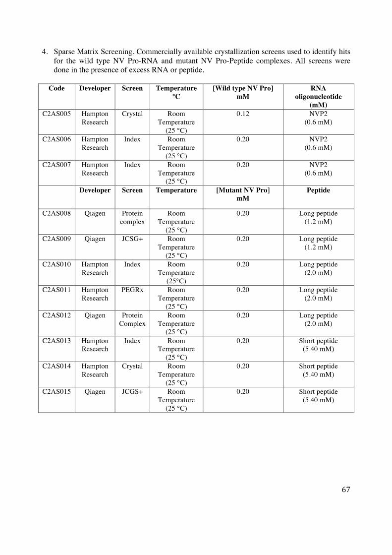
67
4. Sparse Matrix Screening. Commercially available crystallization screens used to identify hits for the wild type NV Pro-RNA and mutant NV Pro-Peptide complexes. All screens were done in the presence of excess RNA or peptide.
Code Developer Screen
Temperature °C
[Wild type NV Pro] mM
RNA oligonucleotide
(mM) C2AS005 Hampton
Research Crystal Room
Temperature (25 °C)
0.12 NVP2 (0.6 mM)
C2AS006 Hampton Research
Index Room Temperature
(25 °C)
0.20 NVP2 (0.6 mM)
C2AS007 Hampton Research
Index Room Temperature
(25 °C)
0.20 NVP2 (0.6 mM)
Developer Screen Temperature [Mutant NV Pro]
mM Peptide
C2AS008 Qiagen Protein complex
Room Temperature
(25 °C)
0.20 Long peptide (1.2 mM)
C2AS009 Qiagen JCSG+ Room Temperature
(25 °C)
0.20 Long peptide (1.2 mM)
C2AS010 Hampton Research
Index Room Temperature
(25°C)
0.20 Long peptide (2.0 mM)
C2AS011 Hampton Research
PEGRx Room Temperature
(25 °C)
0.20 Long peptide (2.0 mM)
C2AS012 Qiagen Protein Complex
Room Temperature
(25 °C)
0.20 Long peptide (2.0 mM)
C2AS013 Hampton Research
Index Room Temperature
(25 °C)
0.20 Short peptide (5.40 mM)
C2AS014 Hampton Research
Crystal Room Temperature
(25 °C)
0.20 Short peptide (5.40 mM)
C2AS015 Qiagen JCGS+ Room Temperature
(25 °C)
0.20 Short peptide (5.40 mM)

68
5. Structural Comparison of HuNV Pro dimers.
Figure 32: Structural comparison of HuNV Pro dimers obtained in the presence of RNA (Red) and RNA/Peptide
(Green). The structures are identical considering that the RMSD difference is only 0.2 Å.
6. Mass Spectroscopy data was interpreted using Mascot software (Matrix Science). The protein
score is used to rank the proposed proteins according to the total amount of evidence supporting the identification of each protein. A higher score indicates more evidence. The exponentially modified protein abundance index (emPAI) offers approximate, label free, quantitation for the proteins in a mixture based on the number of peptides observed per protein [92].
Description Protein Score Non-duplicate Match emPAI Norwalk chymotrypsin-like cysteine protease
776 11 3.11
Enterobacteria phage HK620 cII
31 3 0.29
Pseudomonas aeruginosa KlaB
19 1 0.07

69
ELSEVIER LICENSETERMS AND CONDITIONS
Aug 09, 2015
This is a License Agreement between Asfandyar Sikandar ("You") and Elsevier ("Elsevier")provided by Copyright Clearance Center ("CCC"). The license consists of your order details,the terms and conditions provided by Elsevier, and the payment terms and conditions.
All payments must be made in full to CCC. For payment instructions, please seeinformation listed at the bottom of this form.
Supplier Elsevier LimitedThe Boulevard,Langford LaneKidlington,Oxford,OX5 1GB,UK
Registered CompanyNumber
1982084
Customer name Asfandyar Sikandar
Customer address 225b, Olympus Hall
Calgary, AB t2n 4v7
License number 3685080041684
License date Aug 09, 2015
Licensed content publisher Elsevier
Licensed content publication Virology
Licensed content title Enzyme kinetics of the human norovirus protease control viruspolyprotein processing order
Licensed content author Jared May,Brent Korba,Alexei Medvedev,Prasanth Viswanathan
Licensed content date September 2013
Licensed content volumenumber
444
Licensed content issuenumber
1-2
Number of pages 7
Start Page 218
End Page 224
Type of Use reuse in a thesis/dissertation
Intended publisher of newwork
other
Portion figures/tables/illustrations
Number offigures/tables/illustrations
2
Format both print and electronic

70
Are you the author of thisElsevier article?
No
Will you be translating? No
Original figure numbers Table 1 and 2
Title of yourthesis/dissertation
Structural Studies of Human Norovirus Protease with RNA andPeptides
Expected completion date Aug 2015
Estimated size (number ofpages)
70
Elsevier VAT number GB 494 6272 12
Permissions price 0.00 USD
VAT/Local Sales Tax 0.00 USD / 0.00 GBP
Total 0.00 USD
Terms and Conditions
INTRODUCTION
1. The publisher for this copyrighted material is Elsevier. By clicking "accept" inconnection with completing this licensing transaction, you agree that the following termsand conditions apply to this transaction (along with the Billing and Payment terms andconditions established by Copyright Clearance Center, Inc. ("CCC"), at the time that youopened your Rightslink account and that are available at any time athttp://myaccount.copyright.com).
GENERAL TERMS
2. Elsevier hereby grants you permission to reproduce the aforementioned material subject tothe terms and conditions indicated.
3. Acknowledgement: If any part of the material to be used (for example, figures) hasappeared in our publication with credit or acknowledgement to another source, permissionmust also be sought from that source. If such permission is not obtained then that materialmay not be included in your publication/copies. Suitable acknowledgement to the sourcemust be made, either as a footnote or in a reference list at the end of your publication, asfollows:
"Reprinted from Publication title, Vol /edition number, Author(s), Title of article / title ofchapter, Pages No., Copyright (Year), with permission from Elsevier [OR APPLICABLESOCIETY COPYRIGHT OWNER]." Also Lancet special credit - "Reprinted from TheLancet, Vol. number, Author(s), Title of article, Pages No., Copyright (Year), withpermission from Elsevier."
4. Reproduction of this material is confined to the purpose and/or media for whichpermission is hereby given.
5. Altering/Modifying Material: Not Permitted. However figures and illustrations may bealtered/adapted minimally to serve your work. Any other abbreviations, additions, deletionsand/or any other alterations shall be made only with prior written authorization of Elsevier

71
2015-08-09, 6:40 PMRightslink Printable License
Page 1 of 8https://s100.copyright.com/App/PrintableLicenseFrame.jsp?publish…2-6e99-48c5-a5fd-18c40ad9275a%20%20&targetPage=printablelicense
ELSEVIER LICENSETERMS AND CONDITIONS
Aug 09, 2015
This is a License Agreement between Asfandyar Sikandar ("You") and Elsevier ("Elsevier")provided by Copyright Clearance Center ("CCC"). The license consists of your order details,the terms and conditions provided by Elsevier, and the payment terms and conditions.
All payments must be made in full to CCC. For payment instructions, please seeinformation listed at the bottom of this form.
Supplier Elsevier LimitedThe Boulevard,Langford LaneKidlington,Oxford,OX5 1GB,UK
Registered CompanyNumber
1982084
Customer name Asfandyar Sikandar
Customer address 225b, Olympus Hall
Calgary, AB t2n 4v7
License number 3685060522892
License date Aug 09, 2015
Licensed content publisher Elsevier
Licensed content publication Virology
Licensed content title RNA binding by human Norovirus 3C-like proteases inhibitsproteaseactivity
Licensed content author Prasanth Viswanathan,Jared May,Sunghae Uhm,ChangsuekYon,Brent Korba
Licensed content date 30 March 2013
Licensed content volumenumber
438
Licensed content issuenumber
1
Number of pages 8
Start Page 20
End Page 27
Type of Use reuse in a thesis/dissertation
Portion figures/tables/illustrations
Number of 2

72
2015-08-09, 6:40 PMRightslink Printable License
Page 2 of 8https://s100.copyright.com/App/PrintableLicenseFrame.jsp?publish…2-6e99-48c5-a5fd-18c40ad9275a%20%20&targetPage=printablelicense
figures/tables/illustrations
Format both print and electronic
Are you the author of thisElsevier article?
No
Will you be translating? No
Original figure numbers Fig 5 and 6
Title of yourthesis/dissertation
Structural Studies of Human Norovirus Protease with RNA andPeptides
Expected completion date Aug 2015
Estimated size (number ofpages)
70
Elsevier VAT number GB 494 6272 12
Permissions price 0.00 USD
VAT/Local Sales Tax 0.00 USD / 0.00 GBP
Total 0.00 USD
Terms and Conditions
INTRODUCTION
1. The publisher for this copyrighted material is Elsevier. By clicking "accept" inconnection with completing this licensing transaction, you agree that the following termsand conditions apply to this transaction (along with the Billing and Payment terms andconditions established by Copyright Clearance Center, Inc. ("CCC"), at the time that youopened your Rightslink account and that are available at any time athttp://myaccount.copyright.com).
GENERAL TERMS
2. Elsevier hereby grants you permission to reproduce the aforementioned material subject tothe terms and conditions indicated.
3. Acknowledgement: If any part of the material to be used (for example, figures) hasappeared in our publication with credit or acknowledgement to another source, permissionmust also be sought from that source. If such permission is not obtained then that materialmay not be included in your publication/copies. Suitable acknowledgement to the sourcemust be made, either as a footnote or in a reference list at the end of your publication, asfollows:
"Reprinted from Publication title, Vol /edition number, Author(s), Title of article / title ofchapter, Pages No., Copyright (Year), with permission from Elsevier [OR APPLICABLESOCIETY COPYRIGHT OWNER]." Also Lancet special credit - "Reprinted from TheLancet, Vol. number, Author(s), Title of article, Pages No., Copyright (Year), withpermission from Elsevier."

73
Title: Structural Basis of SubstrateSpecificity and ProteaseInhibition in Norwalk Virus
Author: Zana Muhaxhiri, Lisheng Deng,Sreejesh Shanker et al.
Publication: Journal of VirologyPublisher: American Society for
MicrobiologyDate: Apr 15, 2013Copyright © 2013, American Society for Microbiology
Logged in as: Asfandyar Sikandar Account #: 3000925953
Permissions Request
ASM authorizes an advanced degree candidate to republish the requested material in his/her doctoralthesis or dissertation. If your thesis, or dissertation, is to be published commercially, then you mustreapply for permission.
Copyright © 2015 Copyright Clearance Center, Inc. All Rights Reserved. Privacy statement. Terms and Conditions. Comments? We would like to hear from you. E-mail us at [email protected]
Title: A Norovirus Protease StructureProvides Insights into Active andSubstrate Binding Site Integrity
Author: Kentaro Nakamura, YuichiSomeya, Takashi Kumasaka etal.
Publication: Journal of VirologyPublisher: American Society for
MicrobiologyDate: Nov 1, 2005Copyright © 2005, American Society for Microbiology
Logged in as: Asfandyar Sikandar Account #: 3000925953
Permissions Request
ASM authorizes an advanced degree candidate to republish the requested material in his/her doctoralthesis or dissertation. If your thesis, or dissertation, is to be published commercially, then you mustreapply for permission.
Copyright © 2015 Copyright Clearance Center, Inc. All Rights Reserved. Privacy statement. Terms and Conditions. Comments? We would like to hear from you. E-mail us at [email protected]

74
2015-08-09, 7:14 PMCopyright Clearance Center
Page 1 of 7https://www.copyright.com/printCoiConfirmPurchase.do?operation=defaultOperation&confirmNum=11418861&showTCCitation=TRUE
Step 3: Order Confirmation
Confirmation Number: 11418861Order Date: 08/09/2015
If you paid by credit card, your order will be finalized and your card willbe charged within 24 hours. If you choose to be invoiced, you canchange or cancel your order until the invoice is generated.
Asfandyar Sikandar [email protected] +1 (403) 827-1196 Payment Method: n/a
Note: Copyright.com supplies permissions but not the copyrighted content itself.
Thank you for your order! A confirmation for your order will be sent to your account email address. If you havequestions about your order, you can call us at +1.855.239.3415 Toll Free, M-F between 3:00 AM and 6:00 PM (Eastern),or write to us at [email protected]. This is not an invoice.
Payment Information
Order Details
Permission type: Republish or display contentType of use: Republish in a thesis/dissertation
Requestor type Academic institution
Format Print, Electronic
Portion image/photo
Number ofimages/photosrequested
1
Title or numericreference of theportion(s)
Fig.2
Title of the article or
Order detail ID: 67931867Order License Id: 3685071066542
ISSN: 0022-1317Publication Type: JournalVolume:Issue:Start page:Publisher: SOCIETY FOR GENERAL
MICROBIOLOGYAuthor/Editor: SOCIETY FOR GENERAL
MICROBIOLOGY ; FEDERATION OFEUROPEAN MICROBIOLOGICALSOCIETIES
The Journal of general virology
Permission Status: Granted

75
2015-08-09, 7:14 PMCopyright Clearance Center
Page 2 of 7https://www.copyright.com/printCoiConfirmPurchase.do?operation=defaultOperation&confirmNum=11418861&showTCCitation=TRUE
chapter the portion isfrom
Norovirus gene expressionand replication
Editor of portion(s) N/A
Author of portion(s) Lucy G. Throne and Ian G.Goodfellow
Volume of serial ormonograph N/A
Issue, if republishingan article from a serial 95
Page range of portion 278-291
Publication date ofportion 2014
Rights for Main product
Duration of use Current edition and up to5 years
Creation of copies forthe disabled yes
With minor editingprivileges no
For distribution to Canada
In the followinglanguage(s)
Original language ofpublication
With incidentalpromotional use no
Lifetime unit quantityof new product Up to 499
Made available in thefollowing markets educational
The requestingperson/organization
Asfandyar Sikandar(Graduate Student)
Order reference number

76
2015-08-09, 7:14 PMCopyright Clearance Center
Page 3 of 7https://www.copyright.com/printCoiConfirmPurchase.do?operation=defaultOperation&confirmNum=11418861&showTCCitation=TRUE
Total order items: 1 Order Total: 0.00 USD
Author/Editor Asfandyar Sikandar
The standard identifier 1
Title
Structural Studies ofHuman Norovirus ProteaseComplexes with RNA andPepties
Publisher University of Calgary
Expected publicationdate Aug 2015
Estimated size (pages) 70
Note: This item will be invoiced or charged separately through CCC's RightsLink service. More info $ 0.00
This is not an invoice.

77
Title: Serine Protease Mechanism andSpecificity
Author: Lizbeth HedstromPublication: Chemical ReviewsPublisher: American Chemical SocietyDate: Dec 1, 2002Copyright © 2002, American Chemical Society
Logged in as: Asfandyar Sikandar Account #: 3000925953
PERMISSION/LICENSE IS GRANTED FOR YOUR ORDER AT NO CHARGE
This type of permission/license, instead of the standard Terms & Conditions, is sent to you because nofee is being charged for your order. Please note the following:
Permission is granted for your request in both print and electronic formats, andtranslations.If figures and/or tables were requested, they may be adapted or used in part.Please print this page for your records and send a copy of it to your publisher/graduateschool.Appropriate credit for the requested material should be given as follows: "Reprinted(adapted) with permission from (COMPLETE REFERENCE CITATION). Copyright(YEAR) American Chemical Society." Insert appropriate information in place of thecapitalized words.One-time permission is granted only for the use specified in your request. No additionaluses are granted (such as derivative works or other editions). For any other uses, pleasesubmit a new request.
If credit is given to another source for the material you requested, permission must be obtainedfrom that source.
Copyright © 2015 Copyright Clearance Center, Inc. All Rights Reserved. Privacy statement. Terms and Conditions. Comments? We would like to hear from you. E-mail us at [email protected]