Embed Size (px)
Citation preview

For correspondence rficner
uni-goettingende
Competing interests The
authors declare that no
competing interests exist
Funding See page 22
Received 14 September 2016
Accepted 15 January 2017
Published 16 January 2017
Reviewing editor Timothy W
Nilsen Case Western Reserve
University United States
Copyright Tauchert et al This
article is distributed under the
terms of the Creative Commons
Attribution License which
permits unrestricted use and
redistribution provided that the
original author and source are
credited
Structural insights into the mechanism ofthe DEAH-box RNA helicase Prp43Marcel J Tauchert1 Jean-Baptiste Fourmann2 Reinhard Luhrmann2 Ralf Ficner1
1Department of Molecular Structural Biology Institute for Microbiology andGenetics GZMB Georg-August-University Gottingen Gottingen Germany2Department of Cellular Biochemistry Max Planck Institute for BiophysicalChemistry Gottingen Germany
Abstract The DEAH-box helicase Prp43 is a key player in pre-mRNA splicing as well as the
maturation of rRNAs The exact modus operandi of Prp43 and of all other spliceosomal DEAH-box
RNA helicases is still elusive Here we report crystal structures of Prp43 complexes in different
functional states and the analysis of structure-based mutants providing insights into the unwinding
and loading mechanism of RNAs The Prp43ATP-analogRNA complex shows the localization of
the RNA inside a tunnel formed by the two RecA-like and C-terminal domains In the ATP-bound
state this tunnel can be transformed into a groove prone for RNA binding by large rearrangements
of the C-terminal domains Several conformational changes between the ATP- and ADP-bound
states explain the coupling of ATP hydrolysis to RNA translocation mainly mediated by a b-turn of
the RecA1 domain containing the newly identified RF motif This mechanism is clearly different to
those of other RNA helicases
DOI 107554eLife21510001
IntroductionHelicases are omnipresent enzymes spread through the whole phylogenetic tree of life as well as
viruses since their unwinding capabilities are of striking importance for numerous cellular pathways
of RNA and DNA metabolism Based on sequence alignments helicases were subdivided into six
superfamilies (SF) which come in two different flavors (Singleton et al 2007 Fairman-
Williams et al 2010 Jankowsky and Fairman 2007 Pyle 2011) Most members of the SF1 or
SF2 are monomeric in contrast to the members of SF3 to SF6 which assemble as hexamers
(Singleton et al 2007) The latter are composed of either RecA- or AAA+-like domains thus being
distinct from the SF1 and SF2 helicases which are exclusively composed of two RecA-like domains in
juxtaposition with each other
The SF2 is by far the largest superfamily comprising a vast number of RNA helicases with implica-
tions in numerous RNP metabolism pathways such as translation initiation and termination pre-
mRNA editing and splicing mRNA export as well as ribosome biogenesis (Ozgur et al 2015) The
centerpiece of these helicases is formed by the two RecA-like domains harboring conserved motifs
which are crucial for these NTPase-dependent RNA helicases to accomplish their function The
motifs I II V and VI are required for nucleoside triphosphate binding and hydrolysis motifs Ia Ib
and IV are involved in RNA binding and the motif III participates in coupling the NTPase and the
unwinding activity (Caruthers and McKay 2002 Cordin and Beggs 2013 Cordin et al 2012)
Most SF2 helicases exhibit extensions at the N- and C-termini of the helicase core which is also true
for the DEAH-box proteins a subfamily of the DEAHRHA family The C-terminal extension is highly
conserved in DEAH-box helicases and is composed of three domains a winged helix (WH) a ratchet
and an oligosaccharide binding (OB) fold (Walbott et al 2010 He et al 2010) The ratchet
domain was originally named according to the homologous domain of the DNA helicase Hel308
Tauchert et al eLife 20176e21510 DOI 107554eLife21510 1 of 25
RESEARCH ARTICLE
(Buttner et al 2007) but recent studies on MLE suggested that this domain has no ratcheting func-
tion and should be renamed (Prabu et al 2015) Therefore it is hereinafter referred to as ratchet-
like domain In DEAH-box proteins the degrees of freedom of the helicase core are reduced by the
interactions between the C-terminal domains and the two RecA-like domains in comparison to those
DEAD-box proteins lacking such C-terminal domains The conservation of the N-terminal extension
is distinctly lower and they dramatically differ in length between the members of this subfamily
Reported functions for this N-terminal extension are among others the involvement in the recruit-
ment as shown for Prp16 (pre-mRNA processing factor 16) as well as for Prp22 or for the subcellular
targeting to the nucleus as elucidated for Prp43 (Wang and Guthrie 1998 Schneider and Schwer
2001 Fouraux et al 2002)
The latter mentioned helicase Prp43 is an outstanding member of the DEAH-box subfamily since
it has implications in different substantive cellular processes The first reported function of Prp43 is
its involvement in pre-mRNA splicing In the course of this process Prp43 acts at the latest stage of
the splicing cycle and it is required to dismantle the intron-lariat spliceosome into the excised lariat
and the U2U5U6 snRNPs (Arenas and Abelson 1997 Fourmann et al 2013 2016a) Recently
the target substrate of Prp43 during this process was revealed which is the RNA network between
the U2 snRNP and the branch site of the intron (Fourmann et al 2016b) Indeed in the spliceo-
some Prp43 was crosslinked exclusively to the pre-mRNA and not to any snRNAs Furthermore
Prp43 is also a key player for the disassembly of stalled spliceosomes (Pandit et al 2006
Semlow and Staley 2012 Koodathingal et al 2010) During these spliceosomal processes Prp43
binds RNA in a sequence-independent manner Prp43 action is also essential for ribosome biogene-
sis Here it is involved in the maturation of 18S and 25S rRNAs with several reported binding sites
on intermediates which vary in sequence and thus make a sequenced-based binding mechanism
unlikely for Prp43 (Lebaron et al 2005 Bohnsack et al 2009) Lately the involvement of Prp43 in
a third distinct cellular function was illuminated and it was unraveled that Prp43 is also involved in
the promotion of apoptosis (Heininger et al 2016)
The distribution of Prp43 between these three fundamentally different processes is regulated by
a fine-tuned interplay of the helicase itself and cofactors These cofactors belong to the family of
G-patch proteins which share an intrinsically disordered glycine-rich region the G-patch motif
(Aravind et al 1999) Apart from being involved in recruitment these G-patch proteins also
increase the NTPase and stimulate the helicase activity of Prp43 To date in S cerevisiae five
G-patch proteins were identified out of which four interact with Prp43 In the context of splicing
Prp43 function is modulated by Ntr1 (Nineteen complex-related proteins 1) (Tanaka et al 2007
Tsai et al 2005 2007 Boon et al 2006) During rRNA maturation Prp43 is associated with Pfa1
(Prp forty-three associated 1) or Gno1 (G-patch nucleolar protein 1) (Lebaron et al 2005
2009 Bohnsack et al 2009) Moreover its apoptotic function is implemented by the interaction
with Cmg1 (Cytoplasmic and mitochondrial G-patch protein 1) (Heininger et al 2016) The remain-
ing G-patch protein from yeast is Spp2 (Suppressor of PRP protein 2) which interacts with another
spliceosomal DEAH-box helicase Prp2 (Warkocki et al 2015)
Despite intensive studies the exact modus operandi of the DEAH-box helicases and thus of
Prp43 is still elusive to date This is primarily attributable to a lack of crystal structures of members
of the DEAH-box subfamily in an active state and with bound interaction partners Recently the crys-
tal structure of the related DExH-box helicase MLE (Maleless) which belongs to another DEAHRHA
subfamily provided a first insight into how this helicase might function The complex of MLE with a
U10-RNA and ADPAlF4- led to a proposed mechanism of how RNA is translocated through its inte-
rior tunnel driven by conformational changes of a Hook-Loop in the RecA2 domain (Prabu et al
2015)
So far three crystal structures of Prp43 in the post-catalytic ADP-bound state have been available
from S cerevisiae or C thermophilum (Walbott et al 2010 He et al 2010 Tauchert et al
2016) The latter mentioned organism might be a valuable alternative for structural studies on
DEAH-box proteins since several crystal structures of spliceosomal RNA helicases from this thermo-
philic fungus were solved only recently (Tauchert et al 2016 Tauchert and Ficner 2016
Absmeier et al 2015) Owing to this intensive crystallization trails with Prp43 from C thermophi-
lum were performed leading to three new crystal structures All three structures show Prp43 in an
active state since it was crystallized with a non-hydrolysable ATP-analog (ADPBeF3-) as well as one
of these structures show Prp43 with an additional U7-RNA The Prp43U7-RNAADPBeF3-complex
Tauchert et al eLife 20176e21510 DOI 107554eLife21510 2 of 25
Research article Biochemistry Biophysics and Structural Biology
unveils the structural basis of why Prp43 can bind RNA in a sequence-independent manner The
Prp43ADPBeF3- structures illustrate how Prp43 binds and releases complex RNA substrates due to
massive rearrangements of the C-terminal domains which affect an opening of the interior RNA-
binding tunnel which is formed by the two RecA-like and the C-terminal domains The hydrolysis of
an ATP molecule induces conformational rearrangements of the RecA-like domains which in turn
allow the translocation of the RNA through the interior tunnel via the so far unrecognized RF motif
present in a b-turn of all DEAH-box helicases According to our results the translocation mechanism
of MLE is very unlikely to be compatible for Prp43 All proposed functional mechanisms of Prp43 are
strengthened by functional analyses of structure-based mutants in this study
Results
Crystal structure of Prp43 with bound RNA in an active stateIn order to overcome the lack of crystal structures of DEAH-box helicases in an active state we crys-
tallized Prp43 in the presence of the non-hydrolysable ATP-analog ADPBeF3- as well as a U16-RNA
For these crystallization approaches an N-terminally truncated version of Prp43 from C thermophi-
lum was used (ctPrp43DN) which lacks the first 60 amino acids (Figure 1a) Previously we already
demonstrated that this truncated ctPrp43 variant is fully capable of replacing its yeast ortholog in
spliceosome disassembly assays (Tauchert et al 2016) Crystals of ctPrp43DNADPBeF3- in the
presence of the U16-RNA were obtained in the space group P6122 and diffracted up to 262 A In
addition to the ADPBeF3- and a central magnesium ion at the active site seven out of the 16
nucleotides of the RNA as well as one additional phosphate group were traceable in the electron
density (for an omit map of the RNA see Figure 1mdashfigure supplement 1a) Hence this complex is
referred to here as ctPrp43DNU7ADPBeF3- The refinement of this complex converged at Rwork
and Rfree values of 1970 and 2297 (Table 1)
By this ctPrp43DNU7ADPBeF3- complex structure (Figure 1bndash1e) insights into the sequence-
unspecific RNA-binding mechanism of a genuine DEAH-box helicase were obtained ctPrp43 con-
sists of six different domains an N-terminal extension (residues 1ndash96) the RecA1 (97ndash273) and
RecA2 (274ndash458) domains and the C-terminal domains which were assigned as degenerate winged
helix domain (459ndash526) ratchet-like domain (527ndash640) and oligosaccharide binding-fold domain
(641ndash764) (Walbott et al 2010 He et al 2010)
The RecA-like domains and the C-terminal domains appear to be stably associated by multiple
interactions and form a tunnel inside the Prp43 molecule (Figure 1d) which is the binding site of the
U7-RNA The 5rsquo end of the RNA is located at the RecA2 domain and the 3rsquo RNA end is situated at
the RecA1 domain For the first three nucleotides at the 5rsquo end (U1ndashU3) two alternative conforma-
tions are observed (Figure 1mdashfigure supplement 2) The structure reveals the basis for the
sequence-independent RNA binding of Prp43 which is in line with previous biochemical data
(Fourmann et al 2016b Bohnsack et al 2009 Tanaka and Schwer 2006)
Prp43 binds RNA in a sequence-independent fashionInteractions between Prp43 and the U7-RNA are sparsely present Depending on which of the two
alternative RNA conformations is analyzed only 11 or 13 hydrogen bonds and ionic interactions
between the RNA and Prp43 can be detected (Figure 1c and e) Most of these interactions are
formed between the sugar-phosphate backbone of the RNA and the RecA1 RecA2 or the ratchet-
like domain On the contrary the uracil moieties do not interact with the helicase with the exception
of a hydrogen bond of U3 with the Ser 555 main-chain carboxyl group from the ratchet-like domain
and a base stack between the U1 and Arg 562 which only occurs in one of the two alternative con-
formations These Prp43-RNA interactions are however by far not enough for sequence-specific
binding of a substrate RNA to Prp43 In addition to the unspecific RNA-binding mode of Prp43
RNA binding appears to be transient as only four nucleotides are more tightly associated with
Prp43 In the crystal structure presented this corresponds to the nucleotides U4 to U7 At the 5rsquo
end the first three nucleotides are weakly bound by Prp43 since only 2 (conf B) and 3 (conf A) inter-
actions are detectable respectively The weak interaction is additionally highlighted by elevated B-
factors for this RNA region compared to the four other nucleotides (see Figure 1mdashfigure supple-
ment 3) After the first two nucleotides a kink is introduced in the RNA backbone and the number
Tauchert et al eLife 20176e21510 DOI 107554eLife21510 3 of 25
Research article Biochemistry Biophysics and Structural Biology
Figure 1 Crystal structure of Prp43 in complex with U7-RNA and the ATP-analog ADPBeF3- (a) Domain overview of ctPrp43 The bottom bar indicates
the N-terminally truncated construct (DN 61ndash764) used for crystallization (b) Overall structure of ctPrp43DNU7ADPBeF3- Domains are colored
according to a and the bound U7-RNA is shown in gray with two alternative conformations for nucleotides U1-U3 The ADPBeF3- is bound in the cleft
between the RecA domains (c) Close-up of the bound U7-RNA Residues involved in interactions are labeled according to the wild-type ctPrp43
Figure 1 continued on next page
Tauchert et al eLife 20176e21510 DOI 107554eLife21510 4 of 25
Research article Biochemistry Biophysics and Structural Biology
of Prp43-RNA interactions gets more numerous and more intense At the 3rsquoend the RNA does not
interact with Prp43 as soon as the end of the RNA-binding tunnel is reached The phosphate group
of the eighth nucleotide is exactly located at the end of this tunnel Compared to the nucleotides U4
to U7 this phosphate exhibits elevated B-factors The U8 nucleoside was not defined in the electron
density map and thus might indicate the presence of multiple RNA conformations at this site out of
which none is clearly favorable due the lack of interactions with Prp43
Having determined the crystal structure of ctPrp43DNU7ADPBeF3- another question concerning
Prp43rsquos modus operandi did arise which is directly linked to its cellular role In the context of pre-
mRNA splicing or rRNA biogenesis Prp43 has to bind to or to be released from short singled-
stranded RNA stretches within larger folded RNAs or RNPs In the light of the fact that the RNA-
binding site of Prp43 is located inside a tunnel RNA loading appears to be non-trivial and thus
requires further illumination
Crystal structure of Prp43 in the ATP-bound stateIn pursuance of analyzing how Prp43 binds to ssRNA regions within folded RNAs we targeted at
determining the pre-catalytic state of Prp43 before RNA binding For this purpose we subjected
ctPrp43DN to crystallization trials in the presence of the ATP analog ADPBeF3- The
ctPrp43DNADPBeF3- complex crystallized in two very distinct crystallization conditions and in two
different space groups The structure of the complex which crystallized in P212121 was refined at a
resolution of 178 A to Rwork and Rfree values of 1767 and 1988 (Figure 2a) and is referred to
here as ctPrp43DNADPBeF3-(HR) (high resolution) The second crystal form belongs to the hexago-
nal space group P65 which exhibited a resolution limit of 324 A (Figure 2b) and thus is referred to
as ctPrp43DNADPBeF3-(LR) (low resolution) This structure was refined to Rwork Rfree values of
1822 and 2178 respectively
In comparison to the ctPrp43DNU7ADPBeF3- complex structure the conformation of the two
RecA-like domains in the ctPrp43DNADPBeF3- complex is almost identical and they superpose very
well with rmsd values of 034 A (312 Ca) in case of ctPrp43DNADPBeF3-(HR) and with 039 A
(318 Ca) for the ctPrp43DNADPBeF3-(LR) structure (Figure 2c) However a large rearrangement of
the C-terminal domains is seen in both ctPrp43DNADPBeF3- complex structures (Figure 3) which
leads to the disruption of the interaction between two b-hairpin loops from the RecA2 domain and
the OB-fold As a consequence the RNA-binding tunnel of ctPrp43DNU7ADPBeF3- is opened and
transformed into a shallow groove In the closed state the C-terminal domains of the
ctPrp43DNU7ADPBeF3- complex and of ctPrp43DNADP superpose well (rmsd of 055 A for
267 Ca)
Figure 1 continued
sequence The 5rsquo and 3rsquo end of the RNA is indicated (d) Cross-section of the Prp43 RNA-binding tunnel Prp43 is shown in surface representation and
the RNA in ball-and-sticks mode (e) Schematic figure of the Prp43-RNA interactions Residues which interact with the RNA via their main chain are
shown as triangles and residues which exhibit side chain interactions are presented as ellipses The coloring of the residues corresponds to a The
alternative conformation of the first three nucleotides is shown light gray Stacking interactions are highlighted by double lines and polar interactions by
dotted lines
DOI 107554eLife21510002
The following figure supplements are available for figure 1
Figure supplement 1 Omit maps of a fraction of the U7-RNA from the ctPrp43DNU7ADPBeF3- complex structure and of the active site of
ctPrp43DNADPBeF3-(HR) (Figure 2a)
DOI 107554eLife21510003
Figure supplement 2 The two alternative conformations of the U7-RNA in the ctPrp43DNU7ADPBeF3- complex structure
DOI 107554eLife21510004
Figure supplement 3 Overview of the B-factors of the U7-RNA in the ctPrp43DNU7ADPBeF3- complex structure
DOI 107554eLife21510005
Figure supplement 4 Schematic representation of the NS3 HCV- and MLE-RNA interaction networks
DOI 107554eLife21510006
Tauchert et al eLife 20176e21510 DOI 107554eLife21510 5 of 25
Research article Biochemistry Biophysics and Structural Biology
RNA loading mechanism of Prp43The opening of the RNA-binding tunnel appears to be the key mechanism allowing Prp43 and very
likely numerous other DEAH-box helicases to bind to complex folded RNA substrates Thus these
rearrangements of the C-terminal domains are expected to be crucial for their unwinding function
The opening is feasible due to a large movement of the ratchet-like and the OB-fold domains Rear-
rangements of the WH domain are less pronounced owing to the fact that the WH rather functions
as a hinge region between the RecA2 and the ratchet-like domain Comparison between the
ctPrp43DNU7ADPBeF3- and the ctPrp43DNADPBeF3
-(HR) complex structures after alignment of
the RecA-like domains revealed that the center of mass of the ratchet-like domain is shifted by 162
A and by 141 A for the OB-fold domain In the ctPrp43DNADPBeF3-(LR) complex structure these
Table 1 Data collection and refinement statistics
ctPrp43DNU7ADPBeF3- ctPrp43DNADPBeF3
-(HR) ctPrp43DNADPBeF3-(LR)
PDBid 5lta 5ltj 5ltk
Data collection
Space group P6122 P212121 P65
Cell dimensions
a b c (A) 10639 10639 35670 8883 10564 11905 18434 18434 8232
a b g (˚) 900 900 1200 900 900 900 900 900 1200
Resolution (A) 4856 ndash 262(270 ndash 262)
7902 ndash 178(189 ndash 178)
9217 ndash 324(339 ndash 324)
Rmeas () 59 (903) 72 (1235) 98 (762)
Is(I) 2216 (185) 1488 (164) 1465 (226)
CC12 () 999 (652) 999 (619) 998 (638)
Completeness () 985 (868) 996 (988) 988 (949)
Redundancy 514 (534) 471 (466) 400 (359)
Refinement
Resolution (A) 4856 ndash 262 6799 ndash 178 9217 ndash 324
No reflections 36887 107276 25304
Rwork Rfree 19702297 17671988 18222178
No atoms
Protein 5605 5730 5622
RNA 204
Ligand Ion 37 105 42
Water 52 688 4
B-factors(A2)
Protein 7283 3441 8887
RNA 8865
Ligand Ion 5504 4783 8389
Water 6051 4429 6219
Rms deviations
Bond length (A) 00028 00041 00026
Bond angles (˚) 078 087 067
Ramachandran Plot
Favored 9598 9734 9628
Outlier 00 00 00
Values in parentheses are for the highest resolution shell
DOI 107554eLife21510007
Tauchert et al eLife 20176e21510 DOI 107554eLife21510 6 of 25
Research article Biochemistry Biophysics and Structural Biology
values are equal to 170 A (ratchet-like) and 117 A (OB-fold) respectively The
ctPrp43DNADPBeF3-(HR) structure is in a marginally more closed conformation hence the
ctPrp43DNADPBeF3- complex structures are not completely identical They represent two slightly
different snapshots of Prp43 which both might exist in solution and presumably are favored by the
different crystallization conditions or different crystal packing These two states of
ctPrp43DNADPBeF3- might also give a hint at how Prp43 switches into the closed conformation
since in the ctPrp43DNADPBeF3-(HR) complex structure one inter-domain contact is present
between the RecA2 (Asp 486) and the ratchet-like domain (Lys 605) These interactions between
sequentially distal residues might trigger the switching from the open into the closed conformation
after the binding to RNA The circumstance that both structures of the ctPrp43DNADPBeF3- com-
plex are in a similar open conformation strongly suggests the possibility that the complex structures
presented reflect the main conformation of Prp43 in an active state in solution Owing to the fact
that Prp43ADPBeF3- crystallized in different space groups and in divergent crystallization condi-
tions the open conformation in crystallo is highly unlikely to be a crystallization artefact This
assumption is additionally supported by the low number of crystal contacts for the
ctPrp43DNADPBeF3-(HR) (3553 A2 of buried surface11 of the total surface) and the
ctPrp43DNADPBeF3-(LR) (15243 A247) complex structure
To further strengthen the hypothesis that the opening of the tunnel by the displacement of the
C-terminal domains is crucial for the helicase function of Prp43 we designed a mutant of Prp43
which allows us to trap the closed conformation by the introduction of an internal disulfide bond
(ctPrp43-IDSB) For this purpose one cysteine was introduced into the RecA1 domain and another
one into the ratchet-like domain at exposed positions to maximize the number of formed disulfide
bonds (Figure 4mdashfigure supplement 1) The functional impact of these mutations was analyzed by
an ATPase assay a fluorescence-based helicase assay and an intron-lariat spliceosome (ILS) disas-
sembly assay First of all the percentage of formed disulfide bonds was experimentally estimated
Figure 2 Structures of Prp43 with the bound ATP-analog ADPBeF3- in different crystal forms (a) Overall structure of ctPrp43DNADPBeF3
- at high and
(b) low resolution ctPrp43DNADPBeF3-(HR) crystalized in the orthorhombic space group P212121 and ctPrp43DNADPBeF3
-(LR) in the hexagonal
space group P65 Domains are colored according to Figure 1a ADPBeF3- is shown at the binding cleft between the two RecA-like domains in ball-
and-stick mode (c) Superposition of ctPrp43DNADPBeF3-(HR) (orange) and ctPrp43DNADPBeF3
-(LR) (blue) with ctPrp43DNU7ADPBeF3- (green)
Structures were superimposed via their helicase core (RecA1 and RecA2 domains) and are shown in ribbon representation
DOI 107554eLife21510008
The following figure supplement is available for figure 2
Figure supplement 1 The main chain of the Hook-Turn (RF motif) is present in two alternative conformations
DOI 107554eLife21510009
Tauchert et al eLife 20176e21510 DOI 107554eLife21510 7 of 25
Research article Biochemistry Biophysics and Structural Biology
The wild-type protein contains nine cysteines the ctPrp43-IDSB mutant two additional ones Via Ell-
man reaction the number of cysteines was determined to be 85 plusmn 01 (ctPrp43) and 93 plusmn 01
(ctPrp43-IDSB) respectively This allows us to conclude that the majority of ctPrp43-IDSB exhibits
the internal disulfide bridge because oxidized cysteines cannot be detected by this method and only
the nine cysteines which are also present in the wild-type protein were determined
Prp43 trapped in the closed conformation is impaired in its helicaseactivityHaving determined that ctPrp43-IDSB is mainly present in the oxidized and therefore closed state
this mutant was subsequently analyzed concerning its helicase activity using a synthetic dsRNA with
a 3rsquossRNA overhang (Figure 4a and Table 2) Since wild-type Prp43 exhibited a very low activity
(054 nM of unwound RNAmin) in the absence of any stimulatory G-patch (GP) protein we tested
its activity in the presence of the ctNtr1-GP (198 nMmin) and the ctPfa1-GP (6003 nMmin) Since
the ctPfa1-GP could increase the Prp43 activity to a much higher level the ctPfa1-GP was used for
the in vitro helicase activity analysis of all Prp43 mutants ctPrp43-IDSB exhibited an activity of 1218
Figure 3 Conformational changes between the ctPrp43DNADP complex structure (5d0u) (Tauchert et al 2016) ctPrp43DNU7ADPBeF3- (5lta)
ctPrp43DNADPBeF3-(HR) (5ltj) and ctPrp43DNADPBeF3
-(LR) (5ltk) Structures were superposed via their RecA1 domains In the top panel the back
view is presented (rotated by 180˚ with respect to Figure 1b Figure 2a and Figure 2b) and in the bottom panel the side view (90˚ rotation around a
vertical axis) Structures are ordered according to the degree of C-terminal displacement and opening of the RNA-binding tunnel The location of the
RNA-binding tunnel which is present in ctPrp43DNADP and ctPrp43DNU7ADPBeF3- is indicated
DOI 107554eLife21510010
Tauchert et al eLife 20176e21510 DOI 107554eLife21510 8 of 25
Research article Biochemistry Biophysics and Structural Biology
Figure 4 Helicase activity and RNA-binding assays of ctPrp43 and mutants (a) The maximal unwinding velocity
(nMmin) for a dsRNA with a 3rsquo overhang is shown (b) RNA binding of 5rsquo6FAM-U16-RNA by ctPrp43 and ctPrp43-
HT was determined via fluorescence anisotropy measurements Error bars indicate the standard deviation from
three independent measurements for a and b
DOI 107554eLife21510011
The following figure supplements are available for figure 4
Figure supplement 1 Position of the two introduced cysteine residues in the ctPrp43-IDSB mutant
DOI 107554eLife21510012
Figure supplement 2 ATPase activity of Prp43 and mutants (a) without further stimulation (b) in the presence of a
G-patch (c) in the presence of a U16-RNA and (d) in the presence of a G-patch and a U16-RNA
DOI 107554eLife21510013
Figure supplement 3 Raw data of exemplary helicase activity measurements
DOI 107554eLife21510014
Tauchert et al eLife 20176e21510 DOI 107554eLife21510 9 of 25
Research article Biochemistry Biophysics and Structural Biology
nMmin which equals about 15th of the wild-type Prp43 activity and thus is clearly impaired con-
cerning its ability to unwind dsRNA The remaining helicase activity of the mutant is most likely
caused by the fraction of ctPrp43-IDSB which does not contain the internal disulfide bond The activ-
ity of ctPrp43 and the ctPrp43-IDSB mutant were also analyzed for a physiological and more com-
plex substrate RNP by using the S cerevisiae ILS disassembly assays (Figure 5 and Figure 5mdashfigure
supplement 1) In previous work it was shown that ctPrp43 is capable of functionally replacing
scPrp43 in the spliceosome (Tauchert et al 2016) ctPrp43 can release about 60 of intron-lariat
RNA from scILS in the presence of ATP and scNtr(12) and dissociate the remaining spliceosomal
RNP core into the free U6 snRNA 18S U5 and 20S U2 snRNP (Figure 5c) This is close to the 65
efficiency of recombinant scPrp43 (Figure 5b) In case of the ctPrp43-IDSB mutant these assays
were performed in the presence and absence of DTT which can reduce the disulfide bridge
between the RecA1 and ratchet-like domain and thus free ctPrp43-IDSB from its locked conforma-
tion Without DTT the efficiency of ILS disassembly by ctPrp43-IDSB is clearly impaired compared to
the wild-type protein since only 10 of all ILSs are dissociated which equals the amount of the nega-
tive control (Figure 5a and d) In the presence of DTT the ILS dissociation efficiency of ctPrp43-
IDSB can be fully restored to the wild-type activity level of 60 (Figure 5e) This clearly hints at the
fact that locking Prp43 in the closed conformation distinctly influences its activity as deduced from
the crystal structures of ctPrp43DNADPBeF3- and thus proving the importance of adopting the
open conformation for the helicase activity of Prp43 also in the spliceosome (Video 1)
To exclude the possibility that the results of the helicase or ILS disassembly assays differ between
ctPrp43 and ctPrp43-IDSB due to a reduced ATPase activity of the latter ATPase activity measure-
ments were performed For all ctPrp43 variants the ATPase activity was analyzed for ctPrp43 itself
after the addition of a GP or U16-RNA or in the presence of both Since this was the first enzymatic
characterization of Prp43 from C thermophilum several general results are worth mentioning
(Table 3 and Figure 4mdashfigure supplement 2) The basal ATPase activity of ctPrp43 (181 min1) is
very similar to its ortholog from S cerevisiae (243 min1) (Christian et al 2014) ctPrp43 is stimu-
lated by both the ctNtr1-GP and the ctPfa1-GP but the effect by the latter is more pronounced A
clear difference between C thermophilum and S cerevisiae Prp43 is that ctPrp43 does not exhibit
RNA-stimulated ATPase activity in the absence of a GP The ctPrp43 ATPase activity can only be
induced by RNA if an additional G-patch protein is present
The intrinsic ATPase activity of ctPrp43-IDSB (504 min1) is similar to the one determined for
wild-type ctPrp43 ctPrp43-IDSB is also stimulated by ctPfa1-GP and by U16-RNA in the presence of
the ctPfa1-GP but in the contrast to wild-type Prp43 also just by U16-RNA These findings confirm
that the difference in the helicase and the ILS disassembly assay between ctPrp43 and ctPrp43-IDSB
is solely caused by the impairment of the unwinding capabilities The RNA-induced stimulation of
ctPrp43-IDSB in the trapped conformation might still be feasible due to the fact that a short single-
stranded U16-RNA without any secondary structures was used for these assays Binding to the entry
or exit site of the RNA-binding tunnel which does not require an opening of the tunnel might be
sufficient for the RNA-induced stimulation of the ATPase activity (see below) Furthermore Prp43 in
the trapped closed conformation appears to be more prone for the stimulation of the ATPase
Table 2 Helicase activity
nMmin plusmn
ctPrp43 054 018
ctPrp43 + ctPfa1-GP 6003 275
ctPrp43 + ctNtr1-GP 198 045
ctPfa1-GP 046 028
ctPrp43-IDSB + ctPfa1-GP 1218 023
ctPrp43-HT+ ctPfa1-GP 068 016
ctPrp43-HL + ctPfa1-GP 6187 714
ctPrp43-HTampHL+ ctPfa1-GP 088 003
DOI 107554eLife21510017
Tauchert et al eLife 20176e21510 DOI 107554eLife21510 10 of 25
Research article Biochemistry Biophysics and Structural Biology
Figure 5 Intron-lariat spliceosome (ILS) disassembly assays 10ndash30 glycerol gradient sedimentation of purified yeast ILS (scILS) incubated in solution
with ATP plus (a) no recombinant protein (b) scPrp43 and cofactors scNtr(12) (c) ctPrp43 and scNtr(12) (d) ctPrp43-IDSB and scNtr(12) (e) ctPrp43-
IDSB scNtr(12) and 05 mM DTT (f) ctPrp43-HT and scNtr(12) (g) ctPrp43-HL and scNtr(12) (h) ctPrp43-HTampHL and scNtr(12) U2 U5 and U6 snRNAs
were visualized by Northern blotting followed by autoradiography RNA identities are indicated on the left Quantifications were performed with
Figure 5 continued on next page
Tauchert et al eLife 20176e21510 DOI 107554eLife21510 11 of 25
Research article Biochemistry Biophysics and Structural Biology
activity by RNA since the ctPrp43-IDSB mutant (2499 min1) is stimulated by U16-RNA while the
wild-type protein is not (208 min1) It is conceivable that the GP increases the number of Prp43
molecules in a closed conformation since in the presence of a GP also the wild-type protein is stimu-
lated by RNA
Having shown the importance of the opening of Prp43rsquos C-terminal domains for its unwinding
capability we asked how Prp43 couples ATP hydrolysis and RNA translocation leading to its heli-
case activity Since high-resolution structures of the pre- and post-catalytic state of a DEAH-box heli-
case are both now available conformational rearrangements of Prp43 in the course of nucleotide
hydrolysis were analyzed
Conformational rearrangements at the helicase coreTo investigate the effect of ATP hydrolysis on the Prp43 conformation the structures of the ADP
and the ADPBeF3- bound state were compared Since the conformations of the RecA-like domains
of the three structures ctPrp43DNU7ADPBeF3- ctPrp43DNADPBeF3
-(HR) and
ctPrp43DNADPBeF3-(LR) are very similar the analysis of the conformational switches was restricted
to the comparison of ctPrp43DNADPBeF3-(HR) with the previously published ctPrp43DNADP
structure
The individual RecA-like domains of ctPrp43DNADP and ctPrp43DNADPBeF3-(HR) superimpose
well with rmsd values of 036 A (139 Ca) for the RecA1 domains and 044 A (142 Ca) for the
RecA2 domains Superimposing the helicase cores consisting of both RecA-like domains shows that
the RecA1 and RecA2 domains are rotated by ca 4˚ and ca 14˚ respectively There is also a slight
translational displacement of the RecA2 domain by a 27 A shift of its center of mass with regard to
the RecA1 domain Depending on the ADPATP state conformational changes of several of the con-
served SF2 helicase motifs most strikingly of
those in the RecA2 domain are observed (Fig-
ure 6) The conformations of the motifs I Ia Ib
and II which are all located in the RecA1
domain are similar between the ATP-bound and
the ADP-bound state of Prp43 This ATP-bound
state mimicked by the bound ADPBeF3- also
allows to deduce how Prp43 does hydrolyze
ATP The Glu 219 of the eponymous DEAH-motif
(motif II) binds a water molecule (H2O 388) and
positions it in close spatial proximity to the
BeF3- which corresponds to the ATP g-phos-
phate Presumably this H2O 388 performs the
nucleophilic attack on the phosphorus atom of
the g-phosphate since it is in an almost perfect
orientation for an SN2 substitution mechanism at
180˚ to the leaving group In the ADPBeF3-
complex structure this water molecule is addi-
tionally bound via the interactions with Gln 428
and Arg 435 from motif VI The region contain-
ing the motif VI undergoes one of the most pro-
nounced conformational rearrangements of the
helicase core and it is shifted in the ADP-bound
Figure 5 continued
ImageQuant software (Molecular Dynamics) Numbers represent the percentage of intron-lariat RNA released in the top fractions (sum of fractions 1ndash
11) or associated with the ILS (unreleased sum of fractions 12ndash23) relative to the intron-lariat RNA distributed in all 23 fractions the sum of which was
set to 100
DOI 107554eLife21510015
The following figure supplement is available for figure 5
Figure supplement 1 Isolation of intron-lariat spliceosomes (ILSs)
DOI 107554eLife21510016
Video 1 Prp43 adopts an open conformation after
ATP binding and switches into the closed conformation
after binding to RNA This morphing movie between
the ADP-bound state of Prp43 (PDBid 5d0u) the two
ADPBeF3- bound states (PDBids 5ltk and 5ltj) and the
ctPrp43DNU7ADPBeF3- complex structure (PDBid
5lta) illustrates the RNA-binding mode of Prp43
DOI 107554eLife21510018
Tauchert et al eLife 20176e21510 DOI 107554eLife21510 12 of 25
Research article Biochemistry Biophysics and Structural Biology
state 51 A apart from the bound nucleotide In the ATP-bound state also interactions between Arg
432 and Arg 435 from motif VI and the phosphate(-mimic) groups are detectable In the ADP-bound
state only Arg 435 of motif VI interacts with the 3rsquoOH of the ribose Another noticeable rearrange-
ment can be observed for Phe 360 which is not part of a classical SF2 helicase motif In the ADP-
bound state the adenine moiety interacts with Phe 360 via p-electron stacking and by cation-p-inter-
actions with Arg 162 In the ATP-bound state Phe 360 is shifted 54 A apart from the base which is
now solely stabilized by Arg 162 The rearrangements of the other conserved motifs are less distinct
but still notably In the ADP-bound state the motifs IV and V are shifted by 33 A and 29 A respec-
tively apart from the C-terminal domains
In the course of NTP hydrolysis the loop harbouring the conserved motif III which is known to
couple ATPase and unwinding activity (Schwer and Meszaros 2000 Banroques et al 2010
Fitzgerald et al 2016) also undergoes a pronounced rearrangement The transition of this motif
might be required to induce the global conformational rearrangements leading to the rotation of
the RecA domains The rotation leads to a movement of a b-turn in the RecA1 domain and of a b-
hairpin in the RecA2 domain which both are directly located at the RNA-binding tunnel and thus
might be involved in RNA translocation (Figure 7a Video 2) The b-turn in the RecA1 domain con-
sists of the amino acids Arg 180 and Phe 181 and the b-hairpin of the RecA2 domain is composed
of the amino acids Tyr 348 Gly 349 and Thr 350 Interestingly this second loop has previously been
identified to be crucial for the helicase activity of the DExH-box helicase MLE (Prabu et al 2015) In
this study by Prapu and co-workers a triple-mutant of the MLE was generated in which all residues
located in this loop were mutated to glycine residues This loop was termed Hook-Loop as it hooks
between the bases of the bound RNA It was assumed that in the course of NTP hydrolysis the
movement of this loop causes the translocation of the bound RNA Replacing the wild-type Hook-
Loop sequence by GGG led to a complete loss of the helicase activity of MLE Notably in contrast
Table 3 ATPase activity
kcat (min1) plusmn KM (mM) plusmn
ctPrp43 181 003 4723 340
ctPrp43 + U16-RNA 208 005 375 068
ctPrp43 + ctNtr1-GP 416 004 549 032
ctPrp43 + ctNtr1-GP + U16-RNA 1628 009 487 019
ctPrp43 + ctPfa1-GP 3628 036 890 058
ctPrp43 + ctPfa1-GP + U16-RNA 37220 291 2611 121
ctPrp43-IDSB 504 012 1623 238
ctPrp43-IDSB + ctPfa1-GP 19935 159 2904 135
ctPrp43-IDSB + U16-RNA 2499 037 342 038
ctPrp43-IDSB + ctPfa1-GP + U16-RNA 60938 615 8814 350
ctPrp43-HT 1321 018 9123 589
ctPrp43-HT + ctPfa1-GP 6095 074 1329 103
ctPrp43-HT + U16-RNA 1058 034 5392 827
ctPrp43-HT + ctPfa1-GP + U16-RNA 9861 184 1319 152
ctPrp43-HL 619 005 1431 071
ctPrp43-HL + ctPfa1-GP 8650 084 1438 089
ctPrp43-HL + U16-RNA 616 006 538 035
ctPrp43-HL + ctPfa1-GP + U16-RNA 53355 953 4949 386
ctPrp43-HTampHL 761 011 1672 149
ctPrp43-HTampHL + ctPfa1-GP 11318 103 2517 137
ctPrp43-HTampHL + U16-RNA 553 007 559 051
ctPrp43-HTampHL + ctPfa1-GP + U16-RNA 8228 131 1188 118
DOI 107554eLife21510019
Tauchert et al eLife 20176e21510 DOI 107554eLife21510 13 of 25
Research article Biochemistry Biophysics and Structural Biology
Figure 6 Active site of Prp43 in the ATP- and ADP-bound state (a Left panel) Active site of Prp43 with the bound ATP-analog ADPBeF3- as present in
ctPrp43DNADPBeF3-(HR) (PDBid 5ltj) The RecA1 domain is colored in light gray the RecA2 domain in dark gray carbon atoms of the ADP in yellow
oxygen in red nitrogen in blue phosphorus in orange beryllium in chartreuse fluoride in light blue magnesium in light green and water molecules in
pale pink Nucleotide-interacting motifs are shown in blue nucleic acid-binding motifs in ruby and motif III which couples ATP hydrolysis to RNA
Figure 6 continued on next page
Tauchert et al eLife 20176e21510 DOI 107554eLife21510 14 of 25
Research article Biochemistry Biophysics and Structural Biology
to MLE the corresponding residues of the Prp43 Hook-Loop do not contact the bound RNA In order
to analyze the impact of both b-hairpin loops on RNA unwinding several mutants of Prp43 were
generated In the Hook-Turn mutant (ctPrp43-HT) the amino acids 180ndash181 were mutated to GG
and in the Hook-Loop mutant (ctPrp43-HL) the residues 348 and 350 were replaced by glycines In
addition to this a double mutant in which both the Hook-Turn and the Hook-Loop contain the gly-
cine substitutions (ctPrp43-HTampHL) was also analyzed
Prp43 translocates RNA via its Hook-TurnThe Hook-Turn and Hook-Loop mutants were tested for their dsRNA unwinding capabilities As
shown in Figure 4a the ctPrp43-HT (068 nMmin) and the ctPrp43-HTampHL (088 nMmin) mutants
exhibit a strongly impaired RNA unwinding activity compared with wild-type ctPrp43 (6003 nMmin)
which corresponds to an almost complete loss of function as seen for the negative control (046 nM
min) Intriguingly the ctPrp43-HL (6187 nMmin) mutant shows an activity being almost equal to the
wild-type protein This high helicase activity of the ctPrp43-HL mutant is in clear contrast to the
results for MLE and thus indicates a different RNA translocation mechanism between the two subfa-
milies of DEAH-box and DExH-box helicases This difference in the translocation mechanism is con-
sistent with the ctPrp43DNU7ADPBeF3- complex structure An interaction between the Hook-Loop
and the RNA as seen in MLE is unlikely in Prp43 because the Hook-Loop residues point at the
opposite direction and do not contact the RNA (Figure 7b) In contrast Arg 180 and Phe 181 of the
Hook-Turn directly point at the RNA and after ATP-hydrolysis these residues are moving towards
the RNA (Figure 7b) and presumably hook in between the bases The Hook-Turn exhibits some flexi-
bility since its main chain is present in two alternative conformations in the
ctPrp43DNADPBeF3-(HR) complex structure According to this structure this b-turn is present as
type I (47) and type II (53) (Figure 2mdashfigure supplement 1)
The assumption that the Hook-Turn is the key player of RNA translocation in Prp43 is additionally
strengthened by the results of the ILS disassembly assay (Figure 5f g and h) The ctPrp43-HL
mutant dissociates ILSs at the wild-type protein level ~60 (Figure 5c and g) while the activity of
both ctPrp43-HT and ctPrp43-HTampHL is abolished and is similar to the background level of scILS
at ~10 (Figure 5a f and h) These data confirmed that also in the spliceosome only the Hook-Turn
residues are crucial for the function of Prp43 while the Hook-Loop residues are not required
In order to address the possibility that the reduced helicase activity of the HT mutant is caused
by a reduced affinity for RNA the RNA binding was measured by means of fluorescence anisotropy
revealing that this mutant is able to bind U16-RNA even with a higher affinity than the wild-type pro-
tein (KD 084 plusmn 005 mM vs 498 plusmn 057 mM) (Figure 4b)
Furthermore the loss of helicase activity of the HT mutant is not caused by a loss of ATPase activ-
ity since all three mutants (ctPrp43-HT 1321 min1 ctPrp43-HL 619 min1 ctPrp43-HTampHL 761
min1) exhibit a level of intrinsic ATPase activity comparable to wild-type Prp43 (Table 3) The
increased activity of the HT mutant is presumably caused by the fact that the arginine from the
Hook-Turn is involved in hydrogen-bonding which has to be disrupted in the course of ATP
Figure 6 continued
unwinding in green Residues which are involved in base-stacking with the adenine moiety are presented in light orange Residues from motif I which
are involved in Mg2+ coordination (Thr 126) and from motif II which coordinate water molecules at the active site (Asp 218 and Glu 219) are presented
as sticks The water molecule 388 is perfectly positioned for the nucleophilic attack on the g-phosphate and thus its hydrolysis The conserved SF2
helicase motifs I-VI are numbered according to convention (a Right panel) Schematic representation of the active site in ctPrp43DNADPBeF3-(HR)
Motifs and residues are labeled as introduced in the left panel Water molecules are numbered according to the PDB entry Stacking interactions are
shown by double lines polar interactions between ADPBeF3- and Prp43 via red dotted lines interactions between Prp43 and water molecules via light
blue lines and the coordination of the central Mg2+ by dark green lines (b) Active site of Prp43 with bound ADP (PDBid 5d0u) (Tauchert et al 2016)
(Left and right panel) Labeling and numbering according to a Numerous rearrangements and conformational changes are noticeable between the
ATP- and ADP-bound state
DOI 107554eLife21510020
The following figure supplement is available for figure 6
Figure supplement 1 Superposition of the RecA-like domains of the ctPrp43DNADPBeF3- (HR) structure with chain A of the MLEU10ADPAlF4
-
complex (PDB 5aor) (Prabu et al 2015)
DOI 107554eLife21510021
Tauchert et al eLife 20176e21510 DOI 107554eLife21510 15 of 25
Research article Biochemistry Biophysics and Structural Biology
hydrolysis In the corresponding Hook-Turn mutants these contacts are not present due to the sub-
stitution by glycines and therefore less energy is consumed by the conformational rearrangements
Furthermore all of the three Hook-Turn or Hook-Loop mutants are stimulated by G-patch proteins
but it is remarkable that the ctPrp43-HT and ctPrp43-HTampHL mutants are not additionally stimulated
by U16-RNA in contrast to the ctPrp43-HL mutant The differences between the ATPase activity of
ctPrp43-HT and ctPrp43-HTampHL in the presence of the G-patch protein and of the G-patch protein
with RNA are not significant The lack of RNA-induced stimulation for these two constructs might
hint at the fact that the ATPase activity stimulation of Prp43 by RNA occurs at the interior tunnel via
the interaction with the Hook-Turn
Figure 7 Position of the Hook-Turn and Hook-Loop in ctPrp43 (a) The localization of the Hook-Turn in the RecA1 domain and of the Hook-Loop in the
RecA2 domain in the ctPrp43DNU7ADPBeF3- complex structure is shown Domains are colored according to Figure 1a (b) Superpositions of the
RecA1 and RecA2 domains of the ctPrp43DNU7ADPBeF3- and the ctPrp43DNADP (PDB 5d0u) complexes for the Hook-Turn and Hook-Loop
respectively The superpositions indicate that the Hook-Loop remains in a highly similar conformation after ATP hydrolysis in contrast to the Hook-Turn
which is shifted towards the RNA in the ADP-bound state
DOI 107554eLife21510022
The following figure supplement is available for figure 7
Figure supplement 1 Partial sequence alignment of Prp43 from C thermophilum to all DEAH-box RNA helicases from S cerevisiae and of MLE from
D melanogaster to all yeast DExH-box RNA helicases
DOI 107554eLife21510023
Tauchert et al eLife 20176e21510 DOI 107554eLife21510 16 of 25
Research article Biochemistry Biophysics and Structural Biology
DiscussionPrp43 was known to bind RNA in a sequence-
independent manner which is explained by the
ctPrp43DNU7ADPBeF3- complex structure
revealing the almost entire absence of interac-
tions between the bases of the RNA and Prp43
The observed KD value in the mM range suggests
that the RNA binding of Prp43 is transient which
is in line with the low number of hydrogen bonds
primarily formed by four out of the seven nucleo-
tides and the high B-factors for U1-U3 and their
alternative conformations However this unselec-
tive and transient binding mode of RNA by Prp43
is perfectly in line with its molecular function
Prp43 helicase activity is required in several dis-
tinct cellular processes Target sites in rRNA mat-
uration and pre-mRNA splicing are very different
and thus selective RNA binding would not be
compatible with Prp43rsquos mode of function Fur-
thermore a tight binding of substrate RNA would
be undesirable since it would decrease the effi-
ciency of Prp43 and prolong the required time
for the release of substrate RNA
The comparison of the RNA-binding mode of
Prp43 with that of MLE a DExH-box protein from D melanogaster that was previously crystallized
with a U10-RNA and the transition state analog ADPAlF4- (Prabu et al 2015) reveals several differ-
ences but also striking similarities Despite the fact that the architecture of the conserved domains is
almost identical and the relative position of the bound RNA in the helicase is similar MLE binds
poly-uridine RNA in a sequence-dependent manner Numerous bases are specifically recognized by
MLE and a tighter and much more elaborated interaction network between MLE and the RNA is
present Nonetheless there are also noticeable similarities concerning the RNA interactions In
Prp43 four main-chain amide interaction interactions between residues of the RecA1 or RecA2
domains and the RNA are present one with each of the tightly bound nucleotides (U4-U7) Glu 316
interacts with U4 Gly 349 with U5 Arg 153 with U6 and Arg 180 with U7 (Figure 1e) There are also
two threonine residues of conserved motifs that interact with the RNA Thr 195 from motif Ib and
Thr 381 from motif V are involved in hydrogen bonding via their sides chains with the phosphates of
the nucleotides U7 and U5 respectively Interestingly these main-chain amide interactions are
completely conserved in MLE Despite the fact that the residues themselves are not conserved in
MLE the position at which the interaction takes place is virtually identical (Figure 1mdashfigure supple-
ment 4) The corresponding interacting residues and nucleotides in MLE are Trp 663-U6 Ser 692-
U7 Arg 443-U8 and Arg 470-U9 Furthermore the interactions with the conserved threonine resi-
dues are also present in MLE (Thr 486-U9 and Thr 717-U7) Moreover there are three additional side
chain interactions which are conserved between Prp43 and MLE The interaction of Arg 201 (motif
Ib) with the ribose of the U7 the hydrogen bonding of Asn 382 with the ribose of U4 and the inter-
action of Lys 403 with the phosphate moiety of U4 in Prp43 have identical counterparts in MLE
These counterparts are Arg 492-U9 Asn 718-U6 and Arg 739-U6
Having unraveled the conserved interactions with RNA between the two members of the DEAH
RHA subfamilies the question arose whether these interactions are present also in the related family
of viral NS3NPH-II helicases Members of the NS3NPH-II family also contain a helicase core com-
posed of two RecA-like domains with the protruding b-hairpin in the RecA2 domain In the NS3
NPHII helicase family there is only one C-terminal domain present which is not homologous to any
C-terminal domain of the DEAHRHA helicases and no RNA-binding tunnel is formed Owing to the
fact that numerous NS3 helicases are encoded by human pathogenic viruses such as hepatitis C
Zika or yellow fever these helicases have been subject of intensive studies and various crystal struc-
tures are available The comparison with Prp43 was restricted to the NS3 hepatitis C virus (HCV)
Video 2 Local conformational rearrangements at the
active site induce global conformational changes in
Prp43 which are coupled with the unwinding activity
The morphing between the ctPrp43DNU7ADPBeF3-
(PDBid 5lta) complex structure and ctPrp43DNADP
(PDBid 5d0u) reveals conformational rearrangements
in the course of ATP hydrolysis mainly of the Hook-
Turn which is in close spatial proximity to the RNA
DOI 107554eLife21510024
Tauchert et al eLife 20176e21510 DOI 107554eLife21510 17 of 25
Research article Biochemistry Biophysics and Structural Biology
helicase as NS3 HCV binds RNA also in a sequence-independent manner and a crystal structure
with a bound ATP-analog (ADPBeF3-) and U8-RNA is available making a direct comparison with
Prp43 very feasible (PDBid 3o8r) (Appleby et al 2011) The comparison between Prp43 and NS3
HCV revealed that the unspecific interactions between the RNA backbone and the main-chain
amides as well as the two interactions with the conserved threonines from motif Ib and V are also
present in NS3 HCV (Figure 1mdashfigure supplement 4) In NS3 HCV the main-chain amides of the
residues Lys 371 Arg 393 Val 232 and Gly 255 interact with the nucleotides U4-U7 and Thr 269 as
well as Thr 411 are involved in hydrogen bonding via their site chains with U7 and U5 respectively
Hence this RNA-binding mode appears to be conserved for the sequence-unspecific interaction of
the RecA-like domains of several members of the DEAHRHA and NS3 families However there are
also differences in the RNA binding between the DEAHRHA and NS3 families eg a functionally
important tryptophan of NS3 members (Trp 501 in NS3 HCV) which stacks on the nucleotide at the
3rsquo end is not conserved in DEAH-box helicases
With the set of new Prp43 structures novel insights into the mechanism of RNA loading are pro-
vided Considering the fact that the RNA-binding site of Prp43 is in its interior tunnel and the circum-
stance that the substrates of Prp43 are highly folded RNA networks RNA loading to Prp43 is not
trivial The Prp43 target sites in the spliceosome and in the pre-ribosomal subunits are surrounded
by complex tertiary structures without single-stranded overhangs Here we could demonstrate that
in the ATP-bound state Prp43 is capable of adopting an open conformation in which numerous inter-
actions between the RecA-like domains and the C-terminal domains are disrupted converting the
RNA-binding tunnel into a groove and thus enabling Prp43 to bind to single-stranded RNA regions
within folded RNA stretches (Figure 3) This rearrangement of the C-terminal domains of Prp43 with
bound ADPBeF3- has not been observed in any other crystal structure of a DEAH- or DExH-box heli-
case which demonstrates that Prp43 exhibits a higher degree of flexibility as initially expected from
the previously determined crystal structures of Prp43ADP complexes
Since structures of both the ATP- and ADP-bound state of Prp43 are now available an in-depth
analysis of conformational rearrangements which occur at the active site is possible One can assume
that these rearrangements almost exclusively occur after the release of the g-phosphate owing to
the fact that the helicase cores of Prp43 with the ATP-analog ADPBeF3- and of MLE (chain A) with
the transition state analog ADP-AlF4- superpose very well with an rmsd value of 070 A for 235 Ca
(Figure 6mdashfigure supplement 1) ATP hydrolysis causes the movement of one b-turn and one loop
located at the RNA-binding site of the RecA1 and RecA2 domain respectively (Figure 7a) The b-
turn in the RecA1 domain the Hook-Turn was shown to be of crucial importance for Prp43rsquos heli-
case activity its capability to disassemble ILSs and also for its RNA-induced ATPase activity In con-
trast to this the Hook-Loop located in the RecA2 domain does neither affect the unwinding activity
nor the ATPase activity of Prp43 Remarkably the integrity of this Hook-Loop was known to be
essential for the helicase activity of MLE and it was therefore proposed that the Hook-Loop is pre-
sumably required in all DExH- but also all DEAH-box helicases for the translocation of RNA
(Prabu et al 2015) Unexpectedly but according to our results the difference in the mechanism of
how DEAH-box and DExH-box helicases do translocate RNA through their interior tunnel seems to
be tremendous For Prp43 exclusively the interaction between the Hook-Turn and the RNA is
required for successful translocation of the RNA The mutation of the Hook-Loop which is essential
in MLE is effectless in Prp43 Furthermore the Hook-Turn is not required for RNA binding but it is
involved in the RNA-induced stimulation of the ATPase activity This is supported by the finding that
the ctPrp43-IDSB mutant still exhibits RNA-stimulated ATPase activity For this mutant RNA binding
in the tunnel is unlikely but the stimulation might be caused by the binding of the single stranded
RNA only to the tunnel exit as this is formed among others by the Hook-Turn
To further elaborate the difference in the RNA-translocation mechanism between the DEAH- and
DExH-box subfamilies the conservation of the Hook-Turn and the Hook-Loop residues in family
members from S cerevisiae ctPrp43 and MLE was analyzed (Figure 7mdashfigure supplement 1) The
RF motif the Hook-Turn is conserved in all DEAH-box helicases while in the DExH-box helicases this
motif is only present in MLE Between DEAH-box and DExH-box helicases the Hook-Loop is not
conserved (Yxx vs Hxx) but conservation among the individual members is noticeable These find-
ings are perfectly in line with our interpretation of the activity of the Hook-Turn and Hook-Loop
mutants which clearly hint at the fact that the RNA-translocation mechanism between DEAH- and
DExH-box helicases is strikingly different The Hook-Turn is presumably the key feature of genuine
Tauchert et al eLife 20176e21510 DOI 107554eLife21510 18 of 25
Research article Biochemistry Biophysics and Structural Biology
DEAH-box helicases for RNA translocation through the interior binding tunnel and the Hook-Turn RF
motif should be added to the list of conserved DEAH-box helicase motifs Like for MLE the structure
of the Prp43-RNA complex does not provide any indication for a ratcheting function of the C-termi-
nal helical-bundle domain which supports the idea of renaming this domain
Materials and methods
Protein purificationctPrp43 and ctPrp43(61ndash764) (ctPrp43DN) were expressed and purified as previously described
(Tauchert et al 2016) The mutants ctPrp43(F181C N623C) ctPrp43(R180G F181G) ctPrp43
(Y348G T350G) and ctPrp43(R180G F181G Y348G T350G) were expressed and purified under
identical conditions These mutants are referred to here as ctPrp43-IDSB ctPrp43-HT ctPrp43-HL
and ctPrp43-HTampHL The only deviation from the wild-type protein purification protocol was done
for ctPrp43-IDSB This mutant was subjected to overnight stirring during removal of the GST-tag to
increase the formation of intramolecular disulfide bonds
The homologs of the G-patch proteins Pfa1 and Ntr1 from C thermophilum were identified by
BLAST search against the complete C thermophilum genome (Altschul et al 1990) The identified
homologs are annotated as lsquohypothetical protein CTHT_0048220rsquo and lsquohypothetical protein
CTHT_0020180rsquo and here termed as ctPfa1 and ctNtr1 respectively The truncated G-patch con-
structs ctPfa1(662ndash742) (ctPfa1-GP) and ctNtr1(242ndash305) (ctNtr1-GP) were cloned from genomic
DNA of C thermophilum var thermophilum DSM 1495 into pGEX-6P-1 via BamHISalI and EcoRI
SalI restriction sites Both constructs were expressed in and purified from E coli BL21(DE3) (Agilent
Technologies) Cells were lysed in 50 mM TrisHCl pH 75 500 mM NaCl and 10 mM EDTA by micro-
fluidization and soluble protein was separated by ultracentrifugation The N-terminally tagged GST-
fusion proteins were loaded onto Glutathione Sepharose 4B (GE Healthcare) in lysis buffer and thor-
oughly washed with an additional 2 M LiCl After protein elution in 50 mM TrisHCl pH 75 500 mM
NaCl 2 mM MgCl2 and 30 mM reduced glutathione GST-tag removal was realized by incubation
with PreScission Protease (1100 (ww) GE Healthcare) Final polishing was obtained by size exclu-
sion chromatography (Superdex 75 GE Healthcare) and a second GST affinity chromatography step
both in 10 mM TrisHCl pH 75 200 mM NaCl and 2 mM MgCl2
Crystallization and data collectionComplexes of ctPrp43DN were crystallized via the sitting-drop vapor-diffusion technique by mixing
equal volumes of protein solution and crystallization buffer ctPrp43DNADPBeF3-(LR) was crystal-
lized at a concentration of 5 mg ml1 in 40 (vv) pentaerythritol propoxylate (54 POOH) 6 (vv)
ethanol 16 (vv) glycerine and 100 mM TrisHCl pH 80 at 20˚C ctPrp43DNADPBeF3-(HR) crystal-
lized at 45 mg ml1 in 35 (vv) 2-methyl-24-pentanediol 167 (wv) PEG 4000 and 100 mM Na
HEPES pH 70 at 4˚C Crystals of the ctPrp43DNU7-RNAADPBeF3- complex were obtained at 20˚C
by mixing ctPrp43DN at 3 mg ml1 with a 25-fold molar excess of poly-U16-RNA (AXOlabs Ger-
many) and 25 (wv) PGA-LM 13 (wv) PEG 8000 and 100 mM Na Cacodylate pH 65 For all crys-
tallization attempts ADP was used at a tenfold molar excess with respect to ctPrp43DN BeSO4 with
a twentyfold and NaF at a sixtyfold excess
Prior to data collection crystals of the RNA-complex structure were cryo-protected in 26 (vv)
glycerine Both ctPrp43DNADPBeF3- crystals did not require additional cryo-protection Diffraction
data were collected at 100 K on beamline P13 PETRA III DESY (Hamburg Germany) and were proc-
essed with the XDS package (Kabsch 2010) Data collection statistics are tabularized in Table 1
Structure determination refinement and analysisThe structure of ctPrp43DNU7-RNAADPBeF3
- was solved by molecular replacement using Phaser
as implemented in the CCP4 suite and the ctPrp43DNADP (PDBid 5d0u) structure as a search
model (Tauchert et al 2016 McCoy et al 2007) It was crucial to split this protein into three
search models to obtain a reasonable molecular replacement solution The RecA1 the RecA2 and
the C-terminal domains had to be placed independently from each other After initial model
improvement in PHENIX and manual adjustment in Coot the ADPBeF3- and seven nucleotides of
the U16-RNA which was present in the crystallization reaction could be fitted into the difference
Tauchert et al eLife 20176e21510 DOI 107554eLife21510 19 of 25
Research article Biochemistry Biophysics and Structural Biology
density (Adams et al 2010 Emsley et al 2010) Subsequent to additional refinement in PHENIX
and Coot the final model was refined to Rwork and Rfree of 1970 and 2297 (for refinement statis-
tics see Table 1) and validated with MolProbity (Chen et al 2010) 9598 of all residues are in
favored regions of the Ramachandran plot and 00 are indicated as outliers
Both crystal structures of ctPrp43DNADPBeF3-(HR and LR) were determined by means of molec-
ular replacement with Phaser using the atomic coordinates of ctPrp43DNU7-RNAADPBeF3- as a
search model which was split in three distinct parts The RecA1 with the RecA2 domain the ratchet-
like domain linked to the OB-fold and the WH domain as last item were placed in a stepwise fashion
to obtain a successful replacement solution Subsequent iterative cycles of automated refinement
with PHENIX and manual model building in Coot were carried out before model quality assessment
in MolProbity The structure of ctPrp43DNADPBeF3-(HR) was refined to Rwork and Rfree values of
1767 and 1988 and of ctPrp43DNADPBeF3-(LR) to 1822 and 2178 with 9734 and
9628 of all residues in the most-favored regions of the Ramachandran plot respectively Both
structures do not exhibit any Ramachandran outlier Detailed refinement statistics are summarized in
Table 1
Figures were prepared with PyMOL (v13 Schrodinger) and Chimera (Pettersen et al 2004)
ATPase activity assaysThe ATPase activity of ctPrp43 and all of its mutants was monitored in an NADH-dependent enzyme
coupled assay by recording the decrease of NADH absorption at 340 nm as initially described by
Agarwal and coworkers (Agarwal et al 1978) All reactions were performed as a set of triplicates in
25 mM TrisHCl pH 75 150 mM KCl and 3 mM MgCl2 at 25˚C on an Ultrospec 2100 pro UVVis
spectrophotometer (GE Healthcare) Reactions were supplemented with 250 nM NADH 500 nM
phosphoenolpyruvate 6ndash83 Uml pyruvate kinase (Sigma-Aldrich UK) and 9ndash14 Uml lactic dehy-
drogenase (Sigma-Aldrich UK) ctPrp43 and mutants were used at a concentration range between
025ndash10 mM G-patch proteins and U16-RNA (Sigma-Aldrich UK) were each added in five-fold excess
over ctPrp43 kcat and KM values were determined according to Michaelis and Menten equation
Helicase activity assaysHelicase activity was monitored with a fluorescence-based unwinding assay (Belon and Frick 2008)
using a dsRNA substrate with a 3rsquossRNA overhang consisting of 5rsquo-GCG CCU ACG GAG CUG GUG
GCG UAG GCG CAA AAA AAA AAA AAA AAA AAA-3rsquo and 5rsquo-(Cy5)-GCG CCU ACG CCA CCA
GCU CCG UAG GCG C-(BBQ)-3rsquo as previously established for scPrp43 (Christian et al 2014) In
this assay a decrease of fluorescence is measured which is caused by the quenching of BBQ on Cy5
Quenching only occurs if the dsRNA is disrupted and an internal hairpin is formed within the labeled
RNA strand positioning BBQ and Cy5 in close spatial proximity Assays were performed in 25 mM
TrisHCl pH 75 150 mM KCl 3 mM MgCl2 and 1 mM ATP at 20˚C and recorded on a Fluoromax III
(Horiba Jobin Yvon) ctPrp43 and ctPrp43 mutants were used at a concentration of 250 nM G-patch
proteins at 125 mM and the dsRNA (AXOlabs Germany) at 500 nM The excitation wavelength was
set to 643 nm and the emission was measured at 673 nm with a slit setting of 25 nm20 nm The ini-
tial slope (DFs) of each reaction which is the maximum reaction velocity was determined in three
independent measurements per sample (for example measurements see Figure 4mdashfigure supple-
ment 3) To determine the amount of unwound RNA in nMmin data were normalized with regard
to the fluorescence signals at 667 nm for the intact dsRNA and the completely quenched ssRNA
respectively The mean value of the initial slope (nMmin) for the reactions with ctPrp43 and all
ctPrp43 mutants was plotted with the corresponding standard deviation
Determination of reduced cysteine residuesThe number of reduced cysteines was determined using Ellmanrsquos reagent (55rsquo-dithiobis(2-nitroben-
zoic acid)) (Ellman 1959) Reactions were carried out under denaturing conditions in 8 M urea in the
presence of 400 mM Ellmanrsquos reagent and 5 mM ctPrp43 or ctPrp43-IDSB The number of sulfhydryl
groups was calculated by measuring the absorption at 412 nm using an Ultrospec 2100 pro UVVis
spectrophotometer (GE Healthcare) and applying an extinction coefficient of 14290 M1 cm1
Tauchert et al eLife 20176e21510 DOI 107554eLife21510 20 of 25
Research article Biochemistry Biophysics and Structural Biology
RNA-binding assayRNA binding of ctPrp43 and ctPrp43-HT was analyzed via fluorescence anisotropy measurements
using a Fluoromax VI (Horiba Jobin Yvon) 5rsquo 6-carboxyfluorescein-labeled U16-RNA (Sigma-Aldrich
UK) was used at a concentration of 01 mM and ctPrp43 or ctPrp43-HT at concentrations between
001 mM to 50 mM in the presence of a 100-fold excess of AMPPNP (Jena Bioscience Germany)
RNA-binding assays were performed in 25 mM Tris pH 75 150 mM KCl and 3 mM MgCl2 at 25˚CThe excitation wavelength was set to 490 nm and the emission was measured at 517 nm with a slit
setting of 5 nm5 nm Data points were analyzed as a set of triplicates and for each sample and the
anisotropy was determined as the mean value of ten individual measurements The measured anisot-
ropy was normalized with respect to the sample with a concentration of 0 mM ctPrp43 or ctPrp43-
HT
Spliceosome purification and reconstitutionYeast Bact DPrp2 complexes were assembled in heat-inactivated extracts from the yeast strain prp2-1
(Yean and Lin 1991) by incubating with Actin7 pre-mRNA containing MS2 aptamers at 23˚C for 45
min (Figure 5mdashfigure supplement 1) Samples were centrifuged for 10 min at 9000 rpm and loaded
onto columns containing 200 ml of amylose matrix equilibrated with GK75 buffer (20 mM HEPES-
KOH pH 79 15 mM MgCl2 75 mM KCl 5 glycerol 001 NP40) The matrix was washed twice
with 10 ml GK75 buffer To obtain ILSs (Fourmann et al 2013) Bact DPrp2 complexes bound to the
amylose matrix were supplemented with a 10-fold molar excess of recombinant proteins (Prp2
Spp2 Cwc25 Prp16 Slu7 and Prp18) and the reaction volume was adjusted to 400 ml with GK75
buffer then 40 ml of 10x lsquorescuersquo solution (200 mM KPO4 (pH 74) 10 mM MgCl2 20 mM ATP 10
PEG 8000) were added to the reaction that was incubated at 23˚C for 45 min After thorough mix-
ing the reaction was incubated at 23˚C for 45 min Matrices were subsequently washed 3 times with
10 column volumes of GK75 buffer Then a 10-fold molar excess of recombinant Prp22 was added
and the volume was adjusted to 400 ml with 1x lsquorescuersquo solution prepared in GK75 buffer After thor-
ough mixing the reaction was incubated at 23˚C for 15 min The supernatant (containing the
released ILS) was collected and GK75 buffer was added to the matrix to a final volume of 400 ml
After gentle mixing and repeated centrifugation for 1 min at 2000 rpm the supernatant was col-
lected and then loaded onto linear 10ndash30 (vv) glycerol gradients containing GK75 buffer Samples
were centrifuged for 16 h at 21500 rpm in a TH660 rotor (Thermo Scientific) and harvested manually
from the top in 23 fractions of 175 ml Fractions were analyzed by Cherenkov counting in a scintilla-
tion counter Peak fractions containing ILSs were pooled and the glycerol concentration was
adjusted to 5 with GK75 buffer without glycerol
Spliceosomal disassembly assaysTo dismantle purified ILSs (Fourmann et al 2016b) samples were incubated with distinct combina-
tions of a 10-fold molar excess over the spliceosome of recombinant scPrp43 ctPrp43 ctPrp43-
IDSB ctPrp43-HT ctPrp43-HL ctPrp43-HTampHL and scNtr(12) An additional step of incubation was
performed for the variant ctPrp43-IDSB in the presence of 05 mM DTT for 5 min at 23˚C The vol-
ume was adjusted to 400 ml with 1 lsquorescuersquo solution prepared in GK75 buffer containing ATP and
only for the variant ctPrp43-IDSB 05 mM DTT was added to the reaction After thorough mixing
the mixture was incubated at 23˚C for 15 min and then subjected to glycerol gradient centrifugation
for 2 h at 60000 rpm in a TH660 rotor and harvested manually from the top in 23 fractions of 175 ml
Each fraction was digested with Proteinase K followed by phenol-chloroform-isoamyl alcohol (PCI)
extraction RNA was precipitated with ethanol and then analyzed by PAGE on 8 polyacrylamide 8
M urea gels (PAGE) and visualized by autoradiography or Northern blot analysis
Accession codesCoordinates and structure factors of ctPrp43DNU7ADPBeF3
- (PDBid 5lta)
ctPrp43DNADPBeF3-(HR) (PDBid 5ltj) and ctPrp43DNADPBeF3
-(LR) (PDBid 5ltk) have been
deposited in the Protein Data Bank
Tauchert et al eLife 20176e21510 DOI 107554eLife21510 21 of 25
Research article Biochemistry Biophysics and Structural Biology
AcknowledgementsWe acknowledge access to the EMBL beamline P13 PETRA III DESY Hamburg This work was sup-
ported by grants from the Deutsche Forschungsgemeinschaft (DFG) to RF and RL (SFB 860 TPA2
and TPA1) Furthermore we are thankful to Dr Piotr Neumann for support during data collection
and structure validation
Additional information
Funding
Funder Grant reference number Author
Deutsche Forschungsge-meinschaft
SFB860 TPA2 Ralf Ficner
Deutsche Forschungsge-meinschaft
SFB860 TPA1 Reinhard Luhrmann
The funders had no role in study design data collection and interpretation or the decision tosubmit the work for publication
Author contributions
MJT Conceptualization Data curation Formal analysis Validation Investigation Visualization Writ-
ingmdashoriginal draft Writingmdashreview and editing J-BF Formal analysis Investigation Visualization
Writingmdashreview and editing RL Supervision Funding acquisition Writingmdashreview and editing RF
Conceptualization Resources Data curation Formal analysis Supervision Funding acquisition Vali-
dation Project administration Writingmdashreview and editing
Author ORCIDs
Reinhard Luhrmann httporcidorg0000-0002-6403-4432
Ralf Ficner httporcidorg0000-0002-1739-6086
Additional files
Major datasets
The following datasets were generated
Author(s) Year Dataset title Dataset URL
Database licenseand accessibilityinformation
Tauchert MJ FicnerR
2016 Crystal structure of the Prp43-ADP-BeF3-U7-RNA complex
httpwwwrcsborgpdbexploreexploredostructureId=5LTA
Publicly available atthe RCSB ProteinData Bank (accessionno 5LTA)
Tauchert MJ FicnerR
2016 Crystal structure of the Prp43-ADP-BeF3 complex (in orthorhombicspace group)
httpwwwrcsborgpdbexploreexploredostructureId=5LTJ
Publicly available atthe RCSB ProteinData Bank (accessionno 5ltj)
Tauchert MJ FicnerR
2016 Crystal structure of the Prp43-ADP-BeF3 complex (in hexagonal spacegroup)
httpwwwrcsborgpdbexploreexploredostructureId=5LTK
Publicly available atthe RCSB ProteinData Bank (accessionno 5LTK)
The following previously published datasets were used
Tauchert et al eLife 20176e21510 DOI 107554eLife21510 22 of 25
Research article Biochemistry Biophysics and Structural Biology
Author(s) Year Dataset title Dataset URL
Database licenseand accessibilityinformation
Tauchert MJ FicnerR
2016 Crystal structure of the RNA-helicase Prp43 from Chaetomiumthermophilum bound to ADP
httpwwwrcsborgpdbexploreexploredostructureId=5D0U
Publicly available atthe RCSB ProteinData Bank (accessionno 5D0U)
Prabu JR Conti E 2015 Structure of MLE RNA ADP AlF4complex
httpwwwrcsborgpdbexploreexploredostructureId=5AOR
Publicly available atthe RCSB ProteinData Bank (accessionno 5AOR)
Appleby TC Somo-za JR
2011 Visualizing ATP-dependent RNATranslocation by the NS3 Helicasefrom HCV
httpwwwrcsborgpdbexploreexploredostructureId=3O8R
Publicly available atthe RCSB ProteinData Bank (accessionno 3O8R)
ReferencesAbsmeier E Rosenberger L Apelt L Becke C Santos KF Stelzl U Wahl MC 2015 A noncanonical PWI domainin the N-terminal helicase-associated region of the spliceosomal Brr2 protein Acta Crystallographica Section DBiological Crystallography 71762ndash771 doi 101107S1399004715001005
Adams PD Afonine PV Bunkoczi G Chen VB Davis IW Echols N Headd JJ Hung LW Kapral GJ Grosse-Kunstleve RW McCoy AJ Moriarty NW Oeffner R Read RJ Richardson DC Richardson JS Terwilliger TCZwart PH 2010 PHENIX a comprehensive Python-based system for macromolecular structure solution ActaCrystallographica Section D Biological Crystallography 66213ndash221 doi 101107S0907444909052925PMID 20124702
Agarwal KC Miech RP Parks RE 1978 Guanylate kinases from human erythrocytes hog brain and rat liverMethods in Enzymology 51483ndash490 doi 101016s0076-6879(78)51066-5 PMID 211390
Altschul SF Gish W Miller W Myers EW Lipman DJ Pennsylvania T Park U 1990 Basic local alignment searchtool Journal of Molecular Biology 215403ndash410 doi 101016S0022-2836(05)80360-2 PMID 2231712
Appleby TC Anderson R Fedorova O Pyle AM Wang R Liu X Brendza KM Somoza JR 2011 Visualizing ATP-dependent RNA translocation by the NS3 helicase from HCV Journal of Molecular Biology 4051139ndash1153doi 101016jjmb201011034 PMID 21145896
Aravind L Koonin EV 1999 G-patch a new conserved domain in eukaryotic RNA-processing proteins and typeD retroviral polyproteins Trends in Biochemical Sciences 24342ndash344 doi 101016S0968-0004(99)01437-1PMID 10470032
Arenas JE Abelson JN 1997 Prp43 An RNA helicase-like factor involved in spliceosome disassembly PNAS 9411798ndash11802 doi 101073pnas942211798 PMID 9342317
Banroques J Doere M Dreyfus M Linder P Tanner NK 2010 Motif III in superfamily 2 helicases helps convertthe binding energy of ATP into a high-affinity RNA binding site in the yeast DEAD-box protein Ded1 Journalof Molecular Biology 396949ndash966 doi 101016jjmb200912025 PMID 20026132
Belon CA Frick DN 2008 Monitoring helicase activity with molecular beacons BioTechniques 45433ndash442doi 102144000112834 PMID 18855770
Bohnsack MT Martin R Granneman S Ruprecht M Schleiff E Tollervey D 2009 Prp43 bound at different siteson the pre-rRNA performs distinct functions in ribosome synthesis Molecular Cell 36583ndash592 doi 101016jmolcel200909039 PMID 19941819
Boon KL Auchynnikava T Edwalds-Gilbert G Barrass JD Droop AP Dez C Beggs JD 2006 Yeast ntr1spp382mediates prp43 function in postspliceosomes Molecular and Cellular Biology 266016ndash6023 doi 101128MCB02347-05 PMID 16880513
Buttner K Nehring S Hopfner KP 2007 Structural basis for DNA duplex separation by a superfamily-2 helicaseNature Structural amp Molecular Biology 14647ndash652 doi 101038nsmb1246 PMID 17558417
Caruthers JM McKay DB 2002 Helicase structure and mechanism Current Opinion in Structural Biology 12123ndash133 doi 101016S0959-440X(02)00298-1 PMID 11839499
Chen VB Arendall WB Headd JJ Keedy DA Immormino RM Kapral GJ Murray LW Richardson JS RichardsonDC 2010 MolProbity all-atom structure validation for macromolecular crystallography Acta CrystallographicaSection D Biological Crystallography 6612ndash21 doi 101107S0907444909042073 PMID 20057044
Christian H Hofele RV Urlaub H Ficner R 2014 Insights into the activation of the helicase Prp43 by biochemicalstudies and structural mass spectrometry Nucleic Acids Research 421162ndash1179 doi 101093nargkt985PMID 24165877
Cordin O Beggs JD 2013 RNA helicases in splicing RNA Biology 1083ndash95 doi 104161rna22547PMID 23229095
Cordin O Hahn D Beggs JD 2012 Structure function and regulation of spliceosomal RNA helicases CurrentOpinion in Cell Biology 24431ndash438 doi 101016jceb201203004 PMID 22464735
Ellman GL 1959 Tissue sulfhydryl groups Archives of Biochemistry and Biophysics 8270ndash77 doi 1010160003-9861(59)90090-6 PMID 13650640
Tauchert et al eLife 20176e21510 DOI 107554eLife21510 23 of 25
Research article Biochemistry Biophysics and Structural Biology
Emsley P Lohkamp B Scott WG Cowtan K 2010 Features and development of coot Acta CrystallographicaSection D Biological Crystallography 66486ndash501 doi 101107S0907444910007493 PMID 20383002
Fairman-Williams ME Guenther UP Jankowsky E 2010 SF1 and SF2 helicases family matters Current Opinionin Structural Biology 20313ndash324 doi 101016jsbi201003011 PMID 20456941
Fitzgerald ME Rawling DC Potapova O Ren X Kohlway A Pyle AM 2016 Selective RNA targeting andregulated signaling by RIG-I is controlled by coordination of RNA and ATP binding Nucleic Acids Researchgkw816 doi 101093nargkw816 PMID 27625396
Fouraux MA Kolkman MJ Van der Heijden A De Jong AS Van Venrooij WJ Pruijn GJ 2002 The human La (SS-B) autoantigen interacts with DDX15hPrp43 a putative DEAH-box RNA helicase RNA 81428ndash1443 doi 101017S1355838202021076 PMID 12458796
Fourmann JB Dybkov O Agafonov DE Tauchert MJ Urlaub H Ficner R Fabrizio P Luhrmann R 2016b Thetarget of the DEAH-box NTP triphosphatase Prp43 in Saccharomyces cerevisiae spliceosomes is the U2 snRNP-intron interaction eLife 5e15564 doi 107554eLife15564 PMID 27115347
Fourmann JB Schmitzova J Christian H Urlaub H Ficner R Boon KL Fabrizio P Luhrmann R 2013 Dissectionof the factor requirements for spliceosome disassembly and the elucidation of its dissociation products using apurified splicing system Genes amp Development 27413ndash428 doi 101101gad207779112 PMID 23431055
Fourmann JB Tauchert MJ Ficner R Fabrizio P Luhrmann R 2016a Regulation of Prp43-mediated disassemblyof spliceosomes by its cofactors Ntr1 and Ntr2 Nucleic Acids Researchgkw1225 doi 101093nargkw1225PMID 27923990
He Y Andersen GR Nielsen KH 2010 Structural basis for the function of DEAH helicases EMBO Reports 11180ndash186 doi 101038embor201011 PMID 20168331
Heininger AU Hackert P Andreou AZ Boon KL Memet I Prior M Clancy A Schmidt B Urlaub H Schleiff ESloan KE Deckers M Luhrmann R Enderlein J Klostermeier D Rehling P Bohnsack MT 2016 Protein cofactorcompetition regulates the action of a multifunctional RNA helicase in different pathways RNA Biology 13320ndash330 doi 1010801547628620161142038 PMID 26821976
Jankowsky E Fairman ME 2007 RNA helicasesndashone fold for many functions Current Opinion in StructuralBiology 17316ndash324 doi 101016jsbi200705007 PMID 17574830
Kabsch W 2010 XDS Acta Crystallographica Section D Biological Crystallography 66125ndash132 doi 101107S0907444909047337 PMID 20124692
Koodathingal P Novak T Piccirilli JA Staley JP 2010 The DEAH box ATPases Prp16 and Prp43 cooperate toproofread 5rsquo splice site cleavage during pre-mRNA splicing Molecular Cell 39385ndash395 doi 101016jmolcel201007014 PMID 20705241
Lebaron S Froment C Fromont-Racine M Rain JC Monsarrat B Caizergues-Ferrer M Henry Y 2005 Thesplicing ATPase prp43p is a component of multiple preribosomal particles Molecular and Cellular Biology 259269ndash9282 doi 101128MCB25219269-92822005 PMID 16227579
Lebaron S Papin C Capeyrou R Chen YL Froment C Monsarrat B Caizergues-Ferrer M Grigoriev M Henry Y2009 The ATPase and helicase activities of Prp43p are stimulated by the G-patch protein Pfa1p during yeastribosome biogenesis The EMBO Journal 283808ndash3819 doi 101038emboj2009335 PMID 19927118
McCoy AJ Grosse-Kunstleve RW Adams PD Winn MD Storoni LC Read RJ 2007 Phaser crystallographicsoftware Journal of Applied Crystallography 40658ndash674 doi 101107S0021889807021206 PMID 19461840
Ozgur S Buchwald G Falk S Chakrabarti S Prabu JR Conti E 2015 The conformational plasticity of eukaryoticRNA-dependent ATPases FEBS Journal 282850ndash863 doi 101111febs13198 PMID 25645110
Pandit S Lynn B Rymond BC 2006 Inhibition of a spliceosome turnover pathway suppresses splicing defectsPNAS 10313700ndash13705 doi 101073pnas0603188103 PMID 16945917
Pettersen EF Goddard TD Huang CC Couch GS Greenblatt DM Meng EC Ferrin TE 2004 UCSF chimerandashavisualization system for exploratory research and analysis Journal of Computational Chemistry 251605ndash1612doi 101002jcc20084 PMID 15264254
Prabu JR Muller M Thomae AW Schussler S Bonneau F Becker PB Conti E 2015 Structure of the RNAhelicase MLE reveals the molecular mechanisms for uridine specificity and RNA-ATP coupling Molecular Cell60487ndash499 doi 101016jmolcel201510011 PMID 26545078
Pyle AM 2011 RNA helicases and remodeling proteins Current Opinion in Chemical Biology 15636ndash642doi 101016jcbpa201107019 PMID 21862383
Robert X Gouet P 2014 Deciphering key features in protein structures with the new ENDscript server NucleicAcids Research 42W320ndashW324 doi 101093nargku316
Schneider S Schwer B 2001 Functional domains of the yeast splicing factor Prp22p Journal of BiologicalChemistry 27621184ndash21191 doi 101074jbcM101964200 PMID 11283007
Schwer B Meszaros T 2000 RNA helicase dynamics in pre-mRNA splicing The EMBO Journal 196582ndash6591doi 101093emboj19236582 PMID 11101530
Semlow DR Staley JP 2012 Staying on message ensuring fidelity in pre-mRNA splicing Trends in BiochemicalSciences 37263ndash273 doi 101016jtibs201204001 PMID 22564363
Singleton MR Dillingham MS Wigley DB 2007 Structure and mechanism of helicases and nucleic acidtranslocases Annual Review of Biochemistry 7623ndash50 doi 101146annurevbiochem76052305115300PMID 17506634
Tanaka N Aronova A Schwer B 2007 Ntr1 activates the Prp43 helicase to trigger release of lariat-intron fromthe spliceosome Genes amp Development 212312ndash2325 doi 101101gad1580507 PMID 17875666
Tanaka N Schwer B 2006 Mutations in PRP43 that uncouple RNA-dependent NTPase activity and pre-mRNAsplicing function Biochemistry 456510ndash6521 doi 101021bi052656g PMID 16700561
Tauchert et al eLife 20176e21510 DOI 107554eLife21510 24 of 25
Research article Biochemistry Biophysics and Structural Biology
Tauchert MJ Ficner R 2016 Structural analysis of the spliceosomal RNA helicase Prp28 from the thermophiliceukaryote Chaetomium thermophilum Acta Crystallographica Section F Structural Biology Communications 72409ndash416 doi 101107S2053230X16006038
Tauchert MJ Fourmann J-B Christian H Luhrmann R Ficner R 2016 Structural and functional analysis of theRNA helicase Prp43 from the thermophilic eukaryote Chaetomium thermophilum Acta CrystallographicaSection F Structural Biology Communications 72112ndash120 doi 101107S2053230X15024498
The UniProt Consortium2015UniProt a hub for protein information Nucleic Acids Research 43D204ndashD212doi 101093nargku989 PMID 25348405
Tsai RT Fu RH Yeh FL Tseng CK Lin YC Huang YH Cheng SC 2005 Spliceosome disassembly catalyzed byPrp43 and its associated components Ntr1 and Ntr2 Genes amp Development 192991ndash3003 doi 101101gad1377405 PMID 16357217
Tsai RT Tseng CK Lee PJ Chen HC Fu RH Chang KJ Yeh FL Cheng SC 2007 Dynamic interactions of Ntr1-Ntr2 with Prp43 and with U5 govern the recruitment of Prp43 to mediate spliceosome disassembly Molecularand Cellular Biology 278027ndash8037 doi 101128MCB01213-07 PMID 17893323
Walbott H Mouffok S Capeyrou R Lebaron S Humbert O van Tilbeurgh H Henry Y Leulliot N 2010 Prp43pcontains a processive helicase structural architecture with a specific regulatory domain The EMBO Journal 292194ndash2204 doi 101038emboj2010102 PMID 20512115
Wang Y Guthrie C 1998 PRP16 a DEAH-box RNA helicase is recruited to the spliceosome primarily via itsnonconserved N-terminal domain RNA 41216ndash1229 doi 101017S1355838298980992 PMID 9769096
Warkocki Z Schneider C Mozaffari-Jovin S Schmitzova J Hobartner C Fabrizio P Luhrmann R 2015 TheG-patch protein Spp2 couples the spliceosome-stimulated ATPase activity of the DEAH-box protein Prp2 tocatalytic activation of the spliceosome Genes amp Development 2994ndash107 doi 101101gad253070114PMID 25561498
Yean SL Lin RJ 1991 U4 small nuclear RNA dissociates from a yeast spliceosome and does not participate inthe subsequent splicing reaction Molecular and Cellular Biology 115571ndash5577 doi 101128MCB11115571PMID 1833635
Tauchert et al eLife 20176e21510 DOI 107554eLife21510 25 of 25
Research article Biochemistry Biophysics and Structural Biology
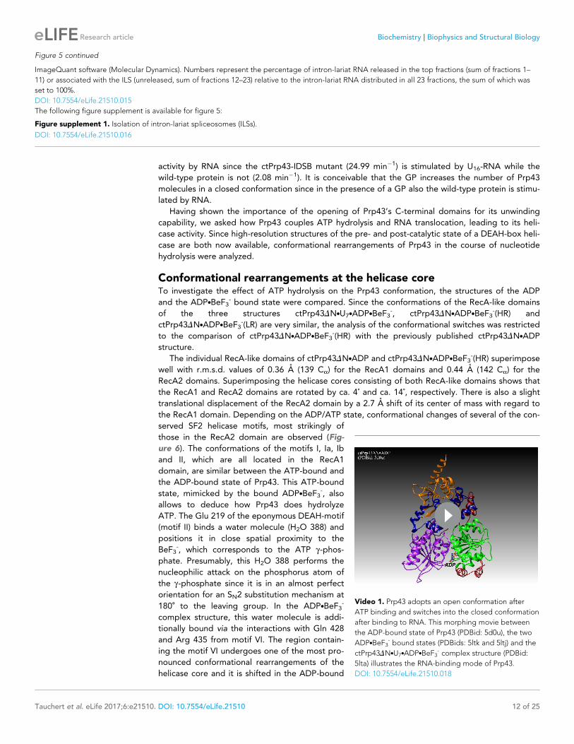
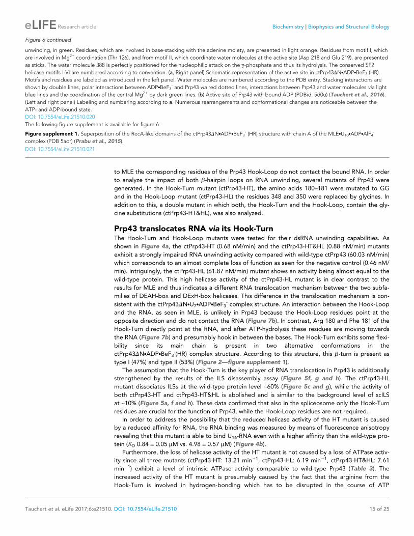
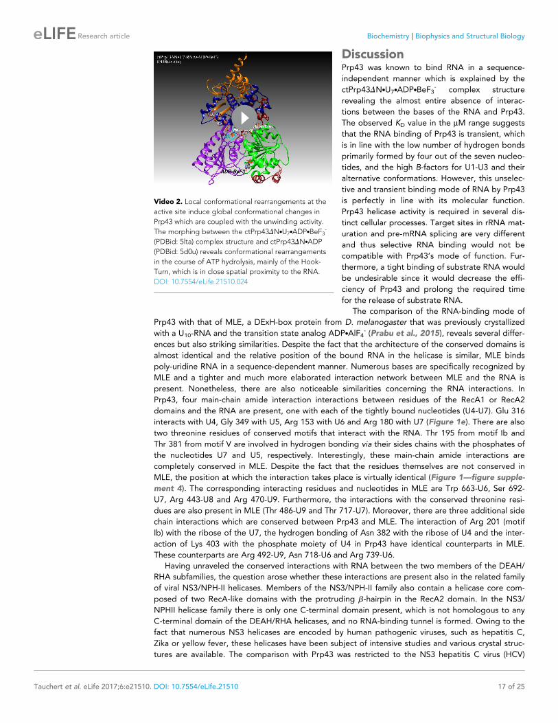
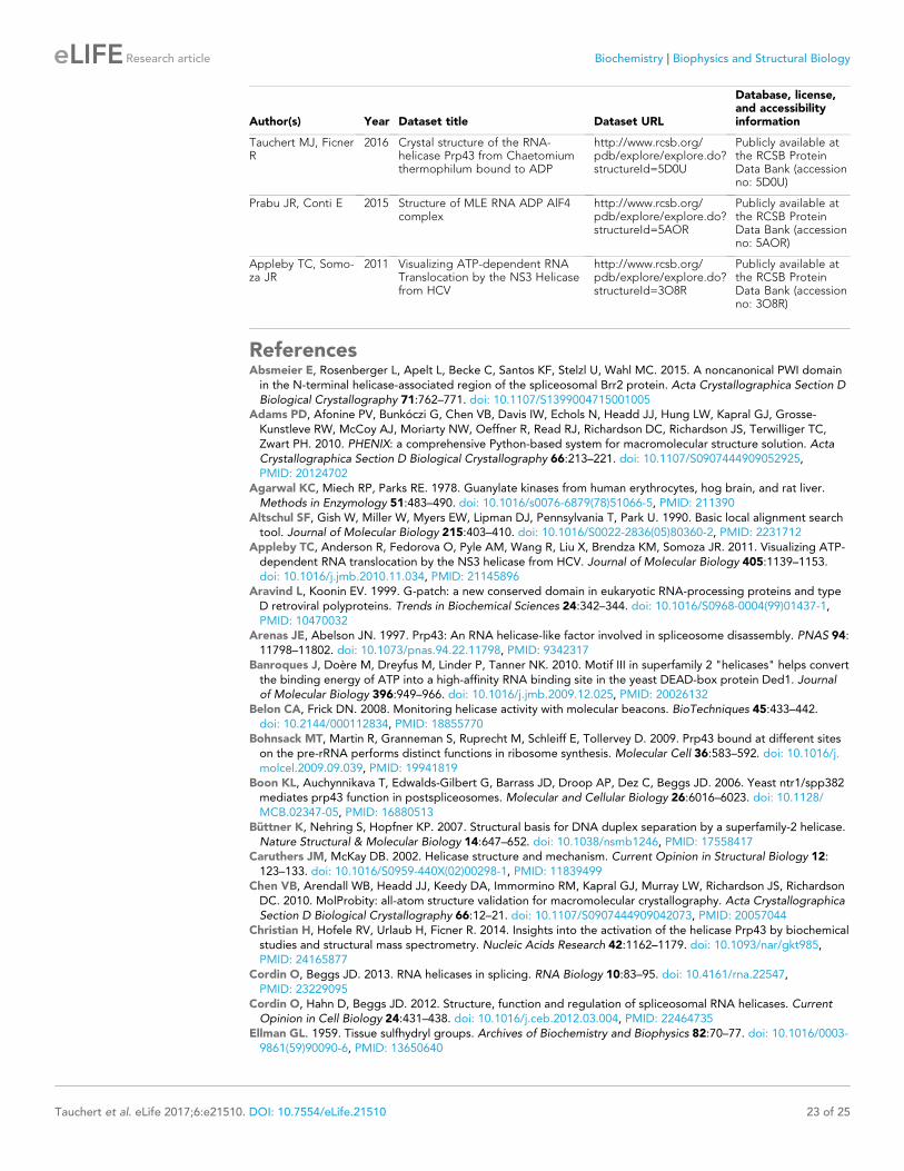

![On the Mechanism of Rifampicin Inhibition of RNA … Mechanism of Rifampicin Inhibition have also found that when the full length RNA transcript of the Pk promoter labeled with [o-32P]CTP](https://img.pdfslide.us/doc/110x75/5b1838ea7f8b9a37258baa04/on-the-mechanism-of-rifampicin-inhibition-of-rna-mechanism-of-rifampicin-inhibition.jpg)