Embed Size (px)
Citation preview
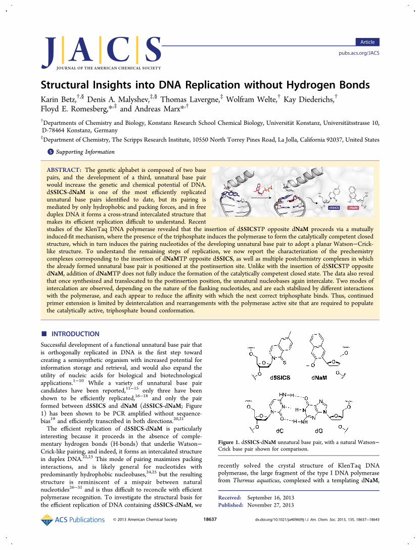
Structural Insights into DNA Replication without Hydrogen BondsKarin Betz,†,§ Denis A. Malyshev,‡,§ Thomas Lavergne,‡ Wolfram Welte,† Kay Diederichs,†
Floyd E. Romesberg,*,‡ and Andreas Marx*,†
†Departments of Chemistry and Biology, Konstanz Research School Chemical Biology, Universitat Konstanz, Universitatsstrasse 10,D-78464 Konstanz, Germany‡Department of Chemistry, The Scripps Research Institute, 10550 North Torrey Pines Road, La Jolla, California 92037, United States
*S Supporting Information
ABSTRACT: The genetic alphabet is composed of two basepairs, and the development of a third, unnatural base pairwould increase the genetic and chemical potential of DNA.d5SICS-dNaM is one of the most efficiently replicatedunnatural base pairs identified to date, but its pairing ismediated by only hydrophobic and packing forces, and in freeduplex DNA it forms a cross-strand intercalated structure thatmakes its efficient replication difficult to understand. Recentstudies of the KlenTaq DNA polymerase revealed that the insertion of d5SICSTP opposite dNaM proceeds via a mutuallyinduced-fit mechanism, where the presence of the triphosphate induces the polymerase to form the catalytically competent closedstructure, which in turn induces the pairing nucleotides of the developing unnatural base pair to adopt a planar Watson−Crick-like structure. To understand the remaining steps of replication, we now report the characterization of the prechemistrycomplexes corresponding to the insertion of dNaMTP opposite d5SICS, as well as multiple postchemistry complexes in whichthe already formed unnatural base pair is positioned at the postinsertion site. Unlike with the insertion of d5SICSTP oppositedNaM, addition of dNaMTP does not fully induce the formation of the catalytically competent closed state. The data also revealthat once synthesized and translocated to the postinsertion position, the unnatural nucleobases again intercalate. Two modes ofintercalation are observed, depending on the nature of the flanking nucleotides, and are each stabilized by different interactionswith the polymerase, and each appear to reduce the affinity with which the next correct triphosphate binds. Thus, continuedprimer extension is limited by deintercalation and rearrangements with the polymerase active site that are required to populatethe catalytically active, triphosphate bound conformation.
■ INTRODUCTION
Successful development of a functional unnatural base pair thatis orthogonally replicated in DNA is the first step towardcreating a semisynthetic organism with increased potential forinformation storage and retrieval, and would also expand theutility of nucleic acids for biological and biotechnologicalapplications.1−10 While a variety of unnatural base paircandidates have been reported,11−15 only three have beenshown to be efficiently replicated,16−18 and only the pairformed between d5SICS and dNaM (d5SICS-dNaM; Figure1) has been shown to be PCR amplified without sequence-bias19 and efficiently transcribed in both directions.20,21
The efficient replication of d5SICS-dNaM is particularlyinteresting because it proceeds in the absence of comple-mentary hydrogen bonds (H-bonds) that underlie Watson−Crick-like pairing, and indeed, it forms an intercalated structurein duplex DNA.22,23 This mode of pairing maximizes packinginteractions, and is likely general for nucleotides withpredominantly hydrophobic nucleobases,24,25 but the resultingstructure is reminiscent of a mispair between naturalnucleotides26−31 and is thus difficult to reconcile with efficientpolymerase recognition. To investigate the structural basis forthe efficient replication of DNA containing d5SICS-dNaM, we
recently solved the crystal structure of KlenTaq DNApolymerase, the large fragment of the type I DNA polymerasefrom Thermus aquaticus, complexed with a templating dNaM,
Received: September 16, 2013Published: November 27, 2013
Figure 1. d5SICS-dNaM unnatural base pair, with a natural Watson−Crick base pair shown for comparison.
Article
pubs.acs.org/JACS
© 2013 American Chemical Society 18637 dx.doi.org/10.1021/ja409609j | J. Am. Chem. Soc. 2013, 135, 18637−18643

and with or without bound d5SICSTP (KTQdNaM‑d5SICSTP andKTQdNaM, respectively).
22 The structures of these prechemistryd5SICSTP incorporation complexes revealed that the pairing ofd5SICSTP with dNaM drives the open-to-closed conforma-tional change characteristic of a natural base pair,32−34 andinterestingly, once in the closed environment, the pairingunnatural nucleotides adopt a planar, Watson−Crick-likegeometry.22 Thus, we demonstrated that not only is thepolymerase able to select for pairs that form a correct Watson−Crick structure, but at least with hydrophobic analogues it isable to enforce the correct structure. This mutually induced fitmechanism highlights what might be a fundamental advantageof using hydrophobicity and packing forces to mediatereplication, as they are sufficiently strong to mediate pairing,but also sufficiently plastic to adapt to the structure required bythe polymerase. However, the insertion of d5SICSTP oppositedNaM is a particularly efficient step of replication,16 and themechanism by which dNaMTP is inserted opposite d5SICSand the mechanism by which the primer containing eitherunnatural nucleotide is further elongated, which actually limitsreplication, remained unclear.Here, to fully characterize the mechanism of unnatural base
pair replication, we report the crystal structures of theprechemistry incorporation complexes leading to the insertionof dNaMTP opposite d5SICS: the binary complex of KlenTaqwith a DNA template containing d5SICS at the templatingposition (KTQd5SICS) and the corresponding ternary complexwith dNaMTP bound (KTQd5SICS‑dNaMTP). We also report thestructure of four postincorporation complexes: the binarycomplexes of KlenTaq and either a primer terminating withd5SICS paired opposite dNaM in a template (KTQdNaM‑d5SICS)in three different sequence contexts, or a primer terminatingwith dNaM paired opposite d5SICS (KTQd5SICS‑dNaM). Alongwith our previously reported structures, these structuresprovide key insights into the replication of the unnatural basepair and elucidate a mechanism that is based on a balance ofintercalation and deintercalation and structural rearrangementsof the polymerase active site.
■ RESULTS
Prechemistry dNaMTP Incorporation Complexes.KlenTaq was first crystallized bound to a DNA primer/template with d5SICS at the templating position (position n).In the resulting complex (KTQd5SICS; Figure 2A), the d5SICSnucleoside adopts an extrahelical position that is similar to thatobserved for a natural dG in KTQdG (PDB ID 3SZ2).22
However, compared to the previously described binarystructures KTQdNaM (PDB ID 3SYZ) and KTQdT (PDB ID3SV4), the single-stranded DNA of the template adopts adifferent arrangement (Figure S1, Supporting Information). Itis likely that the single-stranded portion of the template isflexible in the binary structures, and that the differences are notfunctionally relevant. Structural heterogeneity of the template isalso implied by the absence of well-defined electron density forthe d5SICS nucleobase.To determine whether the addition of dNaMTP to
KTQd5SICS drives the same conformational change observedupon d5SICSTP binding to KTQdNaM, we next determined thestructure of KTQd5SICS‑dNaMTP by soaking KTQd5SICS crystalswith dNaMTP. The structure of KTQd5SICS‑dNaMTP reveals thatthe unnatural triphosphate is bound to the O-helix (Figure 2B),which is rotated and only partially closed. The position of thetriphosphate appears to be stabilized by ionic interactions withArg659 and Lys663 of the O-helix, as well as with His639 of theN-helix and Arg587 from the N-terminal end of the thumbdomain K-helix. In addition, along with three water molecules,the triphosphate moiety coordinates a Mg2+ ion. The electrondensities for the sugar and the nucleobase moieties of dNaMTPare less well-defined than that for the triphosphate moiety,suggesting an increased level of disorder and/or flexibility. TheN- and O-helices of the fingers domain adopt a conformationintermediate between the open and closed states (Figure 2C)(the root-mean-square deviation (rmsd) of residues 637−700 is1.59 and 2.23 Å relative to KTQd5SICS and KTQdNaM‑d5SICSTP,respectively). In addition, Tyr671 is slightly displaced from itsopen conformation position in the insertion site (Figure S2),and the templating unnatural nucleobase moves from itsextrahelical position toward the insertion site, again representa-tive of a state intermediate between the open and closedconformations.
Figure 2. Open binary and precatalytic ternary complex of KTQd5SICS and KTQd5SICS‑dNaMTP, respectively. (A) The natural base pair at thepostinsertion site, the templating d5SICS, and Tyr671 are shown as sticks, and the O- and N-helices are shown as cartoon. Simulated annealingmFo-DFc omit map around d5SICS is shown, contoured at 3σ. (B) Same arrangement as in (A) but for the ternary complex KTQd5SICS‑dNaMTP.Simulated annealing mFo-DFc omit map around the bound dNaMTP and the coordinated Mg2+ ion (green sphere) and associated water molecules(red spheres) is shown, contoured at 3σ. (C) Superposition of KTQd5SICS (cyan), KTQd5SICS‑dNaMTP (orange), and KTQdNaM‑d5SICSTP (PDB ID 3SZ2,purple) shows the open, partially closed, and closed state of the enzyme.
Journal of the American Chemical Society Article
dx.doi.org/10.1021/ja409609j | J. Am. Chem. Soc. 2013, 135, 18637−1864318638

Postchemistry Extension Complexes. We next sought toinvestigate the structures of the postchemistry complexes, withthe unnatural base pair positioned in the postinsertion site,where it is poised for continued primer elongation (i.e.,extension of the unnatural base pair). We first characterized thestructure of KTQdNaM‑d5SICS with d5SICS at the primerterminus paired opposite dNaM at the n-1 position, withthree different primer/template complexes (E1−E3, Table 1).
In each binary complex, the polymerase adopts the expectedopen conformation, similar to that observed in KTQdNaM,KTQd5SICS, or KlenTaq bound to a fully natural primer/template.22 However, the presence of the unnatural base pairhas a significant effect on the structure of the primer/template.In the structure of KTQ(E1)dNaM‑d5SICS, the template dNaMcross-strand intercalates into the primer strand betweend5SICS and the 5′ dC (dCn‑2) (Figures 3 and S3). Toaccommodate this intercalation, relative to their positionsobserved with natural substrates, the C1′ of the primerunnatural nucleotide moves 4.7 Å toward the template andthe C1′ of the unnatural nucleotide in the template shifts 4.5 Åin the direction of translocation (Figure 4A and C). The extentof intercalation is evident by the sugar C1′−C1′ distance of 8.4Å, compared to the ∼10.5 Å distance that is typical for a naturalpair in the postinsertion site.22,33 This degree of intercalation iseven greater than in the free duplex, where the C1′−C1′distance is 9.1 Å,22 likely reflecting a decreased level ofstructural restraints when the unnatural base pair is positionedat the end of a duplex, as opposed to the middle. Intercalationalso positions the templating nucleobase proximal to the primerterminus, and the N1 and C2 amino group of dGn form H-bonds with the phosphate backbone of the primer (Figure S4).
Although perturbations are apparent with the n-2 and n-3template nucleotides, they are smaller, and the remainder of theenzyme-bound template is widely unperturbed, relative to itsfully natural counterpart.22 In contrast, at least minordistortions are apparent throughout the primer (Figure 4A).Examination of the polymerase reveals that, relative to its
open form, the presence of the unnatural base pair at thepostinsertion site induces the thumb domain to rotate, withhelices H, I, and K, which interact with the 3′ side of theprimer, moving closer to the active site, and helices H1 and H2,which interact with the 5′ side of the primer, moving away(Figure 4B). The position of the fingers domain is lessperturbed. The intercalated state appears to be accommodatedby a network of protein residues of the fingers domain,including Asn750, Tyr671, Gln754, and Glu615, which pack onthe free 3′ face of the d5SICS nucleobase (Figure S3), and theprimer terminus appears further stabilized by H-bonds betweenthe 3′ OH and phosphate backbone with His784 and Arg587,respectively. While the latter interaction is also observed with anatural substrate,22 the former is not (by analogy to thehomologous large fragment of polymerase I from Bacillusstearothermophilus (Bacillus fragment, BF), the 3′ OH of a fullynatural substrate forms an H-bond with Asp78535). Further-more, the sulfur atom of d5SICS engages in a water mediatedH-bond with Thr571. The phosphate of the template dNaMinteracts with Arg746 (Figure S3), and an analogous interactionis observed with a fully natural substrate.In the structure of KTQ(E2)dNaM‑d5SICS, the unnatural base
pair again forms via intercalation, but in this case, by the primerd5SICS nucleobase inserting between the template dNaM andits 3′ dG (dGn‑2) (Figure 3). The extent of intercalation appearssomewhat less than in KTQ(E1)dNaM‑d5SICS, with a C1′-C1′distance of 9.4 Å (compared to 8.4 Å). The overall structure ofthe primer/template is similar to that observed with KTQ-(E1)dNaM‑d5SICS, but despite the decreased intercalation, it issomewhat more distorted, with the C1′ of the primer unnaturalnucleotide moving 5.8 Å toward the template and the C1′ ofthe unnatural nucleotide in the template shifting 5.4 Å in thedirection of translocation, relative to their positions observedwith natural substrates (Figure 4A and D). In addition, dNaMshields the templating nucleobase from interacting with theprimer strand, and possibly as a result, neither the downstreamnucleotides nor Arg587 are well resolved in the KTQ-(E2)dNaM‑d5SICS structure. While the overall structure of thepolymerase is similar in KTQ(E2)dNaM‑d5SICS and KTQ-
Table 1. Primer/Template Sequences of PostchemistryComplexes Characterized
postchemistry complex primer/template sequence PDB ID
KTQ(E1)dNaM‑d5SICS 5′-ACC ACG GCG C 5SICS 4C8L3′-TGG TGC CGC G NaM GA
KTQ(E2)dNaM‑d5SICS 5′-GCC ACG GCG C 5SICS 4C8O3′-CGG TGC CGC G NaM CTT
KTQ(E2)d5SICS‑dNaM 5′-GCC ACG GCG C NaM 4C8M3′-CGG TGC CGC G 5SICS CTT
KTQ(E3)dNaM‑d5SICS 5′-ACC ACG GCG C 5SICS 4C8N3′-TGG TGC CGC G NaM GTT
Figure 3. Primer/template arrangement of open binany complexes with dNaM-d5SICS in the postinsertion site. KTQ(E1)dNaM‑d5SICS,KTQ(E2)dNaM‑d5SICS, and KTQ(E2)d5SICS‑dNaM are labeled and shown in green, blue, and red, respectively. The intercalated unnatural base pair isshown in dark green, dark blue, and pink, respectively, surrounded by their simulated annealing mFo-DFc omit maps contoured at 3σ. C1′−C1′distances (Å) within each unnatural pair are shown.
Journal of the American Chemical Society Article
dx.doi.org/10.1021/ja409609j | J. Am. Chem. Soc. 2013, 135, 18637−1864318639
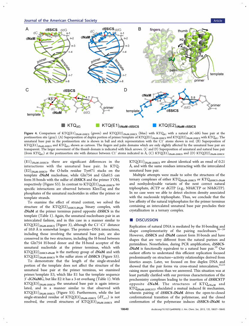
(E1)dNaM‑d5SICS, there are significant differences in theinteractions with the unnatural base pair. In KTQ-(E2)dNaM‑d5SICS, the O-helix residue Tyr671 stacks on thetemplate dNaM nucleobase, while Gln754 and Glu615 canform H-bonds with the sulfur of d5SICS and the primer 3′OH,respectively (Figure S3). In contrast to KTQ(E1)dNaM‑d5SICS, nospecific interactions are observed between KlenTaq and thephosphates of the unnatural nucleotides in either the primer ortemplate strands.To examine the effect of strand context, we solved the
structure of the KTQ(E2)d5SICS‑dNaM binary complex, withdNaM at the primer terminus paired opposite d5SICS in thetemplate (Table 1). Again, the unnatural nucleobases pair in anintercalated fashion, and in this case in a manner similar toKTQ(E2)dNaM‑d5SICS (Figure 3), although the C1′-C1′ distanceof 10.0 Å is somewhat longer. The protein−DNA interactions,including those involving the unnatural base pair, are alsoconserved in the two structures, including the H-bond betweenthe Gln754 H-bond donor and the H-bond acceptor of theunnatural nucleotide at the primer terminus, which withKTQ(E2)d5SICS‑dNaM is the methoxy group of dNaM and withKTQ(E2)dNaM‑d5SICS is the sulfur atom of d5SICS (Figure S3).To demonstrate that the length of the single-stranded
portion of the template does not affect the structure of theunnatural base pair at the primer terminus, we examinedprimer/template E3, which like E1 has the template sequence3′-dGNaMG, but like E2 it has a 3-nt overhang (Table 1). WithKTQ(E3)dNaM‑d5SICS, the unnatural base pair is again interca-lated, and in a manner similar to that observed withKTQ(E1)dNaM‑d5SICS (Figure S3). Furthermore, while the lastsingle-stranded residue of KTQ(E3)dNaM‑d5SICS (dTn+2) is notresolved, the overall structures of KTQ(E3)dNaM‑d5SICS and
KTQ(E1)dNaM‑d5SICS are almost identical with an rmsd of 0.21Å, and with the same residues interacting with the intercalatedunnatural base pair.Multiple attempts were made to solve the structures of the
ternary complexes of either KTQdNaM‑d5SICS or KTQd5SICS‑dNaMand nonhydrolyzable variants of the next correct naturaltriphosphate, dCTP or dGTP (e.g., NHdCTP or NHdGTP).In no case were we able to detect electron density associatedwith the nucleoside triphosphate. Thus, we conclude that thelow affinity of the natural triphosphates for the primer terminuscontaining an intercalated unnatural base pair precludes theircrystallization in a ternary complex.
■ DISCUSSION
Replication of natural DNA is mediated by the H-bonding andshape complementarity of the pairing nucleobases.36−39
However, d5SICS and dNaM cannot form H-bonds and haveshapes that are very different from the natural purines andpyrimidines. Nonetheless, during PCR amplification, d5SICS-dNaM is functionally equivalent to a natural base pair.19 Ourearliest efforts to understand this efficient replication focusedpredominantly on structure−activity relationships derived fromkinetics assays. Later, we focused on free duplex DNA andshowed that the pair forms via cross-strand intercalation,22,23
raising more questions than we answered. This situation was atleast partially clarified with our previous characterization of theprechemistry complexes leading to the insertion of d5SICSTPopposite dNaM . The structures of KTQdNaM andKTQdNaM‑d5SICSTP elucidated a mutual induced fit mechanism,wherein pairing of d5SICS-dNaM drives the open-to-closedconformational transition of the polymerase, and the closedconformation of the polymerase induces d5SICS-dNaM to
Figure 4. Comparison of KTQ(E1)dNaM‑d5SICS (green) and KTQ(E2)dNaM‑d5SICS (blue) with KTQdG with a natural dC-ddG base pair at thepostinsertion site (gray). (A) Superposition of duplex portion of primer/template of KTQ(E1)dNaM‑d5SICS and KTQ(E2)dNaM‑d5SICS with KTQdG. Theunnatural base pair in the postinsertion site is shown in ball and stick representation with the C1′ atoms shown in red. (B) Superposition ofKTQ(E1)dNaM‑d5SICS and KTQdG, shown as cartoon. The fingers and palm domains which are only slightly affected by the unnatural base pair aretransparent. The larger movement of the thumb domain is indicated with black arrows. (C and D) Superposition of unnatural and natural base pair(from KTQdG) at the postinsertion site with distance between C1′ atoms indicated in Å, (C) KTQ(E1)dNaM‑d5SICS and (D) KTQ(E2)dNaM‑d5SICS.
Journal of the American Chemical Society Article
dx.doi.org/10.1021/ja409609j | J. Am. Chem. Soc. 2013, 135, 18637−1864318640

adopt a Watson−Crick-like structure. With these results, ourattention turned to the mechanisms underlying the remainingsteps of replication, including the insertion of dNaMTPopposite d5SICS, and the subsequent continued primerelongation after incorporation of either unnatural triphosphate.Unlike with the addition of d5SICSTP to KTQdNaM,
22 theaddition of dNaMTP to KTQd5SICS did not induce thecanonical open-to-closed conformational change observedduring the synthesis of a natural base pair, but rather resultedin the formation of a structure wherein the DNA polymerasefingers domain remains in a partially open conformation andthe dNaMTP is bound via its triphosphate moiety to the O-helix. A similar conformation has been described by Beese andWu for BF polymerase with a dG-dTTP or a dG-ddTTPmismatch.40 In this structure, the polymerase remains in apartially open conformation, referred to as “ajar,” and thetemplating nucleotide displaces the “gate keeping” residue Y714(Y671 in KlenTaq) from the insertion site. It has beensuggested that this ajar conformation allows the DNApolymerase to test for complementarity between the incomingand templating nucleotides before the enzyme transitions to theclosed catalytically competent state. While Y761 remains in thetemplating position in KTQd5SICS‑dNaMTP, both it and thenucleobase of d5SICS appear strained toward the same switchobserved in the BF structure. A similar configuration has beenobserved with KlenTaq with an abasic site at the templatingposition.41,42 Thus, the KTQd5SICS‑dNaMTP complex appearstrapped in an intermediate state between the open binarycomplex and the ajar state observed with BF, similar to apartially closed state observed with the homologous E. colipolymerase I via biophysical studies.43,44 Regardless, it is clearthat incorporation of dNaMTP would require significantrearrangement of the polymerase to reach the catalyticallycompetent closed state, while KTQdNaM‑d5SICSTP spontaneouslyforms the catalytically competent closed complex. Thisdifference likely explains why the insertion of dNaMTPopposite d5SICS is often less efficient than the insertion ofd5SICSTP opposite dNaM.16
In all four postincorporation complexes characterized, thenucleobases pair in an intercalated manner, similar to theirpairing in free duplex DNA.23 However, two modes ofintercalation are observed. With primer/template complexes
E1 and E3, a common mode of intercalation is observed(Figures 3 and S3), which demonstrates that the mode ofpairing is unlikely to depend on the length of the singlestranded template. In this mode of intercalation, the templatedNaM inserts between its pairing d5SICS and the flankingdCn‑2, which allows for the template dGn to form stabilizinginteractions with the primer terminus (Figure S4). In contrast,in both complexes with primer/template E2, dCn is unable tomediate such interactions, and the intercalated structure isformed by insertion of the primer d5SICS (KTQ-(E2)dNaM‑d5SICS) or dNaM (KTQ(E2)d5SICS‑dNaM) between itspairing unnatural nucleobase and its flanking dGn‑2 of thetemplate, which likely optimizes packing interactions. Surpris-ingly, the mode of intercalation appears to depend most onsequence-specific interactions of the flanking nucleotides, withthe specific packing interactions between the intercalatingnucleobases being of secondary importance.Interestingly, the polymerase appears to be able to provide
unique stabilizing interactions to the two types of intercalatedstructures at the primer terminus. In the KTQ(E1)dNaM‑d5SICSand KTQ(E3)dNaM‑d5SICS structures, the observed intercalatedstate leaves one face of the primer d5SICS unpacked by aflanking nucleobase, and its position is stabilized by packinginteractions with Asn750, Tyr671, Gln754 and Glu615, an ionicinteraction between its phosphate and Arg587, a water-mediated H-bond between its sulfur and Thr571, and by anH-bond between its 3′OH and His784. The position of dNaMin the template is stabilized by an ionic interaction between itsphosphate and Arg746. In the KTQ(E2)dNaM‑d5SICS andKTQ(E2)d5SICS‑dNaM structures, the intercalated state adoptedleaves one face of the unnatural nucleobase in the template(dNaM and d5SICS, respectively) unpacked by a flankingnucleobase, and its position is stabilized by packing interactionswith O-helix residue Tyr671. In this case, the position of theprimer terminus can be stabilized by an H-bond between its3′OH and Gln615 and by an H-bond between Gln754 and theH-bond acceptor ortho to the glycosidic linkage (methoxy indNaM and sulfur in d5SICS). Unlike with E1 and E3, neitherthe template nor primer strand in either complex with E2 isstabilized via interactions with their backbone phosphates. Therather different interactions by which the two intercalatedstructures are accommodated reveals that the polymerase is
Figure 5. Proposed mechanism of replication. Intermediates not yet validated by structural studies (i.e., extension complexes) are shown in lightercolor. The steps corresponding to incorporation of the unnatural triphosphate and subsequent extension of the nascent unnatural base pair areindicated. The O-helix of the protein is shown, phosphates are indicated with open circles, natural nucleotides are indicated with open rectangles,and the unnatural nucleotides are indicated with gray and black rectangles.
Journal of the American Chemical Society Article
dx.doi.org/10.1021/ja409609j | J. Am. Chem. Soc. 2013, 135, 18637−1864318641

surprisingly plastic. Regardless, both structures of the post-insertion complexes require deintercalation and significantremodeling of the polymerase active site for incorporation ofthe next dNTP, likely explaining why structures with the nextcorrect natural triphosphate could not be obtained and alsowhy extension of the unnatural base pair is less efficient thanincorporation of the unnatural triphosphate.Based on this and previously reported structural
data,22,23,32,36,39,40 we propose the following mechanism ofreplication (Figure 5). The unnatural triphosphate initiallybinds to the O-helix, producing a flexible complex that samplesdifferent conformations, and when sufficiently stabilizinghydrophobic and packing interactions are made, the open-to-closed transition is induced, which induces the unnatural basepair to adopt a planar, Watson−Crick-like pairing, andincorporation of the triphosphate onto the growing primerterminus. With d5SICSTP incorporation, the intermediatestates are populated only transiently, and the closed complexmay only be captured by preventing incorporation with adideoxy primer terminus. However, with dNaMTP incorpo-ration, the series of conformational changes are halted at anajar-like state with the unnatural triphosphate remaining boundto the O-helix, due to either the stability of this complex or theinstability of the corresponding closed complex, and furtherprogress toward the incorporation of dNaMTP requiresthermal fluctuations to populate the closed state. Afterincorporation of either d5SICSTP or dNaMTP, the polymerasereturns to the open conformation and pyrophosphate isreleased.45 However, in this state, the unnatural base pairadopts a cross-strand intercalated structure, similar to thestructure it adopts in free duplex DNA, and continued primerelongation requires thermal fluctuations to both deintercalatethe unnatural base pair and reorganize the polymerase activesite. Because extension consistently limits the replication ofDNA containing the unnatural base pair, the model predictsthat further optimization of d5SICS-dNaM may be possible bymaking changes to the nucleobase analogues that decrease thestability of the intercalated structures. Efforts to test thishypothesis are currently underway.
■ MATERIALS AND METHODSOligonucleotide Synthesis. Natural oligonucleotides were
purchased from IDT (San Diego, CA). dNaM and d5SICSphosphoramidites and nucleosides were obtained from Berry &Associates Inc. (Dexter, MI), and the latter were phosphorylated usingLudwig and Eckstein conditions46 as described.16 Oligonucleotidescontaining an unnatural nucleotide were prepared using standardautomated DNA synthesis methodology with ultramild DNA synthesisphosphoramidites on CPG ultramild supports (1 μmol, GlenResearch; Sterling, VA) and an ABI Expedite 8905 synthesizer. Afterautomated synthesis, the DMT-ON oligonucleotide was first purifiedby Glen-Pak cartridge (Glen Research) and then by 8 M urea 20%PAGE, followed by Synergi Fusion-RP HPLC (Phenomenex,Torrance, CA) to single-band purity (>98%) using a linear gradientof 100 mM triethylammonium bicarbonate buffer (pH 7.5) andacetonitrile (5−30% over 35 min). The fractions containing purifiedoligonucleotides were collected and dried by vacuum centrifugation,and their identity was confirmed by MALDI-ToF with THAP matrix.Protein Production, Crystallization, and Structure Determi-
nation. KlenTaq was prepared using an E. coli codon-optimized geneencoding amino acids 293−832 of Taq polymerase (purchased fromGeneArt, Germany) cloned into the vector pGDR11 and expressed inE. coli strain BL21 (DE3) in LB medium for 4 h after induction with 1mM IPTG. The harvested cell pellet was resuspended in lysis buffer(50 mM Tris HCl pH 8.5, 10 mM MgCl2, 16 mM (NH4)2SO4 0.1%
TritonX-100, 0.1% hydroxypolyethoxydodecane, and 1 mM PMSF)and lysed by the addition of 0.5 mg/mL lysozyme and incubation for 1h at 37 °C. After lysis, a heat denaturation was performed (20 min, 80°C) and the cell debris was pelleted by ultracentrifugation (1 h, 35000g). Bacterial DNA in the supernatant was removed by PEI-precipitation and centrifugation. The resulting supernatant waspurified by anion exchange chromatography (Q Sepharose) in 20mM Tris HCl pH 8.5, 1 mM EDTA, 1 mM β-mercaptoethanol, elutingwith a NaCl gradient. Fractions containing KlenTaq were pooled,concentrated, and further purified by size-exclusion chromatography(Superdex 75) in 20 mM Tris HCl pH 7.5, 1 mM EDTA, 1 mM β-mercaptoethanol, 0.15 M NaCl.
Purified KlenTaq was stored at 4 °C. Primers and templates wereannealed prior to addition of protein and triphosphates. KlenTaq wasmixed with triphosphate and/or primer/template and incubated for 30min at 30 °C. The mixture was then filtered, and crystallizationconditions were screened using the sitting drop vapor diffusionmethod at 18 °C. Hits were reproduced using either the sitting orhanging drop vapor diffusion method. Prior to measurement, crystalswere flash frozen in liquid nitrogen either with or without cryoprotection (see the Supporting Information).
Data was collected at the beamline PXIII (XO6DA) and PXI(XO6SA) at the Swiss Light Source of the Paul Scherrer Institute inVilligen, Switzerland. Data reduction was performed with the XDSpackage.47 Statistics of data collection and refinement for all structuresare given in Table S1. Data was used in refinement up to a resolutionwith a CC1/2 value48 of around 50%. To facilitate comparison withother deposited structures, we also report resolution values at which1/σ = 2 (see Table S1). Data reduction of the KTQd5SICS andKTQd5SICS‑dNaMTP data was done in space group P3121 (for celldimensions, see Table S1), and the structures were solved by rigid-body refinement using a previously published KlenTaq structure(PDB: 3M8S49) as a model. All binary elongation complexes(KTQ(E1), KTQ(E2), and KTQ(E3)) crystallized in space groupC2221 with similar cell dimensions (see Table S1). The KTQ(E1)complex was solved by molecular replacement using the binaryKlenTaq structure 3SZ222 as a search model. The KTQ(E2) andKTQ(E3) structures were solved by rigid body refinement againstKTQ(E1). All structures were improved by refinement in PHENIX50
and model building in COOT.51 During refinement, structures wereevaluated using the MolProbity server.52 Restraint files of dNaM,d5SICS, and dNaMTP for refinement were created using the gradeWeb Server.53 Figures were created with PyMOL.54
■ ASSOCIATED CONTENT*S Supporting InformationSupporting table and figures, crystallization conditions, andoligonucleotide sequences. This material is available free ofcharge via the Internet at http://pubs.acs.org.
■ AUTHOR INFORMATIONCorresponding [email protected]@uni-konstanz.deAuthor Contributions§K.B. and D.A.M.: These authors contributed equally.NotesThe authors declare no competing financial interest.
■ ACKNOWLEDGMENTSWe thank the beamline staff of the Swiss Light Source at thePaul Scherrer Institute for their assistance during datacollection. We thank Dr. Phillip Ordoukhanian for assistancewith oligonucleotide synthesis. This work was supported by theNational Institutes of Health (GM 060005 to F.E.R.) and bythe Konstanz Research School Chemical Biology.
Journal of the American Chemical Society Article
dx.doi.org/10.1021/ja409609j | J. Am. Chem. Soc. 2013, 135, 18637−1864318642

■ REFERENCES(1) Collins, M. L.; Irvine, B.; Tyner, D.; Fine, E.; Zayati, C.; Chang,C.; Horn, T.; Ahle, D.; Detmer, J.; Shen, L. P.; Kolberg, J.; Bushnell,S.; Urdea, M. S.; Ho, D. D. Nucleic Acids Res. 1997, 25, 2979−2984.(2) Johnson, S. C.; Marshall, D. J.; Harms, G.; Miller, C. M.; Sherrill,C. B.; Beaty, E. L.; Lederer, S. A.; Roesch, E. B.; Madsen, G.; Hoffman,G. L.; Laessig, R. H.; Kopish, G. J.; Baker, M. W.; Benner, S. A.; Farrell,P. M.; Prudent, J. R. Clin. Chem. 2004, 50, 2019−2027.(3) Arens, M. Q.; Buller, R. S.; Rankin, A.; Mason, S.; Whetsell, A.;Agapov, E.; Lee, W. M.; Storch, G. A. J. Clin. Microbiol. 2010, 48,2387−2395.(4) Lee, W. M.; Grindle, K.; Pappas, T.; Marshall, D. J.; Moser, M. J.;Beaty, E. L.; Shult, P. A.; Prudent, J. R.; Gern, J. E. J. Clin. Microbiol.2007, 45, 2626−2634.(5) Kimoto, M.; Yamashige, R.; Matsunaga, K.; Yokoyama, S.; Hirao,I. Nat. Biotechnol. 2013, 31, 453−457.(6) Hollenstein, M.; Hipolito, C. J.; Lam, C. H.; Perrin, D. M. NucleicAcids Res. 2009, 37, 1638−1649.(7) Keefe, A. D.; Cload, S. T. Curr. Opin. Chem. Biol. 2008, 12, 448−456.(8) Seeman, N. C. Annu. Rev. Biochem. 2010, 79, 65−87.(9) Wang, H.; Yang, R.; Yang, L.; Tan, W. ACS Nano 2009, 3, 2451−2460.(10) Chen, T.; Shukoor, M. I.; Chen, Y.; Yuan, Q.; Zhu, Z.; Zhao, Z.;Gulbakan, B.; Tan, W. Nanoscale 2011, 3, 546−556.(11) Chelliserrykattil, J.; Lu, H.; Lee, A. H.; Kool, E. T.ChemBioChem 2008, 9, 2976−2980.(12) Brotschi, C.; Mathis, G.; Leumann, C. J. Chem.Eur. J. 2005,11, 1911−1923.(13) Kaul, C.; Muller, M.; Wagner, M.; Schneider, S.; Carell, T. Nat.Chem. 2011, 3, 794−800.(14) Meggers, E.; Holland, P. L.; Tolman, W. B.; Romesberg, F. E.;Schultz, P. G. J. Am. Chem. Soc. 2000, 122, 10714−10715.(15) Minakawa, N.; Ogata, S.; Takahashi, M.; Matsuda, A. J. Am.Chem. Soc. 2009, 131, 1644−1645.(16) Lavergne, T.; Malyshev, D. A.; Romesberg, F. E. Chem.Eur. J.2012, 18, 1231−1239.(17) Yamashige, R.; Kimoto, M.; Takezawa, Y.; Sato, A.; Mitsui, T.;Yokoyama, S.; Hirao, I. Nucleic Acids Res. 2012, 40, 2793−2806.(18) Yang, Z.; Chen, F.; Alvarado, J. B.; Benner, S. A. J. Am. Chem.Soc. 2011, 133, 15105−15112.(19) Malyshev, D. A.; Dhami, K.; Quach, H. T.; Lavergne, T.;Ordoukhanian, P.; Torkamani, A.; Romesberg, F. E. Proc. Natl. Acad.Sci. U.S.A. 2012, 109, 12005−12010.(20) Seo, Y. J.; Matsuda, S.; Romesberg, F. E. J. Am. Chem. Soc. 2009,131, 5046−5047.(21) Seo, Y. J.; Malyshev, D. A.; Lavergne, T.; Ordoukhanian, P.;Romesberg, F. E. J. Am. Chem. Soc. 2011, 133, 19878−19888.(22) Betz, K.; Malyshev, D. A.; Lavergne, T.; Welte, W.; Diederichs,K.; Dwyer, T. J.; Ordoukhanian, P.; Romesberg, F. E.; Marx, A. Nat.Chem. Biol. 2012, 8, 612−614.(23) Malyshev, D. A.; Pfaff, D. A.; Ippoliti, S. I.; Hwang, G. T.;Dwyer, T. J.; Romesberg, F. E. Chem.Eur. J. 2010, 16, 12650−12659.(24) Johar, Z.; Zahn, A.; Leumann, C. J.; Jaun, B. Chem.Eur. J.2008, 14, 1080−1086.(25) Matsuda, S.; Fillo, J. D.; Henry, A. A.; Rai, P.; Wilkens, S. J.;Dwyer, T. J.; Geierstanger, B. H.; Wemmer, D. E.; Schultz, P. G.;Spraggon, G.; Romesberg, F. E. J. Am. Chem. Soc. 2007, 129, 10466−10473.(26) Krahn, J. M.; Beard, W. A.; Wilson, S. H. Structure 2004, 12,1823−1832.(27) Chou, S. H.; Chin, K. H.; Wang, A. H. Nucleic Acids Res. 2003,31, 2461−2474.(28) Chou, S. H.; Zhu, L.; Reid, B. R. J. Mol. Biol. 1994, 244, 259−268.(29) Shepard, W.; Cruse, W. B.; Fourme, R.; de la Fortelle, E.;Prange, T. Structure 1998, 6, 849−861.
(30) Spackova, N. A.; Berger, I.; Sponer, J. J. Am. Chem. Soc. 2000,122, 7564−7572.(31) Sunami, T.; Kondo, J.; Hirao, I.; Watanabe, K.; Miura, K. I.;Takenaka, A. Acta Crystallogr., Sect. D: Biol. Crystallogr. 2004, 60, 90−96.(32) Rothwell, P. J.; Waksman, G. Adv. Protein Chem. 2005, 71, 401−440.(33) Li, Y.; Korolev, S.; Waksman, G. EMBO J. 1998, 17, 7514−7525.(34) Doublie, S.; Tabor, S.; Long, A. M.; Richardson, C. C.;Ellenberger, T. Nature 1998, 391, 251−258.(35) Kiefer, J. R.; Mao, C.; Braman, J. C.; Beese, L. S. Nature 1998,391, 304−307.(36) Echols, H.; Goodman, M. F. Annu. Rev. Biochem. 1991, 60, 477−511.(37) Kool, E. T. Annu. Rev. Biochem. 2002, 71, 191−219.(38) Goodman, M. F. Proc. Natl. Acad. Sci. U.S.A. 1997, 94, 10493−10495.(39) Kunkel, T. A. J. Biol. Chem. 2004, 279, 16895−16898.(40) Wu, E. Y.; Beese, L. S. J. Biol. Chem. 2011, 286, 19758−19767.(41) Obeid, S.; Welte, W.; Diederichs, K.; Marx, A. J. Biol. Chem.2012, 287, 14099−14108.(42) Obeid, S.; Blattner, N.; Kranaster, R.; Schnur, A.; Diederichs, K.;Welte, W.; Marx, A. EMBO J. 2010, 29, 1738−1747.(43) Hohlbein, J.; Aigrain, L.; Craggs, T. D.; Bermek, O.; Potapova,O.; Shoolizadeh, P.; Grindley, N. D. F.; Joyce, C. M.; Kapanidis, A. N.Nat. Commun. 2013, 4, 2131.(44) Berezhna, S. Y.; Gill, J. P.; Lamichhane, R.; Millar, D. P. J. Am.Chem. Soc. 2012, 134, 11261−11268.(45) Golosov, A. A.; Warren, J. J.; Beese, L. S.; Karplus, M. Structure2010, 18, 83−93.(46) Ludwig, J.; Eckstein, F. J. Org. Chem. 1989, 54, 631−635.(47) Kabsch, W. Acta Crystallgr., Sect. D: Biol. Cyrstallogr. 2010, 66,125−132.(48) Karplus, P. A.; Diederichs, K. Science 2012, 336, 1030−1033.(49) Betz, K.; Streckenbach, F.; Schnur, A.; Exner, T.; Welte, W.;Diederichs, K.; Marx, A. Angew. Chem., Int. Ed. 2010, 49, 5181−5184.(50) Adams, P. D.; Afonine, P. V.; Bunkoczi, G.; Chen, V. B.; Davis,I. W.; Echols, N.; Headd, J. J.; Hung, L.-W.; Kapral, G. J.; Grosse-Kunstleve, R. W.; McCoy, A. J.; Moriarty, N. W.; Oeffner, R.; Read, R.J.; Richardson, D. C.; Richardson, J. S.; Terwilliger, T. C.; Zwart, P. H.Acta Crystallogr., Sect. D: Biol. Crystallogr. 2010, 66, 213−221.(51) Emsley, P.; Lohkamp, B.; Scott, W. G.; Cowtan, K. ActaCrystallogr., Sect. D: Biol. Crystallogr. 2010, 66, 486−501.(52) Chen, V. B.; Arendall, W. B., 3rd; Headd, J. J.; Keedy, D. A.;Immormino, R. M.; Kapral, G. J.; Murray, L. W.; Richardson, J. S.;Richardson, D. C. Acta Crystallogr., Sect. D: Biol. Crystallogr. 2010, 66,12−21.(53) Smart, O. S.; Womack, T. O.; Sharff, A.; Flensburg, C.; Keller,P.; Paciorek, W.; Vonrhein, C.; Bricogne, G. grade, version 1.2.2;Global Phasing Limited: Cambridge, U.K., 2011; http://www.globalphasing.com.(54) DeLano, W. L. The PyMOL Molecular Graphics System;Schrodinger, LLC: San Carlos, CA, 2002; http://www.pymol.org.
Journal of the American Chemical Society Article
dx.doi.org/10.1021/ja409609j | J. Am. Chem. Soc. 2013, 135, 18637−1864318643