Embed Size (px)
Citation preview

Structural dynamics inside a functionalizedmetal–organic framework probed by ultrafast 2DIR spectroscopyJun Nishidaa, Amr Tamimia, Honghan Feib, Sonja Pullenc, Sascha Ottc, Seth M. Cohenb,1, and Michael D. Fayera,1
aDepartment of Chemistry, Stanford University, Stanford, CA 94305; bDepartment of Chemistry and Biochemistry, University of California, San Diego, CA92093; and cDepartment of Chemistry, Ångström Laboratory, Uppsala University, 75120 Uppsala, Sweden
Contributed by Michael D. Fayer, November 20, 2014 (sent for review November 4, 2014)
The structural elasticity of metal–organic frameworks (MOFs) isa key property for their functionality. Here, we show that 2D IRspectroscopy with pulse-shaping techniques can probe the ultrafaststructural fluctuations of MOFs. 2D IR data, obtained from a vibra-tional probe attached to the linkers of UiO-66 MOF in low concen-tration, revealed that the structural fluctuations have time constantsof 7 and 670 ps with no solvent. Filling the MOF pores with dime-thylformamide (DMF) slows the structural fluctuations by reducingthe ability of the MOF to undergo deformations, and the dynamicsof the DMF molecules are also greatly restricted. Methodologyadvances were required to remove the severe light scatteringcaused by the macroscopic-sizedMOF particles, eliminate interferingoscillatory components from the 2D IR data, and address Förstervibrational excitation transfer.
2D IR spectroscopy | metal–organic framework | UiO-66 MOF |ultrafast structural fluctuations | solvent confinement effect
Metal–organic frameworks (MOFs) are molecular archi-tectures in which metal clusters are connected by organic
linkers to yield relatively regular 3D coordination polymers withnanometer-sized pores (1, 2). MOFs have been investigated fora wide variety of chemical applications, such as adsorption ofgases (3) and heterogeneous catalysts (4). Among many types ofporous materials, MOFs are unique because of their structuralelasticity coexisting with a high degree of spatial regularity. Insome sense, MOFs are like crystals and polymers. They haverelatively regular structures like a crystal, but the metal clustersare joined by organic linkers that, in some systems, producesignificant structural mobility. The elasticity of MOFs is in-timately related to their physical properties and behavior (5).An important goal for understanding the nature of MOFs and
how their chemical composition influences their properties andapplications is to develop and apply an experimental methodthat can measure the ultrafast structural motions of MOFs. Therelatively slow motions of the framework occurring in sub-microsecond to millisecond scales have been studied by NMRand neutron scattering (6). However, until now, quantitativemeasurements that can characterize the time dependence ofMOF structural motions in ultrafast regimes have not beenpossible because of the lack of appropriate techniques. Here,we have accomplished the goal by applying ultrafast 2D IRspectroscopy (7) to the study of MOF structural dynamics. 2D IRis akin to 2D NMR, but it operates on the ultrafast timescalesnecessary to characterize the time dependence of MOF struc-tural motions. In addition, there is the important question of theeffects on MOF dynamics when guest molecules fill the MOFnanopores. The guest molecules will affect the structural fluc-tuations of the framework by interacting with the linkers and themetal units. Furthermore, because of the confinement of guestmolecules in MOF nanopores, the dynamics of these moleculesare expected to be very different from their bulk liquid behavior(8). These issues can also be addressed with 2D IR spectroscopy.
The line shape observed in the FTIR absorption spectrum isrelated to the variety of environments that a vibrational probeexperiences. The different environments produce a range ofvibrational frequencies; this range of frequencies is called in-homogeneous broadening. Dynamics cause environments andtherefore vibrational frequencies to change over time, butthe time dependence of structural evolution cannot be ex-tracted from the line shape. 2D IR experiments make thetime evolution of the frequency of the vibrational probes andthus the time evolution of the structure a direct observable(9). The time-dependent evolution of the frequency is calledspectral diffusion.A useful way of thinking about the influence of environmental
interactions on the frequencies of vibrational probe molecules isin terms of the electric field produced by the vibrational probes’surroundings (10). The atoms and molecular groups makingup the environments around the vibrational probes have partialelectrical charges. These partial charges produce a resultantelectric field experienced by the probe vibration. The strengthand direction of the electric field affect the vibrational frequencythrough the Stark effect. The vibrational probes that we studywith 2D IR are chemically introduced into the MOFs. BecauseMOFs are not perfect crystals, the environment (electric field)experienced by different vibrational probe molecules will differ.Therefore, there will be a range of vibrational frequencies (theinhomogeneous broadening) that reflect the range of environ-ments. The MOF structure is not static, and the metal clustersand the linkers are constantly moving. Relative to the vibrationalprobe, the distances and orientations of the various components
Significance
A unique aspect of metal–organic frameworks (MOFs) is theirstructural flexibility coexisting with a degree of regularity.Adsorbed guest molecules can cause MOF pore shapes to de-form. Pore shape changes may be related to the high capacityand selectivity of the MOFs for gas adsorption and other pro-cesses. MOF flexibility and other properties are influencedby fast dynamics of the framework. Direct measurements tocharacterize fast motions of the MOFs have not been appliedpreviously. We show that 2D IR spectroscopy can be performedon functionalized MOFs. We use 2D IR and other ultrafast IRmethods to elucidate the timescales for ultrafast structuralfluctuations and how they are influenced by a solvent fillingthe pores.
Author contributions: J.N., S.O., S.M.C., and M.D.F. designed research; J.N., A.T., H.F., andS.P. performed research; H.F., S.P., S.O., and S.M.C. contributed new reagents; J.N. andM.D.F.analyzed data; and J.N. and M.D.F. wrote the paper.
The authors declare no conflict of interest.1To whom correspondence may be addressed. Email: [email protected] or [email protected].
This article contains supporting information online at www.pnas.org/lookup/suppl/doi:10.1073/pnas.1422194112/-/DCSupplemental.
18442–18447 | PNAS | December 30, 2014 | vol. 111 | no. 52 www.pnas.org/cgi/doi/10.1073/pnas.1422194112
in the framework that give rise to the electric field are changing.Thus, the frequency of each vibrational probe is time-dependent,and the time dependence of the frequencies of the vibrationalprobes (spectral diffusion) reflects the time dependence of thestructural motions of the MOFs.Recently, Pullen et al. (4) showed that a di-iron carbonyl complex
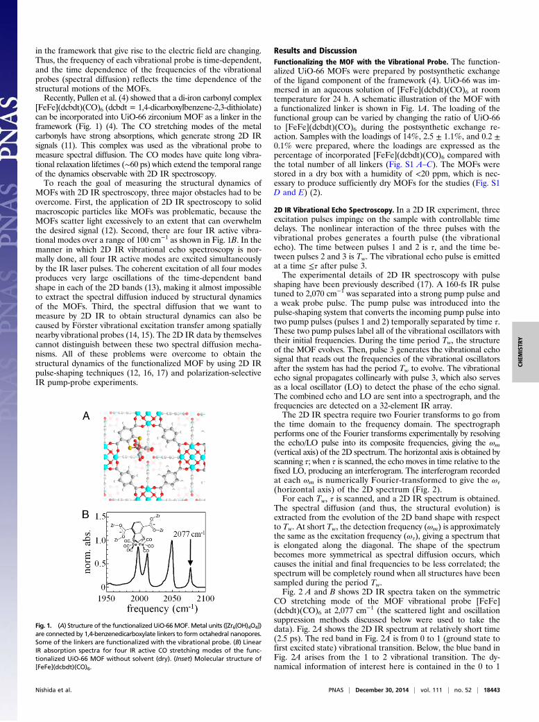
[FeFe](dcbdt)(CO)6 (dcbdt = 1,4-dicarboxylbenzene-2,3-dithiolate)can be incorporated into UiO-66 zirconium MOF as a linker in theframework (Fig. 1) (4). The CO stretching modes of the metalcarbonyls have strong absorptions, which generate strong 2D IRsignals (11). This complex was used as the vibrational probe tomeasure spectral diffusion. The CO modes have quite long vibra-tional relaxation lifetimes (∼60 ps) which extend the temporal rangeof the dynamics observable with 2D IR spectroscopy.To reach the goal of measuring the structural dynamics of
MOFs with 2D IR spectroscopy, three major obstacles had to beovercome. First, the application of 2D IR spectroscopy to solidmacroscopic particles like MOFs was problematic, because theMOFs scatter light excessively to an extent that can overwhelmthe desired signal (12). Second, there are four IR active vibra-tional modes over a range of 100 cm−1 as shown in Fig. 1B. In themanner in which 2D IR vibrational echo spectroscopy is nor-mally done, all four IR active modes are excited simultaneouslyby the IR laser pulses. The coherent excitation of all four modesproduces very large oscillations of the time-dependent bandshape in each of the 2D bands (13), making it almost impossibleto extract the spectral diffusion induced by structural dynamicsof the MOFs. Third, the spectral diffusion that we want tomeasure by 2D IR to obtain structural dynamics can also becaused by Förster vibrational excitation transfer among spatiallynearby vibrational probes (14, 15). The 2D IR data by themselvescannot distinguish between these two spectral diffusion mecha-nisms. All of these problems were overcome to obtain thestructural dynamics of the functionalized MOF by using 2D IRpulse-shaping techniques (12, 16, 17) and polarization-selectiveIR pump-probe experiments.
Results and DiscussionFunctionalizing the MOF with the Vibrational Probe. The function-alized UiO-66 MOFs were prepared by postsynthetic exchangeof the ligand component of the framework (4). UiO-66 was im-mersed in an aqueous solution of [FeFe](dcbdt)(CO)6 at roomtemperature for 24 h. A schematic illustration of the MOF witha functionalized linker is shown in Fig. 1A. The loading of thefunctional group can be varied by changing the ratio of UiO-66to [FeFe](dcbdt)(CO)6 during the postsynthetic exchange re-action. Samples with the loadings of 14%, 2.5 ± 1.1%, and 0.2 ±0.1% were prepared, where the loadings are expressed as thepercentage of incorporated [FeFe](dcbdt)(CO)6 compared withthe total number of all linkers (Fig. S1 A–C). The MOFs werestored in a dry box with a humidity of <20 ppm, which is nec-essary to produce sufficiently dry MOFs for the studies (Fig. S1D and E) (2).
2D IR Vibrational Echo Spectroscopy. In a 2D IR experiment, threeexcitation pulses impinge on the sample with controllable timedelays. The nonlinear interaction of the three pulses with thevibrational probes generates a fourth pulse (the vibrationalecho). The time between pulses 1 and 2 is τ, and the time be-tween pulses 2 and 3 is Tw. The vibrational echo pulse is emittedat a time ≤τ after pulse 3.The experimental details of 2D IR spectroscopy with pulse
shaping have been previously described (17). A 160-fs IR pulsetuned to 2,070 cm−1 was separated into a strong pump pulse anda weak probe pulse. The pump pulse was introduced into thepulse-shaping system that converts the incoming pump pulse intotwo pump pulses (pulses 1 and 2) temporally separated by time τ.These two pump pulses label all of the vibrational oscillators withtheir initial frequencies. During the time period Tw, the structureof the MOF evolves. Then, pulse 3 generates the vibrational echosignal that reads out the frequencies of the vibrational oscillatorsafter the system has had the period Tw to evolve. The vibrationalecho signal propagates collinearly with pulse 3, which also servesas a local oscillator (LO) to detect the phase of the echo signal.The combined echo and LO are sent into a spectrograph, and thefrequencies are detected on a 32-element IR array.The 2D IR spectra require two Fourier transforms to go from
the time domain to the frequency domain. The spectrographperforms one of the Fourier transforms experimentally by resolvingthe echo/LO pulse into its composite frequencies, giving the ωm(vertical axis) of the 2D spectrum. The horizontal axis is obtained byscanning τ; when τ is scanned, the echo moves in time relative to thefixed LO, producing an interferogram. The interferogram recordedat each ωm is numerically Fourier-transformed to give the ωτ
(horizontal axis) of the 2D spectrum (Fig. 2).For each Tw, τ is scanned, and a 2D IR spectrum is obtained.
The spectral diffusion (and thus, the structural evolution) isextracted from the evolution of the 2D band shape with respectto Tw. At short Tw, the detection frequency (ωm) is approximatelythe same as the excitation frequency (ωτ), giving a spectrum thatis elongated along the diagonal. The shape of the spectrumbecomes more symmetrical as spectral diffusion occurs, whichcauses the initial and final frequencies to be less correlated; thespectrum will be completely round when all structures have beensampled during the period Tw.Fig. 2 A and B shows 2D IR spectra taken on the symmetric
CO stretching mode of the MOF vibrational probe [FeFe](dcbdt)(CO)6 at 2,077 cm−1 (the scattered light and oscillationsuppression methods discussed below were used to take thedata). Fig. 2A shows the 2D IR spectrum at relatively short time(2.5 ps). The red band in Fig. 2A is from 0 to 1 (ground state tofirst excited state) vibrational transition. Below, the blue band inFig. 2A arises from the 1 to 2 vibrational transition. The dy-namical information of interest here is contained in the 0 to 1
Fig. 1. (A) Structure of the functionalized UiO-66MOF.Metal units ([Zr6(OH)4O4])are connected by 1,4-benzenedicarboxylate linkers to form octahedral nanopores.Some of the linkers are functionalized with the vibrational probe. (B) LinearIR absorption spectra for four IR active CO stretching modes of the func-tionalized UiO-66 MOF without solvent (dry). (Inset) Molecular structure of[FeFe](dcbdt)(CO)6.
Nishida et al. PNAS | December 30, 2014 | vol. 111 | no. 52 | 18443
CHEM
ISTR
Y

band. The dashed line through the data in Fig. 2A is the di-agonal. At short time, the spectrum is elongated along the di-agonal, because there has been little spectral diffusion. Fig. 2Bshows the spectrum at long time (100 ps). It is clear that theshape has changed substantially and the band is almost round,indicating that diffusion is almost complete by 100 ps.To quantitatively extract the dynamical information, the cen-
ter line slope (CLS) method was used (9). The CLS analysisprovides a decay curve that contains the time constants andrelative amplitudes of the components of the spectral diffusion.The CLS has been theoretically shown to be proportional to thefrequency–frequency correlation function (FFCF). The absoluteFFCF can be obtained from the CLS and the linear absorptionspectrum as previously described (9).
Scattering Removal and Coherent Oscillation Suppression Methods.As mentioned above, one of the main obstacles to performing2D IR experiments on an MOF powder is severe light scattering.The dominant interference with the signal is scatter from thepump pulses that is heterodyned by the probe beam. Shim et al.(12) showed that such contamination can be removed by a four-shot phase-cycling scheme. For a given τ, the phases of the twopump pulses (pulses 1 and 2) are varied to have relative phases(0, 0), (0, π), (π, 0), and (π, π). This approach is useful for a samplethat produces reasonably small amounts of scattered light.We applied the four-shot phase-cycling sequence to the
functionalized UiO-66 MOF dry powder to probe the symmetricstretching mode at 2,077 cm−1 (Fig. S2). The standard polar-izations (<XXXX>) were used, in which the three input pulsesand the detected echo pulse all had the same horizontal polar-izations. This polarization scheme produces the largest signal.The result is shown in Fig. 2C. Scattered light is manifested asa large amplitude band along the diagonal that is overwhelming
a resonant signal from the vibrational probe. As discussed in SIText, the four-shot phase cycle does not eliminate scattered lightfrom pulse 1 heterodyning with scattered light from pulse 2. Toeliminate this problem, the pulse sequence is changed in twoways. First, the pump pulses 1 and 2 polarizations are rotated tovertical. Although this polarization scheme (<XXYY>) reducesthe echo signal by a factor of ∼3 (18), it eliminates the con-tamination from the scattered pump beams by a factor of >300.Second, an eight-shot cycle is used, in which pulse 3, the probepulse, is chopped every other shot. The 2D IR spectra in Fig. 2 Aand B were both taken with this scattering removal scheme. Thescattered light artifacts are eliminated, which permits the accu-rate determination of the time evolution of the 2D IR spectrum.The same polarizations and phase-cycling pulse sequence wasrecently used in a different application (2D IR microscopy) (19).With the scattered light artifacts eliminated, we can take high-
quality 2D IR data, which are analyzed and plotted as CLSdecays. The second major problem is the broad spectrum of theshort pulses that excite all four IR active modes of the vibrationalprobe (Fig. 1B). Fig. 3A shows the spectrum of pump pulses 1and 2 (Fig. 3A, black curve). Fig. 3B shows the first few pico-seconds of the CLS data taken with the full pump spectrum (Fig.3B, black circles and dashed curve). The large oscillations arecaused by the coherent excitation of all four modes (Figs. S3 andS4) (13). The oscillations continue well past 10 ps, which inter-feres with the extraction of the CLS decay measurement ofspectral diffusion. The Fourier transform of these oscillationsgives the modes’ frequency splittings (Fig. S3C).To eliminate the oscillations, the pulse shaper was used to
modify the spectrum of the IR pulses 1 and 2 as shown as the redcurve in Fig. 3A. The change in the spectrum reduces the timeresolution of the experiments, but the resulting time resolutionis still more than sufficient to measure the spectral diffusion (SIText and Fig. S5). By using the modified spectrum, the symmetric
ωτ (cm-1)
Tw= 2.5ps
Tw= 100ps
2070
2075
2080
ωm(cm-1)
2070
2075
2080
2070
2075
2080
2070 2075 2080
<XXXX>+ 4 shots cycle
A
B
C
Fig. 2. (A and B) 2D IR spectra of the functionalized UiO-66 MOF (14%loading) at short time and long time taken with the scattering removal andthe selective pumping schemes discussed in the text. The dashed line inA is thediagonal. The dynamical information is contained in the Tw-dependent shapesof the 2D IR spectra. (C) Data taken with standard polarizations and four-shotsphase cycling. The spectrum is completely dominated by scattered light andnot usable to obtain dynamical information.
A
B
Fig. 3. (A) Spectra of IR pulses 1 and 2 in the vibrational echo pulse se-quence. Black shows the full laser spectrum. Red shows the spectrum mod-ified by the pulse shaper to give the selective pump that only excites thedesired symmetric CO stretch. (B) CLS decay extracted from the 2D IR spectrafor the 2.5% loading sample. The data taken with the full laser spectrum(black points and curve) display large-amplitude oscillations. In the datataken with the selective pump (red points and curve), the oscillations areeliminated.
18444 | www.pnas.org/cgi/doi/10.1073/pnas.1422194112 Nishida et al.

stretching mode is selectively excited by the pump pulses. The redcircles and curve in Fig. 3B are the short time portion of the CLSdata taken with the modified pump pulses. As is clear from Fig. 3B,the oscillations are eliminated, and it is now possible to measurethe desired CLS decay.
MOF Dynamics Without Solvent. Two of three hurdles that must beovercome to reach our goal of measuring MOF dynamics havenow been dealt with. We can obtain high-quality 2D IR dy-namical data without interference from scattered light or CLSoscillations. Data were taken on dry (no solvent or absorbedwater) functionalized MOFs with vibrational probe loadings of14%, 2.5%, and 0.2%. Fig. 4A shows CLS decays for three typesof samples. Clearly, the loading strongly influences the decays.For 14% loading, one in seven linkers has a vibrational probe.This loading puts the vibrational probes very close together,which can result in Förster vibrational excitation transfer (14,15). If a vibrational excitation starts on one vibrational probethat has a certain vibrational frequency and it hops to anotherprobe with a different frequency, the result is a time-dependentchange in the frequency. The change in frequency is excitationtransfer-induced spectral diffusion (ETISD), which is distinctfrom structural spectral diffusion caused by the structuralevolution of the system. The rate of Förster transfer decreases as1/R6, where R is the distance between vibrational probes. Thehighly concentration-dependent decays in Fig. 4A indicate thatETISD may be involved as a spectral diffusion mechanism.When an excitation hops from one vibrational probe to an-
other, in general, the transition dipoles of the two probes willpoint in different directions. Therefore, excitation hoppingresults in changes of the transition dipole directions of the ex-cited vibrational probes. This process can be investigated usingpolarization-selective IR pump-probe experiments. The anisot-ropy decay, r(t), was obtained by measuring the IR pump-probesignal with the probe polarization parallel [SkðtÞ] and perpendicular
[S⊥ðtÞ] to the pump polarization, with rðtÞ= ½SkðtÞ− S⊥ðtÞ�=½SkðtÞ+ 2S⊥ðtÞ� (18). The initial excitation of vibrational probestends to be along the direction of the electric field of the IRpump pulse. Excitation transfer causes the initial distribution ofangles to become random. As a result, the anisotropy [r(t)] willdecay in time (20, 21). The anisotropy decay can be induced bythe physical reorientation of the vibrational probe as well (ori-entational relaxation). However, in the MOFs, the vibrationalprobe is rigidly bonded to the linker and can undergo, at most,very restricted small-angle orientational relaxation.Fig. 4B shows the anisotropy decays for three samples. Note
that these data were taken with the selective pumping schemedescribed above (Fig. 3A). The 14% and 2.5% samples havesubstantial decays. However, the 0.2% sample has a very smallamount of decay at short time, and then, the anisotropy curvebecomes horizontal, which indicates that there is no additionalanisotropy decay. This small amplitude decay to a constantplateau is consistent with the well-known wobbling-in-a-coneorientational relaxation, in which probes have restricted angularmotions (22). They can sample a limited range of angles, namelythe cone. After they have sampled this restricted range of angles,there is no additional decay of the anisotropy.Because orientational relaxation is not concentration-
dependent, the anisotropy decays of the 14% and 2.5% samplesare attributed to excitation transfer. This conclusion is furthersupported by the fact that the anisotropy decays for 14% and2.5% samples were fit well in the form of Ae−ðt=τETÞ
1=2, which
describes the excitation transfer-induced anisotropy decay insystems with transition dipoles that have random orientationsand separations (23). The solid curves in Fig. 4B through the14% and 2.5% data are fits to this function. In the UiO-66MOFs, there is a large variety of angles and distances, makingAe−ðt=τETÞ
1=2a useful approximation. As the probe loading is re-
duced (2.5%), the anisotropy decay is slower, because the aver-age separation between vibrational probes is increased, and theexcitation transfer slows down; τET is inversely proportional tothe square of the loading. The τET for the 2.5% is, indeed, slowerby the square of the ratio of the concentrations within experi-mental error (Fig. S6). Thus, the source of the spectral diffusionin Fig. 4A for the 14% and 2.5% samples is ETISD. When theconcentration is further reduced to 0.2%, we calculate τET to belonger than 50 ns. Therefore, excitation transfer is negligible inthe 0.2% sample. The solid curve through the 0.2% sample isa single exponential decay with a constant offset. Therefore, forthe 0.2% sample, both the anisotropy decay and the spectraldiffusion are not contaminated by vibrational excitation transfer.The initial decay in the anisotropy of the 0.2% sample is causedby the wobbling motion of the probe. The wobbling cone angleand wobbling time constant can be extracted using the wobbling-in-a-cone theory (24). The total cone half-angle, including in-ertial and diffusive wobbling, is 24°, with the wobbling timeconstant of 18 ± 6 ps (Table 1).
A
B
C
Fig. 4. (A) CLS data (normalized) obtained from time-dependent 2D IRspectra for three vibrational probe concentrations showing the substantialconcentration dependence of the decays. (B) The anisotropy decay measuredby IR pump-probe experiments for three concentrations. The change withconcentration shows that the higher concentrations have substantial Förstervibrational excitation transfer. The 0.2% sample is free of excitation transfer.(C) The CLS decay for the 0.2% sample arises fromMOF structural fluctuations.
Table 1. Dynamical parameters from polarization-selectivepump-probe experiments on MOFs
Sample τr1 (ps) τr2 (ps) r(0) r(∞) θint (°) θcone (°) τw (ps)
0.2% (dry) 5.2 61 0.34 0.30 19 24 18 ± 60.2% (DMF) 3.8 43 0.34 0.30 19 25 42 ± 21
τr1 and τr2 are the vibrational relaxation time constants obtained by a biex-ponential fit of the population decay. The faster component is caused by theequilibration of the vibrational population among four vibrational modes.r(0) and r(∞) are the initial value and the long-time offset in the anisot-ropy decay, respectively. θint and θcone are the inertial and wobbling coneangles extracted from r(0) and r(∞), respectively. τw is the wobbling timeconstant. Details are in SI Text.
Nishida et al. PNAS | December 30, 2014 | vol. 111 | no. 52 | 18445
CHEM
ISTR
Y

The problem of ETISD is eliminated by examining the 0.2%sample. The CLS decay for this sample, which reports on thestructural spectral diffusion, is displayed in Fig. 4C withoutnormalization (Fig. 4C, red circles). The solid red curve in Fig.4C is a biexponential fit to the CLS data. The CLS togetherwith a linear absorption spectrum permit the full FFCF to beobtained, including the homogeneous contribution to the totalline width (9). The parameters are given in Table 2. The spectraldiffusion time constants are τ1 = 7 ps and τ2 = 670 ps. The slowcomponent amplitude (Δ2 = 2.7 cm−1) is approximately twotimes that of the fast component (Δ1 = 1.5 cm−1). The observedspectral diffusion is too slow to be induced by the intramolecularvibrations of the probe (Fig. 1B, Inset) and is attributed toa larger-scale motion of the MOF framework. There are at leasttwo types of framework structural fluctuations that are likely tocontribute to the observed spectral diffusion. The wobbling ofthe probe shows that the framework is undergoing undulatorymotions that can involve many MOF unit cells. MOFs can alsohave breathing motions, in which each unit cell expands, con-tracts, and changes shape in a concerted manner (25). Thecharacteristic time constants for these structural fluctuations arethe spectral diffusion time constants. In addition, there is anultrafast motionally narrowed homogeneous contribution to theline width: Γ = 0.41 cm−1. These ultrafast motions can arise fromvibrations of small groups, including the vibrational probe, ona sub–100-fs timescale.UiO-66 MOF is considered to be relatively rigid (2), and it is
important that, even in a rigid MOF, there is fast structuralevolution. Because the measurements were only made to 100 ps(because of the finite vibrational lifetime), we cannot rule outthe possibility that an even slower process with relatively lowamplitude exists. If there is a very slow component, the 7-psdecay time is not affected, but slow component fit (670 ps) wouldbecome somewhat faster. It is also possible that there is non-evolving structural inhomogeneity in the UiO-66 framework.Recently, several studies have indicated that UiO-66 MOF, inparticular, has more linker defects (missing linkers) than othertypes of MOFs (26), potentially leading to a static local in-homogeneity, which would produce an offset at very long time inthe CLS decay.
Effect of Dimethylformamide in MOF Pores on the Dynamics. The 2DIR and IR pump-probe experiments were also performed on the0.2% loading sample immersed in dimethylformamide (DMF).On the immersion in DMF, the absorption peak position of theCO symmetric stretching mode shifted to ∼2,071.5 cm−1 from2,077 cm−1 for the dry sample (Fig. S2), and the line width Δtotwas significantly broadened (Table 1). Also, the vibrational re-laxation lifetime was reduced to 40 ps from 60 ps (Fig. S7 andTable S1). These observations show that the vibrational probesare in close contact with DMF molecules, and thus, the DMFmolecules are absorbed into the framework pores.The CLS decay of the 0.2% sample in DMF is plotted in Fig. 5.
For comparison, the CLS decay for the 0.2% with no solvent is
also plotted (expanded view in Fig. S8). In addition, the analogof the vibrational probe that is lacking the carboxylate groups,[FeFe](bdt)(CO)6 (bdt = benzene-1,2-dithiolate), was dissolvedin bulk DMF as a 3.3 mM solution, and the CLS decay wasmeasured in the same manner as used for the MOF samples. Thedynamical parameters extracted from the CLS decays are givenin Table 2. It is clear from Fig. 5 that the addition of DMF to theMOF pores slowed the structural dynamics of the MOF. Thefast component slowed from 7 ps to 23 ps, and the 670-ps slowcomponent without solvent became too slow to measure (Table2). In contrast, the vibrational probe analog [FeFe](bdt)(CO)6dissolved in bulk DMF has an enormous homogeneous compo-nent, and the spectral diffusion is a single exponential decay witha 16-ps time constant. The very large homogeneous width iscaused by ultrafast motions of the bulk DMF molecules. Theseultrafast solvent motions are dominant dynamics experiencedby the vibrational probe analog in bulk DMF. The minor in-homogeneous component undergoes complete spectral diffusionwith the 16-ps time constant.Filling the MOF pores with DMF has a major impact on the
structural dynamics of both the MOF and DMF. The MOFstructural evolution is substantially slower when the pores arefilled with DMF. The slowing may be rationalized as arising fromresistance to pore structural fluctuations because of the need forthe DMF molecules in the pores to respond to motions of theframework. Framework fluctuations that change both shape andvolume of the pores are inhibited by DMF inside the pores. Inaddition, ultrafast motions of DMF are suppressed in the poresas shown by the large difference in the homogeneous line widthsof the probe when it experiences DMF in the pores vs. the bulksolution (Table 2). The slowing of DMF dynamics in nanometer-sized pores is consistent with slowing of dynamics in other nano-scopically confined systems, such as water in small reverse micelles(8, 27). There are several molecular dynamics simulations in-dicating that guest molecules adsorbed in some MOFs experiencestrong confinement effects (28) and that the adsorbed moleculesstrongly interact with the organic linkers to potentially enhance
Table 2. 2D IR FFCF parameters on MOFs and bulk solution
Sample νpeak (cm−1) Δtot (cm−1) Γ (cm−1) Δ1 (cm−1) τ1 (ps) Δ2 (cm−1) τ2 (ps)
0.2% (dry) 2,077.0 3.2 0.41 1.5 7.0 ± 0.5 2.7 670 ± 500.2% (DMF) 2,071.4 5.4 1.0 2.0 23 ± 7 4.4 >2,000Bulk DMF 2,078.9 5.2 4.2 2.4 16 ± 1 — —
νpeak is the peak position of the symmetric stretching mode. Δtot is the total line width of the absorption band.Γ is the Lorentzian homogeneous line width. Δ1 and Δ2 are amplitudes (inhomogeneous Gaussian line widthcomponents) for the faster and slower dynamical processes, respectively. τ1 and τ2 are spectral diffusion timeconstants for faster and slower dynamical processes, respectively. All of the line widths are given in full-width athalf-maximum. The total line width is the convolution of the two Gaussian inhomogeneous components, whichis then convolved with the Lorentzian homogeneous component.
Fig. 5. Comparison of CLS decays (structural spectral diffusion) for the MOFwith no solvent, the MOF with MOF pores filled with DMF, and the vibra-tional probe analog [FeFe](bdt)(CO)6 in bulk DMF.
18446 | www.pnas.org/cgi/doi/10.1073/pnas.1422194112 Nishida et al.
the rigidity of the framework (29). The 2D IR results provideexperimental support for the simulation results.
Concluding RemarksThe main results of this study are the measurement with 2D IRof the UiO-66 MOF structural dynamics. With no solvent, dy-namical time constants of 7 and 670 ps are obtained. The MOFstructural dynamics slow substantially when the pores were filledwith DMF, and the adsorbed DMF dynamics are drasticallydifferent from those of bulk DMF liquid. The measurementswere made possible by the elimination of scattered light (Fig. 2),the elimination of interfering oscillations in the data (Fig. 3), andthe elimination of Förster excitation transfer (Fig. 4B). UiO-66 isconsidered to be a fairly rigid MOF; nonetheless, there are sub-stantial dynamics on the 10-ps and subnanosecond timescales.These structural fluctuations sample all or a large fraction of thestructural configurations that give rise to the vibrational probe’sinhomogeneously broadened absorption line.An intriguing aspect of some MOFs is their ability to change
lattice structure on the introduction of a solvent into the pores(5). This property is sometimes referred to as flexibility (25, 30).In some sense, this deformation is similar to a crystal latticephase transition, in which, for example, a lattice changes frommonoclinic to triclinic when temperature is changed. In a crystallattice-phase transition, certain lattice modes are intimately in-volved in the transition. As the transition temperature isapproached from above, the modes that move the lattice com-ponents into configurations that resemble the new phase haveincreased amplitude and reduced frequency (31). For MOFs,structural changes can be driven by interactions with the solventrather than a change in temperature. It will be interesting andimportant to study flexible MOFs with 2D IR and observechanges in the structural fluctuations with various solvent load-ings as the MOF structural transition is approached and occurs.
Such experiments have the potential to explicate the connectionbetween structural dynamics and solvent-induced structuraltransitions.
Materials and MethodsSample Preparation. Functionalized UiO-66 MOFs are synthesized as reportedin ref. 4. The details can be found in SI Text. DMF used for the dynamicalstudies was obtained from Sigma Aldrich and used without additionalpurifications.
2D IR Spectroscopy/Polarization-Selective Pump-Probe Spectroscopy. The outputof a Ti:Sapphire regenerative amplifier with a pulse energy of ∼680 μJ, durationof ∼100 fs, and repetition rate of 1 kHz was used to pump a near-IR opticalparametric amplifier followed by difference frequency generation to yield∼8-μJ IR pulses with ∼160-fs duration centered at ∼2,070 cm−1. A detailed de-scription for the pulse-shaping system used in this study can be found in ref. 17.In polarization-selective pump-probe spectroscopy, the polarizations of thepump and probe beams were set to 45° and 0°, respectively. The probe beamwas resolved after the sample to either +45° (parallel) or −45° (perpendicular)by a polarizer on a computer-controlled rotator, sent through a polarizer fixedto 0° into the spectrograph, and detected by a 32-element array detector. Theanisotropy decays reported in Fig. 4B are those at the center frequency of the0–1 transition (2,077 cm−1).
ACKNOWLEDGMENTS. J.N. and A.T. acknowledge support from StanfordGraduate Fellowships. This work was funded by Division of ChemicalSciences, Geosciences, and Biosciences, Office of Basic Energy Sciences of theUS Department of Energy (DOE) Grant DE-FG03-84ER13251, which partiallysupported M.D.F., materials, and supplies and contributed to support of theinstrumentation. Air Force Office of Scientific Research (AFOSR) GrantFA9550-12-1-0050 also partially supported M.D.F. and contributed to thedevelopment and support of the instrumentation. When not supported byfellowships, J.N. was supported by the AFOSR, and A.T. was supported by theDOE. The contributions of H.F. and S.M.C. to the research were supported byNational Science Foundation, Division of Materials Research Grant DMR-1262226. The contributions of S.P. and S.O. to the research were supportedby the Swedish Research Council, the Swedish Energy Agency, and the Knutand Alice Wallenberg Foundation.
1. Li H, Eddaoudi M, O’Keeffe M, Yaghi OM (1999) Design and synthesis of an excep-tionally stable and highly porous metal-organic framework. Nature 402(6759):276–279.
2. Cavka JH, et al. (2008) A new zirconium inorganic building brick forming metal or-ganic frameworks with exceptional stability. J Am Chem Soc 130(42):13850–13851.
3. Li JR, Kuppler RJ, Zhou HC (2009) Selective gas adsorption and separation in metal-organic frameworks. Chem Soc Rev 38(5):1477–1504.
4. Pullen S, Fei H, Orthaber A, Cohen SM, Ott S (2013) Enhanced photochemical hy-drogen production by a molecular diiron catalyst incorporated into a metal-organicframework. J Am Chem Soc 135(45):16997–17003.
5. Horike S, Shimomura S, Kitagawa S (2009) Soft porous crystals. Nat Chem 1(9):695–704.
6. Kolokolov DI, et al. (2012) Probing the dynamics of the porous Zr terephthalateUio-66 framework using 2H NMR and neutron scattering. J Phys Chem C 116(22):12131–12136.
7. Park S, Kwak K, Fayer MD (2007) Ultrafast 2D-IR vibrational echo spectroscopy: Aprobe of molecular dynamics. Laser Phys Lett 4(10):704–718.
8. Fayer MD (2012) Dynamics of water interacting with interfaces, molecules, and ions.Acc Chem Res 45(1):3–14.
9. Kwak K, Park S, Finkelstein IJ, Fayer MD (2007) Frequency-frequency correlationfunctions and apodization in two-dimensional infrared vibrational echo spectroscopy:A new approach. J Chem Phys 127(12):124503.
10. Williams RB, Loring RF, Fayer MD (2001) Vibrational dephasing of carbonmonoxymyoglobin. J Phys Chem B 105(19):4068–4071.
11. Rosenfeld DE, Gengeliczki Z, Smith BJ, Stack TDP, Fayer MD (2011) Structural dy-namics of a catalytic monolayer probed by ultrafast 2D IR vibrational echoes. Science334(6056):634–639.
12. Shim SH, Strasfeld DB, Ling YL, Zanni MT (2007) Automated 2D IR spectroscopy usinga mid-IR pulse shaper and application of this technology to the human islet amyloidpolypeptide. Proc Natl Acad Sci USA 104(36):14197–14202.
13. Wong DB, Giammanco CH, Fenn EE, Fayer MD (2013) Dynamics of isolated watermolecules in a sea of ions in a room temperature ionic liquid. J Phys Chem B 117(2):623–635.
14. Cowan ML, et al. (2005) Ultrafast memory loss and energy redistribution in the hy-drogen bond network of liquid H2O. Nature 434(7030):199–202.
15. Rosenfeld DE, Fayer MD (2012) Excitation transfer induced spectral diffusion and theinfluence of structural spectral diffusion. J Chem Phys 137(6):064109.
16. Middleton CT, Woys AM, Mukherjee SS, Zanni MT (2010) Residue-specific structuralkinetics of proteins through the union of isotope labeling, mid-IR pulse shaping, andcoherent 2D IR spectroscopy. Methods 52(1):12–22.
17. Karthick Kumar SK, Tamimi A, Fayer MD (2012) Comparisons of 2D IR measured
spectral diffusion in rotating frames using pulse shaping and in the stationary frame
using the standard method. J Chem Phys 137(18):184201.18. Tao T (1969) Time-dependent fluorescence depolarization and Brownian rotational
diffusion coefficients of macromolecules. Biopolymers 8(5):609–632.19. Baiz CR, Schach D, Tokmakoff A (2014) Ultrafast 2D IR microscopy. Opt Express 22(15):
18724–18735.20. Woutersen S, Bakker HJ (1999) Resonant intermolecular transfer of vibrational energy
in liquid water. Nature 402(6761):507–509.21. Chen H, Wen X, Li J, Zheng J (2014) Molecular distances determined with resonant
vibrational energy transfers. J Phys Chem A 118(13):2463–2469.22. Lipari G, Szabo A (1980) Effect of librational motion on fluorescence depolarization
and nuclear magnetic resonance relaxation in macromolecules and membranes. Bi-
ophys J 30(3):489–506.23. Peterson KA, Zimmt MB, Linse S, Domingue RP, Fayer MD (1987) Quantitative de-
termination of the radius of gyration of poly(methyl methacrylate) in the amorphous
solid state by time-resolved fluorescence depolarization measurements of excitation
transport. Macromolecules 20(1):168–175.24. Tan HS, Piletic IR, Fayer MD (2005) Orientational dynamics of water confined on
a nanometer length scale in reverse micelles. J Chem Phys 122(17):174501.25. Férey G, Serre C (2009) Large breathing effects in three-dimensional porous hybrid
matter: Facts, analyses, rules and consequences. Chem Soc Rev 38(5):1380–1399.26. Wu H, et al. (2013) Unusual and highly tunable missing-linker defects in zirconium
metal-organic framework UiO-66 and their important effects on gas adsorption. J Am
Chem Soc 135(28):10525–10532.27. Moilanen DE, Fenn EE, Wong D, Fayer MD (2009) Water dynamics in large and small
reverse micelles: From two ensembles to collective behavior. J Chem Phys 131(1):
014704.28. Medders GR, Paesani F (2014) Water dynamics in metal-organic frameworks: Effects
of heterogeneous confinement predicted by computational spectroscopy. J Phys
Chem Lett 5(16):2897–2902.29. Grosch JS, Paesani F (2012) Molecular-level characterization of the breathing behavior
of the jungle-gym-type DMOF-1 metal-organic framework. J Am Chem Soc 134(9):
4207–4215.30. Wang Z, Cohen SM (2009) Modulating metal-organic frameworks to breathe: A
postsynthetic covalent modification approach. J Am Chem Soc 131(46):16675–16677.31. Dougherty TP, et al. (1992) Femtosecond resolution of soft mode dynamics in struc-
tural phase transitions. Science 258(5083):770–774.
Nishida et al. PNAS | December 30, 2014 | vol. 111 | no. 52 | 18447
CHEM
ISTR
Y


![Designofa Thiosemicarbazide-Functionalized Calix[4 ... · Calix[4]arene Ligand and Related Transition Metal Complexes: Synthesis, Characterization, and Biological Studies. Front](https://img.pdfslide.us/doc/110x75/605fa24291543724d42f3439/designofa-thiosemicarbazide-functionalized-calix4-calix4arene-ligand-and.jpg)
![Metal–Organic Framework-Functionalized Alumina Membranes ... · Metal–organic frameworks (MOFs) are an emerging group of hybrid nano-porous materials [1,2]. Formed by assembling](https://img.pdfslide.us/doc/110x75/5fc16e685df9db35603021e7/metalaorganic-framework-functionalized-alumina-membranes-metalaorganic-frameworks.jpg)