Embed Size (px)
Citation preview
FEBS Letters 585 (2011) 3020–3025
journal homepage: www.FEBSLetters .org
Structural basis of coagulation factor V recognition for cleavage by RVV-V
Daisuke Nakayama a,b, Youssef Ben Ammar a, Toshiyuki Miyata b, Soichi Takeda a,⇑a Department of Cardiac Physiology, National Cerebral and Cardiovascular Center Research Institute, 5-7-1 Fujishiro-dai, Suita, Osaka 565-8565, Japanb Department of Molecular Pathogenesis, National Cerebral and Cardiovascular Center Research Institute, 5-7-1 Fujishiro-dai, Suita, Osaka 565-8565, Japan
a r t i c l e i n f o
Article history:Received 6 June 2011Revised 1 August 2011Accepted 11 August 2011Available online 23 August 2011
Edited by Christian Griesinger
Keywords:Blood coagulationSnake venomSerine proteinaseSubstrate recognitionInduced fit
0014-5793/$36.00 � 2011 Federation of European Biodoi:10.1016/j.febslet.2011.08.022
⇑ Corresponding author. Fax: +81 6 6835 5416.E-mail address: [email protected] (S. Takeda).
a b s t r a c t
Russell’s viper venom factor V (FV) activator (RVV-V) is a thrombin-like proteinase that specificallycleaves the Arg1545–Ser1546 bond of FV. Here we present the crystal structure of RVV-V in complexwith the FV14 peptide (residues 1533–1546 of human FV) determined at 1.8 Å resolution. The struc-ture reveals multiple interactions between RVV-V and the seven residues, Ile1539 (P7)–Arg1545 (P1),of the cleaved substrate. Comparison with substrate-free structures reveals conformational changesof the RVV-V loops upon substrate binding, suggesting that the multiple interactions are mediatedby an induced-fit mechanism. The results provide an explanation for the narrow specificity of RVV-V.� 2011 Federation of European Biochemical Societies. Published by Elsevier B.V. All rights reserved.
1. Introduction
FV is one of the key components of the blood coagulation cas-cade [1,2]. Human FV is a single-chain glycoprotein of 2190 aminoacid residues and consists of six domains, A1, A2, B, A3, C1, and C2[3]. FV circulates in the blood as a precursor molecule and isconverted into the active form, FVa, after consecutive cleavage ofthe three peptide bonds, Arg709–Ser710, Arg1018–Thr1019, andArg1545–Ser1546, by thrombin or activated factor X (FXa) [3–8].These cleavages remove the highly glycosylated B domain fromFV, resulting in the exposure of the binding site for FXa [9,10].FVa acts as an essential cofactor in thrombin generation: the rateof the prothrombin-to-thrombin conversion by FXa is enhancedby several orders of magnitude in the presence of FVa and Ca2+
on phospholipid membranes [6,11].Snake venoms are rich sources of serine proteinases (SVSP) that
exclusively belong to the MEROPS peptidase family S1, subfamilyS1A (chymotrypsin-A subfamily) [12]. SVSPs interfere mostly withthe hemostatic system upon envenomation [13]. Despite signifi-cantly high sequence identity (50–70%), SVSPs display high speci-ficity toward distinct macromolecular substrates. RVV-V is anFV-activating SVSP isolated from Russell’s viper venom [14].RVV-V, which consists of 236 amino acids [15], cleaves only theArg1545–Ser1546 bond of FV and does not cleave the other twothrombin-susceptible bonds [16]. Therefore, cleavage of FV by
chemical Societies. Published by E
RVV-V does not release its B domain. However, FV cleaved byRVV-V acquires the ability to bind to FXa and shows the same levelof procoagulant activity as FV activated by thrombin [4,17]. Whilethrombin acts on numerous proteins associated with hemostasisother than FV, no protein substrate other than FV has been identi-fied for RVV-V to date. Prolonged incubation of RVV-V with factorVIII, fibrinogen, prothrombin and FX showed no apparent effectson either the structures or activities of these proteins [14,18]. Thislimited specificity of RVV-V toward FV among the components ofblood clotting has made it an extremely useful tool in the investi-gation of FV both in the laboratory and for diagnostic purposes[19,20].
The present study reports the structure of RVV-V in a complexwith the FV peptide and delineates the subsites on RVV-V. This isthe first report of the crystal structure of an SVSP in complex witha fragment of its macromolecular substrate.
2. Materials and methods
Protein preparation and crystallization of the substrate-free andD-Phe-Pro-Arg-chloromethylketone (PPACK)-bound RVV-V wereperformed as described previously [21]. The synthetic N-acetylated14-amino acid peptide (FV-14), Ac-S-R-D-P-D-N-I-A-A-W-Y-L-R-S,was purchased from Sigma–Aldrich, Japan. A 20-fold molar excessamount of FV14 was added to the concentrated RVV-V solution.Crystals were obtained within several days at 277 K by the sittingdrop vapor diffusion method with the reservoir solution containing20% w/v PEG3350 and 0.2 M zinc acetate at pH 6.0. Crystals were
lsevier B.V. All rights reserved.
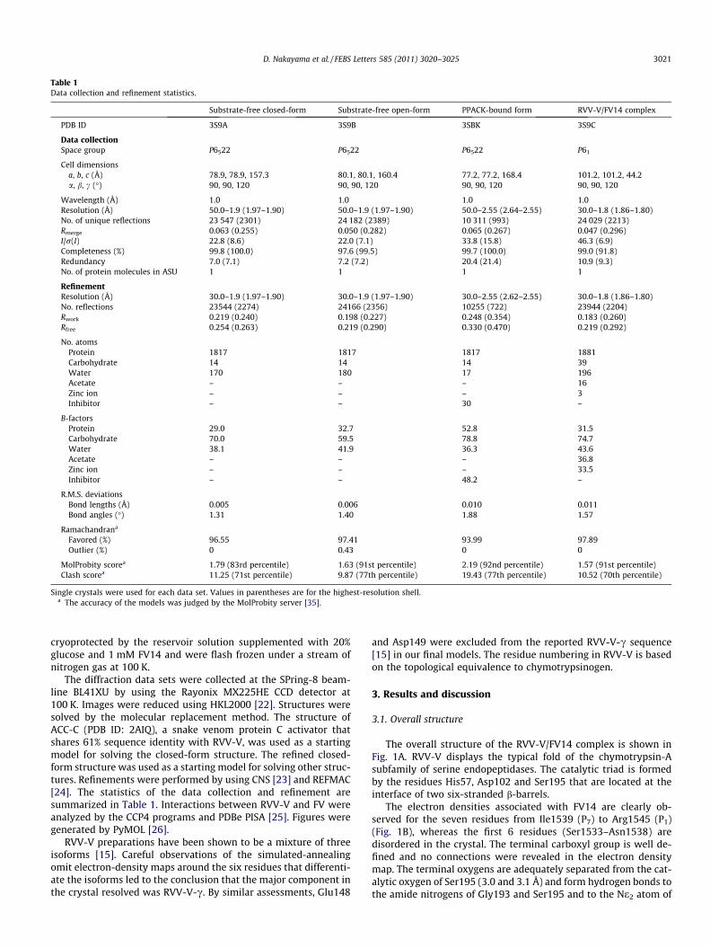
Table 1Data collection and refinement statistics.
Substrate-free closed-form Substrate-free open-form PPACK-bound form RVV-V/FV14 complex
PDB ID 3S9A 3S9B 3SBK 3S9C
Data collectionSpace group P6522 P6522 P6522 P61
Cell dimensionsa, b, c (Å) 78.9, 78.9, 157.3 80.1, 80.1, 160.4 77.2, 77.2, 168.4 101.2, 101.2, 44.2a, b, c (�) 90, 90, 120 90, 90, 120 90, 90, 120 90, 90, 120
Wavelength (Å) 1.0 1.0 1.0 1.0Resolution (Å) 50.0–1.9 (1.97–1.90) 50.0–1.9 (1.97–1.90) 50.0–2.55 (2.64–2.55) 30.0–1.8 (1.86–1.80)No. of unique reflections 23 547 (2301) 24 182 (2389) 10 311 (993) 24 029 (2213)Rmerge 0.063 (0.255) 0.050 (0.282) 0.065 (0.267) 0.047 (0.296)I/r(I) 22.8 (8.6) 22.0 (7.1) 33.8 (15.8) 46.3 (6.9)Completeness (%) 99.8 (100.0) 97.6 (99.5) 99.7 (100.0) 99.0 (91.8)Redundancy 7.0 (7.1) 7.2 (7.2) 20.4 (21.4) 10.9 (9.3)No. of protein molecules in ASU 1 1 1 1
RefinementResolution (Å) 30.0–1.9 (1.97–1.90) 30.0–1.9 (1.97–1.90) 30.0–2.55 (2.62–2.55) 30.0–1.8 (1.86–1.80)No. reflections 23544 (2274) 24166 (2356) 10255 (722) 23944 (2204)Rwork 0.219 (0.240) 0.198 (0.227) 0.248 (0.354) 0.183 (0.260)Rfree 0.254 (0.263) 0.219 (0.290) 0.330 (0.470) 0.219 (0.292)
No. atomsProtein 1817 1817 1817 1881Carbohydrate 14 14 14 39Water 170 180 17 196Acetate – – – 16Zinc ion – – – 3Inhibitor – – 30 –
B-factorsProtein 29.0 32.7 52.8 31.5Carbohydrate 70.0 59.5 78.8 74.7Water 38.1 41.9 36.3 43.6Acetate – – – 36.8Zinc ion – – – 33.5Inhibitor – – 48.2 –
R.M.S. deviationsBond lengths (Å) 0.005 0.006 0.010 0.011Bond angles (�) 1.31 1.40 1.88 1.57
Ramachandrana
Favored (%) 96.55 97.41 93.99 97.89Outlier (%) 0 0.43 0 0
MolProbity scorea 1.79 (83rd percentile) 1.63 (91st percentile) 2.19 (92nd percentile) 1.57 (91st percentile)Clash scorea 11.25 (71st percentile) 9.87 (77th percentile) 19.43 (77th percentile) 10.52 (70th percentile)
Single crystals were used for each data set. Values in parentheses are for the highest-resolution shell.a The accuracy of the models was judged by the MolProbity server [35].
D. Nakayama et al. / FEBS Letters 585 (2011) 3020–3025 3021
cryoprotected by the reservoir solution supplemented with 20%glucose and 1 mM FV14 and were flash frozen under a stream ofnitrogen gas at 100 K.
The diffraction data sets were collected at the SPring-8 beam-line BL41XU by using the Rayonix MX225HE CCD detector at100 K. Images were reduced using HKL2000 [22]. Structures weresolved by the molecular replacement method. The structure ofACC-C (PDB ID: 2AIQ), a snake venom protein C activator thatshares 61% sequence identity with RVV-V, was used as a startingmodel for solving the closed-form structure. The refined closed-form structure was used as a starting model for solving other struc-tures. Refinements were performed by using CNS [23] and REFMAC[24]. The statistics of the data collection and refinement aresummarized in Table 1. Interactions between RVV-V and FV wereanalyzed by the CCP4 programs and PDBe PISA [25]. Figures weregenerated by PyMOL [26].
RVV-V preparations have been shown to be a mixture of threeisoforms [15]. Careful observations of the simulated-annealingomit electron-density maps around the six residues that differenti-ate the isoforms led to the conclusion that the major component inthe crystal resolved was RVV-V-c. By similar assessments, Glu148
and Asp149 were excluded from the reported RVV-V-c sequence[15] in our final models. The residue numbering in RVV-V is basedon the topological equivalence to chymotrypsinogen.
3. Results and discussion
3.1. Overall structure
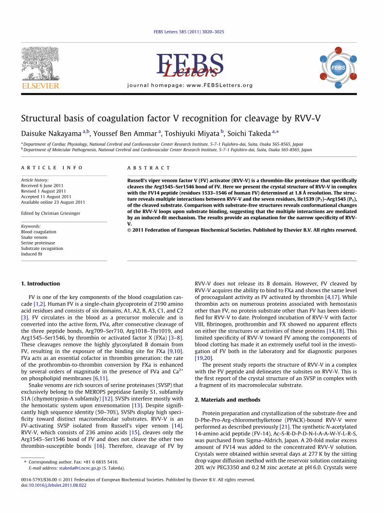
The overall structure of the RVV-V/FV14 complex is shown inFig. 1A. RVV-V displays the typical fold of the chymotrypsin-Asubfamily of serine endopeptidases. The catalytic triad is formedby the residues His57, Asp102 and Ser195 that are located at theinterface of two six-stranded b-barrels.
The electron densities associated with FV14 are clearly ob-served for the seven residues from Ile1539 (P7) to Arg1545 (P1)(Fig. 1B), whereas the first 6 residues (Ser1533–Asn1538) aredisordered in the crystal. The terminal carboxyl group is well de-fined and no connections were revealed in the electron densitymap. The terminal oxygens are adequately separated from the cat-alytic oxygen of Ser195 (3.0 and 3.1 Å) and form hydrogen bonds tothe amide nitrogens of Gly193 and Ser195 and to the Ne2 atom of
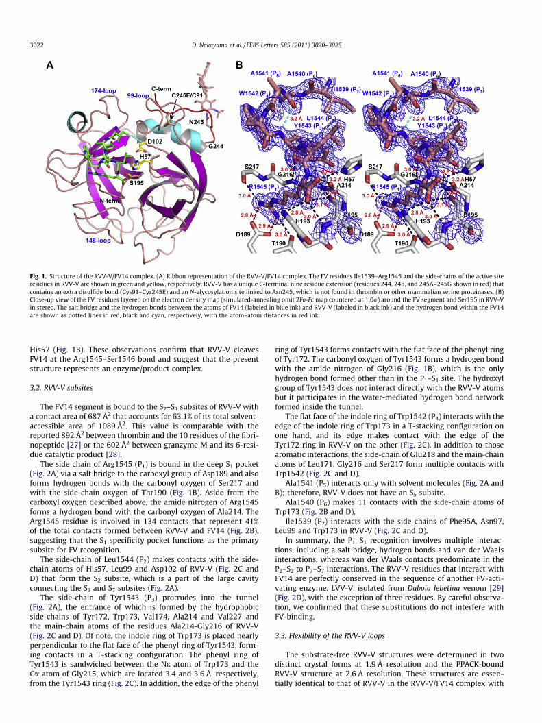
Fig. 1. Structure of the RVV-V/FV14 complex. (A) Ribbon representation of the RVV-V/FV14 complex. The FV residues Ile1539–Arg1545 and the side-chains of the active siteresidues in RVV-V are shown in green and yellow, respectively. RVV-V has a unique C-terminal nine residue extension (residues 244, 245, and 245A–245G shown in red) thatcontains an extra disulfide bond (Cys91–Cys245E) and an N-glycosylation site linked to Asn245, which is not found in thrombin or other mammalian serine proteinases. (B)Close-up view of the FV residues layered on the electron density map (simulated-annealing omit 2Fo-Fc map countered at 1.0r) around the FV segment and Ser195 in RVV-Vin stereo. The salt bridge and the hydrogen bonds between the atoms of FV14 (labeled in blue ink) and RVV-V (labeled in black ink) and the hydrogen bond within the FV14are shown as dotted lines in red, black and cyan, respectively, with the atom–atom distances in red ink.
3022 D. Nakayama et al. / FEBS Letters 585 (2011) 3020–3025
His57 (Fig. 1B). These observations confirm that RVV-V cleavesFV14 at the Arg1545–Ser1546 bond and suggest that the presentstructure represents an enzyme/product complex.
3.2. RVV-V subsites
The FV14 segment is bound to the S7–S1 subsites of RVV-V witha contact area of 687 Å2 that accounts for 63.1% of its total solvent-accessible area of 1089 Å2. This value is comparable with thereported 892 Å2 between thrombin and the 10 residues of the fibri-nopeptide [27] or the 602 Å2 between granzyme M and its 6-resi-due catalytic product [28].
The side chain of Arg1545 (P1) is bound in the deep S1 pocket(Fig. 2A) via a salt bridge to the carboxyl group of Asp189 and alsoforms hydrogen bonds with the carbonyl oxygen of Ser217 andwith the side-chain oxygen of Thr190 (Fig. 1B). Aside from thecarboxyl oxygen described above, the amide nitrogen of Arg1545forms a hydrogen bond with the carbonyl oxygen of Ala214. TheArg1545 residue is involved in 134 contacts that represent 41%of the total contacts formed between RVV-V and FV14 (Fig. 2B),suggesting that the S1 specificity pocket functions as the primarysubsite for FV recognition.
The side-chain of Leu1544 (P2) makes contacts with the side-chain atoms of His57, Leu99 and Asp102 of RVV-V (Fig. 2C andD) that form the S2 subsite, which is a part of the large cavityconnecting the S3 and S7 subsites (Fig. 2A).
The side-chain of Tyr1543 (P3) protrudes into the tunnel(Fig. 2A), the entrance of which is formed by the hydrophobicside-chains of Tyr172, Trp173, Val174, Ala214 and Val227 andthe main-chain atoms of the residues Ala214-Gly216 of RVV-V(Fig. 2C and D). Of note, the indole ring of Trp173 is placed nearlyperpendicular to the flat face of the phenyl ring of Tyr1543, form-ing contacts in a T-stacking configuration. The phenyl ring ofTyr1543 is sandwiched between the Ne atom of Trp173 and theCa atom of Gly215, which are located 3.4 and 3.6 Å, respectively,from the Tyr1543 ring (Fig. 2C). In addition, the edge of the phenyl
ring of Tyr1543 forms contacts with the flat face of the phenyl ringof Tyr172. The carbonyl oxygen of Tyr1543 forms a hydrogen bondwith the amide nitrogen of Gly216 (Fig. 1B), which is the onlyhydrogen bond formed other than in the P1–S1 site. The hydroxylgroup of Tyr1543 does not interact directly with the RVV-V atomsbut it participates in the water-mediated hydrogen bond networkformed inside the tunnel.
The flat face of the indole ring of Trp1542 (P4) interacts with theedge of the indole ring of Trp173 in a T-stacking configuration onone hand, and its edge makes contact with the edge of theTyr172 ring in RVV-V on the other (Fig. 2C). In addition to thosearomatic interactions, the side-chain of Glu218 and the main-chainatoms of Leu171, Gly216 and Ser217 form multiple contacts withTrp1542 (Fig. 2C and D).
Ala1541 (P5) interacts only with solvent molecules (Fig. 2A andB); therefore, RVV-V does not have an S5 subsite.
Ala1540 (P6) makes 11 contacts with the side-chain atoms ofTrp173 (Fig. 2B and D).
Ile1539 (P7) interacts with the side-chains of Phe95A, Asn97,Leu99 and Trp173 in RVV-V (Fig. 2C and D).
In summary, the P1–S1 recognition involves multiple interac-tions, including a salt bridge, hydrogen bonds and van der Waalsinteractions, whereas van der Waals contacts predominate in theP2–S2 to P7–S7 interactions. The RVV-V residues that interact withFV14 are perfectly conserved in the sequence of another FV-acti-vating enzyme, LVV-V, isolated from Daboia lebetina venom [29](Fig. 2D), with the exception of three residues. By careful observa-tion, we confirmed that these substitutions do not interfere withFV-binding.
3.3. Flexibility of the RVV-V loops
The substrate-free RVV-V structures were determined in twodistinct crystal forms at 1.9 Å resolution and the PPACK-boundRVV-V structure at 2.6 Å resolution. These structures are essen-tially identical to that of RVV-V in the RVV-V/FV14 complex with
Fig. 2. Interactions between FV14 and RVV-V. (A) Subsites (S1–S7) are labeled on the molecular surface of RVV-V with the cognate FV residues shown in stick representation(left). Sphere representation with the van der Waal radius of each atom of the FV segment (P1–P7) viewed from the opposite side (right). (B) The number of contacts (pairs ofatoms separated by less than 4.6 Å) between FV and RVV-V are plotted against the FV residues and the sequences of other thrombin-susceptible human proteins, as well asthose of human plasminogen and protein-C, which are susceptible to TSV-PA and ACC-C, respectively. (C) Interactions between FV (shown in yellow and labeled in red ink)and RVV-V (shown in light gray and labeled in black ink) are shown in stereo. (D) The number of contacts between RVV-V and FV14 are plotted against the residues of RVV-V,LVV-V, human thrombin, FXa and SVSPs.
D. Nakayama et al. / FEBS Letters 585 (2011) 3020–3025 3023
the exception of the loop configurations. The structure of theclosed form of RVV-V shows significant differences in both the99- and 174-loops in comparison to the structure of the FV14/RVV-V complex (Fig. 3A). As mentioned, Phe95a and Trp173 are
directly involved in FV recognition by RVV-V and, of note,Trp173 provides the highest number of contacts among theRVV-V residues (Fig. 3D). In the absence of substrate-binding,these two aromatic rings are in proximity to each other and
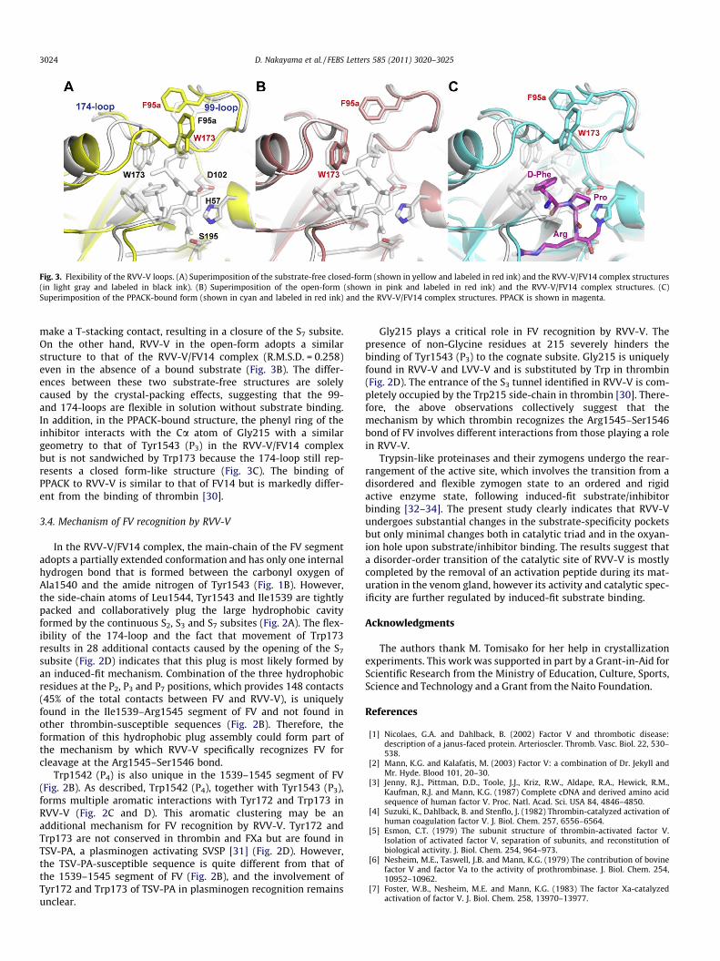
Fig. 3. Flexibility of the RVV-V loops. (A) Superimposition of the substrate-free closed-form (shown in yellow and labeled in red ink) and the RVV-V/FV14 complex structures(in light gray and labeled in black ink). (B) Superimposition of the open-form (shown in pink and labeled in red ink) and the RVV-V/FV14 complex structures. (C)Superimposition of the PPACK-bound form (shown in cyan and labeled in red ink) and the RVV-V/FV14 complex structures. PPACK is shown in magenta.
3024 D. Nakayama et al. / FEBS Letters 585 (2011) 3020–3025
make a T-stacking contact, resulting in a closure of the S7 subsite.On the other hand, RVV-V in the open-form adopts a similarstructure to that of the RVV-V/FV14 complex (R.M.S.D. = 0.258)even in the absence of a bound substrate (Fig. 3B). The differ-ences between these two substrate-free structures are solelycaused by the crystal-packing effects, suggesting that the 99-and 174-loops are flexible in solution without substrate binding.In addition, in the PPACK-bound structure, the phenyl ring of theinhibitor interacts with the Ca atom of Gly215 with a similargeometry to that of Tyr1543 (P3) in the RVV-V/FV14 complexbut is not sandwiched by Trp173 because the 174-loop still rep-resents a closed form-like structure (Fig. 3C). The binding ofPPACK to RVV-V is similar to that of FV14 but is markedly differ-ent from the binding of thrombin [30].
3.4. Mechanism of FV recognition by RVV-V
In the RVV-V/FV14 complex, the main-chain of the FV segmentadopts a partially extended conformation and has only one internalhydrogen bond that is formed between the carbonyl oxygen ofAla1540 and the amide nitrogen of Tyr1543 (Fig. 1B). However,the side-chain atoms of Leu1544, Tyr1543 and Ile1539 are tightlypacked and collaboratively plug the large hydrophobic cavityformed by the continuous S2, S3 and S7 subsites (Fig. 2A). The flex-ibility of the 174-loop and the fact that movement of Trp173results in 28 additional contacts caused by the opening of the S7
subsite (Fig. 2D) indicates that this plug is most likely formed byan induced-fit mechanism. Combination of the three hydrophobicresidues at the P2, P3 and P7 positions, which provides 148 contacts(45% of the total contacts between FV and RVV-V), is uniquelyfound in the Ile1539–Arg1545 segment of FV and not found inother thrombin-susceptible sequences (Fig. 2B). Therefore, theformation of this hydrophobic plug assembly could form part ofthe mechanism by which RVV-V specifically recognizes FV forcleavage at the Arg1545–Ser1546 bond.
Trp1542 (P4) is also unique in the 1539–1545 segment of FV(Fig. 2B). As described, Trp1542 (P4), together with Tyr1543 (P3),forms multiple aromatic interactions with Tyr172 and Trp173 inRVV-V (Fig. 2C and D). This aromatic clustering may be anadditional mechanism for FV recognition by RVV-V. Tyr172 andTrp173 are not conserved in thrombin and FXa but are found inTSV-PA, a plasminogen activating SVSP [31] (Fig. 2D). However,the TSV-PA-susceptible sequence is quite different from that ofthe 1539–1545 segment of FV (Fig. 2B), and the involvement ofTyr172 and Trp173 of TSV-PA in plasminogen recognition remainsunclear.
Gly215 plays a critical role in FV recognition by RVV-V. Thepresence of non-Glycine residues at 215 severely hinders thebinding of Tyr1543 (P3) to the cognate subsite. Gly215 is uniquelyfound in RVV-V and LVV-V and is substituted by Trp in thrombin(Fig. 2D). The entrance of the S3 tunnel identified in RVV-V is com-pletely occupied by the Trp215 side-chain in thrombin [30]. There-fore, the above observations collectively suggest that themechanism by which thrombin recognizes the Arg1545–Ser1546bond of FV involves different interactions from those playing a rolein RVV-V.
Trypsin-like proteinases and their zymogens undergo the rear-rangement of the active site, which involves the transition from adisordered and flexible zymogen state to an ordered and rigidactive enzyme state, following induced-fit substrate/inhibitorbinding [32–34]. The present study clearly indicates that RVV-Vundergoes substantial changes in the substrate-specificity pocketsbut only minimal changes both in catalytic triad and in the oxyan-ion hole upon substrate/inhibitor binding. The results suggest thata disorder-order transition of the catalytic site of RVV-V is mostlycompleted by the removal of an activation peptide during its mat-uration in the venom gland, however its activity and catalytic spec-ificity are further regulated by induced-fit substrate binding.
Acknowledgments
The authors thank M. Tomisako for her help in crystallizationexperiments. This work was supported in part by a Grant-in-Aid forScientific Research from the Ministry of Education, Culture, Sports,Science and Technology and a Grant from the Naito Foundation.
References
[1] Nicolaes, G.A. and Dahlback, B. (2002) Factor V and thrombotic disease:description of a janus-faced protein. Arterioscler. Thromb. Vasc. Biol. 22, 530–538.
[2] Mann, K.G. and Kalafatis, M. (2003) Factor V: a combination of Dr. Jekyll andMr. Hyde. Blood 101, 20–30.
[3] Jenny, R.J., Pittman, D.D., Toole, J.J., Kriz, R.W., Aldape, R.A., Hewick, R.M.,Kaufman, R.J. and Mann, K.G. (1987) Complete cDNA and derived amino acidsequence of human factor V. Proc. Natl. Acad. Sci. USA 84, 4846–4850.
[4] Suzuki, K., Dahlback, B. and Stenflo, J. (1982) Thrombin-catalyzed activation ofhuman coagulation factor V. J. Biol. Chem. 257, 6556–6564.
[5] Esmon, C.T. (1979) The subunit structure of thrombin-activated factor V.Isolation of activated factor V, separation of subunits, and reconstitution ofbiological activity. J. Biol. Chem. 254, 964–973.
[6] Nesheim, M.E., Taswell, J.B. and Mann, K.G. (1979) The contribution of bovinefactor V and factor Va to the activity of prothrombinase. J. Biol. Chem. 254,10952–10962.
[7] Foster, W.B., Nesheim, M.E. and Mann, K.G. (1983) The factor Xa-catalyzedactivation of factor V. J. Biol. Chem. 258, 13970–13977.
D. Nakayama et al. / FEBS Letters 585 (2011) 3020–3025 3025
[8] Thorelli, E., Kaufman, R.J. and Dahlback, B. (1997) Cleavage requirements foractivation of factor V by factor Xa. Eur. J. Biochem. 247, 12–20.
[9] Steen, M. and Dahlback, B. (2002) Thrombin-mediated proteolysis of factor Vresulting in gradual B-domain release and exposure of the factor Xa-bindingsite. J. Biol. Chem. 277, 38424–38430.
[10] Toso, R. and Camire, R.M. (2004) Removal of B-domain sequences from factorV rather than specific proteolysis underlies the mechanism by which cofactorfunction is realized. J. Biol. Chem. 279, 21643–21650.
[11] Rosing, J., Tans, G., Govers-Riemslag, J.W., Zwaal, R.F. and Hemker, H.C. (1980)The role of phospholipids and factor Va in the prothrombinase complex. J. Biol.Chem. 255, 274–283.
[12] Rawlings, N.D., Barrett, A.J. and Bateman, A. (2010) MEROPS: the peptidasedatabase. Nucleic Acids Res. 38, D227–D233.
[13] Serrano, S.M. and Maroun, R.C. (2005) Snake venom serine proteinases:sequence homology vs. substrate specificity, a paradox to be solved. Toxicon45, 1115–1132.
[14] Schiffman, S., Theodor, I. and Rapaport, S.I. (1969) Separation from Russell’sviper venom of one fraction reacting with factor X and another reacting withfactor V. Biochemistry 8, 1397–1405.
[15] Tokunaga, F., Nagasawa, K., Tamura, S., Miyata, T., Iwanaga, S. and Kisiel, W.(1988) The factor V-activating enzyme (RVV-V) from Russell’s viper venom.Identification of isoproteins RVV-V alpha, -V beta, and -V gamma and theircomplete amino acid sequences. J. Biol. Chem. 263, 17471–17481.
[16] Segers, K., Rosing, J. and Nicolaes, G.A. (2006) Structural models of the snakevenom factor V activators from Daboia russelli and Daboia lebetina. Proteins64, 968–984.
[17] Kane, W.H. and Majerus, P.W. (1981) Purification and characterization ofhuman coagulation factor V. J. Biol. Chem. 256, 1002–1007.
[18] Esmon, C.T. and Jackson, C.M. (1973) The factor V activating enzyme ofRussell’s viper venom. Thromb. Res. 2, 509–524.
[19] Marsh, N. and Williams, V. (2005) Practical applications of snake venom toxinsin haemostasis. Toxicon 45, 1171–1181.
[20] Perchuc, A.M. and Wilmer, M. (2010) Diagnostic use of snake venomcomponents in the coagulation laboratory in: Toxins and Hemostasis: FromBench to bedside (Kini, R., Clemetson, K.J., Markland, F.S., McLane, M.A. andMorita, T., Eds.), pp. 747–766, Springer Science+Business Media.
[21] Nakayama, D., Ben Ammar, Y. and Takeda, S. (2009) Crystallization andpreliminary X-ray crystallographic analysis of blood coagulation factor V-activating proteinase (RVV-V) from Russell’s viper venom. Acta Crystallogr.,Sect. F: Struct. Biol. Cryst. Commun. 65, 1306–1308.
[22] Otwinoski, Z. and Minor, W. (1997) Processing of X-ray diffraction datacollected in oscillation mode.
[23] Brunger, A.T. et al. (1998) Crystallography and NMR system: A new softwaresuite for macromolecular structure determination. Acta Crystallogr. D: Biol.Crystallogr. 54 (Pt 5), 905–921.
[24] Murshudov, G.N., Vagin, A.A. and Dodson, E.J. (1997) Refinement ofmacromolecular structures by the maximum-likelihood method. ActaCrystallogr. D: Biol. Crystallogr. 53, 240–255.
[25] Krissinel, E. and Henrick, K. (2007) Inference of macromolecular assembliesfrom crystalline state. J. Mol. Biol. 372, 774–797.
[26] DeLano, W.L. (2002) PyMOL Molecular Viewer. <http://www.pymol.org/>.[27] Martin, P.D., Robertson, W., Turk, D., Huber, R., Bode, W. and Edwards, B.F.
(1992) The structure of residues 7–16 of the A alpha-chain of humanfibrinogen bound to bovine thrombin at 2.3-A resolution. J. Biol. Chem. 267,7911–7920.
[28] Wu, L. et al. (2009) Structural basis for proteolytic specificity of the humanapoptosis-inducing granzyme M. J. Immunol. 183, 421–429.
[29] Siigur, E., Aaspollu, A. and Siigur, J. (1999) Molecular cloning and sequenceanalysis of a cDNA for factor V activating enzyme. Biochem. Biophys. Res.Commun. 262, 328–332.
[30] Bode, W., Mayr, I., Baumann, U., Huber, R., Stone, S.R. and Hofsteenge, J. (1989)The refined 1.9 A crystal structure of human alpha-thrombin: interaction withD-Phe-Pro-Arg chloromethylketone and significance of the Tyr-Pro-Pro-Trpinsertion segment. EMBO J. 8, 3467–3475.
[31] Zhang, Y., Wisner, A., Xiong, Y. and Bon, C. (1995) A novel plasminogenactivator from snake venom. Purification, characterization, and molecularcloning. J. Biol. Chem. 270, 10246–10255.
[32] Bode, W. and Huber, R. (1976) Induction of the bovine trypsinogen–trypsintransition by peptides sequentially similar to the N-terminus of trypsin. FEBSLett. 68, 231–236.
[33] Bode, W., Schwager, P. and Huber, R. (1978) The transition of bovinetrypsinogen to a trypsin-like state upon strong ligand binding. The refinedcrystal structures of the bovine trypsinogen–pancreatic trypsin inhibitorcomplex and of its ternary complex with Ile-Val at 1.9 A resolution. J. Mol. Biol.118, 99–112.
[34] Clausen, T., Kaiser, M., Huber, R. and Ehrmann, M. (2011) HTRA proteases:regulated proteolysis in protein quality control. Nat. Rev. Mol. Cell. Biol. 12,152–162.
[35] Davis, I.W. et al. (2007) MolProbity: all-atom contacts and structure validationfor proteins and nucleic acids. Nucleic Acids Res. 35, W375–W383.


![Thu Aud 09.20 Molecular biology in Malignnat pleural … · 2019. 8. 15. · v Á µ W í ] u u µ v } ] v ] v P ( } Z ] } v Á v^D v ^ W EK J J J J J /RVV RI %$3 QXFOHDU VWDLQLQJ](https://img.pdfslide.us/doc/110x75/6105fc63b4f8af2a146f6411/thu-aud-0920-molecular-biology-in-malignnat-pleural-2019-8-15-v-w-.jpg)


![) [111] cleavage plane](https://img.pdfslide.us/doc/110x75/61c7329341512e61f73ea613/-111-cleavage-plane.jpg)