Embed Size (px)
Citation preview
J. Mol. Biol. (1996) 258 1–5
COMMUNICATION
Structural Bases for Sulfide Recognition inLucina pectinata Hemoglobin I
Menico Rizzi 1, Jonathan B. Wittenberg 2, Alessandro Coda 1
Paolo Ascenzi 3 and Martino Bolognesi 1,4*
The X-ray crystal structure of the sulfide derivative of ferric Lucina pectinata1Dipartimento di Genetica e hemoglobin component I (HbI) has been determined at 1.9 Å resolutionMicrobiologia, Universita’ di (R-factor 0.186). The heme pocket structural organization of HbI is inPavia, Via Abbiategrasso 207 keeping with its ligand binding properties. The fast sulfide association rate
constant can be related to the presence of Gln(64)E7, as the heme distal27100 Pavia, Italyresidue, together with the protein structural properties in the CD-E distal2Department of Physiology region. Moreover, the very high sulfide affinity for HbI is reflected by
and Biophysics, Albert the exceptionally slow ligand dissociation rate. The stabilization of theEinstein College of Medicine heme-bound sulfide molecule is achieved through hydrogen bonding toBronx, NY 10461, USA Gln(64)E7, as well as by finely tuned aromatic-electrostatic interactions
with the clustered residues Phe(29)B10, Phe(43)CD1 and Phe(68)E11. Such3Dipartimento di Biologia,a peculiar arrangement of phenylalanyl residues at the distal ligandTerza Universita’ di Romabinding site has not been observed before in the globin family, and isVia Ostiense 173, 00154unique to HbI, a protein functionally devoted to sulfide transport.Roma, Italy
7 1996 Academic Press Limited4IST, Centro BiotecnologieAvanzate and Dipartimentodi Fisica, Universita’ diGenova, Viale BenedettoXV 10, 16132 Genova, Italy
Keywords: heme protein; monomeric mollusc hemoglobin; sulfide carrier;*Corresponding author crystal structure
Hemoglobins fromsymbiont-harboring molluscs
The symbiosis between molluscs and intracellu-lar chemoautotrophic bacteria is always character-ized by the presence of mollusc cytoplasmic Hbs,most probably located in the cytoplasm of thebacteriocyte gill cells, which are specialized tohouse symbionts fixing carbon dioxide into hexoses.The cytoplasmic Hbs transport oxygen and hydro-gen sulfide from the sea water to the bacterialsymbiont, permitting the bacteria to sustain rapidhexose synthesis, and supply the host animal withits entire carbohydrate nutrition (Arp, 1991;Wittenberg & Kraus, 1991).
Three Hb components have been isolated fromthe modified gill of the bivalve mollusc Lucinapectinata, living in the coastal sedimental area ofPuerto Rico. Components HbII and HbIII, isolatedas homodimers or heterotetramers of two similar(approximately 17 kDa) polypeptide chains, bindO2 non-cooperatively with an affinity (K = 3.5× 10⁶ M−1) similar to that observed for sperm whaleMb (K = 1.2 × 10⁶ M−1), and are responsible for itstransport. The monomeric HbI component, on theother hand, binds O2 with comparable affinity(K = 1.6 × 10⁶ M−1), but is readily oxidized to theferric HbI:sulfide complex in the presence of traceoxygen and hydrogen sulfide concentrations (seeKraus & Wittenberg, 1990; Kraus et al., 1990).
The affinity of the sulfide ligand for HbI isexceptionally high (K = 2.9 × 10⁸ M−1), being about5000-fold higher than that observed for the HbII andHbIII components, and for sperm whale Mb. Sucha high affinity is achieved through fast association(kon = 6.0 × 10⁴ M−1 s−1) and very slow dissociation(koff = 7.8 × 10⁻⁴ s−1) processes (see Kraus & Witten-
Abbreviations used: Hb, hemoglobin; Mb,myoglobin; r.m.s., root-mean-square. Amino acidresidues have been identified by their three-letter code,sequence number (in parentheses), and by theirtopological position in the globin fold. Water moleculeshave been identified by the W prefix (see Rizzi et al.1994).
0022–2836/96/160001–05 $18.00/0 7 1996 Academic Press Limited
Communication2
berg, 1990; Kraus et al., 1990). The sulfide ligand,conversely, is dissociated from HbI upon reductionof the heme iron, a process possibly ascribed in vivoto a bacterial reductase present on the bacteriocytemembrane. More properly therefore, L. pectinataHbI can be considered as a sulfide-reactive Hb,whose functional activity, at variance from ver-tebrate oxygen carrying Hbs or Mbs, is carried onin the ferric state (Wittenberg & Kraus, 1991).
The three-dimensional structure of the aquo-metderivative of HbI has been determined at 1.5 Aresolution by X-ray crystallography (Rizzi et al.,1994). HbI shows a conventional globin fold (lackingthe D helix), with a unique structural organizationof the heme pocket involving residues Phe(29)B10,Phe(43)CD1, Gln(64)E7 and Phe(68)E11. Moreover,the unusual presence and location of severaladditional aromatic residues (ten Phe, four Trp, twoHis and one Tyr) has been observed (Rizzi et al.,1994).
The structural bases for the very high sulfideaffinity of L. pectinata HbI are discussed in thepresent communication in the light of the crystalstructure of its sulfide ferric derivative (at 1.9 Aresolution), and of the ligand binding mechanismsobserved in (non)vertebrate Hbs and Mbs (seePerutz, 1989, 1990; Conti et al., 1993; Rizzi et al.,1994; Brancaccio et al., 1994; Springer et al., 1994;Smerdon et al., 1995).
Structure of the Lucina pectinataHbI:sulfide complex
Upon sulfide† binding, the two water moleculespresent in the distal site of aquo-met HbI (W202 andW208; see Rizzi et al., 1994) are removed. The ligandis co-ordinated to the heme iron, with a coordi-nation bond of 2.3 A. This distance, which has notbeen restrained during refinement, is in fullagreement with those observed in model ironcoordination compounds (Orpen & Brammer, 1989).The heme iron is contained in the pyrrole nitrogenatoms plane, with a mean Fe–N(pyrrole) distanceof 1.9 A. The proximal coordination bond betweenthe His(96)F8 NE2 and the Fe atoms is 2.1 A, andthe His(96)F8 NE2–Fe–S(ligand) angle is 178°.No deviation of the azimuthal orientation of theHis(96)F8 imidazole ring, with respect to theaquo-met protein (Rizzi et al., 1994), has beenobserved upon sulfide binding.
The heme iron bound sulfide ligand is at 3.3 Afrom the Gln(64)E7 side-chain end, this distancebeing compatible with the formation of a hydrogenbond. Moreover, the ligand is surrounded byresidues Phe(29)B10 (present in less than 5% globin
structures; Bashford et al., 1987), Phe(43)CD1(invariant), and Phe(68)E11 (unique to L. pectinataHbs), with closest contacts of 4.0, 3.9 and 4.6 A,respectively, which form a sort of cage adapted tothe ligand size (see Figure 1). This cage is sealed onthe solvent side by Gln(64)E7. The concerted(edge-on) orientation of the aromatic rings contact-ing the ligand results from the juxtaposition ofadditional neighboring Phe residues. In particular,Phe(28)B9 and Phe(33)B14 nestle respectivelyamong the aromatic side-chains of the Phe(29)B10-Phe(68)E11 and Phe(29)B10-Phe(43)CD1 pairs.Residues Trp(21)B2 and Phe(47)CD4 extend thesearomatic clusters to farther regions of the HbImolecule (see Figure 4 of Rizzi et al., 1994).
Sulfide recognition by HbI
The structural observations reported above are inkeeping with the HbI sulfide binding properties.The fast sulfide association rate constant may berelated to the presence of Gln(64)E7 as the distalresidue in HbI (Kraus & Wittenberg, 1990). In fact,Gln(64)E7, longer and more flexible than His, can bemore easily removed from the distal site ‘‘closedgate’’ conformation, allowing displacement of thetwo water molecules there located in the aquo-metderivative, and sulfide association. Other structuralfactors can be expected to control the ligandassociation rate, either through direct influence onGln(64)E7, or through modification of the dynamicproperties of the surrounding distal region. In thisrespect, the presence of Leu(46) at position CD3(usually a charged residue), the observed lack ofsurface-bound water molecules in the CD1-E7region (a potential ligand path to the distal site),and the unique insertion of Ser(44) betweenthe invariant Phe(43)CD1 residue and Gly(45)CD2should be noticed (see Rizzi et al., 1994).
As indicated by analysis of the kinetic constants,the exceptionally high sulfide affinity of HbI isachieved through a very slow ligand dissociationrate (Kraus & Wittenberg, 1990). In this respect, onecontribution to stabilization of the bound sulfide isprovided by the above mentioned Gln(64)E7-- - - S(ligand) hydrogen bond. Besides this, however,a significant role is played by the aro-matic hydrogen atoms of residues Phe(29)B10,Phe(43)CD1 and Phe(68)E11, which are directedtowards the ligand binding site, and providestabilization of the sulfide molecule througharomatic-electrostatic interactions. The relevance ofsuch interactions in model compounds and inproteins has been investigated by quantum-mech-anical methods and statistical studies on thestructural database (Thomas et al., 1982; Burley &Petsko, 1985; Dougherty & Stauffer, 1990). Com-pared to other atomic species, sulfur shows a netpreference for the in-plane interaction with the edgeof aromatic rings, avoiding the above-plane regionsnext to the p electrons (Reid et al., 1985). The originsof these effects, not only for sulfur but also for
† The analysis of the pH dependence of kinetics offerric HbI:sulfide complex formation suggests thatundissociated H2S is the attacking species. In thisrespect, undissociated methanethiol (CH3SH) reactswith ferric HbI with the same second order rateconstant observed for the binding of undissociated H2S(Kraus & Wittenberg, 1990).
Communication 3
dioxygen, have been ascribed to the electrostaticattraction between the electronegative species andthe positively charged aromatic ring hydrogenatoms (Thomas et al., 1982; Reid et al., 1985). In thecase of engineered sperm whale Mb site-specificmutants, aromatic-electrostatic interactions stabilizethe bound dioxygen ligand through the action ofresidue PheB10 by about −1.5 kcal mol−1 (Carveret al., 1991; Brancaccio et al., 1994; Springer et al.,1994; Smerdon et al., 1995).
The presence in HbI of the Phe(68)E11 residueand of an ideally structured aromatic (29)B10-(43)CD1-(68)E11 ‘‘Phe-cage’’ (see Rizzi et al., 1994)is unique among globin structures (Bashford et al.,1987), and reflects the sulfide-carrier role of thismonomeric hemoprotein (Wittenberg & Kraus,1991). On the other hand, the much lower affinityof L. pectinata HbII and HbIII for sulfide (mainlyresulting from faster ligand dissociation) may berelated to perturbations brought about in the ligand
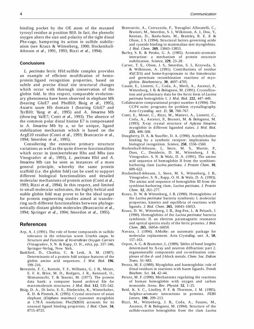
Figure 1. Aquo-met L. pectinataHbI was purified as described byKraus & Wittenberg (1990), modi-fied to include a final gel filtrationstep on a Sephacryl S100HR column.Monoclinic crystals of aquo-met HbIwere grown from ammonium sul-fate solutions as previously de-scribed (crystal form A, a = 50.5,b = 38.8, c = 42.5 A, b = 106.9°;Casale et al., 1991; Rizzi et al., 1994).The ferric HbI:sulfide complex wasprepared by soaking of the aquo-met HbI crystals in solutions con-taining 2.7 M ammonium sulfate(pH 6.0), 10−3 M Na2S, at roomtemperature; the soaking time wasthree hours. Diffracted intensities(a)were collected at 1.9 A resolution ona Rigaku R-Axis II system (28,787reflections were reduced to 12,135independent intensities using MOS-FLM (A. Leslie), Rmerge 0.08, 96.7%completeness in the 15.0 to 1.9 Aresolution range). The structure of L.pectinata HbI, in crystal form A, waspromptly solved by molecular re-placement (Navaza, 1994), using assearch model the refined structureof the same ferric protein crystal-lized in a different crystal form(form B; Rizzi et al., 1994). Themolecular replacement solutionyielded an R-factor value of 0.28 at3.0 A resolution, in the absence ofsolvent. Rigid body refinement andsubsequent restrained crystallo-graphic refinement (Tronrud et al.,1987) of the ferric protein structure,yielded an R-factor value of 0.22, in(b)the 15.0 to 1.9 A resolution range,
with ideal stereochemistry. Subsequently solvent molecules were located using an automated search procedure (CCP4,1994), excluding peaks in the distal site region. Finally the sulfide ligand was located as the main feature in differenceelectron density maps, and refined accordingly, without Fe–S restraints. The final model includes 1038 protein atoms,97 solvent molecules, and the sulfide ligand. The corresponding R-factor, for 12,135 independent reflections in the 15.0to 1.9 A resolution range, is 0.186 with ideal stereochemistry (r.m.s. bond length deviation 0.015 A, r.m.s. bond angledeviation 1.76°). The sulfide ligand has a B factor of 28 A2. Atomic coordinates of the ferric HbI:sulfide complex havebeen deposited with the Brookhaven Protein Data Bank, from which they can be recovered (tracking no. T8220;Bernstein et al., 1977). The r.m.s. deviation between the HbI:sulfide complex main-chain atoms and the correspondingaquo-met HbI structure (Rizzi et al., 1994) is 0.20 A. (a) Stereo view of the heme distal pocket of the ferric HbI:sulfidecomplex, including a ribbon representation of part of the B helix. The main aromatic residues building up thesurrounding of the sulfide ligand (shown as shaded sphere) are displayed together with the His(96)F8 and the Gln(64)E7side-chains. (b) Space-filling representation of the (29)B10-(43)CD1-(68)E11 Phe-cage (blue-green residues) contactingthe sulfide ligand (yellow). The heme group (red) together with the proximal His(96)F8 (green) and the nestlingPhe(28)B9 and Phe(33)B14 resdiues (blue) are displayed.
Communication4
binding pocket by the OE atom of the mutatedtyrosyl residue at position B10. In fact, the phenolicoxygen alters the size and polarity of the tight distalPhe-cage, hampering its action in sulfide stabiliz-ation (see Kraus & Wittenberg, 1990; Hockenhull-Johnson et al., 1991, 1993; Rizzi et al., 1994).
Conclusions
L. pectinata ferric HbI:sulfide complex providesan example of efficient modification of hemo-protein:ligand recognition properties, based onsubtle and precise distal site structural changeswhich occur with thorough conservation of theglobin fold. In this respect, comparable evolution-ary phenomena have been observed in elephant Mb(bearing GlnE7 and PheB10; Bisig et al., 1995),Ascaris suum Hb domain I (bearing GlnE7 andTyrB10; Yang et al., 1995) and A. limacina Mb(showing ValE7; Conti et al., 1993). The absence ofthe common polar distal histine E7 is compensatedin A. limacina Mb by a, so far unique, ligandstabilization mechanism which is based on theArgE10 residue (Conti et al., 1993; Brancaccio et al.,1994; Smerdon et al., 1995).
Considering the extensive primary structurevariations as well as the quite diverse functionalitieswhich occur in (non)vertebrate Hbs and Mbs (seeVinogradov et al., 1993), L. pectinata HbI and A.limacina Mb can be seen as instances of a moregeneral principle, whereby the same proteinscaffold (i.e. the globin fold) can be used to supportdifferent biological functionalities and detailedmolecular mechanisms of protein action (Conti et al.,1993; Rizzi et al., 1994). In this respect, and limitedto small molecular substrates, the highly helical andstable globin fold may prove to be the ideal targetfor protein engineering studies aimed at transfer-ring such different functionalities between phyloge-netically distant globin molecules (Brancaccio et al.,1994; Springer et al., 1994; Smerdon et al., 1995).
ReferencesArp, A. J. (1991). The role of heme compounds in sulfide
tolerance in the echiurian worm Urechis caupo. InStructure and Function of Invertebrate Oxygen Carriers(Vinagradov, S. N. & Kapp, O. H., eds), pp. 337–346,Springer-Verlag, New York.
Bashford, D., Chothia, C. & Lesk, A. M. (1987).Determinants of a protein fold unique features of theglobin amino acid sequences. J. Mol. Biol. 196,199–216.
Bernstein, F. C., Koetzle, T. F., Williams, G. J. B., Meyer,E. F. Jr, Brice, M. D., Rodgers, J. R., Kennard, O.,Shimanouchi, T. & Tasumi, M. (1977). The proteindata bank: a computer based archival file formacromolecule structures. J. Mol. Biol. 112, 535–542.
Bisig, D. A., Di Iorio, E. E., Diederichs, K., Winterhalter,K. H. & Piontek, K. (1995). Crystal structure of asianelephant (Elephans maximus) cyanomet myoglobinat 1.78 A resolution: Phe29(B10) accounts for itsunusual ligand binding properties. J. Biol. Chem. 34,8715–8725.
Brancaccio, A., Cutruzzola, F., Travaglini Allocatelli, C.,Brunori, M., Smerdon, S. J., Wilkinson, A. J., Dou, Y.,Keenan, D., Ikeda-Saito, M., Brantley, R. E. Jr &Olson, J. S. (1994). Structural factors governing azideand cyanide binding to mammalian met myoglobins.J. Biol. Chem. 269, 13843–13853.
Burley, S. K. & Petsko, G. A. (1985). Aromatic-aromaticinteraction: a mechanism of protein structurestabilization. Science, 229, 23–28.
Carver, T. E., Olson, J. S., Smerdon, S. J., Krzywda, S.& Wilkinson, A. (1991). Contributions of residue45(CD3) and heme-6-propionate to the bimolecularand germinate recombination reactions of myo-globin. Biochemistry, 30, 4697–4705.
Casale, E., Lionetti, C., Coda, A., Merli, A., Ascenzi, P.,Wittenberg, J. B. & Bolognesi, M. (1991). Crystalliza-tion and preliminary data for the ferric form of Lucinapectinata hemoglobin I. J. Mol. Biol. 222, 447–449.
Collaborative computational project number 4 (1994). TheCCP4 suite: programs for problem crystallography.Acta Crystallog. sect. D, 50, 760–767.
Conti, E., Moser, C., Rizzi, M., Mattevi, A., Lionetti, C.,Coda, A., Ascenzi, P., Brunori, M. & Bolognesi, M.(1993). X-ray crystal structure of Aplysia limacinamyoglobin in different liganded states. J. Mol. Biol.233, 498–508.
Dougherty, D. A. & Stauffer, D. A. (1990). Acethylcholinebinding by a synthetic receptor: implications forbiological recognition. Science, 250, 1558–1560.
Hockenhull-Johnson, J., Stern, M. S., Martin, P.,Dass, C., Desiderio, D. M., Wittenberg, J. B.,Vinogradov, S. N. & Walz, D. A. (1991). The aminoacid sequence of hemoglobin II from the symbiont-harboring clam Lucina pectinata. J. Protein Chem. 10,609–622.
Hockenhull-Johnson, J., Stern, M. S., Wittenberg, J. B.,Vinogradov, S. N., Kapp, O. H. & Walz, D. A. (1993).The amino acid sequence of hemoglobin III from thesymbiont-harboring clam, Lucina pectinata. J. ProteinChem. 12, 261–277.
Kraus, D. W. & Wittenberg, J. B. (1990). Hemoglobins ofthe Lucina pectinata/bacteria symbiosis: I. molecularproperties, kinetics and equilibria of reactions withligands. J. Biol. Chem. 265, 16043–16053.
Kraus, D. W., Wittenberg, J. B., Jing-Fen, L. & Peisach, J.(1990). Hemoglobins of the Lucina pectinata/bacteriasymbiosis: II. an electron paramagnetic resonanceand optical spectra study of the ferric proteins. J. Biol.Chem. 265, 16054–16059.
Navaza, J. (1994). AMoRe: an automatic package formolecular replacement. Acta Crystallog. sect. A, 50,157–163.
Orpen, A. G. & Brammer, L. (1989). Tables of bond lengthsdetermined by X-ray and neutron diffraction: part 2.organometallic compounds and co-ordination com-plexes of the d- and f-block metals. Chem. Soc. DaltonTrans. S1–S83.
Perutz, M. F. (1989). Myoglobin and haemoglobin: role ofdistal residues in reactions with haem ligands. TrendsBiochem. Sci. 14, 42–44.
Perutz, M. F. (1990). Mechanisms regulating the reactionsof human hemoglobin with oxygen and carbonmonoxide. Annu. Rev. Physiol. 52, 1–25.
Reid, K. S. C., Lindley, P. F. & Thornton, J. M. (1985).Sulphur-aromatic interactions in proteins. FEBSLetters, 190, 209–213.
Rizzi, M., Wittenberg, J. B., Coda, A., Fasano, M.,Ascenzi, P. & Bolognesi, M. (1994). Structure of thesulfide-reactive hemoglobin from the clam Lucina
Communication 5
pectinata: crystallographic analysis at 1.5 A resol-ution. J. Mol. Biol. 244, 86–99.
Smerdon, S. J., Krzywda, S., Brzozowski, A. M., Davies,G. J., Wilkinson, A. J., Brancaccio, A., Cutruzzola', F.,Travaglini Allocatelli, C., Brunori, M., Li, T., Brantley,R. E. Jr, Carver, T. E., Eich, R. F., Singleton, E. &Olson, J. S. (1995). Interactions among residues CD3,E7, E10 and E11 in myoglobins: attempts to simulatethe ligand binding properties of Aplysia limacina.Biochemistry, 34, 8715–8725.
Springer, B. A., Sligar, S. G., Olson, J. S. & Phillips, G. N.Jr (1994). Mechanisms of ligand recognition inmyoglobin. Chem. Rev. 94, 699–714.
Thomas, K. A., Smith, G. M., Thomas, T. B. & Feldmann,R. J. (1982). Electronic distributions within proteinphenylalanine aromatic rings are reflected by thethree dimensional oxygen atom environments. Proc.Natl Acad. Sci. USA, 79, 4843–4847.
Tronrud, D. E., Ten Eyck, L. F. & Matthews, B. W. (1987).An efficient general-purpose least-squares refine-ment program for macromolecular structures. ActaCrystallog. sect. A, 43, 489–501.
Vinogradov, S. N., Walz, D. A., Pohajdak, B., Moens, L.,Kapp, O. H., Suzuki, T. & Trotman, C. N. A. (1993).Adventitious variability? The amino acid sequencesof nonvertebrate globins. Comp. Biochem. Physiol.106B, 1–26.
Wittenberg, J. B. & Kraus, D. W. (1991). Hemoglobins ofeukaryote/prokaryote symbioses. In Structure andFunction of Invertebrate Oxygen Carriers (Vinagradov,S. N. & Kapp, O. H., eds), pp. 323–330, Springer-Verlag, New York.
Yang, J., Kloek, A. P., Goldberg, D. E. & Mathews, F. S.(1995). The structure of Ascaris hemoglobin domainI at 2.2 A resolution: molecular features of oxygenavidity. Proc. Natl Acad. Sci. USA, 92, 4224–4228.
Edited by R. Huber
(Received 10 January 1996; accepted 22 January 1996)