Embed Size (px)
Citation preview

STRUCTURAL AND LIGAND BINDING STUDIES
OF TOLL-LIKE RECEPTOR 4 (TLR4) ASSOCIATED PROTEINS
BY
STACY L. KELLEY
DISSERTATION
Submitted in partial fulfillment of the requirements
for the degree of Doctor of Philosophy in Biochemistry
in the Graduate College of the
University of Illinois at Urbana-Champaign, 2013
Urbana, Illinois
Doctoral Committee:
Associate Professor Richard Tapping, Chair
Professor Satish Nair, Co-Chair
Professor Susan Martinis
Professor James Morrissey

ii
ABSTRACT
The sensing of lipopolysaccharide (LPS) represents a major mechanism of detection of
Gram-negative bacterial infection by the human innate immune system. This pathogen
associated molecular pattern is recognized by the host through the transmembrane pattern
recognition complex comprised of Toll-like receptor 4 (TLR4) and myeloid differentiation
antigen (MD-2). As described in Chapter 1, LPS-dependent complex formation of TLR4/MD-2
heterodimers results in the activation of pro-inflammatory signaling cascades. Although
inflammation is usually host protective, when uncontrolled it can cause host fatality through
sepsis. The function of soluble or membrane anchored human CD14 is to bind and shuttle LPS
to TLR4/MD-2, thereby enhancing host sensitivity of LPS. The mechanism by which CD14
binds and transfers LPS remains unknown, and our understanding of this process would be
advanced by structural information.
Thus, we determined the x-ray crystal structure of human CD14, the results of which
were recently published (Kelley, S.L., et al., (2013). J. Immunol. 190:1304-1311) and are
presented in Chapter 2 of this dissertation. To do so, we cloned, expressed, and purified two
constructs of human CD14 from a mammalian expression system. One of the two crystallized
and resulted in an x-ray crystal structure solution. The structure of human CD14 reveals a bent
solenoid with an N-terminal hydrophobic pocket. Compared to the mouse CD14 crystal
structure, human CD14 reveals an expansion of residues comprising the rim region and an
adjacent, solvent exposed hydrophobic patch, which may facilitate LPS binding.
In Chapter 3 we describe the expression and purification of mouse CD14 as an additional
target for ligand binding crystallization studies. After determining that purified human and
mouse CD14 bind LPS and other ligands using native PAGE gel shift and ForteBio Octet assays,
iii
we confirmed the bioactivity of both purified proteins through cell-based stimulation assays.
Additional small gel filtration mixing studies (Ranoa, D.R., Kelley, S.L., et al. (2013).
J.Biol.Chem. 288: 9729-9741) confirmed the ability of our purified human CD14 to shuttle
triacylated ligands to the Toll-like receptor 2 complex. Chapter 3 also describes our efforts to
produce a ligand bound structure of CD14.
An additional project, described in Chapter 4, focuses on a predicted Toll-like receptor 4
accessory protein called Der p 2. Der p 2 is an aeroallergen from Dermatophagoides
pteronyssinus house dust mites, which has been implicated as an MD-2 analogue. By binding
LPS and TLR4 in place of MD-2, Der p 2 may activate pro-inflammatory signaling and stimulate
IgE production and allergic reaction, most often associated with allergic asthma. In
collaboration with the lab of David Kranz (University of Illinois, Urbana), we used yeast display
to create Der p 2 and MD-2 full length Aga2 fusion proteins. After verifying yeast surface
expression, we assessed protein folding by characterizing the ability of Der p 2 and MD-2 to
bind anti-Derp2 and anti-MD-2 monoclonal antibodies, respectively, in thermal denaturation
studies. Flow cytometry assays indicate that Der p 2 binds biotinylated and non-biotinylated
forms of LPS without detectable binding to purified human TLR4. Clinically, we found that this
system provides a novel assay that reliably and quantitatively detects anti-Der p 2 IgE in serum
and plasma samples of atopic patients.
Chapter 5 summarizes our findings, describes their significance, and suggests avenues for
future work. Although a ligand bound structure of CD14 has not yet been determined, we have
successfully determined the x-ray crystal structure of human CD14, which significantly enhances
our understanding of the LPS binding pocket. The CD14 structure and our ligand binding
crystallization efforts may foster additional ligand binding structural studies, virtual docking
iv
studies, and drug design efforts thus allowing therapeutic intervention at an early step in the
signaling cascade. Additionally, we utilized yeast display and flow cytometry to detect binding
of Der p 2 to LPS and created a novel system for the detection of anti-Der p 2 IgE antibodies in
serum and plasma. Additional mutational analysis of Der p 2 could determine the contribution of
individual amino acids to LPS binding and also natural epitopes of anti-Der p 2 IgE, thereby
creating new targets for drug development to treat allergic asthma. In total, our efforts have
advanced our understanding of host LPS detection during bacterial infection and allergy through
structural and ligand binding studies of the TLR4 accessory proteins human CD14 and Der p 2.

v
To my husband, parents, family, and friends.

vi
ACKNOWLEDGEMENTS
There are many people I would like to thank for their support and assistance along this
journey. First, my sincere thanks goes to all of my advisors, past and present, who had a positive
impact on my life. I wanted to take a moment to specifically thank Dr. Richard Tapping at the
University of Illinois Urbana-Champaign for his encouragement and mentorship, and Dr. Sharon
Weldon at Illinois State University for her advice and steadfast friendship. I would also like to
thank each member of my current committee, Dr. Susan Martinis and Dr. James Morrissey, and
all of my past committee members, Dr. Scott Silverman, Dr. David Kranz, and Dr. David
Shapiro, for their words of wisdom and support. I thank all alumni and current members of the
Tapping, Nair, Huang, and Kranz labs for any encouraging words, friendship, or technical
knowledge imparted. Above all, I thank my husband, my parents, my family, and all of my
friends for their love, friendship, bountiful patience, steadfast encouragement, and support.

vii
TABLE OF CONTENTS
CHAPTER ONE: Innate Immune System ....................................................................................1
1.1 Innate and Adaptive Immunity ..................................................................................................1
1.2 Lipopolysaccharide ....................................................................................................................3
1.3 Lipopolysaccharide Binding Protein (LBP)...............................................................................5
1.4 Cluster of Differentiation Antigen 14 (CD14) ...........................................................................6
1.5 Toll-like Receptor 4 Complex (TLR4/MD-2) .........................................................................10
1.6 Dermatophagoides pteronyssinus allergen (Der p 2) ..............................................................16
CHAPTER TWO: X-ray Crystallography of Human CD14 .......................................................27
2.1 Introduction ..............................................................................................................................27
2.2 Materials and Methods .............................................................................................................28
2.2.1 Reagents ................................................................................................................................28
2.2.2 Cloning and Site Directed Mutagenesis ................................................................................29
2.2.3 Expression and Stable Cell Line Selection ...........................................................................31
2.2.4 Purification ............................................................................................................................32
2.2.5 Initial Crystallization Screening ...........................................................................................34
2.2.6 Crystallization Optimization .................................................................................................35
2.2.7 Cryoprotection Optimization and Crystal Harvesting ..........................................................37
2.2.8 Data Collection and Structure Determination .......................................................................39
2.3 Results ......................................................................................................................................39
2.3.1 Generation of Human CD14 for Crystallization ...................................................................39
2.3.2 Crystal Structure of Human CD14 ........................................................................................41
2.3.3 Comparison of Human and Mouse CD14 Structure .............................................................42
2.4 Discussion ................................................................................................................................44
CHAPTER THREE: Ligand Binding and Bioactivity of Purified CD14 ...................................63
3.1 Introduction ..............................................................................................................................63
3.2 Materials and Methods .............................................................................................................65
3.2.1 Reagents ................................................................................................................................65
3.2.2 Mouse CD14 Cloning, Expression, and Purification ............................................................66
3.2.3 Generation of Human CD14 for Small Gel Filtration Mixing Studies .................................68
3.2.4 Native Polyacrylamide Gel Shift LPS Binding Assay ..........................................................69
3.2.5 ForteBio Octet QK Ligand Binding Assay ...........................................................................69
3.2.6 IL-8 Bioactivity Assay ..........................................................................................................70
3.2.7 Initial Crystallization Screening of Mouse CD14 .................................................................71
3.2.8 Initial Crystallization Screening of Mouse CD14-LPS Complexes .....................................72
3.3 Results ......................................................................................................................................72
3.3.1 Purified Mouse CD14 ...........................................................................................................72
3.3.2 Purified Human and Mouse CD14 Bind E. coli LPS............................................................73
3.3.3 Octet and Small Gel Filtration Mixing Studies Confirm Ligand Binding to Human CD14 74
3.3.4 Purified Human and Mouse CD14 Are Bioactive ................................................................75
3.3.5 Initial Crystallization Screening of Mouse CD14 ................................................................77
3.3.6 Lack of Diffraction Quality Crystals of CD14-LPS Complexes ..........................................78

viii
3.4 Discussion ................................................................................................................................80
CHAPTER FOUR: IgE and Ligand Binding Studies of Der p 2 Allergen .................................92
4.1 Introduction ..............................................................................................................................92
4.2 Materials and Methods .............................................................................................................97
4.2.1 Reagents ................................................................................................................................97
4.2.2 Yeast Display of Der p 2 and MD-2 .....................................................................................98
4.2.3 Detection of Yeast Displayed Der p 2 and MD-2 ...............................................................100
4.2.4 Thermal Denaturation of Yeast Displayed Der p 2 ............................................................101
4.2.5 LPS Binding Studies ...........................................................................................................102
4.2.6 TLR4 Binding Studies ........................................................................................................102
4.2.7 IgE Binding Studies ............................................................................................................103
4.3 Results ....................................................................................................................................103
4.3.1 Yeast Display of Der p 2 and MD-2 ...................................................................................103
4.3.2 Yeast Displayed Der p 2 and MD-2 Bind LPS ...................................................................107
4.3.3 Yeast Displayed MD-2, but not Der p 2, bind TLR4..........................................................109
4.3.4 Detection of Anti-Der p 2 IgE in Human Serum and Plasma Using Yeast Display ...........109
4.4 Discussion ..............................................................................................................................111
CHAPTER FIVE: Conclusion ...................................................................................................128
REFERENCES ...........................................................................................................................133

1
CHAPTER 1:
Innate Immune System
1.1 Innate and Adaptive Immunity
Bacteria, viruses, fungi, and other infectious microorganisms come into contact daily
with potential human hosts. These agents exploit niches in the host in order to acquire resources
for viability and reproduction. To counteract the drain on resources and health hazards created
during these encounters, immune systems capable of detecting and destroying foreign invaders
have evolved in all multicellular organisms. Humans and other vertebrate immune systems can
be subdivided into two branches: innate and adaptive. Both branches of the immune system
work together to detect and remove threats to host health, and to heal the damage inflicted by the
invader and the associated host inflammatory response.
The innate immune system of all multicellular organisms encompasses heritable
mechanisms for sensing microorganisms, provides an immediate host protective response to an
invasion, and helps activate the adaptive immune system. To achieve all of these goals, the
human innate immune system is complex. Physical, chemical, and mechanical barriers, such as
skin, epithelial cell tight junctions, salivary enzymes, low gut pH, and mucosal movements, exist
to prevent invader entry. If these barriers are surpassed, diverse families of germ-line encoded
soluble or transmembrane pattern recognition receptor proteins (PRRs) are present to sense
microbes.
When the idea of PRRs was introduced by Dr. Charles Janeway at a Cold Spring Harbor
symposium, he described the conserved molecular patterns recognized by PRRs as pathogen-
associated molecular patterns (PAMPs) (1). PAMPs are present either in the microbes
2
themselves or in their particulate matter. The PAMP recognition process is considered more
‘non-specific’ than other types of immune recognition processes, because each PRR can
recognize multiple ligands from various microorganisms. PRRs are often subdivided into
families based on structural homology and PAMP recognition. One such PRR family that we
study are the Toll-like receptors (TLRs) (2).
Working in concert with other components of the innate immune system, PRRs trigger
signaling cascades, enabling the activation of a range of innate immune cells, including
macrophages, mast cells, dendritic cells, neutrophils, and a variety of stromal cells. Signaling
stimulates humoral defenses, such as production of cytokines, chemokines, and nuclear
transcription factors, leading to an immediate, yet nuanced immune response that includes host
inflammation to attract and activate additional immune cells for invader killing and clearance
and cellular events leading to the activation of the adaptive immune system (2).
The human adaptive immune system is equally complex. Macrophages and dendritic
cells are primary antigen presenting cells, responsible for the processing and presentation of
antigens derived from the invader. Upon receptor mediated recognition of the antigen, adaptive
immune cells, such as CD4+ helper T cells, CD8+ cytotoxic T cells, and antibody producing B-
cells, can be activated. Although the activation of the adaptive immune response is relatively
slow, initiating days after the innate immune system begins to respond to an invading pathogen,
the adaptive immune response is equally critical for host health because it creates highly specific
humoral effector molecules, such as antibodies, targeted to the invader. Using memory cells,
adaptive immunity also provides a long-term immunological memory response that is rapidly
activated upon reinfection (2).

3
1.2 Lipopolysaccharide
The detection of bacterial lipopolysaccharide (LPS, endotoxin) is the quintessential
example of the power of Toll-like receptor PAMP recognition (Figure 1.1). The discovery of
LPS is rooted in the finding that bacterial components stimulate fever and other symptoms in
mammals. Prior to 1886, no one knew what microbial component was responsible for inducing
the fever response. During that year, Ludwig Brieger discovered heat-labile protein toxins in the
culture supernatant of Gram-positive C. diphtheria bacteria. Because this type of toxin was
secreted thereby physically separated from the original microbe, it was later renamed ‘exotoxin.’
Although Gram-negative Vibrio cholerae infection produced a similar pyrogenic fever response,
studies of V. cholerae cell supernatant did not identify a classical exotoxin. In 1894, while
working with V. cholerae immunized guinea pigs, Richard Pfeiffer found the animals would still
die upon inoculation despite the fact that there was no detectible V. cholerae bacteria in the gut.
Further experiments by Pfeiffer’s showed cell membrane containing insoluble fractions of
heated, lysed V. cholerae maintained the ability to kill, suggesting the existence of a new type of
heat-stable toxin that was liberated from the cell itself during lysis. Pfeiffer called this integrated
microbial toxin ‘endotoxin’. Lipopolysaccharide was revealed as endotoxin following extensive
purification, structure determination, and chemical synthesis experiments (3).
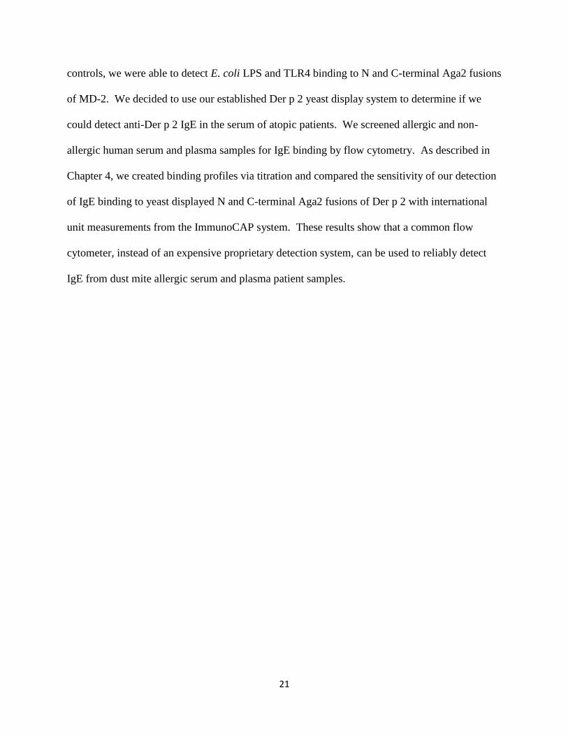
Lipopolysaccharide is essential for the survival of Gram-negative bacteria and is the
major outer membrane lipid component, with each bacterium containing approximately 106
molecules of LPS (4). As an amphipathic molecule, LPS contains hydrophobic fatty acid tails
and hydrophilic sugars and phosphate residues, which are organized into three structural and
functional regions: (a) a highly variable, repeating oligosaccharide (O-antigen), (b) additional
sugars (core), and (c) a glycosylated phospholipid (lipid A) (Figure 1.2A) (5).

4
In the model Gram-negative bacterium species Escherichia coli, lipid A contains a
hexaacylated β(1-6) diglucosamine backbone flanked by two phosphate groups at positions 1 and
4’(Figure 1.2A) (5). Since lipid A is responsible for anchoring LPS in the bacterial outer
membrane, it is typically hidden from immune detection. When lipid A is released during
bacterial cell division or death, it becomes detectable as an endotoxin. In fact, the lipid A portion
of LPS is capable of stimulating human cell activation to the same extent as full length LPS (6).
Injection of picogram levels of purified or synthetic lipid A also stimulates antibody production
and inflammation as much as full length LPS, leading to phenotypic fever and lethality in
C57BL/6 mice (7). Likewise, bacteria defective in lipid A synthesis cause less inflammation and
endotoxic shock in animal models (8).
Although the presence of LPS is conserved in Gram-negative bacteria, the length and
sugar content of the core and O-antigen regions can greatly vary between and within a species.
For example, E. coli and other pathogenic bacteria produce smooth to rough (Ra to Re)
chemotypes, modulating the serological specificity of their cell wall (Figure 1.2A) (5). Smooth
E. coli LPS contains the full complement of O-antigen and core sugars, making colonies formed
by these bacteria on agar appear smooth. Rough mutants of E. coli produce LPS containing lipid
A, but omitting either the complete O-antigen (in the Ra form) or both the O-antigen and
increasing amounts of the core polysaccharide region (in Rb to Re forms) (Figure 1.2A). Rough
mutants form bacterial colonies with rough edges when plated on agar. Most commercially
available preparations of smooth LPS also contain rough precursor forms of LPS (9) and may
contain lipoproteins (10). Variation and modification of the O-polysaccharide can also play a
role in immune evasion (8).

5
Similarly, the length, number and saturation of lipid A fatty acid tails can vary.
Hexaacylated E. coli lipid A most strongly stimulates inflammatory immune signaling (5).
Stimulation using tetraacylated E. coli lipid IVa (Figure 1.2B), a biosynthetic precursor of
hexaacylated E. coli lipid A, produced a differential effect in mice and humans. In mice, lipid
IVa is a weak agonist, but lipid IVa acts as an antagonist in humans (11, 12). Triacylation and
pentacylation are also represented in various bacterial species, resulting in either loss of activity
or antagonism (12-16).
Variation of the fatty acid content of lipid A between different bacterial species, or even
within the same species, is also a bacterial virulence strategy. For example, the human pathogen
Helicobacter pylori subverts complete activation when in the stomach by producing a
pentaacylated form of lipid A that lacks the 4’ phosphate group (17). The most striking example
of modulation of fatty acid content is the causative agent of plague, Yersinia pestis, which is
capable of changing from hexacylated LPS when grown at ambient temperature (26oC) to
tetracylated LPS when grown at 37oC. This temperature shift reflects the change in environment
as Y. pestis moves from the flea vector to the mammalian host and enables this pathogen to evade
sensing by the human innate immune system (18, 19).
1.3 Lipopolysaccharide Binding Protein (LBP)
The central pathway used by cells to detect LPS involves a series of LPS-receptor
interactions (Figure 1.1). The requirement for a receptor-ligand interaction step to sense LPS
was inferred by the exquisite sensitivity of LPS sensitive cells to detect picogram levels of
endotoxin (7, 20). Initial efforts to identify mammalian proteins capable of binding LPS first
revealed lipopolysaccharide binding protein (LBP). LBP is a 60 kD serum glycoprotein

6
produced primarily by liver hepatocytes and secreted in blood at levels of 5-15 g/mL (21, 22).
As an acute-phase protein, the concentration of LBP in serum can increase more than 30 fold
during an infection (23).
In 1990, the sequence of LBP was determined from cDNA clones, making LBP a
founding member of the BPI/LBP/PLUNC-like lipid-binding protein family (22). Although the
structure of LBP has not yet been solved, a 2.4 Å resolution structure of another family member,
bactericidal permeability increasing (BPI) protein, was determined in 1997 by Beamer and
colleagues (24). BPI is produced by neutrophils, and has an anti-inflammatory role of binding
and neutralizing LPS (25, 26). Since BPI and LBP share 50% sequence identity, the structure of
LBP is often modeled off of the structure of BPI (30,32). The structure of BPI revealed an
extended two domain boomerang-shaped protein fold (25, 27) containing the acyl chains of two
co-purified phospholipids bound in two hydrophobic pockets in the N-terminal pocket (24, 26).
Thus, the N-terminal pocket of LBP is hypothesized to accommodate the lipid A region of LPS
and stabilize lipid A phosphates using conserved basic amino acids (24, 25). Biophysical
measurements using fluorescently labeled LPS (28, 29), sucrose density gradients with
radioactively labeled LPS (28), and native polyacrylamide gel shift studies (30) have
demonstrated the capacity of LBP to disaggregate and catalytically deliver LPS to another
molecule called CD14.
1.4 Cluster of Differentiation Antigen 14 (CD14)
The focus of our structure determination effort, described in Chapter 2, is the next
endotoxin receptor in the central LPS sensing pathway, cluster of differentiation antigen 14
7
(CD14). CD14 was initially identified using two different monoclonal antibodies (anti-My4 and
AML-2-23) as a cell differentiation marker in normal myeloid lineage and acute myeloid
leukemia (AML) cells, leading to the initial complication of dual naming as both My 4 (31) and
My23 (32). Isolation of milligram quantities of CD14 protein from the urine of nephrotic
patients allowed Bazil and coworkers to conduct comparative studies, including antibody
binding, N-terminal sequencing, amino acid composition, native polyacrylamide gel molecular
mass and pI estimation, to conclude that My4 and My23 were inherently the same protein (33).
From these findings, the 3rd International Workshop and Conference on Human Leukocyte
Differentiation Antigens adopted the cluster of differentiation nomenclature CD14 (34).
CD14 is encoded by a single gene localized to human chromosome 5 (35) and mouse
chromosome 18 (36). Both species share 73% sequence identity in their coding regions, and
both species are capable of creating both membrane-bound and soluble protein products (Figure
1.1). Membrane CD14 (mCD14) is a ~55 kDa glycophosphatidylinositol (GPI) anchored
glycoprotein (37, 38) that is highly expressed on monocytes, macrophages, and neutrophils with
lower surface expression observed on a variety of other hematopoietic and stromal cells. Soluble
forms of CD14 (sCD14) exist in serum, cerebrospinal, and other body fluids. Soluble CD14 is
generated by at least three different mechanisms, which include bypassing GPI addition,
cleavage of the GPI anchor by phospholipase D, or direct proteolysis from the cell surface (39,
40).
The primary role of CD14 is to concentrate low levels of LPS to increase cellular LPS
sensitivity by binding and delivering LPS directly to the Toll-like receptor 4 (TLR4) signaling
complex (28, 30, 35, 40, 41), which will be discussed in detail in the next section of this chapter.
In concert with LBP, small gel filtration mixing studies have demonstrated a catalytic role for

8
CD14 in the delivery of LPS to the TLR4-MD2 complex (42-45). Membrane CD14 shuttles LPS
to TLR4 complexes in a two dimensional search (40); whereas, sCD14 enables cells that lack
endogenous mCD14, including most epithelial and endothelial cells, to respond to LPS (46).
Both soluble and membrane CD14 are capable of binding LPS over time, or as discussed earlier
in this chapter, CD14-LPS complex formation can be catalyzed by LBP (28-30). The protein-
protein interactions involved and mechanism required for LPS transfer from LBP to CD14 are
still unknown.
CD14-mediated delivery of Gram-negative bacterial LPS stimulates TLR4 activation,
initiating host inflammation by driving cellular production of pro-inflammatory mediators,
including cytokines, chemokines, and cell adhesion molecules (47). However, as a pattern
recognition receptor, CD14 has additional functions beyond LPS-dependent signaling. For
example, CD14 can act as a phagocytic receptor for whole bacteria or aggregates of LPS that
have been opsonized by LBP, thereby clearing infection and reducing inflammation (48-52).
Soluble CD14 is also able to bind and shuttle host phospholipids (53-55). In addition to LPS,
CD14 also enhances cellular inflammatory responses to a wide variety of acylated ligands from
Gram-negative and Gram-positive bacteria, including lipoproteins, lipoteichoic acid (LTA),
mycobacterial lipoarabinomannan (LAM), and atypical lipopolysaccharides, which stimulate
pro-inflammatory signaling through the Toll-like receptor 2 (TLR2) subfamily (56-66).
At the local site of infection and at low levels systemically, inflammatory signaling is
protective and results in the clearance of invading bacteria. However, large scale signaling or
systemic bacterial infection can actively propagate dysregulated pro-inflammatory signaling,
causing fatality through sepsis (65, 66). CD14 deficient mice have greatly diminished levels of
pro-inflammatory cytokines and have lessened symptoms of septic shock, which typically
9
present in mice as lethargy, breathing trouble, and ruffled fur. Likewise, CD14 deficient mice
survive when challenged with otherwise lethal doses of LPS or live E. coli bacteria (67). CD14-
knockout mice affirm the importance of CD14 in the TLR4/MD-2 pro-inflammatory signaling
pathway because both TLR4 and MD-2 deficient mice are also highly resistant to shock induced
by low doses of LPS or Gram-negative bacteria (68). Given this central role in TLR agonist
delivery, CD14 is an obvious drug target for the treatment of sepsis (65, 66).
As described in Chapter 2, we produced and purified human CD14 for x-ray
crystallography. Our solution of the x-ray crystal structure of human CD14 confirms the
presence of a hydrophobic cavity near the N-terminus, which could provide a binding pocket for
LPS. In Chapter 3, we describe efforts produce mouse CD14 protein to obtain a ligand bound
crystal structure of either human or mouse CD14. We used native polyacrylamide gel shift and
ForteBio Octet techniques to confirm the ability of both CD14 proteins to bind ligands and
utilized an IL-8 bioactivity assay to confirm the ability of both proteins to deliver LPS to
stimulate cell signaling. Although we were able to produce single crystals of native CD14,
diffraction quality liganded CD14 crystals did not develop for either construct. Our comparison
of our human CD14 structure and the previously published x-ray crystal structure of mouse
CD14 (69) provided additional insight into the N-terminal LPS binding sites of both proteins.
For example, we identified a hydrophobic surface near the rim of the binding pocket that may
stabilize hydrophobic residues in LPS fatty acid tails not accommodated by the amino terminal
cavity. Additionally, we identified an expanded pocket rim region in our structure that led us to
hypothesize that the previously determined rim region in mouse CD14 is expanded and the
hydrophobic rim residues in human and mouse CD14 may shift to accommodate the fatty acid

10
tails of LPS during ligand binding. These findings may encourage new therapeutic efforts
targeting the structure and function of human CD14 in the treatment of sepsis.
1.5 Toll-like Receptor 4 Complex (TLR4/MD-2)
The terminal receptor in the central pathway of LPS sensing is the TLR4 signaling
complex (Figure 1.1). In 1997, Bruce Beutler discovered the function of TLR4 was to act as the
transmembrane receptor required for LPS sensing (68), earning him the Nobel prize in 2012.
The gene encoding TLR4 and its role to transfer the LPS binding event across the cell membrane
to stimulate signaling were first determined by studying C3H/HeJ mice, which were resistant to
LPS despite the fact they had normal levels of CD14 (70, 71). Genetic mapping studies revealed
the defect in TLR4 protein was due to a single missense point mutation (P712H) in the TLR4
gene (68). Additional evidence for the role of TLR4 was provided by targeted knock out of
TLR4 in mice, which are also hyporesponsive to LPS (68, 72, 73).
TLRs were named Toll-like because they are orthologous to the Toll receptor found in D.
melanogaster, which enables the fruitfly to detect the presence of fungal infections (74) and to
establish the dorsal-ventral axis during embryogenesis (75). Each TLR protein in humans is
encoded by one of ten TLR genes and named TLR1 through TLR10. TLRs are produced in
multiple cell types and tissues, including lymphocytes, macrophages, dendritic cells, lung,
kidney, small intestine, stomach, and reproductive tissues, with varied expression levels for each
TLR (6). For example, the TLR4 gene is expressed in macrophages, but can also be found in
other leukocytes including dendritic cells, mast cells, neutrophils, eosinophils and natural killer
cells (2).

11
The structure of TLR4 represents the strong structure-function relationship typical of all
TLRs. When we initiated our work, only a handful of crystal structures of TLRs or their
accessory proteins had been solved. Now, ~30 human and mouse TLR x-ray crystal structures,
including the ligand bound extracellular domains of TLRs 1, 2, 3, 4, 5, and 6 (76-80) and the TIR
domains of TLRs 1, 2, and 10 (81, 82) have been determined. Although the crystal structure of
the extracellular and transmembrane domains of TLR10 and its natural ligand have not yet been
determined, our lab has discovered that TLR10 can cooperate with TLR2 to sense triacylated
lipoprotein PAMPs from bacteria, though TLR10 does not share common signaling pathways
with TLR2/1 (83).
Generally, TLRs fulfill two major functions: binding PAMP ligands and signaling the
binding event across a membrane. Ligand-dependent dimerization of TLRs stimulates the
production or modulation of pro-inflammatory mediators like interleukins, cytokines,
chemokines, adhesive proteins, tumor necrosis factor alpha (TNF-α), tissue factors, and co-
stimulatory molecules of the adaptive immune system (6, 84, 85). To accomplish these
functions, all TLRs are transmembrane receptors comprised of an N-terminal extracellular
domain, a type I transmembrane domain, and C-terminal cytoplasmic signaling domain (86).
The N-terminus of TLR4 constitutes the extracellular ligand binding domain. This
domain in TLR4 contains 19 leucine rich repeats (LRRs), though other TLRs can have as many
as 25 LRRs. The repetitive leucine rich sequence that creates the bent arc or solenoid LRR
protein folding motif was first identified in 1985 (87, 88). Each LRR of TLR4 forms a single
coil or loop of the solenoid comprised of a leucine rich beta strand, helix, and surrounding loops.
Tandem repeated LRR coils form a solenoid fold which is bent with a leucine rich parallel beta
sheet forming the concave face of the solenoid. Additionally, leucine, phenylalanine and other

12
hydrophobic side chain residues, which vary greatly in sequence in the helical and turn region on
the convex side, are buried in the interior of the coil. The fold is stabilized by hydrophobic beta-
beta interactions and bridging hydrogen bonds between asparagine residues (6). The solenoid
fold can be further subdivided into a central region and two capping regions at the N and C-
terminus of the extracellular domain. These capping regions vary in length and degree of rotation
relative to the central region. The N and C-terminal capping regions contain both hydrophobic
and hydrophilic residues to protect the hydrophobic beta sheet from the hydrophilic extracellular
milieu (89).
The second region of TLR4 is a type I, single pass helical transmembrane domain, which
is present in all TLRs and serves to transfer the signal derived upon PAMP binding across a
membrane. Human TLRs can be subdivided into two groups based upon the cellular location of
the membrane being spanned. TLRs 3, 7, 8 and 9 are found in the membranes of intracellular
compartments, such as endosomes and lysosomes where they sample viral and bacterial nucleic
acid material that has released following phagocytosis and particle destruction (12). TLRs 1, 2,
5, 6, and 10 are present in the plasma membrane and sample the extracellular environment (90).
For most TLRs, trafficking motifs have been identified in the sequences of the transmembrane
helix or adjacent residues to direct the receptor to the appropriate membrane (91). Plasma
membrane TLRs can be internalized to intracellular compartments following receptor mediated
endocytosis. For example, TLR4 is internalized in a membrane CD14-dependent manner (92).
Finally, the third domain in TLR4 is an intracellular, Toll/interleukin-1 Receptor (TIR)
cytoplasmic domain important for propagating the signal derived from PAMP binding. The TIR
domain is named to reflect a shared structural homology with both D. melanogaster Toll and
human interleukin-1 (IL-1), a pro-inflammatory cytokine produced in macrophages responsible

13
for stimulating chemokine induction, cell maturation, proliferation of B cells, and IL-2 release
(93, 94). As mentioned previously, intracellular signaling is propagated by dimerization of the
TIR domains of two TLRs, but the requisite binding partner varies for each TLR. For example,
TLRs 3, 5, 7, 8, and 9 all homodimerize to accommodate their ligands. As a subfamily, TLRs 1,
6, and 10 each form heterodimers with TLR2 during recognition of various di or tri-acylated
bacterial or fungal ligands. The TLR4 signaling complex is once again unique among TLR
family members because it not only contains a dimer of two TLR4 monomers to signal, but also
requires two 25 kD myeloid differentiation antigen (MD-2) accessory proteins in a
heterotetrameric arrangement to bind its acylated PAMPs (95).
MD-2 is essential for LPS binding and TLR4 signaling because it provides a hydrophobic
pocket to accommodate the acyl chains of TLR4 ligands, orienting the ligands in a way that
enables interactions with TLR4 (95). Thus, knocking out MD-2 results in the loss of LPS
signaling (72, 96). Addition of exogenous, soluble MD-2 to human corneal epithelial cells,
which are not capable of producing MD-2, permits sensitization to LPS in vitro (97). Likewise,
transfection of both MD-2 and TLR4 is necessary to enable non-responsive HEK293 or CHO
cell lines to respond to LPS (95, 98).
A discussion of MD-2 structures, with and without bound ligand, will provide a backdrop
for better understanding our solution of the human CD14 crystal structure and how it may
interact with LPS, as described in Chapter 2. The x-ray crystal structure of human MD-2 was
solved in 2007 in the absence (2E56.pdb) and presence of a bound E. coli lipopolysaccharide
ligand precursor called lipid IVa (2E59.pdb) to 2.0 Å and 2.2 Å, respectively (99). Both native
and lipid IVa bound structures show that MD-2 contains an immunoglobulin-like fold that is
conserved among MD-2 like (ML) family members. The fold consists of one sheet comprised
14
of three antiparallel strands and a second sheet with six antiparallel strands. Together these
two sheets produce a deep hydrophobic cavity (1,710 Å3) in a clamshell-like fold (99). MD-2
also contains three disulfide bonds (C25-C51, C37-C148, and C95-C148) and one free cysteine
(C133), which can cause disulfide-bond driven protein aggregation during purification steps
detectable by a ladder-like appearance on protein gels that is abrogated upon addition of a
reducing agent (100). Single point mutations of MD-2 at C95Y and Y102A result in complete
loss of LPS-dependent signaling (98, 101, 102). Comparison of lipid IVa bound and unbound
MD-2 structures show that ligand binding does not significantly alter the structure (0.3 Å
r.m.s.d.). Positively charged residues at the pocket entrance of MD-2 (K122 and R90) help
stabilize the negatively charged phosphates of bound lipid IVa, with all four of its fatty acid
chains completely sequestered inside the pocket.
Numerous ligand-bound TLR4/MD-2 structures were solved between 2007 and 2012
adding to our understanding of TLR4/MD-2 ligand recognition (Figure 1.3). For example, the
crystal structures of mouse MD-2 bound to both TLR4 and lipid IVa (3VQ1.pdb; 2.7 Å) (103)
and human MD-2 bound to lipid IVa (2E59.pdb; 2.2 Å) (Figure 1.3C) (99) have been solved.
Additionally, the crystal structures of mouse TLR4/MD-2 (2Z64.pdb; 2.84 Å) and human
TLR4/MD-2 (2Z65.pdb; 2.7 Å), with and without a synthetic tetraacylated TLR4 antagonist
called Eritoran (Figure 1.3B) (102, 104) were solved. Most importantly, the x-ray crystal
structures of homodimeric complexes of mouse TLR4/MD-2 (3VQ2.pdb; 2.5 Å) (103) and
human TLR4/MD-2 (3FXI.pdb; 3.1 Å) (Figures 1.3A and 1.3D) (77) bound to different
chemotypes of E. coli LPS have been determined.
Both human and mouse species share greater than 60% sequence identity between their
MD-2 or TLR4 proteins, better enabling direct comparison of these structures (102). However,
15
as mentioned in our prior discussion of LPS and lipid IVa, there are species differences
stemming from the recognition of lipid IVa which serves as a weak agonist in mice and an
antagonist in humans (11-13). All of the crystal structures with the tetraacylated ligands Eritoran
and lipid IVa show that the ligand acyl chains are completely sequestered in the pocket of MD-2
(95). However, in the pocket of human MD-2, the antagonists Eritoran and lipid IVa are rotated
by 180 degrees relative to the binding position of agonistic E. coli LPS (77, 102). Intriguingly,
this rotation of lipid IVa does not occur in mouse MD-2 (77, 103), but additional structures need
to be solved to confirm whether this rotation event is characteristic of all tetra-acylated inhibitory
molecules.
The size of the MD-2 pocket does not increase upon binding hexaacylated LPS. Instead,
one additional acyl chain of LPS is accommodated completely inside the pocket of both mouse
and human MD-2 by displacing the backbone and phosphate residues of LPS out of the pocket
and shifting the MD-2 loop containing phenylalanine 126 (F126 loop) by ~5Å, when compared
to its position in the structures of human MD-2 bound to Eritoran or lipid IVa (103). Typically,
MD-2 utilizes charged amino acid residues present just inside the cavity of MD-2 or located in
the adjacent TLR4 of the 1:1 TLR4/MD-2 dimer to orient and stabilize the phosphates of
tetraacylated Eritoran and lipid IVa, as described previously (77, 95, 99, 103). However, due to
the accommodation of the additional acyl chains in LPS, the newly exposed negatively charged
phosphates of LPS are now able to interact with the positively charged residues of the opposing
TLR4 monomer of the 2:2 TLR4/MD-2 dimer. Importantly, the sixth acyl chain of LPS is
partially omitted from the MD-2 pocket and, together with the displaced F126 loop of MD-2,
provides a new surface capable of binding the opposing TLR4 monomer to form the functional

16
TLR4/MD-2 signaling complex (Figures 1.3A through 1.3C). This structural configuration of
TLR4/MD-2 underlies the molecular basis of the strong agonist activity of LPS (77, 103).
1.6 Dermatophagoides pteronyssinus allergen (Der p 2)
An additional project described in Chapter 4 of this thesis focuses on a structural
homologue of MD-2 called Der p 2. Der p 2 is a 15 kD allergen that is found in the gut,
excretory system, and fecal matter of the European house dust mite Dermatophagoides
pteronyssinus (105, 106). House dust mites are a major source of indoor aeroallergenic proteins
(107) and mite extracts contain LPS (1.05 ng LPS/mg extract) (108). Although the geographical
localization of dust mites differ with changes in humidity, Dermatophagoides pteronyssinus
mites are found in every continent (109). Der p 2 was named following allergen naming
conventions determined by the World Health Organization and International Union of
Immunological Societies (WHO/IUIS). “Der” indicates the first three letters of the genus of
origin, “p” indicates the first letter of the species of origin, and the Roman numeral “2” to
indicates the order of purification. There are 15 different natural sequence variants of Der p 2,
each designated by four digits. Allergenic proteins have been divided into 19 different groups
based on structural homology with group members. Der p 2 is a member of the group 2
allergens, and additional group members are listed by the WHO/IUIS in a searchable database,
available at http://www.allergen.org/ (110). The 146 amino acid sequence of Der p 2 revealed
both a 17 amino acid N-terminal secretion signal and less than 16% sequence identity with MD-2
(111). However, other group 2 allergens have highly homologous sequences to Der p 2. For

17
example, Der f 2, a group 2 allergen produced by the American dust mite Dermatophagoides
farinae, shares 87% sequence identity with Der p 2 (112).
Der p 2 and other aeroallergens are processed by antigen presenting cells and displayed
for T cell recognition (Figure 1.4A). In individuals who are predisposed to an allergic response,
these epitopes stimulate helper T cells (TH2 cells) to produce cytokines like IL-4 which activate B
cells to generate IgE antibodies (113). These antigen-specific IgE antibodies bind their high
affinity receptor (FcεRI), which is primarily expressed on the surface of mast cells and basophils.
Upon re-exposure to and binding of the allergen, the FcεRI receptors of mast cells and basophils
are crosslinked and allergy induced signaling is activated. Activation causes release of allergen
mediators, including granules of histamine, and many downstream signaling effects, including
the production of cytokines IL-4, IL-13, and IL-5, which enable the recruitment and activation of
additional immune cells, like helper TH2 cells, macrophages, and eosinophils. Histamine,
cytokines, and other mediators released during the allergy-induced activation of these innate and
adaptive immune cells cause inflammation, smooth muscle cell induced airway constriction, and
formation of goblet cells producing large amounts of mucus, resulting in allergy symptoms.
Prolonged allergic response leads to chronic airway restriction and inflammation characteristic of
the most common form of asthma in the United States, which is called allergic asthma (114).
More than 80% of asthma suffers have detectable levels of IgE antibodies to Der p 2 (115).
To test the structural, functional, and allergenic properties of Der p 2, the allergen has
been purified from dust mite excrement and homogenate, as well as expressed as a recombinant,
soluble protein in many cell lines and in the yeast S. cerevisiae (116) and P. pastoris (117). Both
the NMR (1AV9) (118) and 2.15 Å x-ray crystal structures (1KTJ) (119) of Der p 2 have been
determined. As mentioned previously, the x-ray structure of Der p 2 is highly similar to MD-2
18
(Figure 1.4B), highlighted by the fact the structure of Der p 2 was used to generate an early
molecular model of the structure of MD-2 (120). Much like MD-2, Der p 2 contains an
immunoglobulin-like fold with three beta strands in the first beta sheet and five beta strands in
the second beta sheet. Unlike MD-2, Der p 2 contains only six cysteines, which are all
coordinated in three disulfide bonds leading to greater ease of purification than MD-2. Unlike
other allergenic proteins produced by D. pteronyssinus, Der p 2 is highly thermostable and is
able to accommodate temperatures in excess of 100oC without denaturation (121).
Because of the structural similarity between Der p 2 and MD-2, many pondered the
presence of functional homology. Recent work by Trompette and colleagues has shown that Der
p 2 is able to functionally replace MD-2 in mediating LPS signaling through TLR4 (122). First,
purified Der p 2 was capable of stimulating production of IL-8 in TLR4 positive HEK293 cells
that lacked MD-2 (122). To indirectly show that IL-8 production was due to the ability of Der p
2 to interact with TLR4, an alanine mutant of Der p 2 in tyrosine 91 was made. Der p 2 Y91 is
homologous to Y102, a conserved residue in MD-2. As mentioned during our discussion of the
structure of MD-2 in this chapter, mutation of Y102A in MD-2 resulted in loss of TLR4 binding.
Likewise, mutation of Der p 2 Y91A resulted in a reduction of IL-8. Further, addition of LPS
and Der p 2 to HEK 293 cells containing both MD-2 and TLR4 promoted increased production
of IL-8 compared to LPS stimulation of HEK 293 cells with MD-2 and TLR4 alone, suggesting
Der p 2 may contain an LPS delivery ability and may interact TLR4 and the MD-2/TLR4 induce
pro-inflammatory signaling (122). Thus, Der p 2 is suspected to be a potent allergen by binding
and delivering LPS to TLR4, without a requirement for MD-2.
In support of the functional studies, biochemical evidence shows HA-tagged Der p co-
immunoprecipitates with biotinylated E. coli LPS (122). Both Der p 2 NMR and x-ray crystal

19
structures showed two regions of additional electron density between the two beta sheets, which
in the x-ray structure was shown to accommodate 14 to 16 carbon chains. The authors indicated
this may serve as a binding site for the hydrophobic fatty acid tails of LPS, but noted the beta
sheets would need to open, like a clamshell, in order to more fully accommodate the ligand (118,
119). Additional LPS binding data is limited for Der p 2, but the homologous group 2 allergen
Der f 2 possesses a similar clamshell cavity in the x-ray crystal structure (123). An assay
measuring the intrinsic fluorescence of the only tryptophan present in MD-2 (W23), Der p 2
(W109), and Der f 2 (W109), has been used to measure LPS binding interactions. In all three
structures, this tryptophan residue is located near the entrance of the potential clamshell LPS
binding pocket with its side chain oriented into the pocket. When each protein was mixed with
E. coli 0111:B4 LPS, it resulted in decreased fluorescence and measurable dissociation constants
for MD-2 (65 x 10-9 M) and Der f 2 (6 x 10⁻⁸ M) (124), but Der p 2 had no detectible reduction of
fluorescence (125). NMR analysis of radiolabeled E. coli LPS isolated with Der f 2 revealed
chemical shifts suggestive of a bound glycolipid (124). Moreover, cross-linking studies revealed
a novel band at 32 kD, which Ichikawa and colleagues noted may correspond to Der f 2 cross-
linked with LPS (124). Finally, Der f 2 lysine residues and additional amino acids present in the
hydrophobic clamshell cavity had detectable NMR chemical shifts upon LPS binding (124).
Data also shows Der p 2 may interact with TLR4. Co-immunoprecipitation studies in the
work of Trompette and colleagues showed HA-tagged Der p 2 bound to both Flag-tagged MD-2
and, to a lesser extent, YFP-tagged TLR4. HA-tagged Der p 2 also co-immunoprecipitated with
the HA-tagged MD-2/YFP-tagged TLR4 complex. Further, Flag-tagged CD14 also co-
immunoprecipitated with HA-tagged Der p 2, suggesting Der p 2 may be able to accept LPS

20
from CD14 (122). However, the biophysical conformation of interactions and the molecular
basis for interactions between Der p 2 and TLR4, MD-2 and CD14 have not been determined.
Experimental evidence indicates Der p 2 may play more than one role in the induction of
allergy. For example, challenge of wild type mice with recombinant Der p 2 and picogram levels
of LPS resulted in helper T cell (TH2 cell) stimulated inflammation and increased plasma IgE
levels. Since the same amount of recombinant Der p 2 in TLR4-deficient mice did not induce
either inflammation, increased IgE levels, or an allergic response, TLR4 is required (122). Most
importantly, Trompette and colleagues were able to reconstitute cell infiltration and lung section
staining typical of an allergic asthma mouse model in MD-2 deficient mice by treatment with
Der p 2 (122). Similarly, Der f 2 has been shown to activate downstream TLR4 signaling in
alveolar macrophages, including stimulation of NF-ĸB (126). Thus, the ability of Der p 2 and
other homologous class 2 allergens to mimic the molecular interactions of MD-2 could be one
reason why these proteins are such potent aeroallergens. Thus, not only is Der p 2 the antigenic
target of the adaptive immune system in allergic patients, but Der p 2 also appears to act as an
adjuvant to the allergic response by stimulating inflammation and adaptive immune activation
through the TLR4 LPS sensing complex of the innate immune system.
In Chapter 4, we describe our efforts to better understand the molecular interactions
between Der p 2 and LPS sensing complex components. We used yeast display technology to
create stable, full-length construct of Der p 2 on the surface of S. cerevisiae yeast through N and
C-terminal fusions with the a-agglutinin adhesion subunit, Aga2. We tested the ability of our
Der p 2 Aga2 fusions to associate with anti-Der p 2 antibodies, E. coli LPS, and purified human
TLR4. Although we had limited success in identifying binding of N and C-terminal Aga2
fusions of Der p 2 to LPS, we were unable to reliably detect TLR4 binding to Der p 2. As
21
controls, we were able to detect E. coli LPS and TLR4 binding to N and C-terminal Aga2 fusions
of MD-2. We decided to use our established Der p 2 yeast display system to determine if we
could detect anti-Der p 2 IgE in the serum of atopic patients. We screened allergic and non-
allergic human serum and plasma samples for IgE binding by flow cytometry. As described in
Chapter 4, we created binding profiles via titration and compared the sensitivity of our detection
of IgE binding to yeast displayed N and C-terminal Aga2 fusions of Der p 2 with international
unit measurements from the ImmunoCAP system. These results show that a common flow
cytometer, instead of an expensive proprietary detection system, can be used to reliably detect
IgE from dust mite allergic serum and plasma patient samples.

22
Figure 1.1: Central pathway of LPS sensing
Lipopolysaccharide (LPS; red and white) from the outer membrane of Gram negative bacteria is
shuttled by lipopolysaccharide binding protein (LBP; green) to soluble CD14 [(sCD14-LPS)2;
blue] or membrane CD14 (mCD14-LPS; blue). Soluble CD14 is depicted as a dimer, but our
human CD14 is functional as a monomer. Either form of CD14 shuttles LPS to the MD-2 pocket
of the Toll-like receptor 4 signaling complex (LPS-MD-2-TLR4)2, which induces signaling upon
dimerization induced by LPS binding to MD-2. Reprinted with permission from Elsevier: Peri,
F. and M. Piazza. (2012). Therapeutic targeting of innate immunity with Toll-like receptor 4
(TLR4) antagonists. Biotechnol. Adv. 301 (1): 251-260 (Figure 1).
23
Figure 1.2: Lipopolysaccharide (LPS)
(A), Chemical structure of lipopolysaccharide (LPS) from E. coli, described in detail in this
Chapter. (B), Chemical structure of tetraacylated biosynthetic precursor for lipid A (lipid IVa)
from E. coli, described in detail in this Chapter.
A. E. coli lipopolysaccharide (LPS)
1-PO4
4’-PO4
B. E. coli lipid IVa
1-PO4
4’-PO4
Rough LPS Ra chemotype
Rc chemotype
Re chemotype
O-antigen
Core
Lipid A
Smooth LPS

24
Figure 1.3: Ligand Binding by Human MD-2 and TLR4
(A), The Ra chemotype of E. coli LPS (red) bound to human MD-2 (3FXI.pdb) causes a shift in
the position of the loop containing F126, which enables dimerization with the opposing TLR4.
(B), The synthetic inhibitor Eritoran (yellow) does not rotate in the pocket relative to the position
of E. coli LPS and does not induce a shift of the F126 loop, leaving the loop solvent exposed.
(C), Similar to Eritoran, the E. coli LPS biosynthetic precursor, lipid IVa, does not rotate in the
pocket relative to the position of E. coli LPS and does not induce a shift of the F126 containing
loop, leaving the loop solvent exposed. (D), Upon LPS binding, receptor dimerization occurs
creating an M-shaped dimer capable of activating signaling. Adapted and reprinted with
permission from authors: Manavalan, B., Basith, S., and S. Choi. (2011). “Similar structures but
different roles-an updated perspective on TLR structures.” Front. Physiol. 2: 41., Figure 4.
25
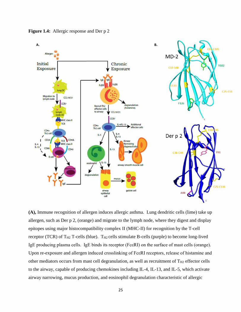
Figure 1.4: Allergic response and Der p 2
(A), Immune recognition of allergen induces allergic asthma. Lung dendritic cells (lime) take up
allergen, such as Der p 2, (orange) and migrate to the lymph node, where they digest and display
epitopes using major histocompatibility complex II (MHC-II) for recognition by the T-cell
receptor (TCR) of TH2 T-cells (blue). TH2 cells stimulate B-cells (purple) to become long-lived
IgE producing plasma cells. IgE binds its receptor (FcεRI) on the surface of mast cells (orange).
Upon re-exposure and allergen induced crosslinking of FcεRI receptors, release of histamine and
other mediators occurs from mast cell degranulation, as well as recruitment of TH2 effector cells
to the airway, capable of producing chemokines including IL-4, IL-13, and IL-5, which activate
airway narrowing, mucus production, and eosinophil degranulation characteristic of allergic

26
asthma. Along the way, the allergen Der p 2 may be capable of inducing pro-inflammatory
signaling through molecular mimicry of MD-2 expressed in these cells with Toll-like receptor 4
(not shown). Adapted and reprinted with permissions from: Holtzman, M. J. (2012). Asthma as
a chronic disease of the innate and adaptive immune systems responding to viruses and allergens.
J Clin Invest. 122 (8): 2741–2748. (B), MD-2 (cyan) and Der p 2 (blue) are structurally similar.
The position of disulfide bonds are indicated (yellow). Mutation of Tyr 91 was shown
previously to disrupt the ability of Der p 2 to induce MD-2/TLR4 responses (122). In Der p 2,
A115 (pink) is in a synonymous position with F126 in MD-2 (green), which lies in the loop that
shifts position upon agonist binding to create a new surface for interaction with TLR4 in the
TLR4 LPS signaling complex.
27
CHAPTER 2:
X-ray Crystallography of Human CD141
2.1 Introduction
CD14 plays a central role in the LPS signaling cascade by acting catalytically in concert
with LBP to deliver Gram-negative bacterial lipopolysaccharide to MD-2/TLR4, increasing the
sensitivity of the immune system to the presence of low levels of LPS (28, 30, 35, 40, 41). LPS
binding to MD-2/TLR4 enhances receptor dimerization, forming a new dimeric TIR interface for
signaling adaptor molecules that are capable of propagating pro-inflammatory signaling in an
effort to clear infection. As discussed in Chapter 1, widespread infection and activation of pro-
inflammatory signaling can cause fatality through sepsis (65, 66) and murine CD14 deficiency
reduced pro-inflammatory cytokine production and protected mice in septic shock models from
lethal doses of LPS (67). Determining the crystal structure of CD14 is an important step to
empower pharmaceutical intervention of CD14 function during sepsis.
The molecular interactions involved in the binding and delivery of structurally diverse
ligands including LPS presumably requires multiple protein-ligand and protein-protein
interaction sites on CD14 that are currently undefined. Additionally, the mechanisms involved
in delivery of these ligands to multiple Toll-like receptor complexes are also unresolved. To
address this gap in knowledge, many groups have purified various recombinant forms of soluble
CD14 using bacteria, yeast, insect, and human cellular expression systems (30, 127-137), often
with the goal of structure determination (135, 136). Currently, only the unliganded crystal
1 Reprinted in part, with permission, from Kelley, S. L., Lukk, Tiit, Nair, S. K., and R. I. Tapping. (2013). “The
crystal structure of human soluble CD14 reveals a bent solenoid with a hydrophobic amino-terminal pocket.” J.
Immunol. 190 (3): 1304-1311.
28
structure of mouse CD14, purified from SF9 insect cells, has been solved (69). Mouse CD14
possesses an N-terminal hydrophobic cavity that provides a putative binding site for LPS and
other acylated ligands. Despite previous efforts, no structure of human CD14 has been
determined and the structural similarity between mouse and human CD14 is unresolved. We
report the crystal structure of human CD14 (4GLP.pdb). Since sepsis is the most common cause
of death in US intensive care units (138), information garnered from the structure of human
CD14 may foster ligand binding structural studies or drug development efforts.
2.2 Materials and Methods
2.2.1 Reagents
Unless otherwise indicated, reagents and chemicals were purchased from Sigma-Aldrich.
DNA polymerases Taq, Platinum Taq, and Platinum Pfx, and bacteriophage T4 DNA ligase were
purchased from Invitrogen Life Technologies (Invitrogen). QIAquick Gel Extraction, QIAprep
Spin Plasmid Miniprep, HiSpeed Plasmid Midi and Maxiprep, and QIAquick PCR Purification
kits were purchased from Qiagen. Chemically competent E. coli DH5α cells and LB-agar plates
containing 100 g/mL ampicillin were purchased from the University of Illinois at Urbana-
Champaign (UIUC) cell Media Facility (kindly produced by Dr. Sandra McMasters). LB media
was produced by mixing 10g tryptone (VWR, Cat. No. 90000-286), 5g yeast extract (Fisher, Cat.
No. df0127179) and 10 g NaCl in 1L total volume of water prior to autoclaving. LB-Amp was a
mixture of LB media and 100 g/mL ampicillin sodium salt (Sigma-Aldrich, Cat. No. A9518).
6xDNA loading dye was prepared by mixing 10 mM Tris HCl, pH 7.6, 0.03% bromophenol
blue, 0.03% xylene cyanol FF, 60% glycerol, and 60 mM EDTA. 5xSDS-PAGE reducing
29
loading dye was prepared by mixing 250 mM Tris-HCl, pH 6.8, 10% SDS, and 30% glycerol
with 10% -mercaptoethanol (-me) or 1 M dithiothreitol (DTT). Polyclonal Goat Anti-Human
CD14 and two Mouse IgG1 Anti-Human CD14 monoclonal antibodies (63D3 and 28C5) were
kindly provided by Dr. Peter Tobias, The Scripps Research Institute, La Jolla, CA (139).
2.2.2 Cloning and Site Directed Mutagenesis
Two constructs with sequence similarity named HsCD14 and HLCD14 (Figure 2.1) were
created for structural studies. Both constructs share a sense primer containing an EcoRI site,
Kozak sequence for enhancing eukaryotic ribosome recognition during translation initiation
(140), and a portion of the N-terminal secretion signal of human CD14 beginning at aa 1, which
is removed during protein expression. Additionally, two antisense primers were designed to
contain varied portions of the C-terminal coding region of human CD14, in addition to a XbaI
cleavage site and thrombin digestion site (LVPRGS) (Figure 2.2A). Primer sets for the HsCD14
construct (aa 1-337) and HLCD14 construct (1-367) were utilized to amplify CD14 encoding
human genomic DNA (kindly provided by a former Tapping lab member, Dr. Katherine Omueti)
using a GeneAmp PCR System 9700 thermocycler (Applied Biosystems) according to the
polymerase manufacturer’s instructions (Invitrogen) for polymerase chain reaction (PCR) and
standard molecular biology techniques (141).
Both PCR products and a modified pDisplay cloning vector (Figure 2.2B), a kind gift
from Dr. David Kranz, University of Illinois, Urbana-Champaign, were digested with EcoRI,
XbaI and NheI restriction enzymes (New England Biolabs) and separated by agarose gel
electrophoresis run alongside a 1kB DNA ladder (Promega) with 6xDNA loading dye on a 1%
agarose gel. Calculated base pair (bp) sizes (Biology Workbench) visualized for products in
30
each reaction are undigested pDisplay vector (6,154 bp), EcoRI and NheI digested pDisplay
vector (5,542 bp & 612 bp), EcoRI and XbaI digested HLCD14 PCR product (1,081 bp), and
EcoRI and XbaI digested HsCD14 PCR product (991 bp). Following digestion, inserts and
vector were ligated using the manufacturer’s protocol for T4 DNA ligase (Invitrogen).
Site directed mutagenesis of the HsCD14 construct to create a cysteine to serine point
mutation (C306S) was completed by primer extension (Figure 2.2C) on a GeneAmp PCR
System 9700 thermocycler (Applied Biosystems). The HsCD14 C306S mutation was confirmed
via two methods. First, the loss of a PvuII digestion site (5’-CAG^CTG-3’) generated by one
nucleotide change (underlined) upon mutagenesis to serine (5’-CAGCAG-3’). Upon 1% agarose
gel electrophoresis of PvuII digested DNA encoding HsCD14 isolated by miniprep (Qiagen)
from heat-shock transformed E. coli DH5α colonies, identification of select colonies containing
the C306S mutation (#1 and #3) was conducted visually by locating two resulting bands (5,542
bp & 612 bp) instead of three resulting bands (3,418 bp, 3,172 bp, & 612 bp) (Figure 2.2D).
Second, DNA sequencing (UIUC Biotechnology Center) confirmed the single nucleotide
thymine to adenine expected result in the HsCD14 serine mutant (Figure 2.2E).
Additionally, pDisplay vectors containing DNA from both HLCD14 and HsCD14 were
also transformed into E. coli DH5α cells heat-shock (Invitrogen). Positive clones were selected
by ampicillin resistance on LB-Ampicillin agar plates. After overnight growth in the 37oC warm
room, single colonies were picked and grown in 3 mL LB-Amp media in 15 mL polystyrene BD
FalconTM round bottom tubes (BD Biosciences) in a 37oC incubator with shaking overnight.
Successful transformation of E. coli DH5α colonies was confirmed using Qiagen DNA
extraction and restriction enzyme digestion with EcoRI and NheI, followed by 1% agarose gel
electrophoresis to visualize the sizing of HLCD14 and HsCD14 inserts. The nucleotide
31
sequences of both constructs were confirmed by capillary electrophoresis DNA sequencing using
the ABI 3730 Genetic Analyzer and Data Collection software Version 3.0 (UIUC Biotechnology
Center).
2.2.3 Expression and Stable Cell Line Selection
After utilizing E. coli DH5α cells to amplify the vector, DNA was extracted with the
QIAprep spin miniprep, followed by ethanol precipitation and QIAquick purification. Human
embryonic kidney cells (HEK 293F) from Invitrogen Life Technologies (FreeStyle 293-F, Cat.
No. R790-07) were cultured, transfected with HLCD14 or HsCD14 containing modified
pDisplay vector, and stably selected using previously described methods (83). Unless otherwise
stated, HEK 293F cells were cultured in a humidified incubator at 37oC with 8% CO2. The
transfection reagent 293fectinTM (Invitrogen) utilized the cationic lipid transfection method (142)
to envelope CD14 containing pDisplay vector and transfer it into 1mL of 3 x 10⁷ cells/mL HEK
293F cells grown adherent in 24 well plates (TPP tissue culture plates, 24 well, flat bottom,
polystyrene; Cat. No. Z707791) with serum-free FreeStyle 293 expression medium (Invitrogen;
Cat. No. 12338-018). Following transfection, cells were placed in 96 well plates (TPP tissue
culture plates, 96 well, flat bottom, polystyrene; Cat. No. 92696) in quantities varied by limiting
dilution. Adherent, stably expressing HEK293F CD14 transfectants were selected by neomycin
resistance by passaging cells for one month in serum-free media containing 0.25 mg/mL G418
sulfate (Gemini BioProducts, Cat. No. #400-111P) stabilized with a 1:1 mixture of culture
supernatant from HEK 293F cells transfected with pDisplay vector alone. Protein expression
levels were qualitatively measured and selection for higher expressing transfected HEK 293F
cells was conducted by a 10% Tris-HCl sodium dodecyl sulfate polyacrylamide gel (10% SDS-

32
PAGE) separation followed by Western blotting with a polyclonal anti-CD14 antibody (data not
shown).
2.2.4 Purification
Both proteins were purified using a four-step chromatographic procedure with slight
variations. First, protein G affinity purification, modified from previously published methods
(83), was used to harvest the Fc-fusion (HLCD14-Fc or HsCD14-Fc) of both constructs from
two liters of HEK 293F culture supernatant seven days following seeding to 0.3 x 10⁶ cells/mL
in serum-free Freestyle 293F media with G418 (0.25 mg/mL) (Figures 2.3A and 2.4A).
Recombinant Protein G sepharose beads (GE Healthcare; 2 mL 50% slurry) were added to the
filtered supernatant with stirring overnight at 4oC. Protein G beads bound to CD14-Fc were
harvested by centrifugation at 2,500 x g, 15 min, 4oC and packed into a disposable PD-10
column (GE Healthcare). The column was washed with 0.02 M sodium phosphate, pH 7.0 and
eluted in 1 mL fractions for ten column volumes of 0.1 M glycine-Cl, pH 2.3 with 1 mL
neutralizing 1 M Tris-HCl, pH 9 buffer. A Precision PlusTM Dual Color molecular weight
marker (Bio-Rad) was used to qualitatively size protein in each elution fraction (5 L), which
was loaded with 5xSDS-PAGE reducing loading dye on a 10% SDS-PAGE (143) for
electrophoresis under reducing conditions. SDS-PAGE gels were subsequently stained with
Coomassie Brilliant Blue R-250, 40% methanol, and 14% acetic acid (stain solution) overnight
with rotation at room temperature, and destained with 40% methanol and 10% acetic acid
(destain solution) in iterative 20 minute incubations at room temperature. Original SDS-PAGE
gels were preserved using hydrated cellophane in a Tut’s Tomb gel tray (Idea Scientific). A
Western blot using anti-human CD14 polyclonal antibody confirmed Protein G elution fractions
33
1-5 contained HLCD14-Fc protein (Figure 2.3B). Similar Western blot analysis was completed
for HsCD14-Fc following Protein G (data not shown).
At this stage, the purification procedure varied between constructs. Thrombin (Novagen)
was added to HLCD14-Fc fusion protein in an effort to remove Fc by overnight incubation at
22oC visualized by 10% SDS-PAGE with Coomassie Brilliant Blue R-250 staining (Figure
2.3C). Additional effort to improve cleavage efficiency included the addition of 0.01% SDS
during thrombin digestion (data not shown) to no avail. HLCD14-Fc thrombin digestion
reactions were purified through an ÄKTAprime™ plus FPLC (GE Healthcare) fitted with two
1mL Hi-Trap Protein A high performance columns (GE Healthcare) run in tandem at a flow rate
of 1ml min-1 in 0.02 M Tris HCl, pH 8.5 buffer. Fractions were visualized by 10% SDS-PAGE
with Coomassie staining (Figure 2.3D). Due to incomplete isolation of thrombin cleaved
HLCD14 by Protein A affinity purification, pooled Protein A fractions containing HLCD14 were
further purified on two 5 mL Hi-Trap Q (GE Healthcare) anion exchange columns run in tandem
at 4oC by injection via a 10 mL loop and elution using linear NaCl gradient (0.02 M - 1 M) in
0.02 M Tris-HCl, pH 8.5 and a 1 mL/min flow rate. Fractions were visualized by 10% SDS-
PAGE with Coomassie staining correspond to the Hi-Trap Q elution profile (Figure 2.3E).
Human soluble CD14 (HsCD14 C306S) protein was similarly digested with thrombin
overnight in the absence of 0.01% SDS. Products of the reaction were separated by injection of
the reaction into ÄKTAprime™ plus FPLC fitted with two tandem 1mL Hi-Trap Protein A high
performance columns (GE Healthcare) run at a 1ml min-1 flow rate in 0.02 M Tris HCl, pH 8.5.
The flow through fractions were collected and re-injected into the FPLC on two tandem Protein
A columns three times consecutively to remove additional Fc. Flow through fractions containing
soluble HsCD14 were identified by 10% SDS-PAGE stained with Coomassie, pooled, and

34
concentrated to 1mL using an Amicon Ultra-15 unit (Millipore). Next, pooled HsCD14 was
injected into two 5 mL Hi-Trap Q anion exchange columns (GE Healthcare) run in tandem with
a linear NaCl gradient (0.02 M - 1 M) in 0.02 M Tris-HCl, pH 8.5 at 4oC using a 1 mL min-1
flow rate to further purify HsCD14 protein. CD14 containing fractions, as analyzed by 10%
SDS-PAGE stained with Coomassie, were concentrated using an Amicon Ultra-15 unit
(Millipore) to 0.5 mL. Finally, pooled HsCD14 was injected for size-exclusion chromatography
using a Superdex 200 column (GE Healthcare) equilibrated with 0.02 M Tris-HCl, pH 8.5 and
0.1 M NaCl at a flow rate of 0.4 mL min-1. Fractions containing HsCD14 were analyzed by 10%
SDS-PAGE stained with Coomassie, pooled, and concentrated to 10 mg ml-1 using an Amicon
Ultra-4 unit (Millipore) as measured by Pierce BCA assay (Rockford, IL). This four column
purification process yielded ~2 mg L-1 soluble, human CD14 with >90% purity ascertained
visually by 10% SDS-PAGE gel stained with Coomassie (Figure 2.4A) corresponding to the
Superdex 200 elution profile (Figure 2.4B).
2.2.5 Initial Crystallization Screening
Human CD14 (HsCD14 C306S) was crystallized using the hanging drop vapor diffusion
method. Crystallization conditions were initially screened using commercially available sparse
matrix kit conditions including Hampton Research Crystal Screens I and II, Emerald Biosystems
Wizard I and II, Hampton Research Natrix, and Hampton Research Salt Reaction, as well as a
handmade crystal screen targeting the previously published mouse CD14 crystallization
condition (1 l protein solution and 1 l of crystallization buffer containing 100 mM sodium
HEPES (pH 7.5), 1.9 M Li2SO4, and 5 mM NiCl2) (69). Each condition was tested on a 12 mm x
0.22 mm siliconized glass cover slide (Hampton Research) over a VDX48 plate with sealant
35
(Hampton Research) by mixing 1 l of each screening solution with 1 µl protein solution
(10 mg ml-1 protein in 0.02 M Tris-HCl, pH 8.5 and 0.1 M NaCl) equilibrated against 300 µl
screening solution in the reservoir. Sparse matrix screening in hanging drop vaporization trays at
22oC, 18oC, and 4oC lead to three conditions with single crystals (Figure 2.5): Emerald
Biosystems Wizard I #28 (20% PEG 3,000, 0.1 M HEPES, pH 7.5, and 0.2 M NaCl) after 2 - 4
days at 22oC; Hampton Research Crystal Screen I #18 (20% PEG 8,000, 0.1 M Na cacodylate,
pH 6.5, and 0.2 M Mg(OAc)) after 6 days at 18oC; and Emerald Biosystems Wizard II #28 (20%
PEG 8,000, 0.1 M MES, pH 6.0, and 0.2 M Ca(OAc)2) after 3 weeks at 22oC (144-146). The
best initial diffraction resolution (6.2 Å) was obtained using Emerald Biosystems Wizard II #28
(Figure 2.6).
2.2.6 Crystallization Optimization
Extensive work was conducted to improve diffraction resolution from 6.2 Å. Improved
purification buffer conditions were identified using a solubility screen (147). A PEG 6,000
sparse matrix screen (Hampton Research) and handmade crystallization buffers were prepared
varying salt, precipitant, and buffer concentrations nearby the initial crystallization conditions,
leading to an optimized hit at K#19 with 4.02 Å resolution (Figure 2.7). Additionally, 1 l of
each initial crystallization solution and 1 µl protein solution (10 mg ml-1 protein in 0.02 M Tris-
HCl, pH 8.5 and 0.1 M NaCl) was combined with 0.1 l of either 96 distinct Additive Screen
reagents (Hampton Research), 96 distinct detergents from Detergent Screens 1-3 (Hampton
Research), 96 additives in Silver Bullets (Hampton Research) and Heavy Atom screens
(Hampton Research). Alternatively, in a 3 l hanging drop, 1.5 l protein and 0.9 l well
36
solution was mixed with 0.6 l additives to test the effects of adding a 5 fold dilution of
Hampton Research additives, detergents, and Silver Bullet reagents.
In vitro proteolytic screening was also conducted through the addition of various
dilutions (10-100 fold) of stock solutions of commercially available proteases (Sigma) including
chymotrypsin (1 mg/mL), trypsin (1.5 mg/mL), Glu C V8 (2.0 mg/mL), subtilisin A (1.0
mg/mL), thermolysin (1.0 mg/mL), and elastase (1.0 mg/mL) to 30 g purified HsCD14 protein
in 5 L reaction volume for 30 and 100 minutes at room temperature prior to the addition of
loading dye and visualization by 10% SDS-PAGE (data not shown) (148, 149). PeptideCutter
(ExPASy; available at http://web.expasy.org/peptide_cutter/) predicted the following number of
cleavage sites for each protease: chymotrypsin (15), trypsin (30), Glu C (34), elastase (74), and
thermolysin (123). A 10 fold dilution of each protease was also added directly to the hanging
drop of the 6.2Å condition to screen proteolysis in situ.
Surface lysine residues were reductively methylated using dimethylamine-borane
complex using the manufacturer’s protocol (Sigma) (148, 149). The methylation reaction
involved a 2 hour incubation at 277K with gentle mixing of small-gel filtration purified HsCD14
protein in 50 mM HEPES (pH 7.5) with 250 mM NaCl buffer with 20 uL freshly prepared 1 M
dimethylamine-borane complex (ABC; Fluka #15584) and 40 uL 1M formaldehyde (Fluka
33220) per mL solution. In two hour increments, 20 uL ABC and 40 uL formaldehyde were
added. Then, 10 uL of ABC was added and the reaction was incubated overnight at 4oC with
rotation. The reaction was quenched with 1M Tris-HCl buffer. The sample was centrifuged to
remove any protein precipitant before an additional round of size exclusion chromatography.
Fractions from the primary elution peak were combined and concentrated to 10 mg/mL and
MALDI-MS (UIUC Mass Spectrometry Lab) using a sinapinic acid matrix to detect methylation.

37
Since glycosylation also contributes to protein heterogeneity, we attempted to inhibit
glycosylation by treating 1 mL cultures of transfected HEK 293F cells with the glycan inhibitors
Streptomyces tunicamycin (T7765) and benzyl-N-acetyl-a-D-galacotsamide (BAG; B4894)
(Sigma), screening 12 inhibitor concentrations (0, 0.25, 0.5, 1, 2, 4, 8, 16, 32, 64, 128, and 256
g/mL). Additionally, the lowest 0.25 g/mL concentrations of both inhibitors tested were
added to 30 mL cultures of transfected HEK 293F cells in a separate test for cell viability upon
treatment (data not shown). In a second method, N-linked glycans were removed enzymatically
by incubating HsCD14 for one hour at 37oC in 1xG7 buffer (0.05 M sodium phosphate, pH 7.5)
containing 1,000 U peptide N-glycosidase F (PNGaseF) (New England Biolabs) prior to Hi-Trap
Q anion exchange chromatography (GE Healthcare), as described previously in Materials and
Methods in this Chapter (Figure 2.4C).
2.2.7 Cryoprotection Optimization and Crystal Harvesting
Cryopreservation reagents were screened using six different crystal handling techniques,
including: (a) movement of the crystal to a hanging drop containing cryo, gentle crystal rolling,
careful harvesting in the loop and freezing (direct transfer); (b) movement of the crystal to
hanging drops containing decreasing cryo between drops varied by a constant dilution factor,
with or without crystal ejection from the loop, and freezing (% stepwise); (c) transfer of the
crystal from the drop to the loop and positioning the loop in a second drop containing the cryo
solution for 1-30 seconds without crystal ejection from the loop (looped); (d) retention of the
crystal in the original drop, which was diluted 2 fold with cryo (dilution); (e) movement of the
crystal to a new hanging drop containing 2.5% glycerol for 30 minutes – 1 week (soaking); and
(f) plunged directly into liquid nitrogen without cryo preservation solution (no cryo). Incubation

38
time of the crystal in the hanging drop containing cryo was also varied for the direct transfer and
% stepwise techniques. Multiple handmade cryo preservation solutions were tested, with
stepwise gradients indicated in parenthesis, including: 10-20% 2,3-butanediol (10%), 10-20%
xylitol (10%), 5-30% trehalose (5%), 5-30% D,L-threitol (5%), 2-30% ethylene glycol (2%), 2-
30% sucrose (2%), 2-30% glucose (2%), 2-30% glycerol (2%), 2-30% MPD (2%), and 2.5-20%
PEG MW 200, 400, 600, 3350, 4k, 5k, 6k, and 8k (2.5%, 5%, and 10%). Commercially
available cryopreservation solutions were also screened, including the Crystal Screen Cryo kit
(Hampton).
Using methods described by Heras and Martin (148), crystals were also dehydrated prior
to vitrification via serial transfer on cover slips exposed to ambient room temperature or on cover
slips exposed to reservoir for various time points. Additionally, macromolecular crystal
annealing after freezing using multiple crystal warming time points and flash annealing after
freezing in a liquid nitrogen stream was attempted for varied durations (148, 150-152). Finally,
room temperature diffraction was conducted at with single crystals harvested on MicroRT
MiTeGen mounts using manfacturer’s protocols (153).
Ultimately, single crystals of purified PNGaseF deglycosylated CD14 were grown at
22oC to a maximum dimension over ~40 days on a 22 mm x 0.22 mm siliconized glass cover
slide (Hampton Research) over a VDX24 plate with sealant (Hampton Research) by mixing
1.5 µl 30% PEG 6,000, 0.1 M MES, pH 6.0, and 0.2 M Ca(OAc)2 crystallization solution with
1.5 µl protein solution (10 mg ml-1 protein in 0.02 M Tris-HCl, pH 8.5 and 0.1 M NaCl)
equilibrated against 1 mL screening solution in the reservoir (Figure 2.8). Crystals were
harvested directly by looping from the drop and plunged immediately into liquid N2 without any
additional cryoprotection or crystal handling.
39
2.2.8 Data Collection and Structure Determination
A native diffraction data set including 130 frames at 1o rotation/sec was collected from a
single vitrified crystal in the optimized 30% PEG 6,000, 0.1 M MES, pH 6.0, and 0.2 M
Ca(OAc)2 crystallization solution on the LS-CAT beamline, Sector 21 ID-F (Advanced Photon
Source, Argonne, IL). Diffraction data were processed to 4.0 Å resolution by a postdoctoral
student in the Nair Lab, Dr. Tiit Lukk. Data were indexed and integrated using XDS, followed
by scaling and merging with XSCALE (154). The structure solution was determined via
molecular replacement with Phaser in the CCP4 suite, using the molecular coordinates of mouse
CD14 (1WWL.pdb), modified with CHAINSAW (155). Iterative cycles of model fitting were
completed in COOT, followed by refinement in REFMAC (156). The final solution of human
CD14 was deposited (http://www.pdb.org/pdb/search/structidSearch.do?structureId=4GLP) and
verified with Molprobity in PHENIX (157). RAMPAGE (University of Cambridge) was used to
create the Ramachandran plot shown in Figure 2.9. Data collection and refinement statistics are
summarized in Table I.
2.3 Results
2.3.1 Generation of Human CD14 for Crystallization
A number of important parameters were considered in generating a soluble form of
human CD14 suitable for crystallization. Since soluble forms of CD14 (aa 20-375, where amino
acid numbering starts with the first methionine of the unprocessed CD14 protein) are naturally
heterogeneous at the C-terminus, we generated an expression construct (HLCD14) which
40
contained the naturally occurring N-terminal secretion signal and the majority of the coding
region of CD14, yet lacked the C-terminal protein sequence required for GPI anchoring. This
was done with the knowledge that C-terminal truncation of CD14 does not affect LPS associated
bioactivity, which resides entirely within the amino-terminal half of the protein (158, 159).
Additionally, since our shorter construct (HsCD14) is truncated between C306 and C352, which
are predicted to form the final disulfide bond, we mutated the unpaired C306 residue to serine.
Both HLCD14 and HsCD14 were expressed as an Fc tagged fusion protein and harvested from
HEK 293F cell supernatant using protein G sepharose beads. An engineered thrombin cleavage
site present in both constructs was utilized for removal of the Fc tag. However, HLCD14
showed incomplete cleavage after thrombin digestion (Figure 2.3C); whereas, the Fc fusion was
more completely removed from HsCD14 upon thrombin digestion. Thrombin digestion in the
presence of 0.01% SDS and further purification steps, including protein A affinity
chromatography and Hi-Trap Q anion exchange chromatography (Figure 2.3D and 2.3E), did
not increase levels of thrombin digestion or effectively separate HLCD14 from the Fc fusion.
So, we set HLCD14 aside and redoubled our efforts purifying HsCD14 for structure studies.
Following thrombin digestion, protein A affinity chromatography and small gel filtration
of HsCD14 resulted in highly pure, soluble protein suitable for crystallization (Figure 2.4A).
LPS binding studies and an ELISA based assay measuring production of IL-8 were used to
determine that our purified protein was bioactive. These studies will be discussed in detail in
Chapter 3. To further ensure improved protein homogeneity for crystallization, we removed
potentially heterogeneous N-linked glycans using PNGase F. This enzymatic deglycosylation
was preferred because mutation of the N-linked glycosylation sites in CD14 prevents cellular
41
secretion (52). As observed by others, PNGase F treatment reduced the molecular weight of
CD14 by approximately 10kD (160, 161) (Figure 2.4C).
2.3.2 Crystal Structure of Human CD14
Human CD14 crystallized in the trigonal space group P3221 (Table I). As listed in
Table I, five percent of reflections were omitted from refinement and used in the cross-validation
calculation of the 32.1% R-free value. A Ramachandran plot was also created for validation,
showing 8.1% outliers (Figure 2.9). The crystal structure begins with aa 26, indicating complete
removal of the leader sequence by HEK 293F cells during secretion along with low electron
density for the first six amino acids, aa 20-25. The overall X-ray crystal structure of human
CD14 reveals a bent solenoid that is formed by 11 leucine rich repeats, each creating a single
turn (Figure 2.10A). This is consistent with the leucine rich repeat sequence motifs of human
CD14 (162), and the expected bent solenoid fold generally characteristic of leucine rich repeat
containing proteins (163). The structure also reveals five alpha helices on the convex side, as
well as eleven beta strands that are coordinated in a parallel beta sheet on the concave side of the
bent solenoid. The pairwise proximity between cysteine residues in the crystal structure at
positions 34 and 51, 187 and 217, as well as 241 and 272 are consistent with the expected
disulfide bonds of human CD14, which have been shown to be important for proper protein
folding (135) (Figure 2.11). Interestingly, recombinant purified human CD14 (HsCD14) lacking
the C306S mutation, a mutation originally designed to avoid unnatural disulfide binding, did not
crystallize and formed aggregates in the crystallization drop (data not shown).
Another key structural feature present in the crystal structure of human CD14 is an N-
terminal pocket or cavity (Figure 2.12A). Although we cannot know with certainty the position

42
of each side chain at this resolution, the overall cavity of human CD14 is comprised largely of
hydrophobic residues present in α1 through α5 and 1 through β6 along with their connecting
loops. It is reasonable to assume that this hydrophobic pocket serves as the binding site for the
lipid chains of LPS and other acylated CD14 ligands, especially as the regions making up the
pocket are entirely contained within the N-terminal region (aa 20-171) of CD14 that has been
shown to be sufficient for bioactivity (158, 159).
It is instructive to examine the rim residues of the CD14 hydrophobic pocket as these
residues likely engage portions of LPS, or other acylated CD14 ligands, that are not
accommodated by the pocket itself. Positively charged residues are located in the rim at K71
and R72 and just outside the rim at R80, K87, and R92 (Figure 2.13A). Hydrophobic residues at
W45, F49, V52, F69, Y82, and L89 encircle the rim and their side chains overlay the entrance to
the pocket. When measured using the program Chimera (164), the pocket diameter of human
CD14 from W45 to L89 is 15.7 Å wide. Interestingly, the hydrophobic pocket entrance is
subdivided by an interaction between the α1 residue F49 and the α3 residue Y82 (Figure 2.13B).
2.3.3 Comparison of Human and Mouse CD14 Structure
Human soluble CD14 purifies and crystallizes as a monomer (Figure 2.4A and 2.4B). In
contrast, the murine protein crystalized as a dimer with the dimer interface comprised of C-
terminal beta strand interactions (69). The mouse and human CD14 structures are comparable at
the C-terminus because both were truncated at similar positions to make them more amenable to
crystallization (69). Different crystallization conditions, constructs, and cellular expression
systems may account for the differences observed in crystal packing between the two proteins.
Nevertheless, it is unlikely that the C-terminal dimerization of truncated mouse CD14 is

43
biologically relevant (69), as numerous biophysical studies support the idea that bioactive
soluble CD14 is a monomeric protein (28, 30, 40, 42-45).
Human and mouse CD14 crystal structures contain a bent solenoid fold and asparagine
ladder, which are structural features typical of leucine rich repeat proteins (163). In fact, both
proteins are highly superimposable with a r.m.s.d. of 1.089 Å (Figure 2.10B). The secondary
structure of human CD14 includes alpha helices and beta strands that are equivalent to α1, α4,
α5, α6, α7, and 3-13 of the published mouse CD14 structure (Figures 2.10B and 2.11) (69).
Although helical coils exist in human CD14 at locations that are synonymous to helices α2 and
α3 of mouse CD14, the structure resolution falls just outside the range needed to determine
helical packing with certainty (Figures 2.10B and 2.11) (165, 166). Human and mouse CD14
both contain a large N-terminal hydrophobic pocket consistent with their similar roles in the
binding and delivery of various lipidated molecules including LPS (Figures 2.12A and 2.12B).
Similar to human CD14, the main pocket and sub-pockets present in mouse CD14 are comprised
of hydrophobic residues within α1-5, 1-6, and their connecting loops (69). Using a structure
based sequence alignment, created using MultiSeq in VMD (167), we compared the general
amino acid characteristics present in the residues at the rim of the proposed binding pocket in
both structures (168). The authors of the mouse CD14 crystal structure (69) identified
hydrophilic and hydrophobic amino acids in rim residue positions indicated in blue (Figure
2.13). Many of these residues, including P39, W41, F45, and Q77 are conserved with rim
residues at the pocket entrance of human CD14. Thus, the N-terminal hydrophobic pocket found
in our crystal structure revealed a conservation of pocket size and hydrophobicity with that of
mouse CD14.

44
Despite the wide ranging structural similarities between mouse and human CD14,
differences do exist. For example, although the amino acids found at equivalent positions of
mouse and human CD14 generally have similar physical properties, there is variation in the
charge of certain rim residues. Namely, the positively charged K38 residue present in the mouse
protein is not retained in the human protein. Likewise, the negatively charged D44 residue in
human CD14 is not found in mouse CD14. Finally, the diameter and subdivision of the entrance
to the pocket differs between species. The mouse crystal structure reveals an 8.0 Å wide
entrance to the pocket that is bounded on one side by a hydrophobic interaction between F45 and
F78 (69) (Figures 2.13A and 2.13B). Residues F45, L49, and I81 in the mouse CD14 structure
create additional hydrophobic interactions which shield this side of the pocket. These
hydrophobic interactions of the mouse CD14 pocket are not present in the human CD14 structure
whose pocket extends to include the rim residues T85 and L89 (Figures 2.13A and 2.13B). The
biological significance of these differences between the pocket and associated rim of mouse and
human CD14 is difficult to define without structural information from ligand bound proteins.
2.4 Discussion
CD14 sensitizes cells to LPS by delivering this bioactive lipid to MD-2; an essential
component of the TLR4 signaling complex. All members of the MD-2 related lipid-recognition
protein superfamily are characterized by two sheets organized in an α cup fold to create a
centralized hydrophobic LPS binding cavity (169). In the crystal structures of human and mouse
MD-2 bound to lipid IVa, all four acyl chains of the ligand are buried inside and occupy the
majority of the hydrophobic pocket volume (99, 103). Crystal structures of MD-2 in the context

45
of the entire mouse and human TLR4/MD-2/LPS homodimeric signaling complex reveals a
narrow and deep (1720 Å3 volume) hydrophobic binding pocket that completely sequesters five
of the six fatty acid chains (77, 170). The remaining unbound acyl chain lies along the surface of
MD-2 and, together with the F126 loop of MD-2, creates a new hydrophobic patch that promotes
homodimer formation by association with TLR4 from an adjacent TLR4/MD-2/LPS complex
(77, 95).
Compared to MD-2, CD14 has been reported to afford less protection against enzymatic
removal of secondary fatty acids of bound LPS (171). This probably reflects that fact that at
about 820 Å3, the hydrophobic pockets of both human and mouse CD14 are considerably smaller
than that of MD-2 (69) (Figures 2.12A and 2.12B). Similar to MD-2, previous circular
dichroism, tryptophan fluorescence, and NMR studies suggest that LPS binding does not induce
large structural changes in human CD14 (136, 172). Given these volume constraints and pocket
rigidity, it is unlikely that CD14 can accommodate all the acyl chains of LPS. This may
contribute to the reduced affinity of CD14 for LPS compared to MD-2 (43, 44).
Given the physical limitations of the hydrophobic pocket, we hypothesize that CD14
stabilizes additional fatty acid chains on a hydrophobic cluster located near α1 just outside of the
pocket entrance. A similar hydrophobic cluster is present in a comparable location in the crystal
structure of mouse CD14 (69) (Figure 2.12B). In human CD14, the hydrophobic cluster is
comprised of the amino acids F32, F49, V52, S53, A54, V55, and L89 (Figure 2.12B). This
cluster and associated acyl chains of LPS could perhaps create a new protein-protein interaction
surface that may help facilitate the transfer of LPS to another molecule such as MD-2.
Our crystal structure confirms that the hydrophobic pocket encompasses the N-terminal
half (aa 20-171) of CD14 that has been shown to be the bioactive portion of the protein (51).

46
Previous literature strongly supports the hypothesis that LPS binds the pocket. For example, aa
26-29 were bound by the anti-CD14 antibody 3C10 neutralizing the ability of CD14 to bind LPS
or activate cell signaling (173). Alanine mutagenesis of this same amino acid region resulted in
the loss of cellular activation (173). Deletion of human CD14 aa 28-31, 41-44, or 78-82 resulted
in diminished LPS binding and partial inhibition of bioactivity (174). Deletion of aa 54-58
caused loss of LPS binding and abrogated SW620 cellular activation (174, 175). Human CD14
aa 57-64 are protected from endoprotease AspN digestion upon binding LPS (172), are bound by
the anti-CD14 neutralizing antibody MEM18, and their deletion resulted a lack of LPS binding
and cellular activation functionality (173, 176). Alanine scanning mutagenesis of the entire N-
terminal region of CD14, encompassing aa 20-171, revealed mutation of aa 28-32 blocks LPS
binding and mutation of aa 58-63 blocks both LPS binding and cellular activation (51). Thus,
extensive deletion mutagenesis, alanine scanning mutagenesis and epitope mapping of inhibitory
anti-CD14 antibodies have revealed four separate regions (aa 26-32, 41-44, 56-64 and 78-83) in
the N-terminal half of CD14 that are critical for LPS binding and cell activation (Figure 2.13A).
We have identified in our crystal structure the location of these four functionally important
regions near β1, within the loop preceding α1, across β3 and within α3, respectively (51, 173-
176). The residues in the first three regions near α1, 1 and 3 appear to fulfill critical roles in
forming the pocket entrance, capping the leucine rich repeat beta sheet, and/or stabilizing proper
folding of the solenoid.
Residues in the fourth region encompass a Y82 loop located on the opposite side of the
pocket entrance. Although Y82 appears to weakly block the entrance of the pocket through
interaction with F49, we believe that upon binding LPS, these hydrophobic residues may change
position to accommodate the acyl chains within the pocket. Although the corresponding

47
interaction is represented by F45 and F78 in mouse CD14, additional hydrophobic residues
present in mouse CD14, but absent in human CD14, close off the pocket entrance (Figures
2.13A and 2.13B). Overall, our human CD14 crystal structure allows for the resolution of an N-
terminal hydrophobic pocket predicted by multiple studies to be the LPS binding site. Compared
to mouse CD14, our work provides an alternative definition of rim residues, reveals an expanded
pocket entrance, and identifies a unique hydrophobic bridge feature that incorporates residues
shown to be important in LPS binding and cell activation (Figures 2.13A and 2.13B).
Hydrophilic residues at the opening of MD-2 have been shown to be important for LPS
binding by properly aligning the negatively charged 1 and 4’ phosphates of LPS (77, 128).
Similar to MD-2, charged rim residues in CD14 may be important for orienting or binding LPS.
For example, previous work has shown that mutation of E47, which falls near the front of the rim
in α1, to either lysine or arginine can block binding of P. gingivalis LPS (177). Solution NMR
spectroscopy analyzing soluble CD14 aa 20-171 bound to the Re chemotype of LPS did not have
structural assignments available upon publication, but does show high average temperature factor
evidence for hydrophilic residues at the rim suggesting local flexibility to accommodate LPS
binding (136). Further work to develop a ligand bound CD14 structure would help to clarify the
roles of both the hydrophobic binding pocket and the hydrophilic rim residues required for ligand
binding.
A number of natural and synthetic acylated agonists and antagonists are known to be
shuttled by CD14. For example, CD14 delivers tri- and di-acylated agonists to complexes
formed by members of the TLR2 subfamily (56-63, 178). Natural antagonists of LPS, including
R. sphaeroides pentacylated lipid A, tetracylated lipid IVa and monophosphoryl lipid A, an
approved vaccine adjuvant in Europe, are shuttled by CD14 to MD-2/TLR4 (12, 170, 179-181).
48
Eritoran, a synthetic LPS antagonist, that is structurally similar to lipid IVa, is also delivered to
MD-2 by CD14 (102, 182). More recently, synthetic LPS inhibitors derived from either
diacylated sugars or tetraacylated sulfate containing compounds have been identified and shown
to competitively block LPS binding to CD14 (183-185). It is highly likely that the hydrophobic
binding pocket in human CD14 accommodates these natural and synthetic agonists and
antagonists especially as they contain fewer acyl chains than hexaacylated enteric bacterial LPS
(180, 186). Since TLR4 inhibitors are largely LPS mimics that are either delivered by CD14 or
directly compete with LPS for interaction with CD14, the structural information presented here
may further the design of drugs for the treatment of sepsis and other inflammatory diseases.

49
Figure 2.1: Human and Mouse CD14 Sequence Alignment
Sequence alignment using the default parameters of ALIGN (SDSC Biology Workbench;
available at http://workbench.sdsc.edu) indicating the sequence of two human (HLCD14 &
HsCD14) and one mouse (MCD14) CD14 construct. DNA sequencing confirmed our constructs
agreed with sequences deposited in SwissProt (available at http://www.uniprot.org/) for human
CD14 (P0857) and mouse CD14 (P10810). Identical (magenta), conserved (light blue) and
cysteine (orange) amino acids are highlighted. Human CD14 and mouse CD14 amino acid
numbering is indicated above and below the alignment, respectively. Residues predicted by
SwissProt to be absent in the mature form of the protein are indicated (gray). The initial and
terminal amino acids (black boxes) of our CD14 constructs are displayed.
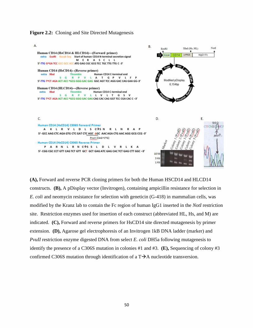
50
Figure 2.2: Cloning and Site Directed Mutagenesis
(A), Forward and reverse PCR cloning primers for both the Human HSCD14 and HLCD14
constructs. (B), A pDisplay vector (Invitrogen), containing ampicillin resistance for selection in
E. coli and neomycin resistance for selection with geneticin (G-418) in mammalian cells, was
modified by the Kranz lab to contain the Fc region of human IgG1 inserted in the NotI restriction
site. Restriction enzymes used for insertion of each construct (abbreviated HL, Hs, and M) are
indicated. (C), Forward and reverse primers for HsCD14 site directed mutagenesis by primer
extension. (D), Agarose gel electrophoresis of an Invitrogen 1kB DNA ladder (marker) and
PvuII restriction enzyme digested DNA from select E. coli DH5a following mutagenesis to
identify the presence of a C306S mutation in colonies #1 and #3. (E), Sequencing of colony #3
confirmed C306S mutation through identification of a TA nucleotide transversion.

51
Figure 2.3: Human HLCD14 Protein Purification
10% SDS-PAGE gels containing Precision Plus Dual Color Marker (BioRad) showing (A),
elution samples #1-5 from Protein G affinity purification detecting HLCD14-Fc fusion protein.
(B), Western blot with anti-Human CD14 polyclonal antibody of Protein G elution fractions #3
and #4 showing bands in gel A are CD14. (C), Pooled Protein G elution fractions #1-5 with and
without thrombin treatment showing incomplete cleavage of HLCD14-Fc. (D), flow through and
elution fractions from Protein A affinity chromatography showing incomplete separation of
HLCD14. (E), flow through and elution fractions of Hi-Trap Q anion exchange chromatography
on AKTA prime plus FPLC, performed as described in Materials and Methods in this Chapter,
corresponding to fractions indicated in the elution profile that show incomplete separation of
HLCD14.

52
Figure 2.4: Human HsCD14 Protein Purification
(A), A 4-20% SDS-PAGE gradient gel stained with Coomassie blue shows major protein bands
observed during various stages of human CD14 purification, including protein G, thrombin
digestion, protein A, HiTrap Q, and size exclusion chromatography as indicated. Fc fusion
removal is observed during protein A purification. (B), Small gel filtration (SGF) purification
profile showing elution of a single peak (elutions #8 & #9) consistent with monomeric human
CD14 protein, when compared to SGF column standards (GE Healthcare Calibration Kit).
Figure 2.1(A), far right lane, shows the purity of this peak. (C), A 4-20% SDS-PAGE gradient
gel stained with Coomassie blue showing purified human CD14 with and without PNGaseF
treatment as indicated.

53
Figure 2.5: Human HsCD14 Initial Sparse Matrix Screen Results
Published conditions from three commercially available sparse matrix screens (Wizard I and II
from Emerald Biosystems and Crystal Screen I from Hampton Research) are listed, which were
utilized during initial crystallization screening without modification to produce single crystals of
HsCD14 shown in photographs of the corresponding hanging drops before crystal harvest.
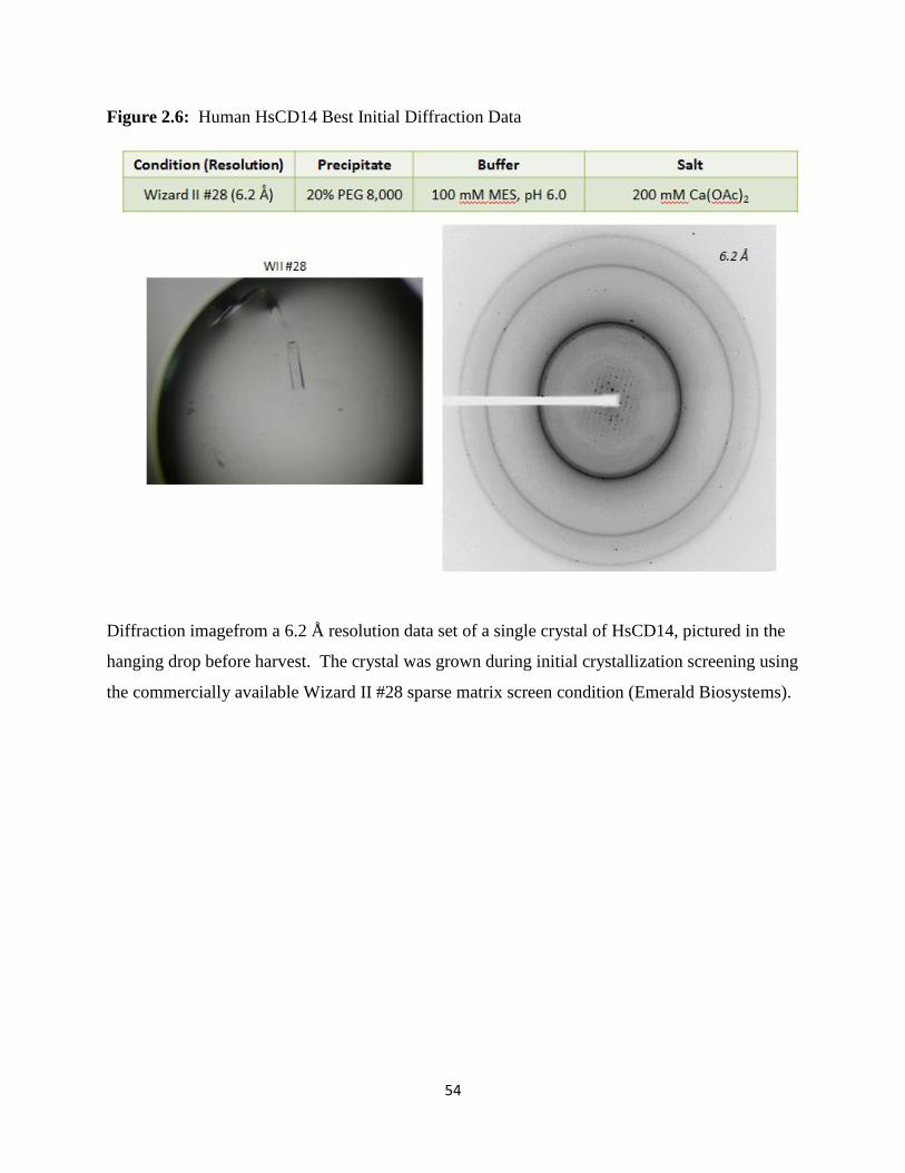
54
Figure 2.6: Human HsCD14 Best Initial Diffraction Data
Diffraction imagefrom a 6.2 Å resolution data set of a single crystal of HsCD14, pictured in the
hanging drop before harvest. The crystal was grown during initial crystallization screening using
the commercially available Wizard II #28 sparse matrix screen condition (Emerald Biosystems).

55
Figure 2.7: Human HsCD14 Diffraction Data after Condition Optimization
Diffraction image from a 4.02 Å resolution data set of a single crystal of HsCD14, pictured in the
hanging drop before harvest (circled). The crystal was grown in a handmade crystallization
condition (indicated) during the crystallization optimization process.

56
Figure 2.8: Human HsCD14 Best Diffraction Data after Full Condition Optimization
Conditions from handmade crystallization conditions (K#25 optimized and K#19 best optimized)
resulted in the best diffraction resolution after the full condition optimization, which is discussed
in detail in the methods section of this chapter. Resolution of diffraction data gathered from each
single crystal, pictured in the hanging drop before harvest (circled), are indicated along with the
crystallization condition. The initial commercially available crystallization hit (Wizard II #28,
Emerald Biosystems) and corresponding single crystal are shown for reference.

57
Table I: Data Collection and Refinement Statistics

58
Figure 2.9: Ramachandran Plot of Human CD14 (4GLP.pdb)
Ramachandran plot (RAMPAGE) showing phi (ɸ) and psi (ψ) angles plotted on the x and y
axis, respectively, for each amino acid in the structure, over top of ‘favored’ and ‘allowed’
smoothed contours determined from high-quality data. In our human CD14 crystal structure
(4GLP.pdb), 69.5% (214/308) residues lie in the ‘favored’ contours (dark blue/dark orange),
22.4% (69/308) lies in the ‘allowed’ contour (light blue/light orange) and 8.1% (25/308) residues
are ‘outliers’ (white).
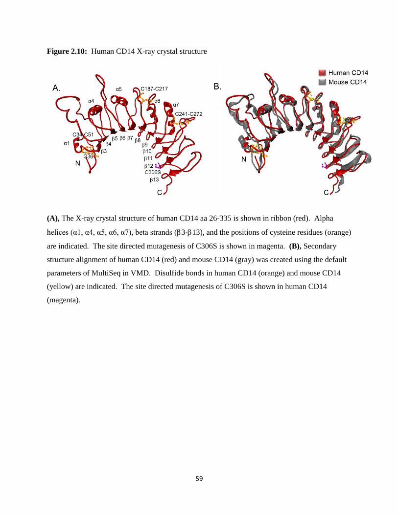
59
Figure 2.10: Human CD14 X-ray crystal structure
(A), The X-ray crystal structure of human CD14 aa 26-335 is shown in ribbon (red). Alpha
helices (α1, α4, α5, α6, α7), beta strands (3-13), and the positions of cysteine residues (orange)
are indicated. The site directed mutagenesis of C306S is shown in magenta. (B), Secondary
structure alignment of human CD14 (red) and mouse CD14 (gray) was created using the default
parameters of MultiSeq in VMD. Disulfide bonds in human CD14 (orange) and mouse CD14
(yellow) are indicated. The site directed mutagenesis of C306S is shown in human CD14
(magenta).
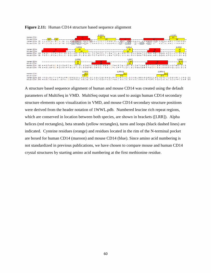
60
Figure 2.11: Human CD14 structure based sequence alignment
A structure based sequence alignment of human and mouse CD14 was created using the default
parameters of MultiSeq in VMD. MultiSeq output was used to assign human CD14 secondary
structure elements upon visualization in VMD, and mouse CD14 secondary structure positions
were derived from the header notation of 1WWL.pdb. Numbered leucine rich repeat regions,
which are conserved in location between both species, are shown in brackets ([LRR]). Alpha
helices (red rectangles), beta strands (yellow rectangles), turns and loops (black dashed lines) are
indicated. Cysteine residues (orange) and residues located in the rim of the N-terminal pocket
are boxed for human CD14 (maroon) and mouse CD14 (blue). Since amino acid numbering is
not standardized in previous publications, we have chosen to compare mouse and human CD14
crystal structures by starting amino acid numbering at the first methionine residue.

61
Figure 2.12: Comparison of Mouse and Human CD14 N-terminal ligand binding pocket.
(A), Mouse (gray) and human (red) CD14 ribbon structures are overlaid over mouse (translucent
gray) and human (translucent red) CD14 space filling structures drawn in VMD. CD14
structures are rotated 90o relative to Figure 2.4(B). The N-terminal hydrophobic binding pocket
is indicated (black arrow). (B), Space filling hydrophobicity structures were drawn in Chimera
for human CD14 (left) and mouse CD14 (right). The Kyte-Doolittle scale was used to compare
hydrophobicity by showing hydrophobic (orange), neutral (white), and hydrophilic (blue)
residues as well as glycosylation sites (red).

62
Figure 2.13: Comparison of Mouse and Human CD14 Pocket Rim Residues
(A), The structure of the rim of the predicted N-terminal ligand binding pocket of both mouse
and human CD14 are overlaid. Rim residues of human CD14 are denoted in red and those of
mouse CD14 are denoted in blue. Regions important for LPS binding, as determined from
previous mutagenesis, blocking antibody, and epitope mapping studies are indicated in black
with gray text. (B), A space filling representation of the residues located on the rim of the
predicted N-terminal ligand binding pocket of human and mouse CD14 are displayed in red and
blue, respectively. Regions important for LPS binding are displayed in black ribbon.

63
CHAPTER 3:
Ligand Binding and Bioactivity of Purified CD142,3
3.1 Introduction
In the work described in this chapter, we developed a source of mouse CD14 for our
ligand binding crystallization studies. We cloned, expressed, and purified a single mouse soluble
CD14 construct (MCD14) from the cell supernatant of HEK 293F cells. The previously
published mouse CD14 x-ray structure encompassed aa 5-313, as numbered from the start of the
mature protein after N-terminal secretion signal removal (69). Our MCD14 construct ended with
the same C-terminal amino acid, but unlike the crystal structure, we maintained the authentic N-
terminal secretion signal (designated aa 1-15). Thus, our purified mouse MCD14 protein
encompassed aa 1-328, as numbered from Met 1.
As described in Chapter 1, soluble CD14 is known to bind a number of acylated TLR4
and TLR2/1 ligands. We characterized the ability of both mouse MCD14 and human HsCD14
proteins utilized in our crystallography studies to bind these acylated ligands employing two
ligand binding detection methods. First, we conducted gel shift analysis using 4-20% native
PAGE. Using this assay, we tested the ability of our purified proteins to bind the Ra chemotype
of E. coli LPS, which signals through TLR4/MD-2 as described in Chapter 1. Upon ligand
binding, the increased negative charge present in bound LPS phosphate groups induces a
2 Reprinted in part with permission from Kelley, S. L., Lukk, Tiit, Nair, S. K., and R. I. Tapping. (2013). “The crystal
structure of human soluble CD14 reveals a bent solenoid with a hydrophobic amino-terminal pocket.” J. Immunol.
190 (3): 1304-1311.
3 Reprinted in part with permission from Ranoa, D. R., Kelley, S. L., and R. I. Tapping. (2013). “Human LBP and CD14 independently deliver triacylated lipoproteins to TLR1 and TLR2 and enhance formation of the ternary signaling complex.” J. Biol. Chem. 288: 9729-9741.

64
downshift of the CD14-LPS complex in a native PAGE, which can be visualized using
Coomassie blue staining. Second, we tested the ability of our purified proteins to bind various
ligands using the Octet QK first generation system (ForteBio). TLR4/MD-2 ligands tested using
this method included the rough Ra chemotype of E. coli LPS and smooth LPS containing the full
O-antigen and core oligosaccharide group from S. Minnesota. Additionally, two TLR2/1 ligands
that are shuttled by CD14 were also tested. A 31 kD N-terminal tripalmitoylated (Pam3)
lipoprotein called Outer Surface Protein A (OspA) (187), which was purified from Borrelia
burgdorferi bacterial extract, and a commercially available synthetic triacylated lipoprotein
mimic of the N-terminus of OspA called Pam3CSK4. Real-time, label-free ligand binding
detection using the Octet QK was conducted on these ligands to corroborate our native
polyacrylamide gel shift ligand binding results.
The ForteBio Octet QK automated system uses up to eight optical biosensors per
experiment in a “Dip and Read” format, where a robotic arm dips the biosensors from one well
to another in a 96-well, black, flat bottom, polypropylene microplate with a 200 L sample
volume. The biosensors have a coated surface for immobilization of the receptor protein being
studied. In our case, we utilized protein A coated biosensor to immobilize the Fc fusion of
HsCD14, though other coating surfaces are commercially available. The thickness of the sensor
(binding, nm) is measured in the Octet QK using bio-layer interferometry, which measures the
change in wavelength between two spectrums (intensity vs. wavelength (nm)) recorded for
reflections of white light off of an internal reference layer and the biosensor tip. As the
HsCD14-Fc protein binds the protein A biosensor, the thickness of the sensor surface (binding,
nm) increases over time (seconds), creating a real-time ligand binding curve.

65
Most importantly, we verified both our human and mouse purified CD14 proteins that
were utilized in crystallography studies are biologically active. As mentioned in Chapter 1,
soluble CD14 functions to enable cells that express TLR4/MD-2, but do not express membrane
bound CD14, to be sensitive to LPS. Since epithelial cells do not produce membrane bound
CD14, we could identify the bioactivity of our purified human and mouse CD14 proteins by
measuring their ability to bind and deliver LPS to stimulate production of the chemokine IL-8 by
TLR4/MD-2 signaling. Thus, we were able to measure the bioactivity of purified human and
mouse CD14 induced production of IL-8 using an ELISA IL-8 detection system. Additionally,
small gel filtration mixing studies using human CD14, which was purified as described in this
Chapter, detected human CD14 bioactivity, because human CD14 was able to bind and deliver
OspA to catalytically form TLR2/1/OspA complexes, independent of LBP (188).
Our efforts to create a ligand-bound crystal of mouse CD14 will also be discussed in this
chapter. We conducted initial crystallization sparse matrix screening for mouse MCD14 similar
to the work described in Chapter 2 for human HsCD14 and determined conditions where our
mouse MCD14 protein crystallized. However, in ligand bound crystallization studies, both
purified proteins did not produce single crystals of sufficient size or quality for diffraction
studies.
3.2 Materials and Methods
3.2.1 Reagents
The same cloning, expression, and protein purification reagents described in the
Materials and Methods section of Chapter 2 were utilized in the cloning, expression and
66
purification of MCD14. Sources of TLR4/MD-2 reagents included smooth Salmonella enterica
serotype minnesota LPS (Sigma L6261) and the Ra chemotype of Escherichia coli EH100 LPS
(Sigma L9641). Sources of TLR 2/1 ligands included recombinant outer surface protein A
(OspA) purified from Borrelia burgdorferi bacterial extract (Recombitek Lyme, purchased from
Merial, Inc. (Athens, GA)) and a synthetic, racemic mixture of N-palmitoyl-S-[2,3-
bis(palmitoyloxy)-propyl]-(R)-cysteinyl-(lysyl)3-lysine (Pam3CSK4; EMC Microcollections,
Tuebingen, Germany). ForteBio Octet wash steps utilized PBS-T buffer (1X PBS with 0.05%
Tween 20 detergent; Sigma P1379). Bioactivity assay reagents included RPMI 1640 media
(Corning; 15040CV), bovine serum albumin (BSA; Sigma A9418), human serum albumin
(Octopharma, Hoboken, NJ), and human serum obtained from the peripheral blood of
anonymous donors from blood draws performed by Dr. Brian Hart and Song Jiang. Monoclonal
anti-CD14 antibodies (28C5 and 63D3) and a polyclonal Goat anti-LBP antibody were kind gifts
from Dr. Peter Tobias, The Scripps Research Institute, La Jolla, CA. Human LBP from Xoma
Corp. (Berkley, CA) was a kind gift from Dr. Jerrold P. Weiss and Dr. Theresa L. Gioannini,
University of Iowa, Iowa City, IA.
3.2.2 Mouse CD14 Cloning, Expression, and Purification
The mouse CD14 constructs was created using the same modified pDisplay vector system
described in Chapter 2 Materials and Methods. Briefly, the forward primer contained an EcoRI
cleavage site, Kozak sequence, and a portion of the N-terminal secretion signal of mouse CD14
beginning at aa 1. The reverse primer consisted of the C-terminal coding region of mouse CD14
from aa 328 in addition to a NheI cleavage site and thrombin digestion site (LVPRGS). This set
of primers (Figure 3.1A) was utilized to amplify mouse CD14 (aa 1-328) from genomic DNA

67
(ATCC) using PCR as described in Chapter 2. After PCR, EcoRI and NheI restriction enzymes
were used to digest the MCD14 PCR product and modified pDisplay vector (Figure 3.1B), a
kind gift from Dr. David Kranz, University of Illinois, Urbana-Champaign. Following agarose
gel electrophoresis, the following sized bands were observed: undigested pDisplay vector (6,154
bp), EcoRI and NheI digested pDisplay vector (5,542 bp & 612 bp), and EcoRI and NheI
digested MCD14 PCR product (976 bp). The vector and insert were ligated by T4 DNA ligase
(Invitrogen) and transformed into E.coli DH5α cells using heat-shock as directed in the
manufacturer’s protocol (Invitrogen). Positively transformed colonies were selected on LB-
Ampicillin plates grown overnight in the 37oC warm room. Conformation of transformation
using DNA extraction, EcoRI and NheI restriction enzyme digestion, agarose gel electrophoresis,
sequencing, transfection of HEK 293F cells, protein expression, and selection of stable lines
were also completed as described in Chapter 2 Materials and Methods.
PNGaseF treated MCD14 protein utilized in later ligand binding crystallization studies
was purified using the identical buffers and method described for HsCD14 in Chapter 2
Materials and Methods. Native Mouse CD14 protein was purified using a four-step
chromatographic procedure as described in Chapter 2 Materials and Methods, with the following
variations. First, Protein G sepharose was used to affinity extracted MCD14-Fc fusion protein
from the supernatant of two liters of HEK 293F cells. Following thrombin cleavage, protein was
injected using a 10 mL loop onto an ÄKTAprime™ plus FPLC (GE Healthcare) fitted with Hi-
Trap Q anion exchange chromatography using a linear NaCl gradient (0.02 M - 1 M) in 0.02 M
HEPES, pH 7.5 run at a 1 ml min-1 flow rate. Pooled fractions containing HsCD14 were
concentrated to 1 mL using an Amicon Ultra-15 unit (Millipore) and injected onto two tandem
Protein A columns run with 0.02 M HEPES, pH 7.5 buffer using a 1 ml min-1 flow rate for free

68
Fc removal. Flow through fractions containing MCD14 were pooled and injected into size-
exclusion chromatography using a Superdex 200 column (GE Healthcare) equilibrated with 0.02
M HEPES, pH 7.5 and 0.1 M NaCl at a flow rate of 0.4 mL min-1. Final SGF fractions
containing MCD14 were pooled and concentrated mg ml-1 using an Amicon Ultra-4 unit
(Millipore) as measured by Pierce BCA assay (Rockford, IL). This four column purification
process yielded ~2-3 mg L-1 soluble, mouse CD14 with >90% purity as ascertained visually by
SDS-PAGE (Figure 3.1C). The SGF elution profile indicated fractions containing MCD14
which was confirmed visually on 10% SDS-PAGE with Coomassie staining (Figure 3.1D).
3.2.3 Generation of Human CD14 for Small Gel Filtration Mixing Studies
Human soluble CD14 protein was cloned and expressed as previously described (189),
and similarly purified with the following exceptions. Briefly, human CD14 (aa 1-337) was
amplified from genomic DNA and cloned into a modified pDisplay vector preceding a thrombin
cleavage site (LVPRGS) and the Fc domain of human IgG1. The final construct was sequenced
(University of Illinois Urbana-Champaign Sequencing Center) following site-directed
mutagenesis (C306S) via primer extension to avoid unnatural disulfide bonding resulting from
the truncated coding region of our construct. Following transfection and stable selection of
human HEK 293F cells (Invitrogen), human soluble CD14 was purified from cell supernatant in
three chromatographic steps, including protein G affinity chromatography, thrombin cleavage,
protein A affinity chromatography, and size exclusion chromatography. Finally, fractions
containing CD14 were pooled and concentrated to 10 mg/mL using an Amicon Ultra-4 unit

69
(Millipore) as measured by Pierce BCA assay. Human soluble CD14 (aa 1-337, C306S) purified
by this method was stored at 4oC for up to six months prior to use (188).
3.2.4 Native Polyacrylamide Gel Shift LPS Binding Assay
The binding of LPS by native mouse CD14 (Figure 3.2A and 3.2B), native human
HsCD14 (Figure 3.3A), or PNGaseF treated human CD14 (Figure 3.3B) was detected by a
native polyacrylamide gel shift method (190). Ten micrograms CD14 was mixed with a 2:1
molar ratio of the Ra chemotype of E. coli LPS (Sigma), with or without a 1:100 molar ratio of
LBP:CD14 (LBP kindly provided by Dr. Jerrold Weiss, University of Iowa). The reactions were
incubated for 1 hr or less as indicated (pre-incubation time) at 37oC in a total volume of 5 L in
PBS buffer containing 0.01M EDTA. 5 L bromophenol blue with glycine dye was added and
each sample was loaded in a 4-20% Mini-PROTEAN TGX precast gel, pH 6.8 (Bio-rad) run at
100V in the manufacturer recommended running buffer without SDS at 4oC for 3 hours.
3.2.5 ForteBio Octet QK Ligand Binding Assay
Protein A biosensors (ForteBio) were equilibrated in a 96 well, flat bottom, black, non-
binding polypropylene microplate (Greiner Bio One; #655900) placed in the sample block of the
ForteBio Octet QK in 0.2 mL PBS for 2 minutes. Immediately after equilibration, the protein A
biosensors were transferred by robotic arm into the adjacent wells to immobilize of 0.03 M
HsCD14-Fc for 30 minutes. After a 20 minute wash in PBS-T buffer, the HsCD14-Fc
containing biosensors were placed in wells containing a 0.2 mL ligand solution with or without
LBP for 1 hour of ligand binding. In Figure 3.4A, well A contained 0.03 M LBP (pink), well B

70
contained 0.03 M OspA and 0.03 M LBP (1:1 molar ratio) (orange), well C contained 0.2 M
the Ra chemotype of E. coli and 0.1 M LBP (2:1 molar ratio) (red), and well D contained 0.2
M S. Minnesota LPS and 0.1 M LBP (2:1 molar ratio) (blue). In Figure 3.4B, well A
contained the nonblocking anti-CD14 63D3 antibody (green) and well B contained the blocking
anti-CD14 28C5 antibody (pink). At this point the procedure differs as indicated below. In
Figure 3.4A, a 30 minute incubation in PBS-T buffer is followed by a 1 hr incubation for LBP
detection using 0.3 M polyclonal anti-LBP, followed by a 30 minute PBS-T buffer wash step
(pink, orange, blue, and green). In Figure 3.4B, a 1 hr incubation with 0.03 M OspA and 0.03
M LBP (1:1 molar ratio) followed ligand binding, followed by a 20 minute PBS-T buffer wash
step.
3.2.6 IL-8 Bioactivity Assay
Various concentrations of the Ra chemotype of E. coli LPS (Sigma) were preincubated
for 1 hr (Figure 3.5A) or 1 hr or overnight (ON) (Figure 3.5B) at 37°C at a 2:1 molar ratio
LPS:CD14 in RPMI media containing 1% human serum albumin for both SGF purified Human
HsCD14 (Figure 3.5A) and Mouse MCD14 (Figure 3.5B). For some reactions, LBP was added
as a catalyst at a 1:100 molar ratio of LBP:CD14. We included a natural source of soluble CD14
(2% human serum; Figure 3.4A) as a positive control. The bioactivity of CD14 in these
reactions was assessed using SW620 human epithelial cells that express TLR4/MD-2 and lack
detectable levels of membrane CD14 (128). SW620 cells were cultured in RPMI complete
media containing 1% BSA, seeded in a 96-well plate at a density of 1.0 x 10⁶ cells/well, and
allowed to adhere for 1 hr at 37°C, 5% CO2. To remove serum, we carefully washed SW620
cells four times with PBS, two times with Life Technologies FreeStyle 293 Expression Media

71
(Invitrogen), and two times with RPMI containing 1% human serum albumin. Pre-incubated
samples containing CD14 and LPS were added to the carefully washed SW620 epithelial cells.
Cell supernatant was harvested 6 hrs later, and IL-8 production was measured using a human IL-
8 Cytoset sandwich ELISA kit (Invitrogen). In the ELISA, mouse anti-human IL-8 capture
antibody was immobilized in a 96 well plate format and blocked with 1% bovine serum albumin
(Sigma) in PBS. Cellular supernatant was applied for two hours. After a wash step with 1%
BSA in PBS, biotinylated anti-human IL-8 detection antibody was applied, followed by a 1%
BSA/PBS wash. Streptavidin conjugated horseradish peroxidase (HRP; R&D Systems) was
applied and developed with the substrate o-phenylenediamine (OPD; Sigma; P9029), which
yields a water soluble yellow-orange color detectable at ~490 nm by microplate reader (BioRad
Model 680).
3.2.7 Initial Crystallization Screening of Mouse CD14
For the initial sparse matrix screening, the hanging drop vapor diffusion methods was
utilized to set out 2 l drops containing native MCD14 (10 mg/mL) combined in equal volume
with commercially available sparse matrix screen conditions (Hampton Research Crystal Screen
I, Crystal Screen II, Natrix, and Salt Reaction and Emerald Biosystems Wizard I and II) or
handmade crystal screen conditions targeting the previously published mouse CD14
crystallization condition (1 l protein solution and 1 l of crystallization buffer containing 100
mM sodium HEPES (pH 7.5), 1.9 M Li2SO4, and 5 mM NiCl2) (69). Crystallization trays were
set up and monitored for microcrystals, crystal clusters, or single crystals (Figure 3.7), using
methodology previously detailed in Chapter 2 Materials and Methods.
72
3.2.8 Initial Crystallization Screening of Mouse CD14-LPS Complexes
Native or PNGaseF deglycosylated MCD14 (and HsCD14; data not shown) were
incubated with a 2:1 molar ratio of the Ra chemotype of E. coli LPS in the presence of a 1:100
molar ratio of LBP:CD14 at 37oC for 1 hr immediately prior to setting out crystallization trays.
This protein mixture was then screened against the same commercially available sparse matrix
crystallization kits utilized in the initial crystallization screening methodology described above.
Each condition was tested on a 12 mm x 0.22 mm siliconized glass cover slide (Hampton
Research) over a VDX48 plate with sealant (Hampton Research) by mixing 1 l of each
screening solution with 1 µl LPS containing protein solution [10 mg ml-1 MCD14 or HsCD14
protein in 0.02 M HEPES, pH 7.5 and 0.1 M NaCl (MCD14) or 0.02 M Tris-HCl, pH 8.5 and 0.1
M NaCl (HsCD14)] equilibrated against 300 µl screening solution in the reservoir and set out at
RT.
3.3 Results
3.3.1 Purified Mouse CD14
We cloned, expressed, and purified mouse CD14 (MCD14) in an effort to obtain a source
of protein useful for ligand bound crystallization studies. Forward and mouse CD14 PCR
primers were created to encompass aa 1-328 (Figure 3.1A), terminating at the same residue as
the x-ray crystal structure of mouse CD14 (69). Ligating the PCR product into a modified
pDisplay vector using EcoRI and NheI digestions sites (Figure 3.1B) enabled protein expression
in eukaryotic cells. Following stable transfection of human embryonic kidney HEK 293F cells,

73
mouse CD14 was purified using protein G sepharose beads, thrombin digestion, HiTrap Q anion
exchange, protein A affinity purification (to further remove free Fc), and small gel filtration
(Figure 3.1C). Purification resulted in >90% pure MCD14 protein, as judged SDS-PAGE
stained with Coomassie blue and SGF purification profile (Figure 3.1D).
3.3.2 Purified Human and Mouse CD14 Bind E. coli LPS
One of our first questions following purification was whether or not purified HsCD14 or
MCD14 is able to bind and shuttle ligands to TLRs. We set out to answer the question of LPS
binding using a native polyacrylamide gel shift method (190). As mentioned previously, when
CD14 binds LPS, the CD14-LPS complex shifts relative to the position of CD14 alone, due to
the increased negative charge present from the bound LPS phosphate groups. As shown in
Figure 3.2A lanes 3-9, 13, and 15, mouse CD14 is capable of binding the Ra chemotype of LPS
indicated by downward shifts in a 4-20% gradient native PAGE. The binding reaction involves a
1 hr incubation at 37oC of CD14 and LPS in the presence of a catalytic amount of LBP (1:100
LBP:LPS molar ratio). A 1.5-2 molar ratio of LPS:MCD14 facilitated the most complete shift or
complex formation. Therefore, we utilized a 2:1 molar ratio of LPS:MCD14 as a loading
condition for CD14 ligand binding crystallization studies. Further gel shift analysis included
optimization of incubation conditions. For example, in Figure 3.2B we varied incubation time.
Thirty minutes incubation also resulted in complex formation (as indicated by a shift in protein
mobility), and in further gel shift analysis (data not shown) we found CD14 and LPS incubated
for five minutes in the presence of LBP was sufficient to drive complex formation. Similarly,
native (Figure 3.3A) and deglycosylated (Figure 3.3B) human HsCD14 bound the Ra

74
chemotype of LPS when mixed with a 2:1 molar ratio of this ligand in the presence of 1:100
LBP:LPS molar ratio. However, without LBP, additional incubation time was needed to drive
complex formation as evidenced by mixing a 2:1 molar ratio of LPS:CD14 with (lane 3) and
without (lane 2) LBP (Figure 3.3B).
3.3.3 Octet and Small Gel Filtration Mixing Studies Confirm Ligand Binding to Human CD14
Numerous ligands capable of binding human HsCD14-Fc fusion were assessed through
Octet QK ligand binding studies (summarized in Figure 3.4A). We found that immobilized
human HsCD14-Fc is able to bind B. burgdorferi OspA (orange), the Ra chemotype of E. coli
LPS (red), and S. minnesota LPS (blue) in the presence of LBP. Although we did not detect
binding of LBP alone (pink) to immobilized HsCD14-Fc, we wondered if the binding observed
was entirely due to OspA or LPS ligand binding, or if LBP was retained in the final complex. To
test for LBP retention, in the final complexes we measured the binding of polyclonal anti-LBP to
the captured complexes. We found complexes formed using rough LPS (from E. coli) retained
bound LBP during the capture stage, as indicated by the presence of a binding curve upon
addition of an anti-LBP antibody. Similarly, complexes containing smooth S. minnesota LPS
also bound the anti-LBP, indicating LBP retention to a lesser extent than rough LPS. However,
complexes formed using the ligand OspA did not show a detectable binding curve with anti-LBP
antibody, indicating that OspA binds immobilized CD14 without LBP retention.
In a separate study shown in Figure 3.4B, we assessed the ability of anti-CD14
antibodies 63D3 and 28C5 to block OspA binding to immobilized HsCD14-Fc fusion. Anti-
CD14 antibody 28C5 binds CD14 near the N-terminal LPS binding site and block the LPS

75
binding and cellular activation functions of CD14 (128). In contrast, the anti-CD14 antibody
63D3 does not bind the N-terminal portion of CD14 and does not block LPS binding to CD14.
In both Octet QK experiments, we immobilized 0.03 M human HsCD14-Fc on a protein A
biosensor. As shown in Figure 3.4B, both 63D3 (green) and 28C5 (pink) were capable of
binding to immobilized HsCD14-Fc as measured in the capture step. As expected, incubation of
the CD14 bound sensors with OspA and LBP (at a 1:1 molar ratio) facilitated complex
formation. The 28C5 antibody blocked OspA binding, but the 63D3 antibody did not. These
results indicate that the binding site of OspA is physically located at or near the binding site of
LPS in the N-terminus of CD14.
3.3.4 Purified Human and Mouse CD14 Are Bioactive
In addition to ensuring that our purified HsCD14 and MCD14 proteins were capable of
binding ligands including LPS, we also measured their ability to stimulate LPS responses in
human cells. As mentioned previously, human epithelial SW620 cells express MD-2 and TLR4,
but lack membrane bound CD14. When CD14 shuttles LPS to the SW620 cells, they are
stimulated to secrete pro-inflammatory cytokines and chemokines, including IL-8, which can be
measured in culture supernatants by ELISA. Using methodology described in detail in the
Materials and Methods section of this chapter, 0.01-100 ng/mL of the Ra chemotype of E. coli
LPS was incubated for 1 hr (or overnight where indicated) at 37oC with and without a 1:100
molar ratio of LBP:LPS in either the presence or absence of 1 g/mL HsCD14 (Figure 3.5A) or
MCD14 (Figure 3.5B). These reactions were then added to SW620 cells and secreted IL-8
chemokine levels were monitored by ELISA. In the absence of LBP or CD14, only the highest

76
concentration of LPS was capable of stimulating IL-8 production (blue). When HsCD14 was
added to LPS and LBP (red), SW620 cells became 100 fold more sensitive to the presence of
LPS compared to LBP alone (orange) (Figure 3.5A). To confirm our assay was capable of
detecting the full activity of LBP and CD14, we tested two percent human serum as a positive
control and a natural source of LBP and soluble CD14 (127). Two percent human serum
induced similar levels of IL-8 as that of LBP and our purified CD14 protein (green).
Likewise, purified mouse CD14 (green or red) was able to stimulate 10-100 fold greater
levels of IL-8 secretion than that of LBP catalysis alone (orange) (Figure 3.5B). In this
experiment, we saw no difference between 1 hr (red) and overnight (green) incubation of
MCD14 with LPS with and without LBP. However, in a similar bioactivity assay with HsCD14,
we saw overnight incubation severely inhibited the ability of the HsCD14 protein to stimulate
cells to produce IL-8, indicating lower protein stability compared to MCD14 following
prolonged incubation at 37oC (data not shown).
A separate small gel filtration mixing study further confirmed the bioactivity of purified
human CD14. In this work, our soluble human CD14, purified as described in the Materials and
Methods in this Chapter, is able to bind and catalytically deliver OspA to form
TLR2/TLR1/OspA complexes without the requirement for LBP (188). A two hour
preincubation at 37oC of OspA, soluble TLR2, and soluble TLR1, in the absence of both CD14
and LBP, was unable to form a TLR2/1/OspA complex detectable by small gel filtration (SGF)
(data not shown). However, addition of our purified human CD14 enabled detectable complex
formation in the SGF elution profile without CD14 retention, which was confirmed by western
blot detection of TLR2, TLR1, and CD14 using anti-TLR2, anti-TLR1, and polyclonal anti-
CD14 antibodies in the corresponding SGF elution fractions (data not shown). Taken together

77
with our ForteBio Octet results showing human CD14 is able to bind OspA without the retention
of LBP, these subsequent small gel filtration mixing studies show CD14 is able to deliver bound
OspA to TLR2/1 in a catalytic manner independent of LBP, which further endorses the
bioactivity of our purified human CD14.
3.3.5 Initial Crystallization Screening of Mouse CD14
We created a handmade crystallization screen that slightly varied the precipitate, buffer,
and salt components around the previously published mouse CD14 crystallization condition (1.9
M Li2SO4, 100 mM HEPES, pH 7.5, and 5 mM NiCl2). Although our purified mouse CD14 was
unable to crystallize at that condition, potentially due to differences in our purification system,
we were able to obtain crystals of MCD14 protein under different experimental conditions. Our
initial screening of MCD14 used commercially available kits, as mentioned in the Materials and
Methods sections of this Chapter and Chapter 2, including Wizard I, Wizard II, and Crystal
Screen I. We also varied crystallization temperature, including an examination of 18oC and
room temperature conditions. The crystallization conditions capable of producing single crystals
or crystal clusters are shown in Figure 3.6. We did not pursue diffraction of these conditions
because the mouse CD14 crystal structure had already been published. However, we did use this
information to target ligand binding crystallization conditions.
78
3.3.6 Lack of Diffraction Quality Crystals of CD14-LPS Complexes
A ligand bound crystal structure of CD14 with LPS will greatly improve our
understanding of the molecular interactions between LPS and this receptor. Ligand bound
crystal structures are difficult to achieve, especially utilizing a non-homogenous, non-small
molecule and highly amphipathic ligand capable of forming aggregates like LPS. However,
since the Ra chemotype of LPS which lacks the O-antigen was successfully crystallized in
complex with MD-2/TLR4 (77), we began our ligand binding crystallization studies using this
LPS chemotype. In setting up these experiments, we wished to load CD14 with LPS and then
purify the CD14-LPS complexes away from unbound LPS. Since loading CD14 with LPS prior
to Protein G affinity purification was not an option due to the large cell supernatant volume
(~2L) we were utilizing, we began by loading CD14 with LPS immediately following Protein G
affinity purification (just prior to thrombin cleavage and Hi-Trap Q anion exchange), prior to
protein A affinity purification, and prior to small gel filtration with a 2:1 molar ratio of
LPS:CD14, as measured by BCA assay, and a catalytic amount of LBP (1:100 LBP:CD14 molar
ratio). We found that incubation of CD14 with LPS prior to thrombin digestion reduced the
cleavage reaction efficiency (data not shown). Introduction of LPS prior to Protein A
purification of CD14 led to a large reduction in protein yield following purification and increased
column pressure, possibly due to the formation of CD14-LPS aggregates during column
chromatography. Similarly, we found that introduction of LPS immediately prior to small gel
filtration led to aggregation of LPS on the Superdex 200 column, which required prolonged
stringent washing and rinsing steps (70% ethanol or 1 M NaOH followed by water and buffer re-
equilibration) prior to beginning the next round of purification.
79
Although we set out crystallization trays of CD14 protein loaded with LPS by LBP after
HiTrap Q anion exchange, after protein A affinity chromatography, and after purification
following a second round of small gel filtration, we did not obtain any promising crystallization
leads. Instead, the data presented in this chapter was produced by the most straightforward way
of introducing LPS, where CD14 was loaded with LPS by LBP immediately following protein
purification (prior to crystallization). In this approach, we incubated LPS with human or mouse
CD14 in a 1:1 molar ratio with LPS with and without LBP at 37oC overnight immediately prior
to setting out trays, in an effort to give CD14 enough time to interact with LPS. Additionally, in
the initial sparse matrix screening results using mouse CD14 (Figure 3.7), we used conditions
we previously found to be capable of producing evidence of LPS binding by native
polyacrylamide gel shift and ForteBio Octet QK analysis (2:1 molar ratio of LPS:CD14 and a
1:100 molar ratio of LBP:CD14) and we pre-incubated the mixture at 37oC for 1 hr before setting
out the initial sparse matrix crystallization screen.
Using PNGaseF treated mouse CD14 protein, purified as described in Materials and
Methods, we observed phase separation and protein aggregation in the hanging drops of Wizard I
#28 and Wizard I #30 after <1 day at room temperature (Figure 3.7). These outcomes were
typical of all of the crystallization trials involving human or mouse CD14 incubated with LPS in
either the presence of or the absence of LBP. The best result from all CD14-LPS crystallization
studies was observed after 45 days in Wizard I #7 (Figure 3.7). Only four microcrystals were
observed, but these crystals were too small to harvest for diffraction analysis. Instead of further
pursing optimization of conditions to enhance these non-diffraction quality crystals, we
refocused our protein production and crystallization efforts to obtain an x-ray crystal structure of
human CD14 without ligand bound (189). The human CD14 crystallization condition we

80
determined was utilized to screen human and mouse CD14 after LPS loading, resulting in no
diffraction quality CD14 crystals with ligand bound.
3.4 Discussion
In this Chapter, we described the cloning, expression, and purification of mouse MCD14
protein. We determined the capacity of this protein and our purified HsCD14 protein to bind
LPS after incubation with LBP at 37oC using native polyacrylamide gel shift analysis. To further
support our native polyacrylamide gel shift data, we confirmed HsCD14-Fc LPS binding using
ForteBio Octet QK. Smooth and rough forms of LPS were capable of producing detectable
binding curves with immobilized human HsCD14. We also found human CD14 bound the
lipoprotein OspA. Similarly, our ForteBio Octet data showed LBP retention in complexes
generated with either form of LPS, but not with OspA lipoprotein. In support of these findings,
small gel filtration mixing studies showed further purified human CD14 was able to deliver
bound OspA to TLR2/1 catalytically without the need for LBP (188). Also using ForteBio
Octet, we also found less LBP was retained with smooth LPS from S. minnesota compared to
rough LPS from E. coli. One possible explanation for the retention of LBP in this in vitro system
is that the equimolar ratio of LBP and CD14 in the presence of a 2 fold excess molar ratio of LPS
may have shifted CD14 from its primary role of shuttling LPS to TLR4 to its alternative function
as a cell surface receptor for LBP opsonized LPS aggregates leading to clearance of LPS (48, 49,
52, 191). Although the mechanism and molecular interactions involved are undetermined,
membrane bound CD14 is known to control LPS-induced endocytosis of TLR4 (192).
81
Wooten and colleagues (187) found that 28C5, an anti-CD14 antibody that inhibits LPS-
mediated cell activation, also inhibits activation induced by OspA, a triacylated lipoprotein of
Borrelia. In support of this we found in our studies that the inhibitory 28C5 antibody blocks the
binding of OspA to HsCD14. Since OspA contains Pam3, we also tried to detect the synthetic
TLR2/1 agonist Pam3CSK4 binding to CD14 using the ForteBio Octet QK Green instrument.
However, Pam3CSK4 binding was not detected, an observation that could be due to the minimum
ligand molecular weight (10 kD) required for detectable bio-layer interferometry signaling
greatly exceeded the molecular weight (1.5 kD) of monomeric Pam3CSK4. This limitation
suggests that even if Pam3CSK4 is able to bind HsCD14-Fc, we would not be able to detect it
unless it bound in large aggregates or retained LBP upon binding. Repeating this experiment
with the second generation ForteBio Octet Red96 system may enable detection of binding
because the minimum ligand molecular weight requirement for this newer instrument is 150 Da.
We also determined both MCD14 and HsCD14 purified proteins are capable of mediating the
responses of SW620 epithelial cells to the Ra chemotype of LPS. Studies also confirmed the
bioactivity of purified human CD14 by its ability to form TLR2/1/OspA complexes in small gel
filtration (188). Taken together, these results suggest purified mouse and human CD14 proteins
are bioactive by their capacity to bind and deliver ligands to TLR4/MD-2 and TLR2/1
complexes. Verifying that our purified CD14 proteins are able to bind ligands, determining
loading conditions, and confirming biologically activity after purification was important before
advancing these proteins through ligand binding crystallization trials.
Our x-ray structure of human CD14 presents a good framework to discuss our ligand
binding crystallization results. As described in Chapter 2, we confirmed the presence of an N-
terminal hydrophobic pocket in human CD14, which is predicted by a number of deletional,

82
mutational, and antibody blocking studies to be the LPS binding site. Our x-ray crystal structure
of human CD14 also advanced our understanding of the residues involved in the rim of this N-
terminal pocket, enabling us to predict rim residue repositioning upon ligand binding.
Additionally, we were able to identify a pocket adjacent hydrophobic patch that may interact
with LPS acyl chains that are not fully sequestered in the pocket. So, we focused on solving a
ligand bound structure to confirm these results and to identify molecular interactions between
CD14 and its ligand.
Despite optimizing loading of human and mouse CD14 with ligand and exploring various
crystallization conditions, we were unable to obtain diffraction quality ligand bound CD14
crystals. Individual and crystal clusters of purified human and mouse CD14 were produced
using commercially available crystallization conditions, which we used to screen crystallization
of human and mouse CD14 in the presence of the hexaacylated Ra chemotype of LPS. One
commercially available condition (Wizard I #7) produced four potential microcrystals of mouse
CD14 in the presence of LPS, which were of insufficient quality or size to carry forward for
diffraction studies. The majority of hanging drops resulted in disordered aggregates and phase
separation. Since we used the same LPS loading conditions that showed CD14-LPS complex
formation in native PAGE, we expect CD14-LPS complexes were formed, but the hexaacylated
Ra chemotype of LPS may not have been an ideal ligand for crystallization studies with CD14,
despite its previous crystallization with TLR4/MD-2 (77, 103) and its biological significance.
Since our crystal structure of human CD14 (189) revealed a relatively smaller N-terminal
hydrophobic pocket compared to MD-2, we believe the hexaacylated LPS ligand may not be
fully accommodated by our pocket. Thus, the disordered aggregates and phase separation we
observed during crystallization may have been induced by the unsequestered acyl chains of our

83
CD14-LPS complexes associating with each other and thwarting our crystallization efforts.
Futures work to obtain a ligand bound crystal structure of CD14 may be made possible through
the use of ligands with smaller numbers of acyl chains, like the synthetic diacylated or
tetraacylated inhibitors discussed in Chapter 2, which are able to competitively block LPS
binding to CD14 (183-185) and may be completely sequestered in the pocket of CD14. A ligand
bound structure of CD14 would confirm the N-terminal ligand binding site and our identification
of the adjacent hydrophobic patch binding site, offering beneficial insight for drug development
by further characterizing important molecular interactions between CD14 and its ligand.
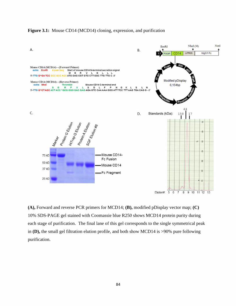
84
Figure 3.1: Mouse CD14 (MCD14) cloning, expression, and purification
(A), Forward and reverse PCR primers for MCD14; (B), modified pDisplay vector map; (C)
10% SDS-PAGE gel stained with Coomassie blue R250 shows MCD14 protein purity during
each stage of purification. The final lane of this gel corresponds to the single symmetrical peak
in (D), the small gel filtration elution profile, and both show MCD14 is >90% pure following
purification.

85
Figure 3.2: Mouse CD14 (MCD14) LPS binding native polyacrylamide gel shift (native PAGE)
(A), In a 4-20% native PAGE gel stained with Coomassie blue R250 we visualized the
interaction of purified mouse CD14 (MCD14; 10 µg) with a 2:1 molar ratio of the Ra chemotype
of E. coli lipopolysaccharide (LPS; Sigma) in the presence of a 1:100 molar ratio of LBP:LPS
after 1 hr incubation at 37oC (pre-incubation) as described in Materials and Methods. A
downshift indicates formation of the MCD14+LPS ligand bound complex. (B), The pre-
incubation time of 10 µg MCD14 with a 2:1 molar ratio of the Ra chemotype of E. coli
lipopolysaccharide (LPS; Sigma) in the absence or presence of a 1:100 molar ratio of LBP:LPS
was varied (30 minutes or 1 hr) to assay the length of sample incubation time required for
complex formation. Although we chose to standardize our pre-incubation time in subsequent
experiments to 1 hr at 37oC, we visualized complex formation after ~5 minutes in the presence of
LBP in similar time course experiments with mouse and human CD14 (data not shown).
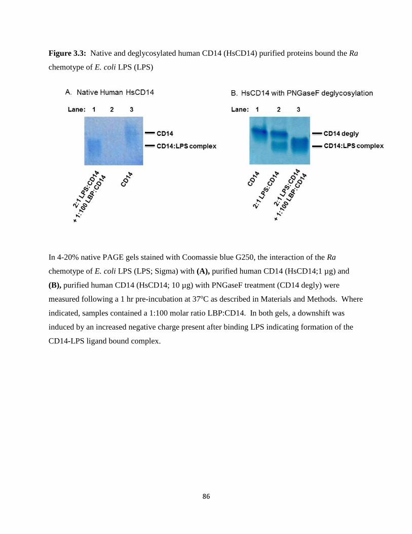
86
Figure 3.3: Native and deglycosylated human CD14 (HsCD14) purified proteins bound the Ra
chemotype of E. coli LPS (LPS)
In 4-20% native PAGE gels stained with Coomassie blue G250, the interaction of the Ra
chemotype of E. coli LPS (LPS; Sigma) with (A), purified human CD14 (HsCD14;1 µg) and
(B), purified human CD14 (HsCD14; 10 µg) with PNGaseF treatment (CD14 degly) were
measured following a 1 hr pre-incubation at 37oC as described in Materials and Methods. Where
indicated, samples contained a 1:100 molar ratio LBP:CD14. In both gels, a downshift was
induced by an increased negative charge present after binding LPS indicating formation of the
CD14-LPS ligand bound complex.
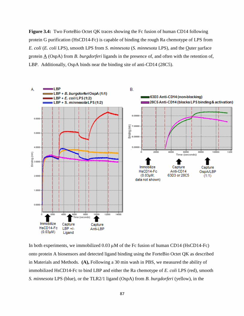
87
Figure 3.4: Two ForteBio Octet QK traces showing the Fc fusion of human CD14 following
protein G purification (HsCD14-Fc) is capable of binding the rough Ra chemotype of LPS from
E. coli (E. coli LPS), smooth LPS from S. minnesota (S. minnesota LPS), and the Outer surface
protein A (OspA) from B. burgdorferi ligands in the presence of, and often with the retention of,
LBP. Additionally, OspA binds near the binding site of anti-CD14 (28C5).
In both experiments, we immobilized 0.03 M of the Fc fusion of human CD14 (HsCD14-Fc)
onto protein A biosensors and detected ligand binding using the ForteBio Octet QK as described
in Materials and Methods. (A), Following a 30 min wash in PBS, we measured the ability of
immobilized HsCD14-Fc to bind LBP and either the Ra chemotype of E. coli LPS (red), smooth
S. minnesota LPS (blue), or the TLR2/1 ligand (OspA) from B. burgdorferi (yellow), in the
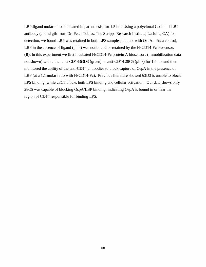
88
LBP:ligand molar ratios indicated in parenthesis, for 1.5 hrs. Using a polyclonal Goat anti-LBP
antibody (a kind gift from Dr. Peter Tobias, The Scripps Research Institute, La Jolla, CA) for
detection, we found LBP was retained in both LPS samples, but not with OspA. As a control,
LBP in the absence of ligand (pink) was not bound or retained by the HsCD14-Fc biosensor.
(B), In this experiment we first incubated HsCD14-Fc protein A biosensors (immobilization data
not shown) with either anti-CD14 63D3 (green) or anti-CD14 28C5 (pink) for 1.5 hrs and then
monitored the ability of the anti-CD14 antibodies to block capture of OspA in the presence of
LBP (at a 1:1 molar ratio with HsCD14-Fc). Previous literature showed 63D3 is unable to block
LPS binding, while 28C5 blocks both LPS binding and cellular activation. Our data shows only
28C5 was capable of blocking OspA/LBP binding, indicating OspA is bound in or near the
region of CD14 responsible for binding LPS.

89
Figure 3.5: Human (HsCD14) and Mouse (MCD14) Bioactivity Assay
(A), Purified human HsCD14 (1 µg/mL) was pre-incubated with a 1:100 molar ratio LBP:CD14
and increasing concentrations LPS for 1 h at 37oC. Alternatively, LPS was incubated with 2%
human serum. (B), Similarly, purified mouse CD14 (1) was pre-incubated with a 1:100 molar
ratio LBP:CD14 and increasing concentrations of LPS for 1 hr or overnight at 37C as described
in Materials and Methods. In both experiments, SW620 cells were stimulated for 6 hours with
these preformed complexes and IL-8 was measured in culture supernatants.

90
Figure 3.6: Initial Sparse Matrix Screen Results for Mouse CD14 (MCD14)
We were unable to obtain crystals utilizing the published crystallization condition for Mouse
CD14 (69) (listed as MCD14 Published above) likely due to the use of different expression
systems, so we conducted an initial sparse matrix crystallization screen with our purified Mouse
CD14 (MCD14) protein using three commercially available kits (Wizard I and II from Emerald
Biosystems and Crystal Screen I from Hampton Research) to look for single crystals. Pictures of
hanging drops yielding the best results from the initial screen, including crystal clusters or single
crystals, are shown and the compositions of the commercially available reagents are indicated.
.

91
Figure 3.7: No diffraction quality crystals resulted from MCD14 (or HsCD14; data not shown)
crystallization with the Ra chemotype of E. coli LPS in the presence of LBP
All three hanging drop conditions are commercially available reagents Wizard I #28, Wizard I
#30, and Wizard I #7 (Emerald Biosystems). For this experiment, 10 mg/mL MCD14 was
purified with PNGaseF treatment as described in Materials and Methods prior to incubation with
the Ra chemotype of E. coli LPS and a catalytic amount of LBP (1:100 LBP:CD14) for 1 hour at
37oC prior to setting out the crystallization tray at RT. In all crystallization efforts with ligand
present, phase separation or protein aggregation were predominant outcomes, resulting in a lack
of crystals for further optimization. The best result was obtained with Wizard I #7 showing the
presence of four potential microcrystals.

92
CHAPTER 4:
IgE and Ligand Binding Studies of Der p 2 Allergen
4.1 Introduction
Der p 2 induces an allergic response by acting as a structural and functional mimic of
MD-2 in the LPS sensing pathway (Chapter 1). Given this functional role of Der p 2, we were
interested in determining if we could express Der p 2 using yeast display and develop an assay
for detecting binding to LPS and TLR4. Yeast display is a powerful technique pioneered by the
Kranz lab to allow rapid screening of large libraries of mutants on the surface of yeast in order to
identify mutations that lead to increased protein stability or higher ligand affinity using a high-
throughput fluorescence activated cell sorting format that circumvents the need for protein
purification (193). Thus, we anticipated that yeast display of Der p 2 would enable us to identify
and characterize the residues of this protein involved in LPS and TLR4 binding.
The yeast display system involves fusion of a protein encoding gene of interest to a gene
encoding the yeast a-agglutinin 2 (Aga2) protein within a yeast expression vector. Since the
Aga2 protein inherently associates with the chromosomally encoded surface displayed Aga1
yeast protein by disulfide bonding, the protein of interest is displayed on the surface of yeast
(Figure 4.1). We created two yeast display constructs in which the yeast Aga2 protein was fused
to either the amino or carboxyl terminus of Der p 2; hereafter named N-Dp2 and C-Dp2,
respectively. Similarly, we constructed a C-terminal Aga2 fusion of MD-2 for comparison
(named C-MD-2).
Alongside our efforts, the N-terminal fusion of MD-2 (N-MD-2) was created by Dr.
Adam Chervin and utilized by Daiva Mattis (Kranz Lab, University of Illinois, Urbana) to
identify residues important for LPS and TLR4 binding. Their work showed yeast displayed N-

93
MD-2 is full-length, properly folded, and able to bind both biotinylated E. coli LPS and the
purified extracellular domain of human TLR4. Therefore, we chose to use N-MD-2 alongside
our N-Dp2, C-Dp2, and C-MD-2 yeast display binding assays as a positive control. We also
used an unrelated N-terminal Aga2 fusion (N-L3) as a negative control in our studies. L3 is a 15
kD TCR V protein, which was kindly provided by Davia Mattis (Kranz Lab, University of
Illinois, Urbana).
As we will discuss in this Chapter, we were able to detect full length expression of our
stable N-Dp2, C-Dp2, and C-MD-2 fusions on the surface of yeast. Because of the thermal
stability of Der p 2 discussed in Chapter 1, difficulties arose when we attempted to use thermal
denaturation to test Der p 2 folding. We tested the ability of our Der p 2 and MD-2 fusions to
bind E. coli LPS and the purified extracellular domain of human TLR4 (kindly provided by
Diana Ranoa, Tapping Lab, University of Illinois, Urbana). Detection of TLR4 binding would
provide additional biophysical evidence of the ability of Der p 2 to act as an adjuvant in the
allergic arm of the MD-2/TLR4/LPS sensing pathway.
As a second goal, we recently shifted our focus to researching the Der p 2 yeast display
system as a rapid, cost effective, and clinically relevant method to detect and quantify levels of
IgE in dust mite allergic patient samples. Historically, two methods have been utilized to detect
the level of an allergic response in an individual. The first method, an in vivo skin-prick test,
uses one or more allergens to the skin with a disposable tool containing fine needles, or
alternatively, injected just below the outer dermal layer. Usually, the allergens are placed on
identifiable marks or in a grid, enabling a physician to identify atopic (or allergic) patients who
develop a raised white patch and surrounding redness from blood vessel dilation typical of a
“wheal and flare” response to the appropriate allergen. Histamine and saline are applied as
94
positive and negative controls for comparison and the physician records the allergic response on
a 0-5 scale as compared to histamine. Although this in vivo method allows for rapid detection in
less than an hour, its drawbacks include the limits of quantitation using a 0-5 scale, the
requirement of highly pure allergens, and the inaccurate results obtained if the patient is taking
anti-allergy medications or has a skin condition capable of interfering with detection. Although
this method is predictive, skin-prick tests do have a higher false-positive rate than other test
methods (194).
The second commonly used in vitro method for diagnosing allergy involves detection of
immunoglobulin E (IgE) antibodies specific to an allergen. As discussed in Chapter 1, IgE
mediates the human allergic response by binding both the allergen and an FcεRI receptor that is
primarily expressed on the surface of mast cells and basophils (~100,000 receptors per cell) in
skin, tissue near mucosal layers and gastrointestinal sites, causing the release of histamine and
other mediators capable of inducing allergic symptoms in affected patients. Since the
concentration of serum IgE is less than 1 g/mL and it binds with high affinity (>10-9 M) to its
receptor FceRI, IgE is the most difficult antibody to detect in human fluids (195).
Various forms of in vitro IgE detection systems have been created by Thermo Scientific.
Since the 1960s, the first IgE detection radioallergosorbent test (RAST) has been used
nationwide in commercial clinical reference labs (196). In RAST, patient serum is applied to a
paper disk or polyurethane cap (RAST-CAP) containing an immobilized allergen for three hours.
Any IgE from the serum that binds is detected using a radioactively labeled anti-human IgE
antibody incubated with the immobilized allergen for 16-30 hours prior to detection. Thus, this
test is neither rapid nor automated, but it produces a quantitative result by comparing the
resulting radioactivity to an anti-birch antibody response to birch allergen standard. The

95
measurements are read in RAST units per milliliter (RU/mL), and results are grouped into
Classes 0-4. One of the major benefits of RAST testing is the lack of patient risk for
anaphylactic shock since the allergen is not being introduced in vivo. Also, allergens can be
coarsely purified or homogenized prior to immobilization, so this system does not require highly
purified allergens. Additionally, patients taking anti-allergy medications at the time of the test or
those with skin conditions can still obtain test results without interference with the testing
method. Finally, this standardized quantitation and classification system allows for a more
straightforward comparison between patients. Improvements to this test have been made
including the use of an enzyme linked anti-human IgE antibody that provides a colormetric result
when a color changing substrate is applied in an enzyme immunoassay (EIA) format or the use
of a fluorescently-labeled anti-human IgE antibody for detection.
A second generation, fully automated allergosorbent system called ImmunoCAP was also
produced by Thermo Scientific and is now considered the standard clinical method of IgE
detection by the National Institutes of Health (197). Much like RAST, ImmunoCAP uses an
immobilized allergen to bind allergen specific IgE from patient sera and detects binding via a
fluorescently-labeled anti-human IgE antibody. However, in ImmunoCAP testing the allergen is
immobilized to a highly-branched, hydrophilic, cellulose derivative in a solid phase instead of a
two dimensional paper or solid support, leading to the capture of more allergens per test and
increasing assay sensitivity. IgE measurements are quantified in this system in international
units per liter (kU/L). These values are determined by comparison of the measurement with a
standard created by the World Health Organization by pooling nine donor serum samples
together at a potency of 5,000 IU (WHO Reference Reagent NIBSC 75/502). Similarly, a house
dust mite D. pteronyssinus extract is available with a potency of 100,000 IU (WHO Reference

96
Reagent NIBSC 82/518). The quantified results correspond to Classes 0-6, where Class 0 (<0.35
kU/L) is considered negative, Class 1 (0.35-0.69 kU/L) is very low, Class 2 (0.70-3.49 kU/L) is
low, Class 3 (3.5-17.49 kU/L) is moderate, Class 4 (17.5-49.99) is high, Class 5 (50-100 kU/L) is
very high and Class 6 (>100 kU/L) is at or above the limit of detection. As mentioned before,
the sensitivity of ImmunoCAP testing is much higher than that achieved with RAST. In fact, the
RAST Classes 0-4 correspond to the 0.35-17.5 kU/L detection range of ImmunoCAP (198). In a
study of 39 adults, the ImmunoCAP system had high sensitivity (94%) and specificity (100%) in
utilizing IgE to detect for the 23 patients suffering from respiratory allergies (198).
A major limitation of the ImmunoCAP method is the inability to test more than one
allergen specifically in each screen, so testing can be expensive. To overcome this limitation,
Thermo Scientific recently created the ImmunoCAP immuno solid-phase allergy chip (Immuno
CAP ISAC). Immuno CAP ISAC uses the same technology as ImmunoCAP, but screens a panel
of 103 allergens in a multi-array chip format, enabling detection of more than one allergy
sensitization and the identification of cross-reaction in a patient sample at the same time (199).
Similarly, physicians can now probe beyond whole dust mite homogenates present in
ImmunoCAP and ImmunoCAP ISAC to detect serum IgE bound to individual allergenic
proteins, like Der p 2, using a more limited panel of purified allergens called the ImmunoCAP
highly refined (ImmunoCAP HR) test.
An additional limitation of all ImmunoCAP methods described previously is the need to
purchase the proprietary ImmunoCAP machine and/or ImmunoCAP ISAC chip required to run
the test, which is an added expense in the clinical setting. Alternatively, clinicians can ship
patient samples to a commercial clinical laboratory for ImmunoCAP testing, resulting in a longer
wait time for results. In an attempt to overcome the wait time limitation, Thermo Scientific

97
recently developed a rapid ImmunoCAP asthma/rhinitis test, allowing the detection of serum IgE
specific for a panel of 10 allergens, not including Der p 2, in patient blood applied to a single-use
testing strip. Although the test strips still have to be purchased and the result is simply
qualitative, physicians can detect IgE specific for the 10 allergens in 20 minutes.
In this Chapter, we utilized our yeast displayed Der p 2 as a novel method to detect IgE
from dust mite allergic and non-allergic patient serum and plasma samples using flow
cytometery. Since flow cytometry is actively utilized in research and clinical laboratories, our
technique would overcome the costly requirement for purchasing proprietary equipment to
conduct IgE detection. A simple extension of our methodology could use mutant forms of Der p
2 in our yeast display system to detect IgE epitopes specific for an individual patient. This same
type of detailed analysis of Der p 2 interactions with IgE is not possible with the RAST,
ImmunoCAP, ImmunoCAP HR, or ImmunoCAP ISAC methods, as they provide simple
quantitation of IgE binding, so our flow cytometry based method would complement, not
replace, existing in vitro detection systems. In this Chapter, we offer proof-of-concept by
successfully displaying Der p 2 on the surface of S. cerevisiae and quantitatively measuring the
binding of IgE from patient serum and samples using our novel in vitro, flow cytometry-based
IgE detection system.
4.2 Materials and methods
4.2.1 Reagents
The yeast display vector pCT302 and two additional N-terminal Aga2 fusions containing
MD-2 and a V-TCR called L3 were kindly provided by Daiva Mattis (Kranz Lab, University of
98
Illinois, Urbana-Champaign). The Y10 yeast display vector was kindly provided by Dr. David
Aggen (Kranz lab, University of Illinois Urbana-Champaign). Additional fluorescent reagents,
antibodies, and purified proteins are described along with methods below. Media and reagents
utilized in this work for subcloning were previously described in Chapter 2. Likewise, we
purchased chemically competent E. coli DH5α cells and LB-agar plates containing 100 g/mL
ampicillin from the University of Illinois at Urbana-Champaign (UIUC) cell Media Facility
(kindly produced by Dr. Sandra McMasters). EBY100 cells grown in YPD media (yeast extract
10 g/L; peptone 20 g/L, and dextrose 20 g/L) were kindly provided by Sheena Smith and Daiva
Mattis (Kranz lab, University of Illinois, Urbana). Two forms of minimal media plus amino
acids were used for yeast cultures. SD-CAA (SD media) contained dextrose (20g/L), yeast
nitrogen base without amino acids (7 g/L), 19 mM Na2HPO4, 36 mM NaH2PO4, and casamino
acids (5 g/L). SG-CAA (SG media) used the same recipe for preparation except dextrose was
replaced with galactose (20 g/L).
4.2.2 Yeast Display of Der p 2 and MD-2
To create the N-terminal fusion of Der p 2 (N-Dp2), we ordered synthetic, codon
optimized DNA (Genescript) encoding a NheI site, aa 18-146 of Der p 2 (omitting its leader
sequence), a c-Myc epitope tag (EQKLISEEDL), two stop codons (TGA and TAA), and an XhoI
site. The yeast display vector pCT302 was restriction enzyme digested with NheI and XhoI and
treated with calf alkaline phosphatase (NEB). Vector digests were visualized on a 2% agarose
gel with a 1 kB DNA ladder (Promega) using 6xDNA loading dye. The pCT302 yeast display
vector contains a galactose inducible promotor (GAL1) prior to an EcoRI restriction site,
upstream of DNA encoding Aga2, followed by the influenza hemagglutinin epitope tag HA

99
(YPYDVPDYA), a Gly-Ser linker region and a NheI restriction site. We ligated the N-Dp2
insert into pCT302 using T4 DNA ligase (Invitrogen), and after transformation of E.coli DH5α
competent cells, we grew transformants on LB-Amp plates overnight at 37oC. We placed single
colonies in 5 mL LB-Amp cultures overnight 37oC with shaking, and harvested DNA from each
culture by miniprep (Qiagen). DNA sequencing (UIUC Biotechnology Center) and restriction
enzyme digestion with EcoRI and NdeI followed by agarose gel electrophoresis were two
methods used to confirm subcloning.
Next, we transformed yeast cultures with our sequenced pCT302 N-Dp2 vector. Multiple
3 mL cultures of 100 L EBY100 yeast were grown in YPD media without antibiotics in a 30oC
incubator with shaking, overnight, and then transformed with 1, 5, or 10 L N-Dp2 pCT302
DNA by LiOAc heat shock. Afterwards, transformed EBY100 yeast were plated on yeast media
containing dextrose (SD) plates without antibiotics and incubated at 30oC for 3 days. After
colony growth, single EBY100 transformed colonies were picked and grown in 3 mL cultures of
SD media with 100 g/mL penicillin/streptomycin (Gibco) in a 30oC incubator with shaking.
DNA extraction was completed using zymolase digestion of the yeast cell wall and alkaline lysis
via Zymoprep Yeast Plasmid Miniprep II (Zymo Research). DNA sequencing (UIUC
Biotechnology Center) and restriction enzyme digestion with EcoRI and NdeI followed by
agarose gel electrophoresis were used to confirm yeast transformation. Stocks of N-Dp2
transformed EBY100 yeast were frozen in media containing 25% glycerol. Unless indicated,
transformed yeast were inoculated into SD media at 30oC from frozen stocks. Expression of the
N-Dp2 protein from EBY100 was induced by growing the cells in yeast media containing
galactose (SG) at 20oC for 48 hours.

100
Similarly, DNA encoding C-Dp2 and C-MD-2 were purchased (Genescript) and encoded
an EcoRI site, the Aga2 signal peptide (aa 1-18), Der p 2 or MD-2, a c-Myc epitope tag
(EQKLISEEDL), and an Nde I site. Using the same subcloning method described in this
Chapter, the synthetic DNA was ligated into a Y10 vector. The Y10 yeast display vector was
previously created from pCT302 by rearranging the orientation of the Aga2 fusion onto the C-
terminus of the target protein (kindly provided by Dr. David Aggen, Kranz lab, University of
Illinois, Urbana). Y10 has an NdeI site, followed by Aga2, an HA epitope tag (YPYDVPDYA),
a stop codon, and an XhoI/BglII site. Following ligation and transformation into Y10, E. coli
cells were used for amplification, sequencing was completed, EBY100 yeast were transformed
and the final transfectants were sequenced as described above for the N-terminal fusion.
Likewise, stocks of C-Dp2 and C-MD-2 transformed EBY100 yeast were frozen in media
containing 25% glycerol. Expression of the C-Dp2 and C-MD-2 protein from EBY100 was
induced by growing the cells in yeast media containing 2% galactose (SG) at 20oC for 48 hours
prior to flow cytometry.
4.2.3 Detection of Yeast Displayed Der p 2 and MD-2
We stained 1x10⁶ N-Dp2, C-Dp2, and C-MD-2 transformed EBY100 yeast cells after
induction in SG media at 20oC with shaking for 48 hours followed by washing. Each washing
stage noted below involved one dilution and two wash steps with 1 mL PBS containing 0.5%
bovine BSA (Sigma), followed by yeast cell pelleting via centrifugation at 3,000 rpm, 3 min,
4oC, and vacuum aspiration of wash reagent. Yeast were washed and stained with mouse IgG1
anti-HA antibody (HA.11 Clone 16B12, MMS-101R, Covance) and chicken anti-c-Myc
antibody (A21281; Invitrogen) incubated in 1:50 dilutions at 4oC for 1 hr. To probe for Der p 2
101
on the cell surface, we used a variety of monoclonal mouse IgG1 anti-Der p 2 antibodies from
Indoor Biotechnologies at 1:100 dilution including DpX (MA-DPX), 1D8 (MA-1D8-1),
biotinylated-7A1 (MA-B-7A1), and 10E11 (MA-10E11), and an anti-Der f 2 antibody (13A4)
that was previously shown to cross react with Der p 2 in literature. To probe for MD-2, we used
two monoclonal anti-human MD-2 antibodies called 4H1 (HM2243) and 18H10 (HM2245) from
Hycult at a 1:100 dilution. Cells were washed following primary antibody staining. Secondary
antibodies used at 1:200 dilution include the Alexa Fluor 647 conjugated goat anti-mouse (#A-
21237; Invitrogen) and APC conjugated goat anti-chicken (A21449; Invitrogen) antibodies. To
detect biotin, Alexa Fluor 647 conjugated streptavidin (S32357; Invitrogen) was used at a 1:200
dilution. All cells were washed and resuspended in 200 L 1xPBS for flow cytometry detection.
In all flow cytometry experiments, fluorescence was detected with an Accuri C6 flow cytometer
where we gated on a positive population of FCS vs. SSC live cells, unless gating parameters are
otherwise indicated, and captured 10,000 events using a slow flow rate with a detection filter of 8
size limit.
4.2.4 Thermal Denaturation of Yeast Displayed Der p 2
We stained 1x10⁶ EBY100 transfected N-Dp2 or C-Dp2 cells after induction in SG media
at 20oC for 48 hours. Cells were washed as previously described. Each eppindorf tube contained
a 1:100 dilution of an anti-Der p 2 (DpX, 1D8, biotinylated 7A1, or 10E11) or anti-Der f 2
antibody (13A4). Yeast were incubated at the temperature indicated in the figure for 1 hour,
followed by washing and resuspension in 200 L 1xPBS prior to flow cytometry detection as
previously described.
102
4.2.5 LPS Binding Studies
To probe for LPS binding, we incubated biotinylated ultrapure E. coli O111:B4 LPS EB-
Biotin from InvivoGen (EB-LPS; catalogue #tlrl-bblps) or a non-biotinylated precursor form of
ultrapure E. coli O111:B4 LPS (O-LPS; catalogue #tlrl-pelps) with 1x10⁶ EBY100 transformed
N-Dp2, C-Dp2, N-MD-2, C-MD-2, N-L3, or empty Y10 vector for 1 hour at 4oC. Following
washing, we detected bound EB-LPS using Alexa Fluor 647 conjugated streptavidin (S32357;
Invitrogen) at a 1:200 dilution incubated for 1 hour at 4oC. To detect bound O-LPS, we used a
serotype specific monoclonal antibody from Hycult (mouse anti-core; WN1222-5) incubated 30
minutes to 1 hour at 4oC. After a final wash and resuspension in 200 L 1xPBS, we detected
fluorescence using flow cytometry as previously described. At times, we preincubated mouse
(MCD14) or human soluble CD14 (HsCD14) described in Chapters 2 and 3 at 37oC with
biotinylated or non-biotinylated LPS (molar ratios indicated), prior to addition to transformed
yeast cells.
4.2.6 TLR4 Binding Studies
We used a purified extracellular domain of human TLR4 (aa 27-527) kindly provided by
Diana Ranoa (Tapping Lab, University of Illinois, Urbana) in our flow cytometry binding
studies. 1x10⁶ EBY100 transformed N-Dp2, C-Dp2, N-MD-2, C-MD-2, N-L3, or empty Y10
vector were mixed with the TLR4 protein for 1 hour at RT. Following washing as previously
described, mouse IgG2a anti-Human CD284/TLR4 antibody (HTA125; EBioscience) at a 1:100
dilution was utilized. Following washing as previously described, the AlexaFluor 647

103
conjugated goat anti-mouse secondary at a 1:100 dilution was used for detection. Following
washing, the cells were resuspended in 200 L 1xPBS for flow cytometry detection as
previously described.
4.2.7 IgE Binding Studies
We purchased dust mite allergic and non-allergic patient serum and plasma samples from
Plasma Lab International. Following washing, we incubated 100 L of each sample (without or
with dilution as indicated in the figure) with 1x10⁶ EBY100 transformed N-Dp2 and C-Dp2
cells. To detect human IgE, we used biotin-labeled affinity purified Goat anti-Human IgE (16-
10-04; KPL) antibody at a 1:100 dilution. Following washing we detected biotin using Alexa
Fluor 647 conjugated streptavidin (S32357; Invitrogen) at a 1:200 dilution. Following washing
and final resuspension in 200 L 1xPBS, flow cytometry was utilized to produce binding
profiles. Binding curves with non-linear least squares fitting were created using GraphPad Prism
6.
4.3 Results
4.3.1 Yeast Display of Der p 2and MD-2
Initially, we chose to display an N-terminal fusion of Der p 2 (N-Dp2) on the surface of
S. cerevisiae EBY100 yeast using the pCT302 yeast display vector (Figure 4.1 A). Following
subcloning and sequencing to confirm proper insertion of DNA into the NheI and XhoI sites on
pCT302, as described in the Materials and Methods section in this Chapter, we induced
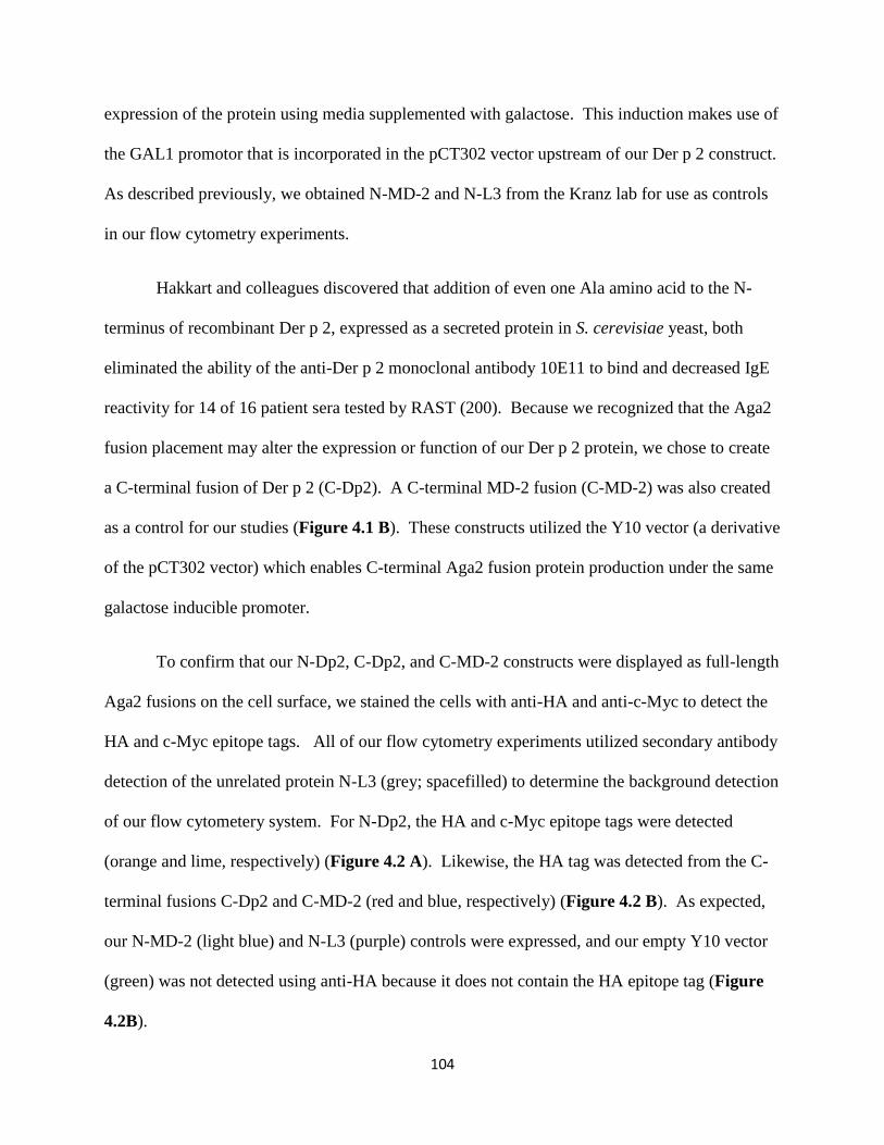
104
expression of the protein using media supplemented with galactose. This induction makes use of
the GAL1 promotor that is incorporated in the pCT302 vector upstream of our Der p 2 construct.
As described previously, we obtained N-MD-2 and N-L3 from the Kranz lab for use as controls
in our flow cytometry experiments.
Hakkart and colleagues discovered that addition of even one Ala amino acid to the N-
terminus of recombinant Der p 2, expressed as a secreted protein in S. cerevisiae yeast, both
eliminated the ability of the anti-Der p 2 monoclonal antibody 10E11 to bind and decreased IgE
reactivity for 14 of 16 patient sera tested by RAST (200). Because we recognized that the Aga2
fusion placement may alter the expression or function of our Der p 2 protein, we chose to create
a C-terminal fusion of Der p 2 (C-Dp2). A C-terminal MD-2 fusion (C-MD-2) was also created
as a control for our studies (Figure 4.1 B). These constructs utilized the Y10 vector (a derivative
of the pCT302 vector) which enables C-terminal Aga2 fusion protein production under the same
galactose inducible promoter.
To confirm that our N-Dp2, C-Dp2, and C-MD-2 constructs were displayed as full-length
Aga2 fusions on the cell surface, we stained the cells with anti-HA and anti-c-Myc to detect the
HA and c-Myc epitope tags. All of our flow cytometry experiments utilized secondary antibody
detection of the unrelated protein N-L3 (grey; spacefilled) to determine the background detection
of our flow cytometery system. For N-Dp2, the HA and c-Myc epitope tags were detected
(orange and lime, respectively) (Figure 4.2 A). Likewise, the HA tag was detected from the C-
terminal fusions C-Dp2 and C-MD-2 (red and blue, respectively) (Figure 4.2 B). As expected,
our N-MD-2 (light blue) and N-L3 (purple) controls were expressed, and our empty Y10 vector
(green) was not detected using anti-HA because it does not contain the HA epitope tag (Figure
4.2B).
105
We wanted to confirm the N-MD-2 protein and C-MD-2 proteins we were producing
were capable of being recognized by anti-MD-2 antibodies. Previous thermal denaturation work
conducted by Daiva Mattis in the Kranz lab showed the anti-MD-2 antibody 18H10 may
recognize a conformational epitope; whereas, binding of the anti-MD-2 antibody 4H1 was
unaffected by changes in temperature, so it may recognize a linear epitope (unpublished). Using
flow cytometry, we verified binding of the 18H10 antibody to yeast displayed N-MD-2,
suggesting that this protein is folded (Figure 4.3 A). As expected, anti-MD-2 18H10 binding to
the either N-L3 (purple) or N-Dp2 (orange) proteins was not detected. Similarly, staining of C-
MD-2 shows 18H10 binding (light blue), indicating the C-terminal fusion we created is also
properly folded upon display (Figure 4.3 B).
Multiple anti-Der p 2 monoclonal antibodies (DpX, 1D8, 7A1, and 10E11) were used to
detect our N and C-terminal fusions of Der p 2 on the surface of yeast. Previously, Hakkaart and
colleagues conducted mutagenesis and deletion studies of secreted recombinant Der p 2
expressed in S. cerevisea and showed that the epitope of the DpX antibody resides within aa 65-
80 (9). The binding of the 7A1 antibody is affected by mutations in amino acid residues 22-26,
45-48, 88-90 (201), and Lys 100 (202). Both DpX and 7A1 anti-Der p 2 antibodies were found
to cross react with the homologous group II allergen Der f 2 (115). Similarly, anti-Der f 2
antibody 13A4 was reported to be slightly cross reactive to Der p 2 (115), and binding of this
antibody was abolished when Der p 2 Lys 100 was mutated (202). We included this anti-Der f 2
antibody in our examination of yeast displayed Der p 2 because epitope mapping NMR studies
showed 13A4 binding depends upon the conformation of Der f 2 (203), so we hypothesized
13A4 binding may support the conclusion that Der p 2 is properly folded upon display.
Additionally, as mentioned previously, anti-Der p 2 antibody 10E11 is capable of differentiating

106
between Der p 2 with and without one additional amino acid on the N-terminus. Mutagenesis of
Der p 2 aa 22-26, 45-48, 67 and 69, and 73-78 decreased binding of 10E11, which may indicate
10E11 recognizes a conformationally dependent epitope that is only formed upon proper folding
(201).
Anti-Der p 2 antibody 1D8 was able to bind N-Dp2 (orange), but, as expected, did not
bind N-MD-2 (light blue) or N-L3 (purple) non-specifically (Figure 4.4 A). 1 D8 was also able
to bind C-Dp2 (red) (Figure 4.4 B). In the same experiment, we were encouraged to see C-Dp2
recognition by 10E11 (pink) without detectable binding to N-Dp2 (green) (Figure 4.4 B). We
also tested the ability of both Der p 2 constructs to bind DpX and 7A1. N-Dp2 (data not shown)
and C-Dp2 (orange) was detected by DpX, similar to detection using 1D8 (green) (Figure 4.4
C). Similarly, we were able to detect 7A1 binding to both fusions (data not shown).
As a whole, this data indicates we properly constructed a yeast display system that
expresses Der p 2 containing linear and possibly conformational epitopes for antibody
recognition. We also tested N-Dp2 and C-Dp2 folding using denaturation studies (204). We
screened the loss of antibody binding upon Der p 2 thermal denaturation for each anti Der p 2
antibody (DpX, 1D8, biotinylated 7A1, and 10E11) as described in Materials and Methods. All
of the antibodies capable of binding N-Dp2 and C-Dp2 still bound both proteins upon heating to
100oC. For example, after heating to 100oC we could detect 1D8 binding to N-Dp2 (Figure 4.5
A). We also screened 10E11 binding to C-Dp2 (Figure 4.5 B) and N-Dp2 (Figure 4.5 C), and
observed similar profiles at all temperatures tested. Finally, we screened the anti-Der f 2
antibody 13A4 and found little to no detection of binding (Figure 4.5 D). As mentioned
previously, Der p 2 is highly thermostable (121) and is only mostly denatured after a 60 minute
incubation at 140oC (205), but the viability of the yeast display scaffold has only been tested to

107
~90oC (204). Since this thermal denaturation method does not appear to denature Der p 2, it is
difficult to confirm proper folding. However, both N-Dp2 and C-Dp2 are properly recognized
by a variety of anti-Der p 2 antibodies.
4.3.2 Yeast Displayed Der p 2 and MD-2 Bind LPS
We tested the ability of Der p 2 and MD-2 to bind LPS by using biotinylated E. coli
O111:B4 (EB-LPS) as described in Materials and Methods. Titration studies of EB-LPS with
the N-terminal Aga2 fusions of MD-2 and Der p 2 indicated binding, but a minor background
level of interaction with the L3 negative control was also detected (Figure 4.6). We did not
expect to see detectable binding of EB-LPS to the N-terminal Aga2 fusion of L3, so we decided
to screen EB-LPS binding to a panel of unrelated N-terminal Aga2 fusion yeast displayed
proteins (kindly provided by Sheena Smith and Daiva Mattis, Kranz lab, University of Illinois,
Urbana). Proteins I, W, and A are all stable, high stability and high affinity engineered 29 kD
single chain T cell receptor V-Vα Aga2 fusions, which differ in their Vs. Protein K is the
single chain variable region of an antibody targeting V and Protein D is a human T cell receptor
V. Both are approximately 13 kD and are fused to Aga2. Protein C is a 25 kD co-stimulatory
molecule expressed as an Aga2 fusion. Binding profiles for each are shown in Figure 4.6.
Proteins K, C, and W all showed interactions with EB-LPS to a greater extent than our N-L3
negative control. All of the unrelated proteins indicated binding to EB-LPS to a lesser extent
than N-MD-2, and most of the unrelated proteins indicated lower detection of EB-LPS binding
than N-Dp2. Further analysis would require additional replicates of this experiment, with the
addition of our C-terminal Der p 2 and MD-2 fusions.

108
We also tested non-biotinylated E. coli O111:B4 (O-LPS) binding to both fusions of Der
p 2 and MD-2 using a serotype specific antibody that binds the core polysaccharide region of
LPS as described in Materials and Methods, as a way to confirm that protein-LPS interactions
were not disrupted or unnaturally aided by biotinylation of the ligand. N-MD-2 (light blue), C-
MD-2 (dark blue), C-Dp2 (red), and N-Dp2 (orange) all indicated binding to O-LPS (Figure 4.6
B). We also detected O-LPS binding to yeast containing the empty Y10 vector (green) and N-L3
(purple). Dr. Adam Chervin in the Kranz lab showed that addition of our purified human
HsCD14 protein reduced EB-LPS binding to an unrelated control protein while not affecting
binding of EB-LPS to N-MD-2 (unpublished). We tested the ability of CD14 to reduce binding
to N-L3 by pre-incubating a 2:1 molar ratio of human soluble CD14 (HsCD14) or mouse CD14
(data not shown) with O-LPS for 1 hr at 37oC before incubation with our yeast fusions. CD14
decreased O-LPS binding to C-Dp2 (red to pink) and N-Dp2 (orange to maroon) (Figure 4.6 C).
Similar to the reduction of LPS binding to an unrelated control in Dr. Chervin’s work, we also
observed a reduction of O-LPS binding to our unrelated N-L3 control (dark purple to light
purple) and our empty Y10 vector yeast cells (dark green to lime). In other LPS binding studies,
we added a catalytic amount of purified lipopolysaccharide binding protein (LBP) (kindly
provided by Dr. Jerrold Weiss, University of Iowa). As described in Chapter 1, LBP
catalytically loads CD14 with LPS, enabling LPS delivery to the hydrophobic pocket of MD-2 in
the LPS sensing cascade. However, in our yeast display system, addition of LBP did not
increase the level of EB-LPS binding we detected (data not shown).

109
4.3.3 Yeast Displayed MD-2, but not Der p 2, bind TLR4
We used flow cytometry to detect the ability of our N and C-terminal Aga2 fusions to
bind purified human TLR4. As described in Chapter 1, human TLR4 interacts with MD-2 and
upon ligand binding forms a heterodimeric complex with LPS bound to the hydrophobic binding
pocket of MD-2. Since Der p 2 has structural similarity to MD-2, the current hypothesis is that
Der p 2 may interact directly with TLR4, as supported by pull down data from Trompette and
colleagues (122). In our data, the N-terminal fusion of MD-2 exhibited binding to human TLR4
at two different concentrations of TLR4 (dark and light blue), but N-Dp2 did not show any
detectable level of binding at either concentration of TLR4 (orange and yellow) (Figure 4.7 A).
Similarly, C-MD-2 exhibited binding to purified human TLR4 (dark blue), but in this and
replicate experiments, C-Dp2 did not bind TLR4 (red) (Figure 4.7 B).
4.3.4 Detection of Anti-Der p 2 IgE in Human Serum and Plasma Using Yeast Display
We studied the ability of C-Dp2 to bind IgE from a Der p 2 specific IgE Class 0 (0.02
kU/L) non-allergic patient serum sample and a Class 5 (67.3 kU/L) allergic patient sample, as
measured by ImmunoCAP. Detection of IgE in patient samples utilized a biotin conjugated anti-
Human IgE antibody, which was detected with streptavidin conjugated Alexa Fluor 647. As
expected, we did not detect IgE binding to C-Dp2 in the non-allergic patient sample (Figure 4.8
A), but C-Dp2 readily detected IgE from the allergic patient serum sample (Figure 4.8 B). As
the level of IgE decreased, through dilution of the serum sample, the fluorescence signal also
decreased in a titratable manner (Figure 4.8 C). Using this yeast display assay, the limit of
detection for IgE in patient serum fell between 0.58 and 1.17 kU/L. The histogram for the 1.17

110
kU/L sample exhibited a slight tail indicating detectable binding above the N-L3 negative control
(red) (Figure 4.8 B).
We wanted to make sure that the fibrinogen and clotting factors present in patient plasma
would not interfere with IgE detection in our system. Therefore, we titrated a patient plasma
sample containing the highest ImmunoCAP classification (Class 6) of Der p 2 specific IgE
(238.09 kU/L) and assessed binding to N-Dp2 and C-Dp2. Binding profiles were created by
plotting triplicate relative fluorescence data of IgE binding in our system versus the ImmunoCAP
level of IgE in the each sequential sample dilution. The level of IgE binding to N-Dp2 saturated
near 119.05 kU/L (dark blue) and the limit of detection fell between 0.93 kU/L (sky blue) and
0.47 kU/L (black) (Figure 4.9A). At the same concentrations of IgE, binding to our C-Dp2 did
not saturate even when exposed to 238.09 kU/L IgE and the limit of detection fell near 0.93
kU/L (sky blue) (Figure 4.9B).
Since the above assays rely upon theoretical ImmunoCAP numbers obtained from diluted
serum and plasma, we assayed our yeast display system using multiple patient plasma samples
with a range of ImmunoCAP values for Der p 2 specific IgE. Each sample is designated by a
one or two letter label followed by an ImmunoCAP value of IgE specific to D. pteronyssinus
dust mite homogenate (DPT) and an ImmunoCAP value of IgE specific to Der p 2. We included
one patient sample as a control (GM 42.5 kU/L and 0.01 kU/L), because, although it was positive
for DPT, it was negative for Der p 2 IgE (dark blue) (Figures 4.10A and B). All of the other
samples A through G were positive for DPT and Der p 2. For example, plasma A had 1.07 kU/L
DPT and 0.79 kU/L Der p 2 (purple) (Figures 4.10A and B). Multiple patient samples on N-
Dp2 produced a binding curve that is nearly saturating for plasma G used in the previous titration
studies (Figure 4.9A). Samples were also screened against C-Dp2 (Figure 4.10B), and similar

111
to the C-Dp2 titration with plasma G (Figure 4.9B), we did not observe saturation of the
fluorescence signal. Importantly, all of the IgE specific Der p 2 detected in plasma A-G samples
by the ImmunoCAP system, also had detectable fluorescence in our system. Moreover, there is a
good correlation between the ImmunoCAP values and the relative level of fluorescence in our
system. Both N-Dp2 and C-Dp2 detection systems had a similar detection limit near the plasma
A sample (0.79 kU/L), which is near the limit of detection of the ImmunoCAP system (0.35
kU/L). Thus, the yeast display system appears to be capable of identifying patients with allergies
to Der p 2 just as well as the clinically accepted ImmunoCAP system.
4.4 Discussion
We were able to successfully create N-terminal and C-terminal Aga2 fusions of Der p 2,
and for comparision purposes, a C-terminal Aga2 fusion of MD-2. Detection of the HA and c-
Myc epitope tags allowed us to determine that N-Dp2, C-Dp2, and C-MD-2 are all expressed as
a full-length, stable proteins on the surface of yeast. Conformationally dependent anti-MD-2
antibody 18H10 was able to detect C-MD-2, suggesting proper folding of this protein upon
display. Multiple anti-Der p 2 antibodies (DpX, 1D8, and 7A1) bound both N-Dp2 and C-Dp2
showing that yeast display did not disrupt these antibody epitopes. Anti-Der f 2 antibody 13A4
is a conformational antibody in the Der f 2 system and was previously reported to contain slight
cross-reactivity with Der p 2 as discussed previously in this Chapter. 13A4 produced little to no
detectable binding to N-Dp2 or C-Dp2. N-Dp2 was not detected using anti-Der p 2 10E11, but
C-Dp2 was detected as expected. Anti-Der p 2 antibody 10E11 may be a measure of proper
conformation or the epitope recognized by this antibody may be disrupted by changes to the N-

112
terminus of the protein. Thus, we have produced two forms of full-length, stable recombinant
Der p 2 in a yeast display format that appear to be properly folded but without unequivocal
evidence of proper folding.
We probed the functional homology of Der p 2 and MD-2 using flow cytometry to detect
binding of E. coli LPS and purified human TLR4. LPS binding to Der p 2 is indicated,
regardless of the presence of biotin, though we do not detect purified human TLR4 binding. We
do not have evidence suggesting soluble human CD14 or purified LBP (data not shown) are
actively delivering LPS to Der p 2 in our experiments. Rather, it appears that soluble CD14 is
reducing the amount of LPS associated with yeast expressing either Der p 2, the negative control
L3, or the vector alone. Our negative control panel suggests LPS is able to associate with other
Aga2 containing yeast displayed proteins. When we added CD14 to our negative control panel,
we also saw reduced detection of LPS binding (data not shown). Thus, CD14 may be acting
more as a sink for LPS, instead of actively shuttling LPS to Der p 2. The one hour 37oC pre-
incubation time that we are using to associate human HsCD14 and LPS prior to addition of yeast
may also be insufficient for the formation of CD14-LPS complexes. Our native PAGE gel shift
efforts, like those described in Chapter 2, have shown that the addition of LBP enables CD14-
LPS complex formation in one hour, but the lack of LBP limits the completeness of CD14-LPS
complexation in that same time frame, suggesting extended incubation time is needed for
samples lacking LBP. However, overnight incubation of HsCD14 at 37oC decreased IL-8
production in our bioactivity assays (data not shown). So, repetition of LPS binding studies
using the yeast display system may require a more prolonged incubation of LPS and human
HsCD14, ideally in the presence of a catalytic amount of LBP, to enable production of CD14-
LPS complexes prior to incubation with yeast displayed Der p 2.
113
Our thermal denaturation studies reaffirm Der p 2 is highly thermostable. Previous work
showed Der p 2 proteins are also insensitive to changes in pH or denaturation with 6M guanidine
HCl, and that reduction of Der p 2 reduces detection of IgE binding, suggesting that a portion of
IgE epitopes may be conformationally dependent (121). Previous work screening dust mite
allergic patient sera with fragments of Der p 2 expressed as glutathione S-transferase fusions
showed detectable binding of IgE in only 15 of the 57 patient serum samples tested, suggesting
the majority of IgE epitopes are not linear (206). Likewise, applying dust mite allergic patient
sera to overlapping, short synthetic peptides covering the sequence of Der p 2, which were
coupled to sepharose, showed detection of IgE binding only when the peptide included aa 1-15
or aa 65-78 (207).
The fact that we were able to use our yeast display N- and C-terminal Der p 2 Aga2
fusions to detect IgE from allergic patient serum and plasma samples, we have provided
additional support that our N-Dp2 and C-Dp2 Aga2 fusions are properly folded on the surface of
yeast. Our binding curves measure total binding of Der p 2 specific IgE which includes both
linear and conformational epitopes. The C-Dp2 Aga2 fusion was not saturated at the highest
dose of IgE, suggesting that it has the ability to bind additional IgE compared to the N-Dp2
fusion. Thus, the C-Dp2 Aga2 fusion may be binding additional IgE to conformational epitopes
that are not available on N-Dp2 or are disrupted by the presence of Aga2 at the N-terminus.
Most importantly, we have created a novel in vitro IgE detection method utilizing flow
cytometric measurements of yeast particles in a quantitative format that can be useful in clinical
or research settings. Our predictive capacity is similar to ImmunoCAP in that all of the samples
deemed to be positive for IgE were also detected in our system as positive. Titration studies with
N-Dp2 and C-Dp2 showed the C-Dp2 fusion has a higher capacity to bind IgE, due to the fact

114
that its binding curves are not saturating with Class 6 IgE plasma samples. Our assays limit of
detection (between 0.93 and 0.47 kU/L) is slightly higher than that of ImmunoCAP (0.35 kU/L),
which may only affect quantitation for patients in the very low (Class 1) and the lower end of
Class 2. Our method is cost effective, uses flow cytometers, a common instrument in clinical
labs, and allows for rapid turnaround of samples (~2-3 hours). Thus, the in vitro yeast display
system we have created for Der p 2 appears to be useful as a first step diagnostic tool for patients
with moderate to severe Der p 2 allergies.
115
Figure 4.1: Yeast Display of Der p 2 and MD-2 Aga2 Fusions
(A), N-terminal Aga2 fusion of Der p 2 (N-Dp2) in the yeast display vector pCT302. DNA
encoding Der p 2 (green), a c-Myc epitope tag (blue; EQKLISEEDL), and two stop codons was
purchased (Genescript) and subcloned into pCT302 using restriction sites NheI to XhoI. The
pCT302 yeast display vector contains a gene encoding Aga2, one of the subunits of the yeast a-
agglutinin mating receptor that disulfide bonds to Aga-1 on the yeast cell surface, under a
galactose inducible promoter (GAL1). The pCT302 vector also contains an influenza
hemagglutinin epitope tag (HA; YPYDVPDYA) followed by a (GGGGS)3 linker region. A
cartoon depicts N-Dp2 on the yeast cell surface during flow cytometric detection of biotinylated
E. coli LPS (EB-LPS) with a streptavidin (SA) conjugated Alexa Fluor 647. The pCT302 yeast
display vector, N-MD-2, and a V-TCR N-L3 were kindly provided by Daiva Mattis (Kranz
Lab, University of Illinois, Urbana-Champaign). (B), C-terminal Aga2 fusions of Der p 2 (C-
Dp2) and MD-2 (C-MD-2) in the yeast display vector Y10. DNA between the restriction sites
EcoRI and NheI encoding for the Aga2 secretion leader sequence (aa 1-18; black), Der p 2 or
MD-2 (green), and a c-Myc epitope tag (blue) were purchased from Genescript and subcloned

116
from a pUC57 vector into the Y10 yeast display vector (kind gift from Dr. David Aggen of the
Kranz lab, University of Illinois Urbana-Champaign). Y10 enables the production of C-terminal
Aga2 fusion proteins followed by the HA epitope tag (red) and two stop codons. A cartoon
depicts the arrangement of these C-terminal Aga2 fusion proteins on the yeast cell surface during
detection of LPS binding.

117
Figure 4.2: HA and c-Myc Epitope Tag Detection Confirms Full-Length Expression of Der p 2
and MD-2
(A), Flow cytometry detection of anti-HA (orange) and anti-c-Myc (lime) epitope tags expressed
in N-terminal Aga2 fusion of Der p 2 (N-Dp2) using flow cytometry. (B), Flow cytometry
detection of anti-HA bound to C-Dp2 (red), C-MD-2 (dark blue), and the empty Y10 vector (C-
vector; green) compared to anti-HA stained N-Dp2 (orange), N-MD-2 (light blue), and N-L3
(purple). Negative control used in all flow cytometry experiments is the N-terminal Aga2 fusion
of L3 (N-L3) stained with secondary antibody AlexaFluor647 goat anti-mouse (GαM; grey
filled).

118
Figure 4.3: MD-2 Fusion Detection using Anti-MD-2 Antibody (18H10)
(A), Flow cytometry detection using anti-MD-2 antibody (18H10) of N-MD-2 (light blue), but
not N-Dp2 (orange) or N-L3 (purple). (B), Flow cytometry detection using anti-MD-2 antibody
(18H10) of both N-MD-2 (light blue) and C-MD-2 (dark blue). Negative control used in all flow
cytometry experiments is the N-terminal Aga2 fusion of L3 (N-L3) stained with secondary
antibody AlexaFluor647 goat anti-mouse (GαM; grey filled).
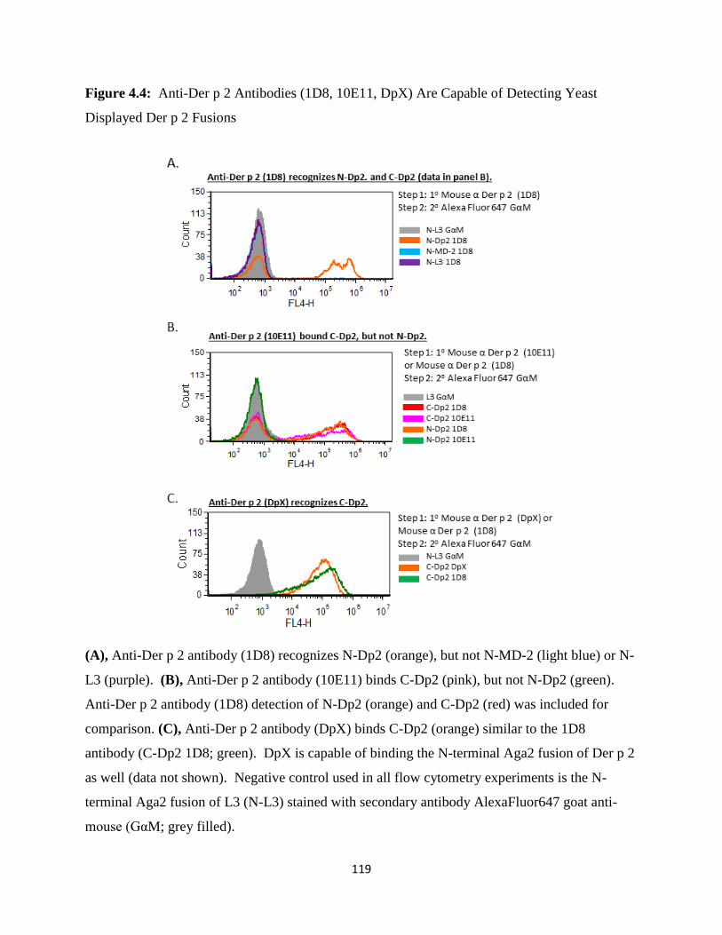
119
Figure 4.4: Anti-Der p 2 Antibodies (1D8, 10E11, DpX) Are Capable of Detecting Yeast
Displayed Der p 2 Fusions
(A), Anti-Der p 2 antibody (1D8) recognizes N-Dp2 (orange), but not N-MD-2 (light blue) or N-
L3 (purple). (B), Anti-Der p 2 antibody (10E11) binds C-Dp2 (pink), but not N-Dp2 (green).
Anti-Der p 2 antibody (1D8) detection of N-Dp2 (orange) and C-Dp2 (red) was included for
comparison. (C), Anti-Der p 2 antibody (DpX) binds C-Dp2 (orange) similar to the 1D8
antibody (C-Dp2 1D8; green). DpX is capable of binding the N-terminal Aga2 fusion of Der p 2
as well (data not shown). Negative control used in all flow cytometry experiments is the N-
terminal Aga2 fusion of L3 (N-L3) stained with secondary antibody AlexaFluor647 goat anti-
mouse (GαM; grey filled).
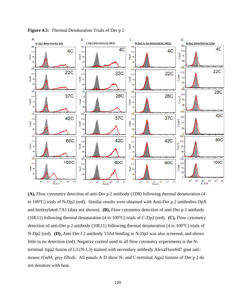
120
Figure 4.5: Thermal Denaturation Trials of Der p 2
(A), Flow cytometry detection of anti-Der p 2 antibody (1D8) following thermal denaturation (4
to 100oC) trials of N-Dp2 (red). Similar results were obtained with Anti-Der p 2 antibodies DpX
and biotinylated-7A1 (data not shown). (B), Flow cytometry detection of anti-Der p 2 antibody
(10E11) following thermal denaturation (4 to 100oC) trials of C-Dp2 (red). (C), Flow cytometry
detection of anti-Der p 2 antibody (10E11) following thermal denaturation (4 to 100oC) trials of
N-Dp2 (red). (D), Anti-Der f 2 antibody 13A4 binding to N-Dp2 was also screened, and shows
little to no detection (red). Negative control used in all flow cytometry experiments is the N-
terminal Aga2 fusion of L3 (N-L3) stained with secondary antibody AlexaFluor647 goat anti-
mouse (GαM; grey filled). All panels A-D show N- and C-terminal Aga2 fusions of Der p 2 do
not denature with heat.
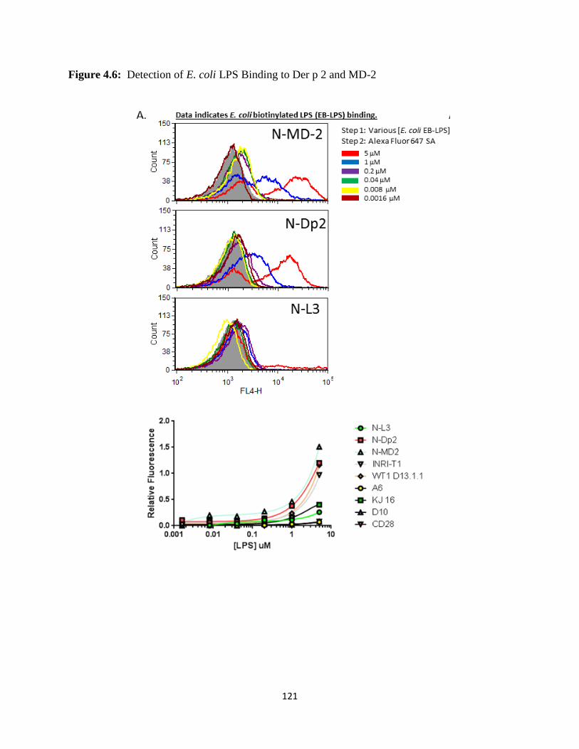
121
Figure 4.6: Detection of E. coli LPS Binding to Der p 2 and MD-2
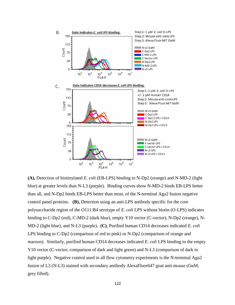
122
(A), Detection of biotinylated E. coli (EB-LPS) binding to N-Dp2 (orange) and N-MD-2 (light
blue) at greater levels than N-L3 (purple). Binding curves show N-MD-2 binds EB-LPS better
than all, and N-Dp2 binds EB-LPS better than most, of the N-terminal Aga2 fusion negative
control panel proteins. (B), Detection using an anti-LPS antibody specific for the core
polysaccharide region of the O111:B4 serotype of E. coli LPS without biotin (O-LPS) indicates
binding to C-Dp2 (red), C-MD-2 (dark blue), empty Y10 vector (C-vector), N-Dp2 (orange), N-
MD-2 (light blue), and N-L3 (purple). (C), Purified human CD14 decreases indicated E. coli
LPS binding to C-Dp2 (comparison of red to pink) or N-Dp2 (comparison of orange and
maroon). Similarly, purified human CD14 decreases indicated E. coli LPS binding to the empty
Y10 vector (C-vector; comparison of dark and light green) and N-L3 (comparison of dark to
light purple). Negative control used in all flow cytometry experiments is the N-terminal Aga2
fusion of L3 (N-L3) stained with secondary antibody AlexaFluor647 goat anti-mouse (GαM;
grey filled).
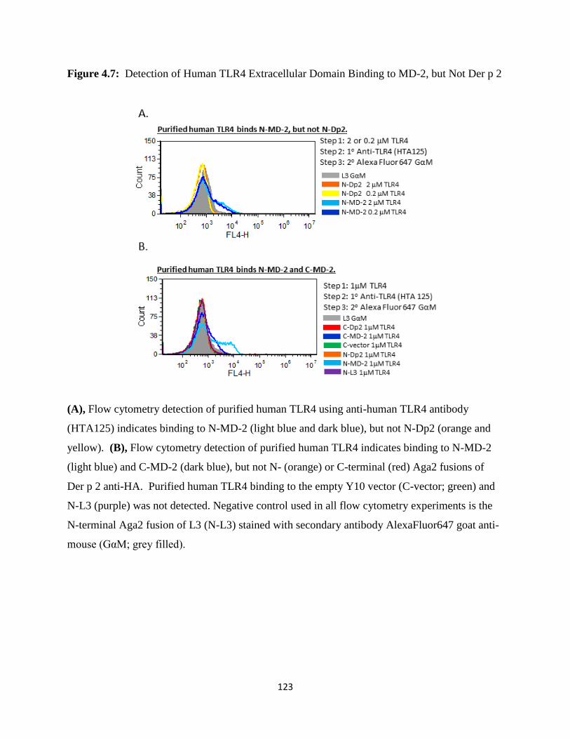
123
Figure 4.7: Detection of Human TLR4 Extracellular Domain Binding to MD-2, but Not Der p 2
(A), Flow cytometry detection of purified human TLR4 using anti-human TLR4 antibody
(HTA125) indicates binding to N-MD-2 (light blue and dark blue), but not N-Dp2 (orange and
yellow). (B), Flow cytometry detection of purified human TLR4 indicates binding to N-MD-2
(light blue) and C-MD-2 (dark blue), but not N- (orange) or C-terminal (red) Aga2 fusions of
Der p 2 anti-HA. Purified human TLR4 binding to the empty Y10 vector (C-vector; green) and
N-L3 (purple) was not detected. Negative control used in all flow cytometry experiments is the
N-terminal Aga2 fusion of L3 (N-L3) stained with secondary antibody AlexaFluor647 goat anti-
mouse (GαM; grey filled).
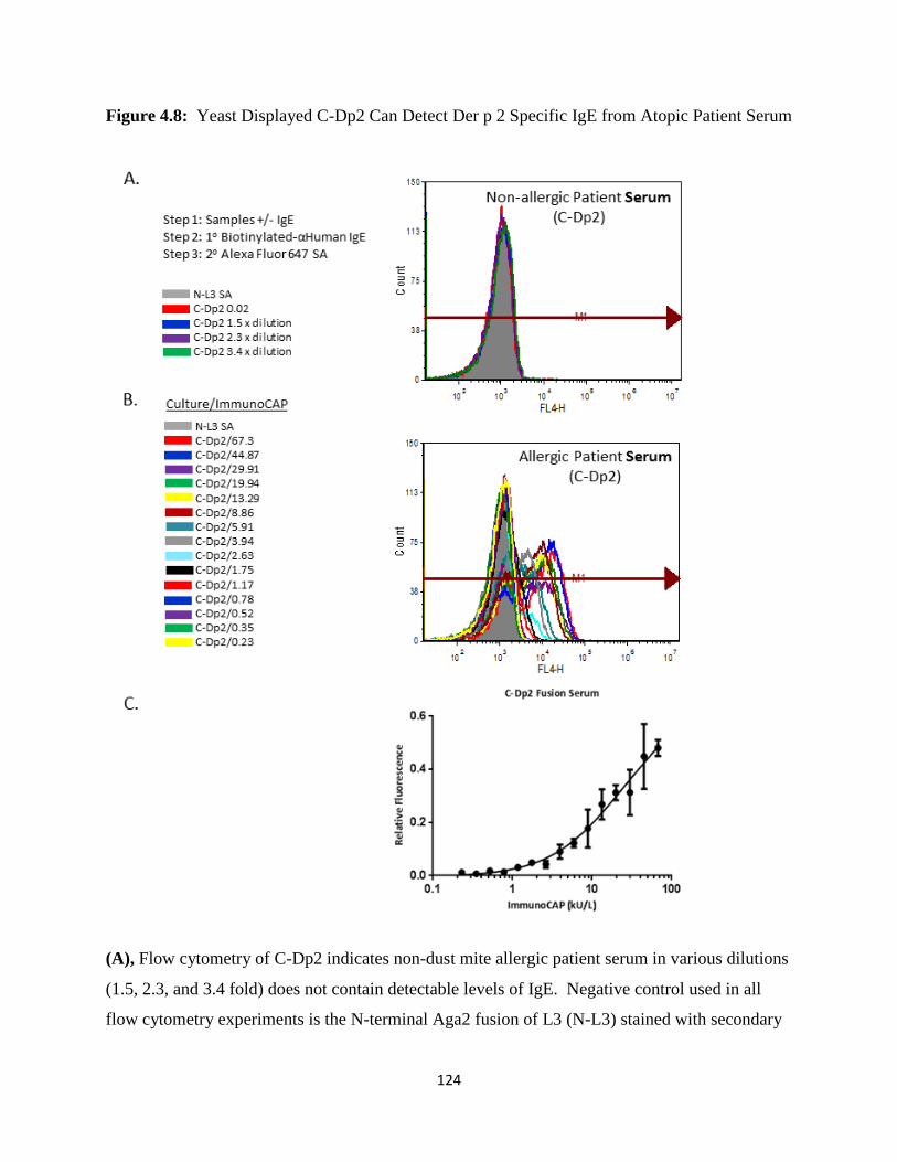
124
Figure 4.8: Yeast Displayed C-Dp2 Can Detect Der p 2 Specific IgE from Atopic Patient Serum
(A), Flow cytometry of C-Dp2 indicates non-dust mite allergic patient serum in various dilutions
(1.5, 2.3, and 3.4 fold) does not contain detectable levels of IgE. Negative control used in all
flow cytometry experiments is the N-terminal Aga2 fusion of L3 (N-L3) stained with secondary

125
antibody AlexaFluor647 goat anti-mouse (GαM; grey filled). (B), Flow cytometry of C-Dp2 was
capable of detecting binding of human IgE from one patient serum sample (sample MN; 67.3
kU/L ImmunoCAP). (C), Relative fluorescence measurements versus ImmunoCAP values are
plotted and non-linear least squares fit is shown (n=3).
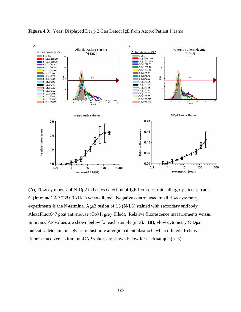
126
Figure 4.9: Yeast Displayed Der p 2 Can Detect IgE from Atopic Patient Plasma
(A), Flow cytometry of N-Dp2 indicates detection of IgE from dust mite allergic patient plasma
G (ImmunoCAP 238.09 kU/L) when diluted. Negative control used in all flow cytometry
experiments is the N-terminal Aga2 fusion of L3 (N-L3) stained with secondary antibody
AlexaFluor647 goat anti-mouse (GαM; grey filled). Relative fluorescence measurements versus
ImmunoCAP values are shown below for each sample (n=3). (B), Flow cytometry C-Dp2
indicates detection of IgE from dust mite allergic patient plasma G when diluted. Relative
fluorescence versus ImmunoCAP values are shown below for each sample (n=3).

127
Figure 4.10: Yeast Displayed Der p 2 Can Detect IgE from Multiple Atopic Patients
(A), Flow cytometry N-Dp2 indicates detection of IgE from multiple patient samples. MFI
measurements versus ImmunoCAP values are shown below for each sample (n=3). Negative
control used in all flow cytometry experiments is the N-terminal Aga2 fusion of L3 (N-L3)
stained with secondary antibody AlexaFluor647 goat anti-mouse (GαM; grey filled). (B), Flow
cytometry C-Dp2 indicates detection of IgE from multiple patient samples. MFI measurements
versus ImmunoCAP values are shown below for each sample (n=3).
128
CHAPTER 5:
Conclusion
In this Chapter, we will summarize the major findings of our work, describe the
significance of our findings, and propose future studies. Our primary project goal, which we
achieved, was to solve the x-ray crystal structure of human CD14. Each stage of crystallography
from construct design to protein expression and purification to optimizing crystals has a
multitude of roadblocks that are only heightened by difficulties unique to the expression and
purification of soluble eukaryotic targets like human CD14. For example, although E. coli is the
expression system of choice for crystallographers due to rapid cell growth, low production costs,
and high purification yield, it was not an option for our work. During their efforts to determine
the structure of human CD14, Meng and colleagues detected soluble human CD14 in insoluble
aggregates with LPS in E. coli, which were not useful for structural studies (135). Thus, to
accomplish our goal, we chose to produce human and mouse CD14 proteins using a mammalian
expression system, which is not a trivial process. Human cells grow much slower by comparison
with E. coli, requiring weekly cycles of protein expression and purification over many years to
obtain sufficient quantities of protein for all of our studies. Others have also experienced the
challenge of structure determination of a human target, as evidence in the PDB, where only 24%
of the 91,190 x-ray structures deposited as of June 2013 are human in origin. Despite the
technical challenges, structural studies have long provided a guide to the interpretation of
functional, biophysical, and mutational data, in order to more fully understand the role of a
protein in a biological process. As described in Chapter 1, the discovery of lipopolysaccharide
as a potent endotoxin sparked studies to determine the order of action and role of each protein in

129
the LPS sensing cascade, which benefitted greatly from extensive x-ray structure studies.
Information gleaned from the native and ligand bound x-ray structures of BPI, mouse CD14, and
multiple human and mouse TLR4/MD-2 complexes had a profound impact on our understanding
of the LPS sensing cascade.
Similarly, our x-ray structure of human CD14 described in Chapter 2 extended the
foundational knowledge provided by the x-ray structure of mouse CD14 (69). Determining the
x-ray crystal structure of human CD14 allowed us to determine similarities and differences
between the two proteins. As with mouse CD14, we found human CD14 contained 11 leucine
rich repeats that form a bent solenoid fold. Although the resolution of our structure fell just
outside the limit of secondary structure detection, we identified five alpha helices and a parallel
beta sheet containing eleven beta strands. Human CD14 also crystallized as a monomer
counteracting a growing misconception of the need for C-terminal dimerization during LPS
delivery, which is illustrated by (CD14-LPS)2 in Figure 1.1. At the N-terminus, we also
identified a hydrophobic pocket formed by residues in α1 through α5 and 1 through β6 that was
similar in size to mouse CD14. During our analysis of this pocket region, we noted an expanded
pocket diameter and subdivided entrance formed by F49 in α1 and Y82 in α3 rim residues in
human CD14, which were not present in mouse CD14. We propose that these rim residues shift
upon LPS binding. The fact that the MD-2 pocket is larger relative to CD14, along with the
knowledge that the MD-2 pocket cannot contain all six acyl chains of E. coli LPS, we predicted
human CD14 would also be incapable of entirely sequestering all six acyl chains in its N-
terminal hydrophobic pocket. With that in mind, we analyzed our human CD14 x-ray structure
and discovered a hydrophobic patch adjacent to the pocket entrance, which may have the
capacity to stabilize exposed LPS acyl chains. We also found a solvent exposed hydrophobic

130
patch in the same region of mouse CD14, which may play an analogous role upon ligand
binding. Thus, our extensive efforts to solve the x-ray crystal structure of human CD14 have
provided many important advances in our understanding of the structure itself, the structural
similarity with mouse CD14, and important properties of the predicted N-terminal LPS binding
pocket and an adjacent hydrophobic patch, which may spark additional efforts to target CD14
function as a means to modulate devastating diseases like sepsis.
Remaining structural questions include the identification of all molecular interactions
between LPS and CD14 required for LPS binding and the protein-protein interactions between
CD14, LBP, and MD-2 necessary for LPS transfer. To tackle the first gap in knowledge, we
attempted to obtain a ligand bound crystal structure of human or mouse CD14 as described in
Chapter 3. As a prerequisite of the ligand bound structural studies, we showed our purified
human and mouse CD14 are able to bind LPS and other ligands as measured by native PAGE gel
shift and ForteBio Octet. We found both proteins are also bioactive because they are able to
deliver LPS and stimulate TLR4 dependent IL-8 production in SW620 cells, and human CD14
was also shown in small gel filtration mixing studies to enable complex formation of
TLR2/1/OspA catalytically without the need for LBP (188). Although our purified CD14
proteins were unable to crystallize in the LPS bound form, we were able to offer these pure,
bioactive proteins for use in other collaborative studies, including the resolution of molecular
interactions in MD-2/TLR4/LPS using yeast display (unpublished) and the characterization of
the catalytic role of CD14 in TLR2/1 agonist delivery (188). In the future, additional ligands
could be explored for ligand bound structural studies.
We also studied the ligand binding ability of the allergen Der p 2 from D. pteronyssinus,
which is reported to be an additional TLR4 accessory protein. As described in Chapter 1, Der p

131
2 is not only processed and displayed for TH2 recognition, stimulating an allergic response, but
may also act as an adjuvant by mimicking the function of MD-2 in the LPS sensing cascade
thereby stimulating inflammation. Since Der p 2 induced allergy and inflammation are both
implicated in the development of allergic asthma, characterizing the interactions between Der p
2, LPS, and TLR4 would advance our understanding of the allergic process. As a first step we
established two forms of Der p 2 on the surface of yeast, which vary in the location of their
agglutinin fusion on the N or C-terminus (N-Dp2 and C-Dp2). One C-terminal MD-2 Aga2
fusion (C-MD-2) was also established for yeast expression. We confirmed full length protein
expression by detecting HA and c-Myc epitope tags using flow cytometry. We also confirmed
anti-MD-2 monoclonal antibodies (4H1 and 18H10) were capable of specifically binding yeast
displayed C-MD-2. Likewise, a multitude of anti-Der p 2 antibodies recognize both the N- and
C-terminal Aga2 fusions of Der p 2 with specificity. We observed detectable interactions
between E. coli LPS and either N-Dp2, C-Dp2, or C-MD-2 proteins. We did not reliably detect
interactions between purified human TLR4 and N-Dp2 or C-Dp2, but we did detect binding to C-
MD-2. We also attempted to determine if Der p 2 is properly folded on the yeast cell surface
using thermal denaturation followed by anti-Der p 2 antibody binding studies. However, we
could not directly confirm folding using this method because of the thermostability of Der p 2.
Using our novel in vitro flow cytometry based system, we showed both N-Dp2 and C-
Dp2 displayed on the surface of yeast were capable of detecting IgE in both allergic patient
serum and plasma. Similar to ImmunoCAP IgE detection, we showed our system reliably
produces a greater fluorescence intensity relative to the amount of IgE in each sample.
ImmunoCAP can detect Class 1 to Class 6 levels. Our system has the ability to sense Class 2 to
Class 6 levels of Der p 2 specific IgE. C-Dp2 may contain additional IgE binding sites relative
132
to N-Dp2, because binding profiles with N-Dp2 showed saturation and C-Dp2 did not show
saturation using the patient sample with the highest Class 6 level of IgE. Thus, our novel
detection method may offer clinical and research laboratories a cost effective way to monitor
allergy in patient samples. Using the power of yeast display to screen large libraries of Der p 2
mutants could better define IgE or LPS binding epitopes, which may spark pharmaceutical
intervention in allergic asthma.

133
REFERENCES
1. Janeway, C. A., Jr. 1989. Approaching the asymptote? Evolution and revolution in
immunology. Cold Spring Harbor symposia on quantitative biology 54 Pt 1: 1-13.
2. Janeway, C. A., Jr., and R. Medzhitov. 2002. Innate immune recognition. Annual review
of immunology 20: 197-216.
3. Beutler, B., and E. T. Rietschel. 2003. Innate immune sensing and its roots: the story of
endotoxin. Nature reviews. Immunology 3: 169-176.
4. Galloway, S. M., and C. R. Raetz. 1990. A mutant of Escherichia coli defective in the
first step of endotoxin biosynthesis. The Journal of biological chemistry 265: 6394-6402.
5. Raetz, C. R., and C. Whitfield. 2002. Lipopolysaccharide endotoxins. Annual review of
biochemistry 71: 635-700.
6. Rietschel, E. T., H. Brade, L. Brade, K. Brandenburg, U. Schade, U. Seydel, U.
Zahringer, C. Galanos, O. Luderitz, O. Westphal, and et al. 1987. Lipid A, the endotoxic
center of bacterial lipopolysaccharides: relation of chemical structure to biological
activity. Progress in clinical and biological research 231: 25-53.
7. Galanos, C., O. Luderitz, E. T. Rietschel, O. Westphal, H. Brade, L. Brade, M.
Freudenberg, U. Schade, M. Imoto, H. Yoshimura, and et al. 1985. Synthetic and natural
Escherichia coli free lipid A express identical endotoxic activities. European journal of
biochemistry / FEBS 148: 1-5.
8. Backhed, F., S. Normark, E. K. Schweda, S. Oscarson, and A. Richter-Dahlfors. 2003.
Structural requirements for TLR4-mediated LPS signalling: a biological role for LPS
modifications. Microbes and infection / Institut Pasteur 5: 1057-1063.
9. Gutschow, M. V., J. J. Hughey, N. A. Ruggero, B. T. Bajar, S. D. Valle, and M. W.
Covert. 2013. Single-Cell and Population NF-kappaB Dynamic Responses Depend on
Lipopolysaccharide Preparation. PloS one 8: e53222.
10. Farhat, K., A. J. Ulmer, and T. W. Jungi. 2012. A potential test system for detecting
contaminations by bacterial lipoproteins. Veterinary immunology and immunopathology
145: 66-73.
11. Kovach, N. L., E. Yee, R. S. Munford, C. R. Raetz, and J. M. Harlan. 1990. Lipid IVA
inhibits synthesis and release of tumor necrosis factor induced by lipopolysaccharide in
human whole blood ex vivo. The Journal of experimental medicine 172: 77-84.
12. Golenbock, D. T., R. Y. Hampton, N. Qureshi, K. Takayama, and C. R. Raetz. 1991.
Lipid A-like molecules that antagonize the effects of endotoxins on human monocytes.
The Journal of biological chemistry 266: 19490-19498.
13. Lynn, W. A., and D. T. Golenbock. 1992. Lipopolysaccharide antagonists. Immunology
today 13: 271-276.
14. Takayama, K., N. Qureshi, B. Beutler, and T. N. Kirkland. 1989. Diphosphoryl lipid A
from Rhodopseudomonas sphaeroides ATCC 17023 blocks induction of cachectin in
macrophages by lipopolysaccharide. Infection and immunity 57: 1336-1338.
15. Rallabhandi, P., A. Awomoyi, K. E. Thomas, A. Phalipon, Y. Fujimoto, K. Fukase, S.
Kusumoto, N. Qureshi, M. B. Sztein, and S. N. Vogel. 2008. Differential activation of
human TLR4 by Escherichia coli and Shigella flexneri 2a lipopolysaccharide: combined
effects of lipid A acylation state and TLR4 polymorphisms on signaling. Journal of
immunology 180: 1139-1147.
134
16. Teghanemt, A., D. Zhang, E. N. Levis, J. P. Weiss, and T. L. Gioannini. 2005. Molecular
basis of reduced potency of underacylated endotoxins. Journal of immunology 175: 4669-
4676.
17. Tran, A. X., C. M. Stead, and M. S. Trent. 2005. Remodeling of Helicobacter pylori
lipopolysaccharide. Journal of endotoxin research 11: 161-166.
18. Telepnev, M. V., G. R. Klimpel, J. Haithcoat, Y. A. Knirel, A. P. Anisimov, and V. L.
Motin. 2009. Tetraacylated lipopolysaccharide of Yersinia pestis can inhibit multiple
Toll-like receptor-mediated signaling pathways in human dendritic cells. The Journal of
infectious diseases 200: 1694-1702.
19. Montminy, S. W., N. Khan, S. McGrath, M. J. Walkowicz, F. Sharp, J. E. Conlon, K.
Fukase, S. Kusumoto, C. Sweet, K. Miyake, S. Akira, R. J. Cotter, J. D. Goguen, and E.
Lien. 2006. Virulence factors of Yersinia pestis are overcome by a strong
lipopolysaccharide response. Nature immunology 7: 1066-1073.
20. Galanos, C., M. A. Freudenberg, F. Jay, D. Nerkar, K. Veleva, H. Brade, and W.
Strittmatter. 1984. Immunogenic properties of lipid A. Reviews of infectious diseases 6:
546-552.
21. Tobias, P. S., K. Soldau, and R. J. Ulevitch. 1986. Isolation of a lipopolysaccharide-
binding acute phase reactant from rabbit serum. The Journal of experimental medicine
164: 777-793.
22. Schumann, R. R., S. R. Leong, G. W. Flaggs, P. W. Gray, S. D. Wright, J. C. Mathison,
P. S. Tobias, and R. J. Ulevitch. 1990. Structure and function of lipopolysaccharide
binding protein. Science (New York, N.Y.) 249: 1429-1431.
23. Schumann, R. R. 2011. Old and new findings on lipopolysaccharide-binding protein: a
soluble pattern-recognition molecule. Biochemical Society transactions 39: 989-993.
24. Beamer, L. J., S. F. Carroll, and D. Eisenberg. 1997. Crystal structure of human BPI and
two bound phospholipids at 2.4 angstrom resolution. Science (New York, N.Y.) 276:
1861-1864.
25. Weiss, J. 2003. Bactericidal/permeability-increasing protein (BPI) and
lipopolysaccharide-binding protein (LBP): structure, function and regulation in host
defence against Gram-negative bacteria. Biochemical Society transactions 31: 785-790.
26. Elsbach, P., and J. Weiss. 1998. Role of the bactericidal/permeability-increasing protein
in host defence. Current opinion in immunology 10: 45-49.
27. Peri, F., M. Piazza, V. Calabrese, G. Damore, and R. Cighetti. 2010. Exploring the
LPS/TLR4 signal pathway with small molecules. Biochemical Society transactions 38:
1390-1395.
28. Tobias, P. S., K. Soldau, J. A. Gegner, D. Mintz, and R. J. Ulevitch. 1995.
Lipopolysaccharide binding protein-mediated complexation of lipopolysaccharide with
soluble CD14. The Journal of biological chemistry 270: 10482-10488.
29. Yu, B., and S. D. Wright. 1996. Catalytic properties of lipopolysaccharide (LPS) binding
protein. Transfer of LPS to soluble CD14. The Journal of biological chemistry 271:
4100-4105.
30. Hailman, E., H. S. Lichenstein, M. M. Wurfel, D. S. Miller, D. A. Johnson, M. Kelley, L.
A. Busse, M. M. Zukowski, and S. D. Wright. 1994. Lipopolysaccharide (LPS)-binding
protein accelerates the binding of LPS to CD14. The Journal of experimental medicine
179: 269-277.
135
31. Griffin, J. D., J. Ritz, L. M. Nadler, and S. F. Schlossman. 1981. Expression of myeloid
differentiation antigens on normal and malignant myeloid cells. The Journal of clinical
investigation 68: 932-941.
32. Maliszewski, C. R., E. D. Ball, R. F. Graziano, and M. W. Fanger. 1985. Isolation and
characterization of My23, a myeloid cell-derived antigen reactive with the monoclonal
antibody AML-2-23. Journal of immunology 135: 1929-1936.
33. Bazil, V., V. Horejsi, M. Baudys, H. Kristofova, J. L. Strominger, W. Kostka, and I.
Hilgert. 1986. Biochemical characterization of a soluble form of the 53-kDa monocyte
surface antigen. European journal of immunology 16: 1583-1589.
34. Bazil, V., V. Horejsi, I. Hilgert, and M. J. McMichael (eds). 1982. Leucocyte Typing III.
White Cell Differentiation Antigens. The Workshop: Myeloid Panel Antibodies
Recognizing the 53-kDa Molecular Weight Monocyte Antigen (CD14). Oxford, Oxford
University Press.: p. 611-613.
35. Goyert, S. M., E. Ferrero, W. J. Rettig, A. K. Yenamandra, F. Obata, and M. M. Le Beau.
1988. The CD14 monocyte differentiation antigen maps to a region encoding growth
factors and receptors. Science (New York, N.Y.) 239: 497-500.
36. Matsuura, K., M. Setoguchi, N. Nasu, Y. Higuchi, S. Yoshida, S. Akizuki, and S.
Yamamoto. 1989. Nucleotide and amino acid sequences of the mouse CD14 gene.
Nucleic acids research 17: 2132.
37. Haziot, A., S. Chen, E. Ferrero, M. G. Low, R. Silber, and S. M. Goyert. 1988. The
monocyte differentiation antigen, CD14, is anchored to the cell membrane by a
phosphatidylinositol linkage. Journal of immunology 141: 547-552.
38. Simmons, D. L., S. Tan, D. G. Tenen, A. Nicholson-Weller, and B. Seed. 1989.
Monocyte antigen CD14 is a phospholipid anchored membrane protein. Blood 73: 284-
289.
39. Bazil, V., and J. L. Strominger. 1991. Shedding as a mechanism of down-modulation of
CD14 on stimulated human monocytes. Journal of immunology 147: 1567-1574.
40. Wright, S. D., R. A. Ramos, P. S. Tobias, R. J. Ulevitch, and J. C. Mathison. 1990.
CD14, a receptor for complexes of lipopolysaccharide (LPS) and LPS binding protein.
Science (New York, N.Y.) 249: 1431-1433.
41. Pugin, J., I. D. Heumann, A. Tomasz, V. V. Kravchenko, Y. Akamatsu, M. Nishijima, M.
P. Glauser, P. S. Tobias, and R. J. Ulevitch. 1994. CD14 is a pattern recognition receptor.
Immunity 1: 509-516.
42. Gioannini, T. L., A. Teghanemt, D. Zhang, N. P. Coussens, W. Dockstader, S.
Ramaswamy, and J. P. Weiss. 2004. Isolation of an endotoxin-MD-2 complex that
produces Toll-like receptor 4-dependent cell activation at picomolar concentrations.
Proceedings of the National Academy of Sciences of the United States of America 101:
4186-4191.
43. Prohinar, P., F. Re, R. Widstrom, D. Zhang, A. Teghanemt, J. P. Weiss, and T. L.
Gioannini. 2007. Specific high affinity interactions of monomeric endotoxin.protein
complexes with Toll-like receptor 4 ectodomain. The Journal of biological chemistry
282: 1010-1017.
44. Teghanemt, A., P. Prohinar, T. L. Gioannini, and J. P. Weiss. 2007. Transfer of
monomeric endotoxin from MD-2 to CD14: characterization and functional
consequences. The Journal of biological chemistry 282: 36250-36256.
136
45. Teghanemt, A., R. L. Widstrom, T. L. Gioannini, and J. P. Weiss. 2008. Isolation of
monomeric and dimeric secreted MD-2. Endotoxin.sCD14 and Toll-like receptor 4
ectodomain selectively react with the monomeric form of secreted MD-2. The Journal of
biological chemistry 283: 21881-21889.
46. Tapping, R. I., and P. S. Tobias. 2000. Soluble CD14-mediated cellular responses to
lipopolysaccharide. Chemical immunology 74: 108-121.
47. Ostuni, R., I. Zanoni, and F. Granucci. 2010. Deciphering the complexity of Toll-like
receptor signaling. Cellular and molecular life sciences : CMLS 67: 4109-4134.
48. Jack, R. S., U. Grunwald, F. Stelter, G. Workalemahu, and C. Schutt. 1995. Both
membrane-bound and soluble forms of CD14 bind to gram-negative bacteria. European
journal of immunology 25: 1436-1441.
49. Grunwald, U., X. Fan, R. S. Jack, G. Workalemahu, A. Kallies, F. Stelter, and C. Schutt.
1996. Monocytes can phagocytose Gram-negative bacteria by a CD14-dependent
mechanism. Journal of immunology 157: 4119-4125.
50. Fan, X., F. Stelter, R. Menzel, R. Jack, I. Spreitzer, T. Hartung, and C. Schutt. 1999.
Structures in Bacillus subtilis are recognized by CD14 in a lipopolysaccharide binding
protein-dependent reaction. Infection and immunity 67: 2964-2968.
51. Stelter, F., M. Bernheiden, R. Menzel, R. S. Jack, S. Witt, X. Fan, M. Pfister, and C.
Schutt. 1997. Mutation of amino acids 39-44 of human CD14 abrogates binding of
lipopolysaccharide and Escherichia coli. European journal of biochemistry / FEBS 243:
100-109.
52. Schiff, D. E., L. Kline, K. Soldau, J. D. Lee, J. Pugin, P. S. Tobias, and R. J. Ulevitch.
1997. Phagocytosis of gram-negative bacteria by a unique CD14-dependent mechanism.
Journal of leukocyte biology 62: 786-794.
53. Wurfel, M. M., and S. D. Wright. 1997. Lipopolysaccharide-binding protein and soluble
CD14 transfer lipopolysaccharide to phospholipid bilayers: preferential interaction with
particular classes of lipid. Journal of immunology 158: 3925-3934.
54. Yu, B., E. Hailman, and S. D. Wright. 1997. Lipopolysaccharide binding protein and
soluble CD14 catalyze exchange of phospholipids. The Journal of clinical investigation
99: 315-324.
55. Wang, P. Y., R. L. Kitchens, and R. S. Munford. 1998. Phosphatidylinositides bind to
plasma membrane CD14 and can prevent monocyte activation by bacterial
lipopolysaccharide. The Journal of biological chemistry 273: 24309-24313.
56. Nakata, T., M. Yasuda, M. Fujita, H. Kataoka, K. Kiura, H. Sano, and K. Shibata. 2006.
CD14 directly binds to triacylated lipopeptides and facilitates recognition of the
lipopeptides by the receptor complex of Toll-like receptors 2 and 1 without binding to the
complex. Cellular microbiology 8: 1899-1909.
57. Dziarski, R., R. I. Tapping, and P. S. Tobias. 1998. Binding of bacterial peptidoglycan to
CD14. The Journal of biological chemistry 273: 8680-8690.
58. Gupta, D., T. N. Kirkland, S. Viriyakosol, and R. Dziarski. 1996. CD14 is a cell-
activating receptor for bacterial peptidoglycan. The Journal of biological chemistry 271:
23310-23316.
59. Savedra, R., Jr., R. L. Delude, R. R. Ingalls, M. J. Fenton, and D. T. Golenbock. 1996.
Mycobacterial lipoarabinomannan recognition requires a receptor that shares components
of the endotoxin signaling system. Journal of immunology 157: 2549-2554.

137
60. Yu, W., E. Soprana, G. Cosentino, M. Volta, H. S. Lichenstein, G. Viale, and D. Vercelli.
1998. Soluble CD14(1-152) confers responsiveness to both lipoarabinomannan and
lipopolysaccharide in a novel HL-60 cell bioassay. Journal of immunology 161: 4244-
4251.
61. Elass, E., B. Coddeville, Y. Guerardel, L. Kremer, E. Maes, J. Mazurier, and D. Legrand.
2007. Identification by surface plasmon resonance of the mycobacterial lipomannan and
lipoarabinomannan domains involved in binding to CD14 and LPS-binding protein.
FEBS letters 581: 1383-1390.
62. Lien, E., T. J. Sellati, A. Yoshimura, T. H. Flo, G. Rawadi, R. W. Finberg, J. D. Carroll,
T. Espevik, R. R. Ingalls, J. D. Radolf, and D. T. Golenbock. 1999. Toll-like receptor 2
functions as a pattern recognition receptor for diverse bacterial products. The Journal of
biological chemistry 274: 33419-33425.
63. Takeuchi, O., K. Hoshino, T. Kawai, H. Sanjo, H. Takada, T. Ogawa, K. Takeda, and S.
Akira. 1999. Differential roles of TLR2 and TLR4 in recognition of gram-negative and
gram-positive bacterial cell wall components. Immunity 11: 443-451.
64. Cohen, J. 2002. The immunopathogenesis of sepsis. Nature 420: 885-891.
65. Opal, S. M., and J. Cohen. 1999. Clinical gram-positive sepsis: does it fundamentally
differ from gram-negative bacterial sepsis? Critical care medicine 27: 1608-1616.
66. Salomao, R., M. K. Brunialti, M. M. Rapozo, G. L. Baggio-Zappia, C. Galanos, and M.
Freudenberg. 2012. Bacterial sensing, cell signaling, and modulation of the immune
response during sepsis. Shock (Augusta, Ga.) 38: 227-242.
67. Haziot, A., E. Ferrero, F. Kontgen, N. Hijiya, S. Yamamoto, J. Silver, C. L. Stewart, and
S. M. Goyert. 1996. Resistance to endotoxin shock and reduced dissemination of gram-
negative bacteria in CD14-deficient mice. Immunity 4: 407-414.
68. Poltorak, A., X. He, I. Smirnova, M. Y. Liu, C. Van Huffel, X. Du, D. Birdwell, E.
Alejos, M. Silva, C. Galanos, M. Freudenberg, P. Ricciardi-Castagnoli, B. Layton, and B.
Beutler. 1998. Defective LPS signaling in C3H/HeJ and C57BL/10ScCr mice: mutations
in Tlr4 gene. Science (New York, N.Y.) 282: 2085-2088.
69. Kim, J. I., C. J. Lee, M. S. Jin, C. H. Lee, S. G. Paik, H. Lee, and J. O. Lee. 2005. Crystal
structure of CD14 and its implications for lipopolysaccharide signaling. The Journal of
biological chemistry 280: 11347-11351.
70. Michalek, S. M., R. N. Moore, J. R. McGhee, D. L. Rosenstreich, and S. E.
Mergenhagen. 1980. The primary role of lymphoreticular cells in the mediation of host
responses to bacterial endotoxim. The Journal of infectious diseases 141: 55-63.
71. Freudenberg, M. A., D. Keppler, and C. Galanos. 1986. Requirement for
lipopolysaccharide-responsive macrophages in galactosamine-induced sensitization to
endotoxin. Infection and immunity 51: 891-895.
72. Hoshino, K., O. Takeuchi, T. Kawai, H. Sanjo, T. Ogawa, Y. Takeda, K. Takeda, and S.
Akira. 1999. Cutting edge: Toll-like receptor 4 (TLR4)-deficient mice are
hyporesponsive to lipopolysaccharide: evidence for TLR4 as the Lps gene product.
Journal of immunology 162: 3749-3752.
73. Qureshi, S. T., L. Lariviere, G. Leveque, S. Clermont, K. J. Moore, P. Gros, and D. Malo.
1999. Endotoxin-tolerant mice have mutations in Toll-like receptor 4 (Tlr4). The Journal
of experimental medicine 189: 615-625.
138
74. Medzhitov, R., P. Preston-Hurlburt, and C. A. Janeway. 1997. A human homologue of
the Drosophila Toll protein signals activation of adaptive immunity. Nature 388: 394-
397.
75. Lemaitre, B., E. Nicolas, L. Michaut, J.-M. Reichhart, and J. A. Hoffmann. 1996. The
Dorsoventral Regulatory Gene Cassette spätzle/Toll/cactus Controls the Potent
Antifungal Response in Drosophila Adults. Cell 86: 973-983.
76. Jin, M. S., S. E. Kim, J. Y. Heo, M. E. Lee, H. M. Kim, S. G. Paik, H. Lee, and J. O. Lee.
2007. Crystal structure of the TLR1-TLR2 heterodimer induced by binding of a tri-
acylated lipopeptide. Cell 130: 1071-1082.
77. Park, B. S., D. H. Song, H. M. Kim, B. S. Choi, H. Lee, and J. O. Lee. 2009. The
structural basis of lipopolysaccharide recognition by the TLR4-MD-2 complex. Nature
458: 1191-1195.
78. Liu, L., I. Botos, Y. Wang, J. N. Leonard, J. Shiloach, D. M. Segal, and D. R. Davies.
2008. Structural basis of toll-like receptor 3 signaling with double-stranded RNA.
Science (New York, N.Y.) 320: 379-381.
79. Lu, J., and P. D. Sun. 2012. The structure of the TLR5-flagellin complex: a new mode of
pathogen detection, conserved receptor dimerization for signaling. Science signaling 5:
pe11.
80. Kang, J. Y., X. Nan, M. S. Jin, S. J. Youn, Y. H. Ryu, S. Mah, S. H. Han, H. Lee, S. G.
Paik, and J. O. Lee. 2009. Recognition of lipopeptide patterns by Toll-like receptor 2-
Toll-like receptor 6 heterodimer. Immunity 31: 873-884.
81. Nyman, T., P. Stenmark, S. Flodin, I. Johansson, M. Hammarstrom, and P. Nordlund.
2008. The crystal structure of the human toll-like receptor 10 cytoplasmic domain reveals
a putative signaling dimer. The Journal of biological chemistry 283: 11861-11865.
82. Xu, Y., X. Tao, B. Shen, T. Horng, R. Medzhitov, J. L. Manley, and L. Tong. 2000.
Structural basis for signal transduction by the Toll/interleukin-1 receptor domains. Nature
408: 111-115.
83. Guan, Y., D. R. Ranoa, S. Jiang, S. K. Mutha, X. Li, J. Baudry, and R. I. Tapping. 2010.
Human TLRs 10 and 1 share common mechanisms of innate immune sensing but not
signaling. Journal of immunology 184: 5094-5103.
84. Akira, S., and K. Takeda. 2004. Toll-like receptor signalling. Nature reviews.
Immunology 4: 499-511.
85. Muzio, M., G. Natoli, S. Saccani, M. Levrero, and A. Mantovani. 1998. The human toll
signaling pathway: divergence of nuclear factor kappaB and JNK/SAPK activation
upstream of tumor necrosis factor receptor-associated factor 6 (TRAF6). The Journal of
experimental medicine 187: 2097-2101.
86. Jin, M. S., and J. O. Lee. 2008. Structures of the toll-like receptor family and its ligand
complexes. Immunity 29: 182-191.
87. Takahashi, N., Y. Takahashi, and F. W. Putnam. 1985. Periodicity of leucine and tandem
repetition of a 24-amino acid segment in the primary structure of leucine-rich a2-
glycoprotein of human serum. Proceedings of the National Academy of Sciences of the
United States of America 82: 1906-1910.
88. Kobe, B., and A. V. Kajava. 2001. The leucine-rich repeat as a protein recognition motif.
Current opinion in structural biology 11: 725-732.
89. Kang, J. Y., and J. O. Lee. 2011. Structural biology of the Toll-like receptor family.
Annual review of biochemistry 80: 917-941.
139
90. Nishiya, T., and A. L. DeFranco. 2004. Ligand-regulated chimeric receptor approach
reveals distinctive subcellular localization and signaling properties of the Toll-like
receptors. The Journal of biological chemistry 279: 19008-19017.
91. Hart, B. E., and R. I. Tapping. 2012. Cell surface trafficking of TLR1 is differentially
regulated by the chaperones PRAT4A and PRAT4B. The Journal of biological chemistry
287: 16550-16562.
92. Jiang, Z., P. Georgel, X. Du, L. Shamel, S. Sovath, S. Mudd, M. Huber, C. Kalis, S.
Keck, C. Galanos, M. Freudenberg, and B. Beutler. 2005. CD14 is required for MyD88-
independent LPS signaling. Nature immunology 6: 565-570.
93. Gay, N. J., and F. J. Keith. 1991. Drosophila Toll and IL-1 receptor. Nature 351: 355-
356.
94. March, C. J., B. Mosley, A. Larsen, D. P. Cerretti, G. Braedt, V. Price, S. Gillis, C. S.
Henney, S. R. Kronheim, K. Grabstein, P. J. Conlon, T. P. Hopp, and D. Cosman. 1985.
Cloning, sequence and expression of two distinct human interleukin-1 complementary
DNAs. Nature 315: 641-647.
95. Shimazu, R., S. Akashi, H. Ogata, Y. Nagai, K. Fukudome, K. Miyake, and M. Kimoto.
1999. MD-2, a molecule that confers lipopolysaccharide responsiveness on Toll-like
receptor 4. The Journal of experimental medicine 189: 1777-1782.
96. Nagai, Y., S. Akashi, M. Nagafuku, M. Ogata, Y. Iwakura, S. Akira, T. Kitamura, A.
Kosugi, M. Kimoto, and K. Miyake. 2002. Essential role of MD-2 in LPS responsiveness
and TLR4 distribution. Nature immunology 3: 667-672.
97. Lang, L. L., L. Wang, and L. Liu. 2011. Exogenous MD-2 confers lipopolysaccharide
responsiveness to human corneal epithelial cells with intracellular expression of TLR4
and CD14. Inflammation 34: 371-378.
98. Schromm, A. B., E. Lien, P. Henneke, J. C. Chow, A. Yoshimura, H. Heine, E. Latz, B.
G. Monks, D. A. Schwartz, K. Miyake, and D. T. Golenbock. 2001. Molecular genetic
analysis of an endotoxin nonresponder mutant cell line: a point mutation in a conserved
region of MD-2 abolishes endotoxin-induced signaling. The Journal of experimental
medicine 194: 79-88.
99. Ohto, U., K. Fukase, K. Miyake, and Y. Satow. 2007. Crystal structures of human MD-2
and its complex with antiendotoxic lipid IVa. Science (New York, N.Y.) 316: 1632-1634.
100. Kennedy, M. N., G. E. Mullen, C. A. Leifer, C. Lee, A. Mazzoni, K. N. Dileepan, and D.
M. Segal. 2004. A complex of soluble MD-2 and lipopolysaccharide serves as an
activating ligand for Toll-like receptor 4. The Journal of biological chemistry 279:
34698-34704.
101. Kawasaki, K., H. Nogawa, and M. Nishijima. 2003. Identification of mouse MD-2
residues important for forming the cell surface TLR4-MD-2 complex recognized by anti-
TLR4-MD-2 antibodies, and for conferring LPS and taxol responsiveness on mouse
TLR4 by alanine-scanning mutagenesis. Journal of immunology 170: 413-420.
102. Kim, H. M., B. S. Park, J. I. Kim, S. E. Kim, J. Lee, S. C. Oh, P. Enkhbayar, N.
Matsushima, H. Lee, O. J. Yoo, and J. O. Lee. 2007. Crystal structure of the TLR4-MD-2
complex with bound endotoxin antagonist Eritoran. Cell 130: 906-917.
103. Ohto, U., K. Fukase, K. Miyake, and T. Shimizu. 2012. Structural basis of species-
specific endotoxin sensing by innate immune receptor TLR4/MD-2. Proceedings of the
National Academy of Sciences of the United States of America 109: 7421-7426.

140
104. Tidswell, M., and S. P. LaRosa. 2011. Toll-like receptor-4 antagonist eritoran
tetrasodium for severe sepsis. Expert review of anti-infective therapy 9: 507-520.
105. Park, G. M., S. M. Lee, I. Y. Lee, H. I. Ree, K. S. Kim, C. S. Hong, and T. S. Yong.
2000. Localization of a major allergen, Der p 2, in the gut and faecal pellets of
Dermatophagoides pteronyssinus. Clinical and experimental allergy : journal of the
British Society for Allergy and Clinical Immunology 30: 1293-1297.
106. Tovey, E. R., M. D. Chapman, and T. A. E. Platts-Mills. 1981. Mite faeces are a major
source of house dust allergens. Nature 289: 592-593.
107. Voorhorst, R., Spieksma Boezeman, M. I. A. & Spieksma, F. Th. M. . 1964. Is A Mite
(Dermatophagoides sp.) The Producer of The House-Dust Allergen? Allergic Asthma 10:
329-334.
108. Braun-Fahrländer, C., J. Riedler, U. Herz, W. Eder, M. Waser, L. Grize, S. Maisch, D.
Carr, F. Gerlach, A. Bufe, R. P. Lauener, R. Schierl, H. Renz, D. Nowak, and E. von
Mutius. 2002. Environmental Exposure to Endotoxin and Its Relation to Asthma in
School-Age Children. New England Journal of Medicine 347: 869-877.
109. Thomas, W. R. 2010. Geography of house dust mite allergens. Asian Pacific journal of
allergy and immunology / launched by the Allergy and Immunology Society of Thailand
28: 211-224.
110. Chapman, M. D. 2004. Allergen Nomenclature. In Allergens and Allergen
Immunotherapy, 3rd ed. S. C. B. Richard F. Lockey, and Jean Bousquet, ed. Marcel
Dekker, New York. 51-64.
111. Chua, K. Y., C. R. Doyle, R. J. Simpson, K. J. Turner, G. A. Stewart, and W. R. Thomas.
1990. Isolation of cDNA coding for the major mite allergen Der p II by IgE plaque
immunoassay. International archives of allergy and applied immunology 91: 118-123.
112. Mueller, G. A., D. C. Benjamin, and G. S. Rule. 1998. Tertiary structure of the major
house dust mite allergen Der p 2: sequential and structural homologies. Biochemistry 37:
12707-12714.
113. Platts-Mills, T. A. E. 1992. Mechanisms of Bronchial Reactivity: The Role of
Immunoglobulin E. American Review of Respiratory Disease 145: S44-S47.
114. Gandhi, V., C. Davidson, M. Asaduzzaman, D. Nahirney, and H. Vliagoftis. 2013. House
Dust Mite Interactions with Airway Epithelium: Role in Allergic Airway Inflammation.
Curr Allergy Asthma Rep: 1-9.
115. Heymann, P. W., M. D. Chapman, R. C. Aalberse, J. W. Fox, and T. A. Platts-Mills.
1989. Antigenic and structural analysis of group II allergens (Der f II and Der p II) from
house dust mites (Dermatophagoides spp). The Journal of allergy and clinical
immunology 83: 1055-1067.
116. Hakkaart, G. A., M. M. Harmsen, K. Y. Chua, W. R. Thomas, R. C. Aalberse, and R.
Van Ree. 1998. Expression of the house dust mite allergen Der p 2 in the baker's yeast
Saccharomyces cerevisiae. Clinical and experimental allergy : journal of the British
Society for Allergy and Clinical Immunology 28: 45-52.
117. Tanyaratsrisakul, S., N. Malainual, O. Jirapongsananuruk, W. A. Smith, W. R. Thomas,
and S. Piboonpocanun. 2010. Structural and IgE binding analyses of recombinant Der p 2
expressed from the hosts Escherichia coli and Pichia pastoris. Int Arch Allergy Immunol
151: 190-198.
118. Mueller, G. A., A. M. Smith, D. C. Williams, Jr., G. A. Hakkaart, R. C. Aalberse, M. D.
Chapman, G. S. Rule, and D. C. Benjamin. 1997. Expression and secondary structure

141
determination by NMR methods of the major house dust mite allergen Der p 2. The
Journal of biological chemistry 272: 26893-26898.
119. Derewenda, U., J. Li, Z. Derewenda, Z. Dauter, G. A. Mueller, G. S. Rule, and D. C.
Benjamin. 2002. The Crystal Structure of a Major Dust Mite Allergen Der p 2, and its
Biological Implications. Journal of molecular biology 318: 189-197.
120. Gruber, A., M. Mancek, H. Wagner, C. J. Kirschning, and R. Jerala. 2004. Structural
model of MD-2 and functional role of its basic amino acid clusters involved in cellular
lipopolysaccharide recognition. The Journal of biological chemistry 279: 28475-28482.
121. Lombardero, M., P. W. Heymann, T. A. Platts-Mills, J. W. Fox, and M. D. Chapman.
1990. Conformational stability of B cell epitopes on group I and group II
Dermatophagoides spp. allergens. Effect of thermal and chemical denaturation on the
binding of murine IgG and human IgE antibodies. Journal of immunology 144: 1353-
1360.
122. Trompette, A., S. Divanovic, A. Visintin, C. Blanchard, R. S. Hegde, R. Madan, P. S.
Thorne, M. Wills-Karp, T. L. Gioannini, J. P. Weiss, and C. L. Karp. 2009. Allergenicity
resulting from functional mimicry of a Toll-like receptor complex protein. Nature 457:
585-588.
123. Suzuki, M., Y. Tanaka, S. Korematsu, B. Mikami, and N. Minato. 2006. Crystal structure
and some properties of a major house dust mite allergen, Derf 2. Biochemical and
biophysical research communications 339: 679-686.
124. Ichikawa, S., T. Takai, T. Yashiki, S. Takahashi, K. Okumura, H. Ogawa, D. Kohda, and
H. Hatanaka. 2009. Lipopolysaccharide binding of the mite allergen Der f 2. Genes to
Cells 14: 1055-1065.
125. Manček Keber, M., H. Gradišar, and R. Jerala. 2005. MD-2 and Der p 2 — a tale of two
cousins or distant relatives? Journal of endotoxin research 11: 186-192.
126. Chen, C. L., C. T. Lee, Y. C. Liu, J. Y. Wang, H. Y. Lei, and C. K. Yu. 2003. House dust
mite Dermatophagoides farinae augments proinflammatory mediator productions and
accessory function of alveolar macrophages: implications for allergic sensitization and
inflammation. Journal of immunology 170: 528-536.
127. Pugin, J., C. C. Schurer-Maly, D. Leturcq, A. Moriarty, R. J. Ulevitch, and P. S. Tobias.
1993. Lipopolysaccharide activation of human endothelial and epithelial cells is mediated
by lipopolysaccharide-binding protein and soluble CD14. Proceedings of the National
Academy of Sciences of the United States of America 90: 2744-2748.
128. Tapping, R. I., and P. S. Tobias. 1997. Cellular binding of soluble CD14 requires
lipopolysaccharide (LPS) and LPS-binding protein. The Journal of biological chemistry
272: 23157-23164.
129. Haziot, A., G. W. Rong, V. Bazil, J. Silver, and S. M. Goyert. 1994. Recombinant soluble
CD14 inhibits LPS-induced tumor necrosis factor-alpha production by cells in whole
blood. Journal of immunology 152: 5868-5876.
130. Stelter, F., M. Pfister, M. Bernheiden, R. S. Jack, P. Bufler, H. Engelmann, and C. Schutt.
1996. The myeloid differentiation antigen CD14 is N- and O-glycosylated. Contribution
of N-linked glycosylation to different soluble CD14 isoforms. European journal of
biochemistry / FEBS 236: 457-464.
131. Majerle, A., J. Kidric, and R. Jerala. 1999. Expression and refolding of functional
fragments of the human lipopolysaccharide receptor CD14 in Escherichia coli and Pichia
pastoris. Protein expression and purification 17: 96-104.

142
132. Bai, J., J. Yin, Z. D. Wang, W. Wang, Z. Z. Wang, and W. Song. 2001. [Clone and
expression of human soluble CD14 and study of its function]. Sheng wu gong cheng xue
bao = Chinese journal of biotechnology 17: 269-272.
133. Nomura, S., K. Inamori, T. Muta, S. Yamazaki, Y. Sunakawa, S. Iwanaga, and K.
Takeshige. 2003. Purification and characterization of human soluble CD14 expressed in
Pichia pastoris. Protein expression and purification 28: 310-320.
134. Burkhardt, M., A. LopezAcosta, K. Reiter, V. Lopez, and A. Lees. 2007. Purification of
soluble CD14 fusion proteins and use in an electrochemiluminescent assay for
lipopolysaccharide binding. Protein expression and purification 51: 96-101.
135. Meng, J., P. Parroche, D. T. Golenbock, and C. J. McKnight. 2008. The differential
impact of disulfide bonds and N-linked glycosylation on the stability and function of
CD14. The Journal of biological chemistry 283: 3376-3384.
136. Albright, S., B. Chen, K. Holbrook, and N. U. Jain. 2008. Solution NMR studies provide
structural basis for endotoxin pattern recognition by the innate immune receptor CD14.
Biochemical and biophysical research communications 368: 231-237.
137. Vida, A., B. Bardoel, F. Milder, L. Majoros, A. Sumegi, A. Bacsi, G. Vereb, K. P. van
Kessel, J. A. van Strijp, and P. Antal-Szalmas. 2012. Fusion of the Fc part of human
IgG1 to CD14 enhances its binding to Gram-negative bacteria and mediates phagocytosis
by Fc receptors of neutrophils. Immunology letters 146: 31-39.
138. Rangel-Frausto, M. S. 2005. Sepsis: still going strong. Archives of medical research 36:
672-681.
139. Pugin, J., V. V. Kravchenko, J. D. Lee, L. Kline, R. J. Ulevitch, and P. S. Tobias. 1998.
Cell activation mediated by glycosylphosphatidylinositol-anchored or transmembrane
forms of CD14. Infection and immunity 66: 1174-1180.
140. Kozak, M. 1987. At least six nucleotides preceding the AUG initiator codon enhance
translation in mammalian cells. Journal of molecular biology 196: 947-950.
141. Sambrook, J. F., E. F.; Maniatis, T. 1989. Molecular cloning: a laboratory manual, 2nd
ed. Cold Spring Harbor, New York,.
142. Chesnoy, S., and L. Huang. 2000. STRUCTURE AND FUNCTION OF LIPID-DNA
COMPLEXES FOR GENE DELIVERY. Annual Review of Biophysics and Biomolecular
Structure 29: 27-47.
143. Laemmli, U. K. 1970. Cleavage of Structural Proteins during the Assembly of the Head
of Bacteriophage T4. Nature 227: 680-685.
144. McPherson, A. 1990. Current approaches to macromolecular crystallization. European
journal of biochemistry / FEBS 189: 1-23.
145. Jancarik, J., and S.-H. Kim. 1991. Sparse matrix sampling: a screening method for
crystallization of proteins. Journal of Applied Crystallography 24: 409-411.
146. McPherson, A. 1985. Use of polyethylene glycol in the crystallization of
macromolecules. Methods in enzymology 114: 120-125.
147. Jancarik, J., R. Pufan, C. Hong, S. H. Kim, and R. Kim. 2004. Optimum solubility (OS)
screening: an efficient method to optimize buffer conditions for homogeneity and
crystallization of proteins. Acta crystallographica. Section D, Biological crystallography
60: 1670-1673.
148. Heras, B., and J. L. Martin. 2005. Post-crystallization treatments for improving
diffraction quality of protein crystals. Acta crystallographica. Section D, Biological
crystallography 61: 1173-1180.
143
149. Walter, T. S., C. Meier, R. Assenberg, K. F. Au, J. Ren, A. Verma, J. E. Nettleship, R. J.
Owens, D. I. Stuart, and J. M. Grimes. 2006. Lysine methylation as a routine rescue
strategy for protein crystallization. Structure (London, England : 1993) 14: 1617-1622.
150. Sauter, C., J. D. Ng., B. Lorber, G. Keith, P. Brion, M. W. Hosseini, J.-M. Lehn, and R.
Giege. 1999. Effects of additives for the crystallization of proteins and nucleic acids. J.
Crystal Growth 196: 365-376.
151. McPherson, A., S. Koszelak, H. Axelrod, J. Day, L. Robinson, M. McGrath, R. Williams,
and D. Cascio. 1986. The effects of netural detergents on the crystallization of soluble
proteins. J. Crystal Growth 76: 547-553.
152. Garman, E. 2003. 'Cool' crystals: macromolecular cryocrystallography and radiation
damage. Current opinion in structural biology 13: 545-551.
153. Kalinin, Y., and R. Thorne. 2005. Crystal growth in X-ray-transparent plastic tubing: an
alternative for high-throughput applications. Acta crystallographica. Section D,
Biological crystallography 61: 1528-1532.
154. Kabsch, W. 2010. XDS. Acta crystallographica. Section D, Biological crystallography
66: 125-132.
155. Collaborative Computational Project, N. 1994. The CCP4 suite: programs for protein
crystallography. Acta Crystallographica Section D 50: 760-763.
156. Emsley, P., and K. Cowtan. 2004. Coot: model-building tools for molecular graphics.
Acta Crystallographica Section D 60: 2126-2132.
157. Chen, V. B., W. B. Arendall, 3rd, J. J. Headd, D. A. Keedy, R. M. Immormino, G. J.
Kapral, L. W. Murray, J. S. Richardson, and D. C. Richardson. 2010. MolProbity: all-
atom structure validation for macromolecular crystallography. Acta crystallographica.
Section D, Biological crystallography 66: 12-21.
158. Juan, T. S., M. J. Kelley, D. A. Johnson, L. A. Busse, E. Hailman, S. D. Wright, and H.
S. Lichenstein. 1995. Soluble CD14 truncated at amino acid 152 binds lipopolysaccharide
(LPS) and enables cellular response to LPS. The Journal of biological chemistry 270:
1382-1387.
159. Viriyakosol, S., and T. N. Kirkland. 1996. The N-terminal half of membrane CD14 is a
functional cellular lipopolysaccharide receptor. Infection and immunity 64: 653-656.
160. Alexander, S., and J. H. Elder. 1989. Endoglycosidases from Flavobacterium
meningosepticum application to biological problems. Methods in enzymology 179: 505-
518.
161. Goyert, S. M., E. M. Ferrero, S. V. Seremetis, R. J. Winchester, J. Silver, and A. C.
Mattison. 1986. Biochemistry and expression of myelomonocytic antigens. Journal of
immunology 137: 3909-3914.
162. Ferrero, E., and S. M. Goyert. 1988. Nucleotide sequence of the gene encoding the
monocyte differentiation antigen, CD14. Nucleic acids research 16: 4173.
163. Enkhbayar, P., M. Kamiya, M. Osaki, T. Matsumoto, and N. Matsushima. 2004.
Structural principles of leucine-rich repeat (LRR) proteins. Proteins 54: 394-403.
164. Pettersen, E. F., T. D. Goddard, C. C. Huang, G. S. Couch, D. M. Greenblatt, E. C. Meng,
and T. E. Ferrin. 2004. UCSF Chimera--a visualization system for exploratory research
and analysis. Journal of computational chemistry 25: 1605-1612.
165. Creighton, T. E. 1987. Protein structure. Stability of alpha-helices. Nature 326: 547-548.
166. Barlow, D. J., and J. M. Thornton. 1988. Helix geometry in proteins. Journal of
molecular biology 201: 601-619.

144
167. Humphrey, W., A. Dalke, and K. Schulten. 1996. VMD: visual molecular dynamics.
Journal of molecular graphics 14: 33-38, 27-38.
168. Roberts, E., J. Eargle, D. Wright, and Z. Luthey-Schulten. 2006. MultiSeq: unifying
sequence and structure data for evolutionary analysis. BMC bioinformatics 7: 382.
169. Inohara, N., and G. Nunez. 2002. ML -- a conserved domain involved in innate immunity
and lipid metabolism. Trends in biochemical sciences 27: 219-221.
170. Akashi, S., Y. Nagai, H. Ogata, M. Oikawa, K. Fukase, S. Kusumoto, K. Kawasaki, M.
Nishijima, S. Hayashi, M. Kimoto, and K. Miyake. 2001. Human MD-2 confers on
mouse Toll-like receptor 4 species-specific lipopolysaccharide recognition. International
immunology 13: 1595-1599.
171. Gioannini, T. L., A. Teghanemt, D. Zhang, P. Prohinar, E. N. Levis, R. S. Munford, and
J. P. Weiss. 2007. Endotoxin-binding proteins modulate the susceptibility of bacterial
endotoxin to deacylation by acyloxyacyl hydrolase. The Journal of biological chemistry
282: 7877-7884.
172. McGinley, M. D., L. O. Narhi, M. J. Kelley, E. Davy, J. Robinson, M. F. Rohde, S. D.
Wright, and H. S. Lichenstein. 1995. CD14: physical properties and identification of an
exposed site that is protected by lipopolysaccharide. The Journal of biological chemistry
270: 5213-5218.
173. Juan, T. S., E. Hailman, M. J. Kelley, S. D. Wright, and H. S. Lichenstein. 1995.
Identification of a domain in soluble CD14 essential for lipopolysaccharide (LPS)
signaling but not LPS binding. The Journal of biological chemistry 270: 17237-17242.
174. Viriyakosol, S., and T. N. Kirkland. 1995. A region of human CD14 required for
lipopolysaccharide binding. The Journal of biological chemistry 270: 361-368.
175. Viriyakosol, S., J. C. Mathison, P. S. Tobias, and T. N. Kirkland. 2000. Structure-
function analysis of CD14 as a soluble receptor for lipopolysaccharide. The Journal of
biological chemistry 275: 3144-3149.
176. Juan, T. S., E. Hailman, M. J. Kelley, L. A. Busse, E. Davy, C. J. Empig, L. O. Narhi, S.
D. Wright, and H. S. Lichenstein. 1995. Identification of a lipopolysaccharide binding
domain in CD14 between amino acids 57 and 64. The Journal of biological chemistry
270: 5219-5224.
177. Shapiro, R. A., M. D. Cunningham, K. Ratcliffe, C. Seachord, J. Blake, J. Bajorath, A.
Aruffo, and R. P. Darveau. 1997. Identification of CD14 residues involved in specific
lipopolysaccharide recognition. Infection and immunity 65: 293-297.
178. Takeuchi, O., T. Kawai, P. F. Muhlradt, M. Morr, J. D. Radolf, A. Zychlinsky, K.
Takeda, and S. Akira. 2001. Discrimination of bacterial lipoproteins by Toll-like receptor
6. International immunology 13: 933-940.
179. Muroi, M., and K. Tanamoto. 2006. Structural regions of MD-2 that determine the
agonist-antagonist activity of lipid IVa. The Journal of biological chemistry 281: 5484-
5491.
180. Rietschel, E. T., T. Kirikae, F. U. Schade, U. Mamat, G. Schmidt, H. Loppnow, A. J.
Ulmer, U. Zahringer, U. Seydel, F. Di Padova, and et al. 1994. Bacterial endotoxin:
molecular relationships of structure to activity and function. FASEB journal : official
publication of the Federation of American Societies for Experimental Biology 8: 217-225.
181. Mata-Haro, V., C. Cekic, M. Martin, P. M. Chilton, C. R. Casella, and T. C. Mitchell.
2007. The vaccine adjuvant monophosphoryl lipid A as a TRIF-biased agonist of TLR4.
Science (New York, N.Y.) 316: 1628-1632.

145
182. Hawkins, L. D., W. J. Christ, and D. P. Rossignol. 2004. Inhibition of endotoxin response
by synthetic TLR4 antagonists. Current topics in medicinal chemistry 4: 1147-1171.
183. Piazza, M., C. Rossini, S. Della Fiorentina, C. Pozzi, F. Comelli, I. Bettoni, P. Fusi, B.
Costa, and F. Peri. 2009. Glycolipids and benzylammonium lipids as novel antisepsis
agents: synthesis and biological characterization. Journal of medicinal chemistry 52:
1209-1213.
184. Piazza, M., L. Yu, A. Teghanemt, T. Gioannini, J. Weiss, and F. Peri. 2009. Evidence of
a specific interaction between new synthetic antisepsis agents and CD14. Biochemistry
48: 12337-12344.
185. Piazza, M., V. Calabrese, G. Damore, R. Cighetti, T. Gioannini, J. Weiss, and F. Peri.
2012. A synthetic lipid A mimetic modulates human TLR4 activity. ChemMedChem 7:
213-217.
186. Schromm, A. B., K. Brandenburg, H. Loppnow, A. P. Moran, M. H. Koch, E. T.
Rietschel, and U. Seydel. 2000. Biological activities of lipopolysaccharides are
determined by the shape of their lipid A portion. European journal of biochemistry /
FEBS 267: 2008-2013.
187. Wooten, R. M., T. B. Morrison, J. H. Weis, S. D. Wright, R. Thieringer, and J. J. Weis.
1998. The role of CD14 in signaling mediated by outer membrane lipoproteins of
Borrelia burgdorferi. Journal of immunology 160: 5485-5492.
188. Ranoa, D. R., S. L. Kelley, and R. I. Tapping. 2013. Human lipopolysaccharide-binding
protein (LBP) and CD14 independently deliver triacylated lipoproteins to Toll-like
receptor 1 (TLR1) and TLR2 and enhance formation of the ternary signaling complex.
The Journal of biological chemistry 288: 9729-9741.
189. Kelley, S. L., T. Lukk, S. K. Nair, and R. I. Tapping. 2013. The crystal structure of
human soluble CD14 reveals a bent solenoid with a hydrophobic amino-terminal pocket.
Journal of immunology 190: 1304-1311.
190. Neumeister, B., M. Faigle, M. Sommer, U. Zahringer, F. Stelter, R. Menzel, C. Schutt,
and H. Northoff. 1998. Low endotoxic potential of Legionella pneumophila
lipopolysaccharide due to failure of interaction with the monocyte lipopolysaccharide
receptor CD14. Infection and immunity 66: 4151-4157.
191. Wright, S. D., P. S. Tobias, R. J. Ulevitch, and R. A. Ramos. 1989. Lipopolysaccharide
(LPS) binding protein opsonizes LPS-bearing particles for recognition by a novel
receptor on macrophages. The Journal of experimental medicine 170: 1231-1241.
192. Zanoni, I., R. Ostuni, L. R. Marek, S. Barresi, R. Barbalat, G. M. Barton, F. Granucci,
and J. C. Kagan. 2011. CD14 controls the LPS-induced endocytosis of Toll-like receptor
4. Cell 147: 868-880.
193. Richman, S. A., D. M. Kranz, and J. D. Stone. 2009. Biosensor detection systems:
engineering stable, high-affinity bioreceptors by yeast surface display. Methods in
molecular biology (Clifton, N.J.) 504: 323-350.
194. Mullenix, M. C., S. Wiltshire, W. Shao, G. Kitos, and B. Schweitzer. 2001. Allergen-
specific IgE Detection on Microarrays Using Rolling Circle Amplification: Correlation
with in Vitro Assays for Serum IgE. Clinical Chemistry 47: 1926-1929.
195. Saito, H., T. Ishizaka, and K. Ishizaka. 2013. Mast cells and IgE: from history to today.
Allergology international : official journal of the Japanese Society of Allergology 62: 3-
12.
146
196. Wide, L., H. Bennich, and S. G. O. Johansson. 1967. DIAGNOSIS OF ALLERGY BY
AN IN-VITRO TEST FOR ALLERGEN ANTIBODIES. The Lancet 290: 1105-1107.
197. 2010. Guidelines for the Diagnosis and Management of Food Allergy in the United
States: Report of the NIAID-Sponsored Expert Panel. The Journal of allergy and clinical
immunology 126: S1-S58.
198. Boccagni, P., F. Favari, G. Zanoni, A. Pezzini, and G. Tridente. 1994. Comparison of
four in vitro assays for specific IgE detection. International journal of clinical &
laboratory research 24: 102-105.
199. Melioli, G., F. Bonifazi, S. Bonini, E. Maggi, M. Mussap, G. Passalacqua, E. R. Rossi, A.
Vacca, and G. W. Canonica. 2011. The ImmunoCAP ISAC molecular allergology
approach in adult multi-sensitized Italian patients with respiratory symptoms. Clinical
biochemistry 44: 1005-1011.
200. Hakkaart, G. A., R. C. Aalberse, and R. van Ree. 1998. Involvement of the N-terminus of
Der p 2 in IgE and monoclonal antibody binding. Int Arch Allergy Immunol 115: 150-
156.
201. Hakkaart, G. A., R. C. Aalberse, and R. van Ree. 1998. Epitope mapping of the house-
dust-mite allergen Der p 2 by means of site-directed mutagenesis. Allergy 53: 165-172.
202. Smith, A. M., and M. D. Chapman. 1997. Localization of antigenic sites on Der p 2 using
oligonucleotide-directed mutagenesis targeted to predicted surface residues. Clinical and
experimental allergy : journal of the British Society for Allergy and Clinical Immunology
27: 593-599.
203. Ichikawa, S., T. Takai, T. Inoue, T. Yuuki, Y. Okumura, K. Ogura, F. Inagaki, and H.
Hatanaka. 2005. NMR study on the major mite allergen Der f 2: its refined tertiary
structure, epitopes for monoclonal antibodies and characteristics shared by ML protein
group members. Journal of biochemistry 137: 255-263.
204. Orr, B. A., L. M. Carr, K. D. Wittrup, E. J. Roy, and D. M. Kranz. 2003. Rapid method
for measuring ScFv thermal stability by yeast surface display. Biotechnology progress
19: 631-638.
205. Cain, G., A. J. Elderfield, R. Green, F. I. Smillie, M. D. Chapman, A. Custovic, and A.
Woodcock. 1998. The effect of dry heat on mite, cat, and dog allergens. Allergy 53:
1213-1215.
206. Chua, K. Y., W. K. Greene, P. Kehal, and W. R. Thomas. 1991. IgE binding studies with
large peptides expressed from Der p II cDNA constructs. Clinical and experimental
allergy : journal of the British Society for Allergy and Clinical Immunology 21: 161-166.
207. Hof, W. V. t., P. C. Driedijk, M. Van Den Berg, A. G. Beck-sickinger, G. Jung, and R. C.
Aalberse. 1991. Epitope mapping of the Dermatophagoides pteronyssinus house dust
mite major allergen Der p II using overlapping synthetic peptides. Molecular immunology
28: 1225-1232.