Embed Size (px)
Citation preview
Proc. NatI. Acad. Sci. USAVol. 87, pp. 6428-6432, August 1990Developmental Biology
Structural analysis of the Hox-3.1 transcription unit and theHox-3.2-Hox-3.1 intergenic region
(murine/homeobox genes/transcription start sites/CT repeats/cis-regulatory elements)
ALEXANDER AWGULEWITSCH*, CHARLES BIEBERICHtt, LEONARD BOGARADt, COODUVALLI SHASHIKANTt,AND FRANK H. RUDDLEt§*Department of Biochemistry and Molecular Biology, Medical University of South Carolina, Charleston, SC 29425; and Departments of tBiology and §HumanGenetics, Yale University, New Haven, CT 06511
Contributed by Frank H. Ruddle, May 30, 1990
ABSTRACT The mouse Hox gene family is a set of mam-malian homeobox genes that may represent developmentalcontrol genes. Complete information about the primary struc-ture of these genes is a prerequisite for a systematic analysis ofthe mechanisms that determine their complex tempero-spatialexpression patterns. In this report we describe the completesequence of the Hox-3.1 locus and provide evidence for severalclosely spaced transcriptional start sites. Sequence analysis ofthe 5' region of the Hox-3.1 gene extending to its nearestupstream neighbor, Hox-3.2, allowed us to identify sequencesknown to be capable of interactions with transcription factors.Several of these sequence motifs are similar to cis-regulatoryelements found in the regulatory regions of other knowndevelopmentally regulated genes.
The identification of controller genes has been a significantrecent finding in developmental biology. Homeotic genes arean important class of controller genes, first described inDrosophila, where they serve to orchestrate expression ofthe genome during ontogeny (reviewed in ref. 1). Geneshomologous to A-type homeotic loci, so named after theDrosophila gene Antennapedia (Antp), which serves as aparadigm of this group, have been discovered in otherorganisms and show considerable similarity in terms ofstructure, organization, and possibly function (reviewed inrefs. 2-5). Several regions in the A-type homeoproteins arehighly conserved both within and across species. Prominentamong these is the homeodomain that is encoded by a180-base-pair (bp) DNA element termed the homeobox. Thehomeobox specifies a helix-turn-helix motif of 60 residueswith known sequence-specific DNA binding properties (4).Several lines of evidence suggest that homeodomain proteinsfunction as transcription factors mediating developmentalcontrol during ontogeny (1). The A-type homeobox genefamily in the mouse is arranged in four clusters, each con-taining five to nine genes. The clusters Hox-J, -2, -3, and 4map to four different chromosomes-namely, 6, 11, 15, and2, respectively (refs. 6-8 and references therein). Based onsequence comparisons, we have proposed that the Hox genefamily has arisen by gene duplication (9-11). This formula-tion is strengthened by additional similarities between theinsect and vertebrate clusters such as colinear correspon-dence between the arrangement of the genes within theclusters and their expression along the anteroposterior fetalaxis (6, 7). In addition to this, all known mammalian A-typegenes are transcribed in the same direction, suggesting that aclustered organization may have functional significance. Totest this hypothesis, we have begun to accumulate DNAsequence information within the gene clusters that help us to
address several questions such as (i) do common mechanismsof transcriptional regulation exist among Hox genes, (ii) canbinding sites for transcriptional control factors be detectedand are these organized into particular patterns, and (iii) canunique sequence features be identified?The Hox-3.1 gene is one of the most extensively charac-
terized homeobox genes of the mouse (refs. 12-16 andreferences therein). In this report, we provide nucleotidesequence information$ of the genomic region encompassingthe entire Hox-3.1 gene and the homeobox of its nearestupstream neighbor, Hox-3.2 (15). Moreover, we characterizea region of transcriptional initiation of the Hox-3.1 gene andidentify putative cis-regulatory elements.
MATERIALS AND METHODSConstruction and Screening of Libraries. Mouse (CD-1)
adult spinal cord cDNA was synthesized (17) by using about1 gg of poly(A)+ RNA and oligo(dT) primers in a commercialcDNA synthesis system (Amersham, RPN-1256). Approxi-mately 4 x 10⁵ Agtl0 phage clones were screened with the32P-labeled pMoEA insert (12), which resulted in the identi-fication of about 30 positive clones. cDNA inserts weresubcloned into pBluescript (Stratagene) or M13 mpl8/19vectors for further analyses. A mouse genomic library wasconstructed from DNA of LTK- cells (18) in pJEB cosmidvector (19) and screened with the pMoEA insert, whichresulted in the isolation of the cosMoEA clone. cosMoEAcontains an insert of about 40 kilobases (kb) including theHox-3.1 and Hox-3.2 homeoboxes (12, 15).DNA Sequence Analysis. Sets of deletions using the exo-
nuclease III/mung bean nuclease system (Stratagene) wereproduced in the two largest cDNA clones, c235 and c210, andthe nucleotide sequences of both clones were determined(20). Genomic DNA fragments overlapping and flanking thec210 and c235 sequences were derived from cosMoEA,subcloned into M13 mpl8/19 vectors, and sequenced byusing Sequenase (United States Biochemical) and sets ofcustom-made oligonucleotides as primers. Sequence datawere analyzed with software provided by DNAStar (Madi-son, WI) and the University ofWisconsin Genetics ComputerGroup (Madison, WI) (21).RNase Protection and Northern Blot Analyses. RNase pro-
tection analysis was carried out as described (22). Total RNA(10 ,ug) was hybridized overnight at 60°C to 1 x 10⁵ cpm ofRNA probe (-1 ng) generated by SP6 polymerase withpHE400 as a template. Plasmid pHE400 contains a 395-bp
Abbreviations: p.c., postcoitum; nt, nucleotide; TRE, thyroid hor-mone response element.tPresent address: The Jerome H. Holland Laboratory, AmericanRed Cross, 15601 Crabbs Branch Way, Rockville, MD 20850.$The sequence reported in this paper has been deposited in theGenBank data base (accession no. M35603).
6428
The publication costs of this article were defrayed in part by page chargepayment. This article must therefore be hereby marked "advertisement"in accordance with 18 U.S.C. §1734 solely to indicate this fact.
Proc. Natl. Acad. Sci. USA 87 (1990) 6429
HincII-EcoRI fragment, extending from positions 6213-6608in the Hox-3.1 upstream region (see Fig. 2).
Poly(A)+ RNAs from 12.5-day postcoitum (p.c.) mouseembryos and adult spinal cord (7.5 gg each) were analyzed byNorthern blot hybridization following standard procedures(23) with formaldehyde-containing gels and 32P-labeledc210B2 fragment (Fig. 1A) as a probe (5 x 10⁸ cpm/,4g), withthe only exception of adding 10% dextran sulfate to the 50%formamide-containing hybridization solution.
RESULTS AND DISCUSSIONCharacterization of cDNAs. A partial restriction map of the
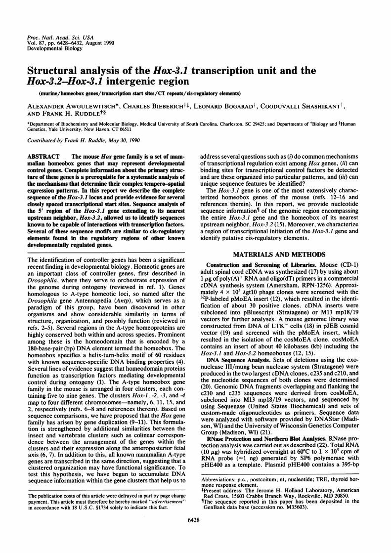
genomic cosMoEA segment analyzed and the structures ofthe two largest cDNAs, c235 (2.35 kb) and c210 (2.1 kb), areshown in Fig. 1A. Both cDNAs contain the complete Hox-3.1protein-coding region of 726 bp, consistent with earlier re-ports (14, 15). In c235, however, this coding sequence isinterrupted by an intervening sequence of about 1350 bp. Thepresence of 5' and 3' splice consensus sequences (25) and theperfect alignment of the c235 restriction map with that one ofthe corresponding genomic DNA segment suggest that c235has been generated from an unspliced precursor RNA (Fig.1A). The intronic nature of this region was also supported byits failure to hybridize with poly(A)+ RNAs isolated fromembryos and adult spinal cord (data not shown). c210 con-tains two perfect poly(A) signals (Fig. 2). A third poly(A)signal was revealed by sequence analyses of genomic DNAabout 160 bp downstream ofthe c210 3' terminus (Fig. 2). Thethird poly(A) signal may be used in transcript processing assuggested by (i) the presence of a G+T-rich sequence ele-ment known to be important for efficient poly(A) addition(26) that is located 17 bp downstream of this signal and absentin the vicinity of the other two poly(A) signals (Fig. 2), and(ii) the detection of a Hox-3.1-specific, 2.7-kb transcript byNorthern blot analysis of embryonic and adult spinal cordRNA with a probe specific for the third poly(A) signal (datanot shown).
AB P E
* poly(A)
Hox 3.2
B
1 2
.. .... ....: ..
*...:.......... .......... ... ..
HSc Sc EPScI I L1
Hc-E frag.
X PS E
poly(A)Hox 3.1
EPSo X PS
c235 's4 I
EPSo S E
c210 I.,,,
(c21 OB2) =
Hox 3.1 mRNA --.,,,-- AAA(=2.7 kb) 1 kb
FIG. 1. Structure of Hox-3.1 locus. (A) A partial restriction mapof the genomic region extending from the Hox-3.2 homeobox to theEcoRI site downstream of the third poly(A) signal of the Hox-3.1gene is shown. The approximate region of transcriptional initiation ofthe Hox-3.1 gene is marked by an angled arrow, protein codingregions are boxed with the homeobox in black, splice sites are
marked by triangles, and the directions of transcription are indicatedby straight arrows. The schematic structures of the c235 and c210cDNAs and of the c210B2 subclone, aligned to the correspondinggenomic regions, are shown below. A diagram of the predicted Hox3.1 mRNA with a presumed standard poly(A) tail of 260-300 bp (24)is shown at the bottom. Restriction endonuclease sites: B = BamHI,E = EcoRI, H = HindIll, Hc = HincII, P = Pst I, Sc = Sac I, S =
Sal I, X = Xho I. (B) Northern blot hybridization of poly(A)+ RNAs(7.5 ,ug each) from 12.5-day p.c. mouse embryos (lane 1) and adultspinal cord (lane 2) with the 32P-labeled c210B2 probe. The size of theHox-3.1 transcript is about 2.7 kb as indicated by the arrowhead.
Northern blot hybridizations using the c210B2 cDNAsubfragment (Fig. 1A) detected a single major hybridizingtranscript of about 2.7 kb in poly(A)+ RNA samples from12.5-day p.c. embryos and adult spinal cord (Fig. 1B). Theadditional 2.1-kb transcript previously detected in the adultspinal cord (12) could not be detected under the conditionsused here, suggesting that earlier results were due to cross-hybridization to a related RNA species.
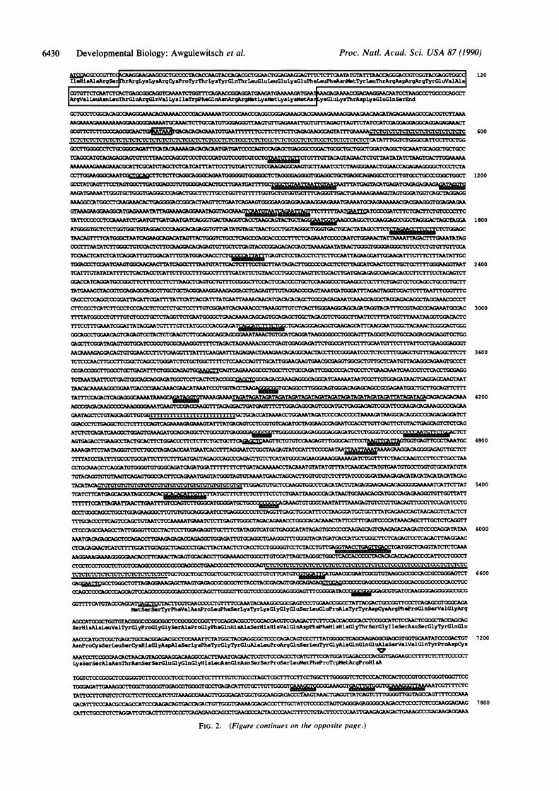
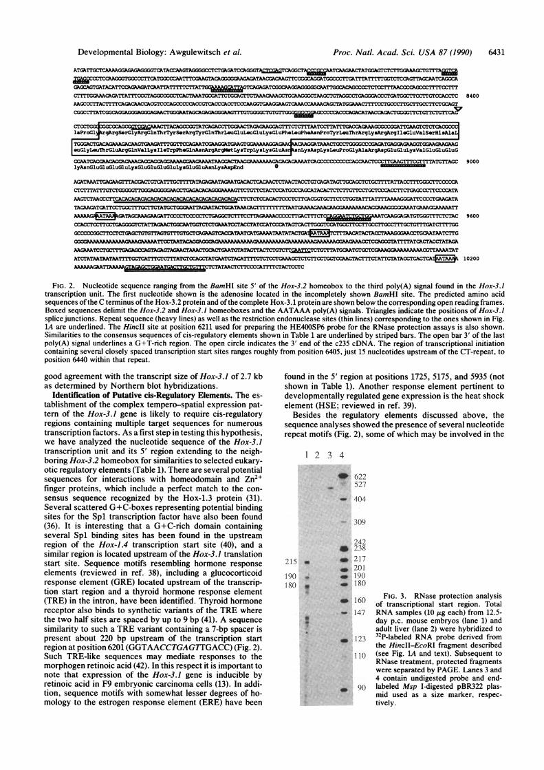
Determination of Transcriptional Start Regions. Northernblot hybridizations using probes derived from the upstreamregion of Hox-3.J provided preliminary evidence for thepresence of a major Hox-3.1 transcription start site in thevicinity of the proximal dinucleotide CT repeat (data notshown). To obtain further evidence for this, we carried outRNase protection analyses using RNA probes derived froma 395-bp HinclI-EcoRI fragment (Figs. 1A and 2) subclonedinto pGEM-2. Transcription with SP6 RNA polymeraseresulted in two distinct RNAs of approximately 472 and 435nucleotides (nt), distinguishable as a major and minor band,respectively, after PAGE (Fig. 3, lane 3). These probes werehybridized to total RNA samples from 12.5-day p.c. em-bryos. Liver RNA from adult mice was used as a negativecontrol. After RNase treatment and PAGE, three majorfragments of about 215, 190, and 180 nt were detected withthe embryonic RNA. These data suggest the presence ofseveral closely spaced transcription start sites in the vicinityof the 5' end of the proximal CT repeat (Fig. 2). A less-well-defined signal in the range of about 135-145 nt may indicatethat some transcripts are initiated within the CT repeat. Twoconsiderably less abundant fragments of about 403 and 365 ntcould correspond to full-length protection of both forms ofthe probe, taking into account the length of the transcribedvector and polylinker regions. Consistent with this interpre-tation, hybridization of the gel-purified larger probe to em-bryonic RNA resulted in the disappearance of the 365-ntprotected fragment (data not shown). This may suggest thatin 12.5-day p.c. embryos, a smaller fraction of the Hox-3.1mRNAs may initiate from another promoter located up-stream of the HincII site. Hybridization of the same probesto RNA samples from other tissues known to express Hox-3.1, including adult spinal cord and kidney, yielded essen-tially the same pattern of protected fragments (data notshown).Primer extension analysis using several unique primers
downstream of the CT repeat failed to yield discerniblespecific products in a standard primer extension assay. Todetermine if specific cDNA products were generated in theprimer extension analyses at levels below the sensitivity ofthe assay, we performed polymerase chain reaction amplifi-cation, followed by cloning and sequencing of the cDNAproducts (27). Sequence analysis of the 20 largest clonesindicated that the 5' ends of the cDNA products were eitherwithin or near the 5' end of the proximal CT repeat, which isin good agreement with the results obtained from RNaseprotection assays. These results indicate that transcriptionalinitiation of the Hox-3.1 gene takes place in the vicinity of anextended CT repeat. No typical TATA boxes are found in thisregion. These results are consistent with the observation thata number of genes that lack TATA boxes contain a homopu-rine/homopyrimidine-rich region whose deletion leads tomarked loss of transcriptional activity (24, 28-30).Taken together, our data suggest a general structure of
Hox-3.1-encoded mRNA prevalent in 12.5-day p.c. embryosand in adult spinal cord as shown in Fig. 1A. The predictedmRNAs contain a 5' untranslated leader region ranging fromabout 420 to 455 nt depending upon the transcriptional startsite used, a protein coding sequence of 726 nt and a 3'untranslated trailer sequence of about 1270 nt with a standardpoly(A) tail of 260-300 nt (26). The resulting lengths of thesemRNAs would range between 2675 and 2750 nt, which is in
Developmental Biology: Awgulewitsch et al.
6430 Developmental Biology: Awgulewitsch et al. Proc. Natl. Acad. Sci. USA 87 (1990)
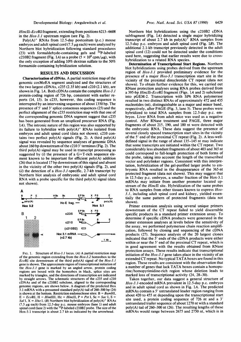
120Ile~is~la~rq~ erhrArg LysLy5ArgCy ProTyrThrLysTyrGlnThrLeuGluLeuGluLysGluPhe.. . ~~~~~wlor la _-L --Su,............................s-ssXs-vxx
C 600
G
T 1200
i
A 1800
T 2400T
T
r
T
C 3000c
r
G
T
A 4200
T
GT
C 4800
A
AG
sT 5400T
'G'TIT
A 6000CA
C
T 6600
:G
'G
LC
;T 7200
AAATCCTCCLyCCArACAaA%,AAsnGACLGyAAflGlAflr.rAOSerLUC TC*A.LrrOGI5ALysearSerAlaAsnThrAsnSerSerGluGlyGlnG]Ly~isLeuAsnGlnAsnSerSerProSerLeutsetPheProTrpMetArgProHi&A
7800
FIG. 2. (Figure continues on the opposite page.)
OGrrTTCNTMCCCAWATgLgML=ACTTCGTCAACC GGGCAGNotS*rSerTyrPheMalAsnProLouPhoSerLysTyrLysGlyGlyGluSerL*uGluProAlaTyrTyrAspCyaArqPheProGlnSerValGlyAr
AGOCATGCGCTGGTGTAC k
SerHisAlaLeuValTyrGlyProGlyGlySerAlaProGlyPhoGInHisAlaSerHisHisValGlnAspPhePheHisHisGlyThrSerGlyIleSerAsnSerGlyTyrGlnGlAACCCAT=CGCTGAGCTGCCACOGAGACGCCTCCAAATTCTATGGCTACGAGGCGCTCCCCAGACAGTCCCTTTATGGGGCTCAGCAAGAGG=GCGTGGTGCAATATCCCGACTGAsnProCyaSerLeuSerCysHisGlyAsphlaSerLysPheTyrGlyTyrGluAlaLeuProhrqGlnSerLeuTyrGlyAlaGlnGlnGluAlaSerValValGlnTyrProAspCy
Proc. Natl. Acad. Sci. USA 87 (1990) 6431
ATGATTGCCAAGGAAAGGCATACCAAATAGGGG~CTCTGA GGATCCAGGGr=ghGC WTCAAGAACTATGG~CTCTGGAAGCTGTGAGCCTCaUkGGTGGCCCTTCATGGCCCAArrTC~kGACA =G~AGAGTAACGACAAGTTC =GCAGGTGOCCTGATTTATTTTTGGTCTCAGTTAGCATAGCGAGCAGTGATACATTCCiGAGAGATCAATTArrrTr~iU~lgiEWTCAGAGATCGCAAGGAGGGGGCAATTGGCACAGCC TCTTAACOCCAGCTTTTCCTTTCTTTTGGAAACAiGATTATTCCCTA=GCC C01CATAAATGCCATTCTGCACTTGTWCAAAAGCTGCAGGTAAGCTGTAGGCCTGAGG;GACC:CTGATGGCTTCCTTGTCCACCTCAAGCCCTTACTTTTCAGACAACCACGTCCCAGCCCCCACCGTCACCCACCTCCCAAGGTGAAGGAAGTCAAACCAAAACAGCTAT_ >T TCGCCTGrTCTTCTGt AGT
;GCCTTATCGCGAGGGGACGGTGAACcCAGACGTAGGGAGTTCTGrTTGTTCAFY
CTZ~laProGl rgArgSerGlyArgGlnThrTyrSerArgTyrGlnThrLouGluLeuGluLyaGluPheLeuPheAsnProTytrLeuThrArgLysArgArgIleGluValSerHisAlaL
TGGGACTGACGAAGCAArGUGTTGTT;AATCGAA~TGAGT A i GGCC1GAG~GAAGAeuGlyLeuThrGluArgGlnValLysIleTrpPheGlnAsnArgArgMetLysTrpLyaLysGluAs nLysAspLyaLeuProGlyAlaArgAspGluGluLysValGluGluGluG
GGA;ATAGG AGAGGGAACATCCCCCCCCACAACTCCCTTGAAGTrTCGTTTTATGTTAGClyAsnGluGluGluGluLysGluGluGluGluLysGluGluAsnLysAspEnd 0
AGATAAATTGAGRAGTTTACGACTGTCA~~TGCTTTTAGhGAAGAATAGACA1CTICAACTAACTACCTGTCAGATAGTTG;CA TGmAACl;GTCCA
AAGTCTAA_ CCTCTTCACGGTGCTTCTCTGGTATrTAn RAAGGTCCCCTGAAG;ATA
TAGAAGATGATT__,L TTTT GAAAGAAAGGAAAMCAAAGGGTGAA AGGAAAATTMMAC 7AqGATAGCAAGMATTCCGCCCTGACTTCiAGATGt;TTCTCTCX C =DTTwAVI=ACCTGCACTT CCTCTGCC = CmrL-Lx;GTTCTTTTTGG(;CCCCCGGCTTCTCT = 1C I IAGCTcAccTAcTm ATATAcTG~iaKrTT~ATAcTACCTAGGAACCGCAATAATCTTG
_A A A A M~~~~~sAGAAAA^ A 1A ~~~~CGGAAAACCTCCAGCGTATTTTATCACTACCTATAGA
AAGAAATCAAAGAGAAccCTCCcCCATAICCTcTrTcrTAGAATTCcTCTGTTTGCrAAACGATATGAAAGGTTAAATAT
8400
9000
9600
ATCTTATAATPrrmATAr;cTATAAICX ATcTcAGATCTraGAAAG ~TCTGTCCTGt;rCC^AGTAcrTTcTATTGTATTAcrTGAr.TcAGTC 10200AAAAAAGAA _TCTATAAC:OCC=TT~~~CCATTTTC'rCTrCC
FIG. 2. Nucleotide sequence ranging from the BamHI site 5' of the Hox-3.2 homeobox to the third poly(A) signal found in the Hox-3.1transcription unit. The first nucleotide shown is the adenosine located in the incompletely shown BamlIl site. The predicted amino acidsequences of the C terminus of the Hox-3.2 protein and of the complete Hox-3.1 protein are shown below the corresponding open reading frames.Boxed sequences delimit the Hox-3.2 and Hox-3.1 homeoboxes and the AATAAA poly(A) signals. Triangles indicate the positions of Hox-3.1splice junctions. Repeat sequence (heavy lines) as well as the restriction endonuclease sites (thin lines) corresponding to the ones shown in Fig.1A are underlined. The HinciI site at position 6211 used for preparing the HE400SP6 probe for the RNase protection assays is also shown.Similarities to the consensus sequences of cis-regulatory elements shown in Table 1 are underlined by striped bars. The open bar 3' of the lastpoly(A) signal underlines a G+T-rich region. The open circle indicates the 3' end of the c235 cDNA. The region of transcriptional initiationcontaining several closely spaced transcription start sites ranges roughly from position 6405, just 15 nucleotides upstream of the CT-repeat, toposition 6440 within that repeat.
good agreement with the transcript size of Hox-3.1 of 2.7 kbas determined by Northern blot hybridizations.
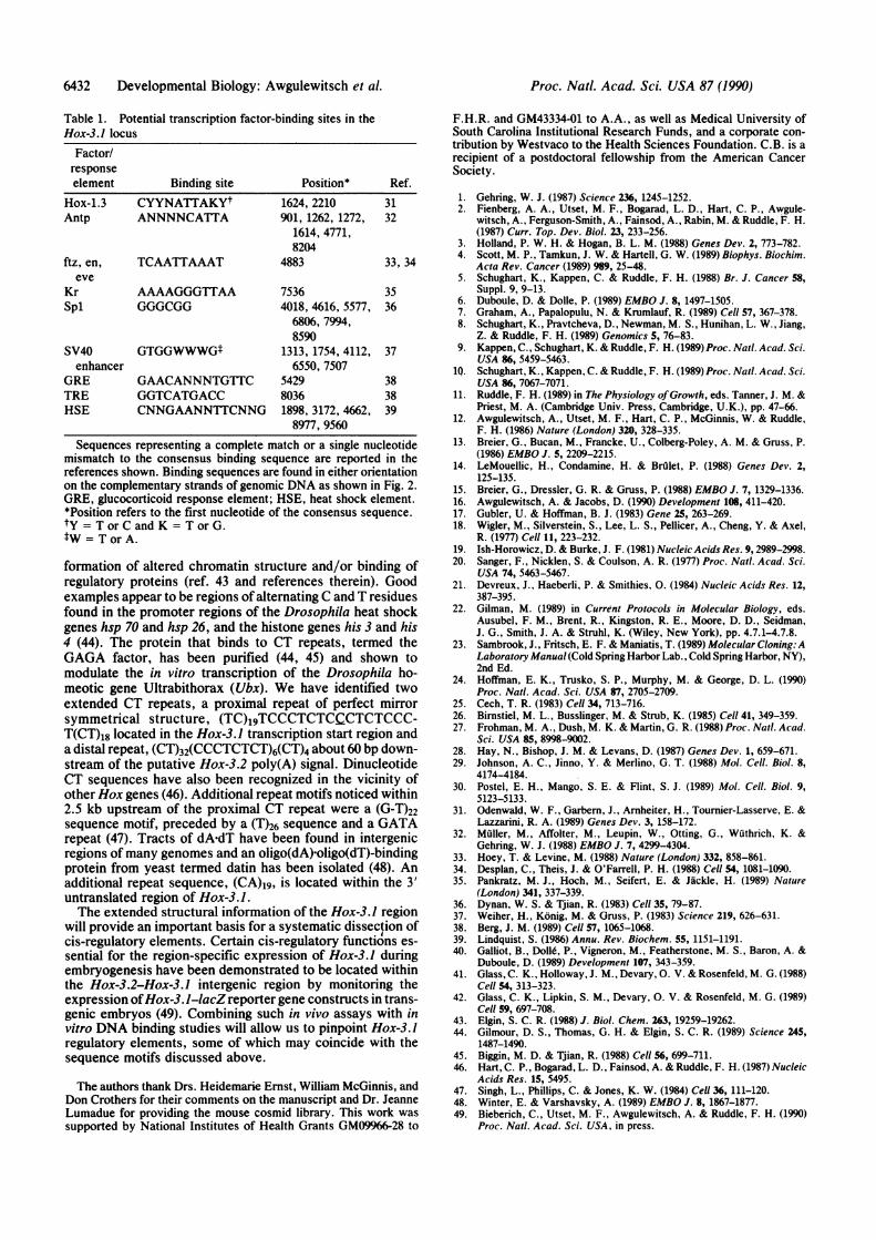
Identification of Putative cis-Regulatory Elements. The es-tablishment of the complex tempero-spatial expression pat-tern of the Hox-3.1 gene is likely to require cis-regulatoryregions containing multiple target sequences for numeroustranscription factors. As a first step in testing this hypothesis,we have analyzed the nucleotide sequence of the Hox-3.1transcription unit and its 5' region extending to the neigh-boring Hox-3.2 homeobox for similarities to selected eukary-otic regulatory elements (Table 1). There are several potentialsequences for interactions with homeodomain and Zn2+finger proteins, which include a perfect match to the con-sensus sequence recognized by the Hox-1.3 protein (31).Several scattered G+C-boxes representing potential bindingsites for the Spl transcription factor have also been found(36). It is interesting that a G+C-rich domain containingseveral Spl binding sites has been found in the upstreamregion of the Hox-1.4 transcription start site (40), and asimilar region is located upstream of the Hox-3.1 translationstart site. Sequence motifs resembling hormone responseelements (reviewed in ref. 38), including a glucocorticoidresponse element (GRE) located upstream of the transcrip-tion start region and a thyroid hormone response element(TRE) in the intron, have been identified. Thyroid hormonereceptor also binds to synthetic variants of the TRE wherethe two half sites are spaced by up to 9 bp (41). A sequencesimilarity to such a TRE variant containing a 7-bp spacer ispresent about 220 bp upstream of the transcription startregion at position 6201 (GGTAACCTGAGTIGACC) (Fig. 2).Such TRE-like sequences may mediate responses to themorphogen retinoic acid (42). In this respect it is important tonote that expression of the Hox-3.1 gene is inducible byretinoic acid in F9 embryonic carcinoma cells (13). In addi-tion, sequence motifs with somewhat lesser degrees of ho-mology to the estrogen response element (ERE) have been
found in the 5' region at positions 1725, 5175, and 5935 (notshown in Table 1). Another response element pertinent todevelopmentally regulated gene expression is the heat shockelement (HSE; reviewed in ref. 39).
Besides the regulatory elements discussed above, thesequence analyses showed the presence of several nucleotiderepeat motifs (Fig. 2), some of which may be involved in the
2 3 4
w 622527
_ 404
- 309
215
190180
242* 238
a _ 217_ 201
* _ 190p _ 180VI.
160 FIG. 3. RNase protection analysis.i 160 of transcriptional start region. Total147 RNA samples (10 ,g each) from 12.5-
day p.c. mouse embryos (lane 1) and-adult liver (lane 2) were hybridized to
l 123 32P-labeled RNA probe derived fromthe HincII-EcoRI fragment described
110 (see Fig. 1A and text). Subsequent toRNase treatment, protected fragmentswere separated by PAGE. Lanes 3 and4 contain undigested probe and end-
o go labeled Msp I-digested pBR322 plas-mid used as a size marker, respec-tively.
Developmental Biology: Awgulewitsch et al.
Ia
6432 Developmental Biology: Awgulewitsch et al.
Table 1. Potential transcription factor-binding sites in theHox-3.1 locus
Factor/responseelement Binding site Position* Ref.
Hox-1.3 CYYNATTAKYt 1624, 2210 31Antp ANNNNCATTA 901, 1262, 1272, 32
1614, 4771,8204
ftz, en, TCAATTAAAT 4883 33, 34eve
Kr AAAAGGGTTAA 7536 35Spi GGGCGG 4018, 4616, 5577, 36
6806, 7994,8590
SV40 GTGGWWWGt 1313, 1754, 4112, 37enhancer 6550, 7507
GRE GAACANNNTGTTC 5429 38TRE GGTCATGACC 8036 38HSE CNNGAANNTTCNNG 1898, 3172, 4662, 39
8977, 9560Sequences representing a complete match or a single nucleotide
mismatch to the consensus binding sequence are reported in thereferences shown. Binding sequences are found in either orientationon the complementary strands of genomic DNA as shown in Fig. 2.GRE, glucocorticoid response element; HSE, heat shock element.*Position refers to the first nucleotide of the consensus sequence.tY = TorC and K = TorG.tW= TorA.
formation of altered chromatin structure and/or binding ofregulatory proteins (ref. 43 and references therein). Goodexamples appear to be regions of alternating C and T residuesfound in the promoter regions of the Drosophila heat shockgenes hsp 70 and hsp 26, and the histone genes his 3 and his4 (44). The protein that binds to CT repeats, termed theGAGA factor, has been purified (44, 45) and shown tomodulate the in vitro transcription of the Drosophila ho-meotic gene Ultrabithorax (Ubx). We have identified twoextended CT repeats, a proximal repeat of perfect mirrorsymmetrical structure, (TC)19TCCCTCTCCCTCTCCC-T(CT)18 located in the Hox-3.1 transcription start region anda distal repeat, (CT)32(CCCTCTCT)6(CT)4 about 60 bp down-stream of the putative Hox-3.2 poly(A) signal. DinucleotideCT sequences have also been recognized in the vicinity ofother Hox genes (46). Additional repeat motifs noticed within2.5 kb upstream of the proximal CT repeat were a (G-T)22sequence motif, preceded by a (T)26 sequence and a GATArepeat (47). Tracts of dA-dT have been found in intergenicregions of many genomes and an oligo(dA)-oligo(dT)-bindingprotein from yeast termed datin has been isolated (48). Anadditional repeat sequence, (CA)19, is located within the 3'untranslated region of Hox-3.1.The extended structural information of the Hox-3.1 region
will provide an important basis for a systematic dissection ofcis-regulatory elements. Certain cis-regulatory functions es-sential for the region-specific expression of Hox-3.1 duringembryogenesis have been demonstrated to be located withinthe Hox-3.2-Hox-3.1 intergenic region by monitoring theexpression ofHox-3. J-lacZ reporter gene constructs in trans-genic embryos (49). Combining such in vivo assays with invitro DNA binding studies will allow us to pinpoint Hox-3.1regulatory elements, some of which may coincide with thesequence motifs discussed above.
The authors thank Drs. Heidemarie Ernst, William McGinnis, andDon Crothers for their comments on the manuscript and Dr. JeanneLumadue for providing the mouse cosmid library. This work wassupported by National Institutes of Health Grants GM09966-28 to
F.H.R. and GM43334-01 to A.A., as well as Medical University ofSouth Carolina Institutional Research Funds, and a corporate con-tribution by Westvaco to the Health Sciences Foundation. C.B. is arecipient of a postdoctoral fellowship from the American CancerSociety.
1. Gehring, W. J. (1987) Science 236, 1245-1252.2. Fienberg, A. A., Utset, M. F., Bogarad, L. D., Hart, C. P., Awgule-
witsch, A., Ferguson-Smith, A., Fainsod, A., Rabin, M. & Ruddle, F. H.(1987) Curr. Top. Dev. Biol. 23, 233-256.
3. Holland, P. W. H. & Hogan, B. L. M. (1988) Genes Dev. 2, 773-782.4. Scott, M. P., Tamkun, J. W. & Hartell, G. W. (1989) Biophys. Biochim.
Acta Rev. Cancer (1989) 989, 25-48.5. Schughart, K., Kappen, C. & Ruddle, F. H. (1988) Br. J. Cancer 58,
Suppl. 9, 9-13.6. Duboule, D. & Dolle, P. (1989) EMBO J. 8, 1497-1505.7. Graham, A., Papalopulu, N. & Krumlauf, R. (1989) Cell 57, 367-378.8. Schughart, K., Pravtcheva, D., Newman, M. S., Hunihan, L. W., Jiang,
Z. & Ruddle, F. H. (1989) Genomics 5, 76-83.9. Kappen, C., Schughart, K. & Ruddle, F. H. (1989) Proc. Natl. Acad. Sci.
USA 86, 5459-5463.1Q. Schughart, K., Kappen, C. & Ruddle, F. H. (1989) Proc. Natl. Acad. Sci.
USA 86, 7067-7071.11. Ruddle, F. H. (1989) in The Physiology ofGrowth, eds. Tanner, J. M. &
Priest, M. A. (Cambridge Univ. Press, Cambridge, U.K.), pp. 47-66.12. Awgulewitsch, A., Utset, M. F., Hart, C. P., McGinnis, W. & Ruddle,
F. H. (1986) Nature (London) 320, 328-335.13. Breier, G., Bucan, M., Francke, U., Colberg-Poley, A. M. & Gruss, P.
(1986) EMBO J. 5, 2209-2215.14. LeMouellic, H., Condamine, H. & Brnlet, P. (1988) Genes Dev. 2,
125-135.15. Breier, G., Dressler, G. R. & Gruss, P. (1988) EMBO J. 7, 1329-1336.16. Awgulewitsch, A. & Jacobs, D. (1990) Development 108, 411-420.17. Gubler, U. & Hoffman, B. J. (1983) Gene 25, 263-269.18. Wigler, M., Silverstein, S., Lee, L. S., Pellicer, A., Cheng, Y. & Axel,
R. (1977) Cell 11, 223-232.19. Ish-Horowicz, D. & Burke, J. F. (1981) Nucleic Acids Res. 9, 2989-2998.20. Sanger, F., Nicklen, S. & Coulson, A. R. (1977) Proc. Natl. Acad. Sci.
USA 74, 5463-5467.21. Devreux, J., Haeberli, P. & Smithies, 0. (1984) Nucleic Acids Res. 12,
387-395.22. Gilman, M. (1989) in Current Protocols in Molecular Biology, eds.
Ausubel, F. M., Brent, R., Kingston, R. E., Moore, D. D., Seidman,J. G., Smith, J. A. & Struhl, K. (Wiley, New York), pp. 4.7.1-4.7.8.
23. Sambrook, J., Fritsch, E. F. & Maniatis, T. (1989) Molecular Cloning: ALaboratory Manual (Cold Spring Harbor Lab., Cold Spring Harbor, NY),2nd Ed.
24. Hoffman, E. K., Trusko, S. P., Murphy, M. & George, D. L. (1990)Proc. Natl. Acad. Sci. USA 87, 2705-2709.
25. Cech, T. R. (1983) Cell 34, 713-716.26. Birnstiel, M. L., Busslinger, M. & Strub, K. (1985) Cell 41, 349-359.27. Frohman, M. A., Dush, M. K. & Martin, G. R. (1988) Proc. Natl. Acad.
Sci. USA 85, 8998-9002.28. Hay, N., Bishop, J. M. & Levans, D. (1987) Genes Dev. 1, 659-671.29. Johnson, A. C., Jinno, Y. & Merlino, G. T. (1988) Mol. Cell. Biol. 8,
4174-4184.30. Postel, E. H., Mango, S. E. & Flint, S. J. (1989) Mol. Cell. Biol. 9,
5123-5133.31. Odenwald, W. F., Garbern, J., Arnheiter, H., Tournier-Lasserve, E. &
Lazzarini, R. A. (1989) Genes Dev. 3, 158-172.32. Muller, M., Affolter, M., Leupin, W., Otting, G., Wuthrich, K. &
Gehring, W. J. (1988) EMBO J. 7, 4299-4304.33. Hoey, T. & Levine, M. (1988) Nature (London) 332, 858-861.34. Desplan, C., Theis, J. & O'Farrell, P. H. (1988) Cell 54, 1081-1090.35. Pankratz, M. J., Hoch, M., Seifert, E. & Jackle, H. (1989) Nature
(London) 341, 337-339.36. Dynan, W. S. & Tjian, R. (1983) Cell 35, 79-87.37. Weiher, H., Konig, M. & Gruss, P. (1983) Science 219, 626-631.38. Berg, J. M. (1989) Cell 57, 1065-1068.39. Lindquist, S. (1986) Annu. Rev. Biochem. 55, 1151-1191.40. Galliot, B., Dolle, P., Vigneron, M., Featherstone, M. S., Baron, A. &
Duboule, D. (1989) Development 107, 343-359.41. Glass, C. K., Holloway, J. M., Devary, 0. V. & Rosenfeld, M. G. (1988)
Cell 54, 313-323.42. Glass, C. K., Lipkin, S. M., Devary, 0. V. & Rosenfeld, M. G. (1989)
Cell 59, 697-708.43. Elgin, S. C. R. (1988) J. Biol. Chem. 263, 19259-19262.44. Gilmour, D. S., Thomas, G. H. & Elgin, S. C. R. (1989) Science 245,
1487-1490.45. Biggin, M. D. & Tjian, R. (1988) Cell 56, 699-711.46. Hart, C. P., Bogarad, L. D., Fainsod, A. & Ruddle, F. H. (1987) Nucleic
Acids Res. 15, 5495.47. Singh, L., Phillips, C. & Jones, K. W. (1984) Cell 36, 111-120.48. Winter, E. & Varshavsky, A. (1989) EMBO J. 8, 1867-1877.49. Bieberich, C., Utset, M. F., Awgulewitsch, A. & Ruddle, F. H. (1990)
Proc. Natl. Acad. Sci. USA, in press.
Proc. Natl. Acad. Sci. USA 87 (1990)