Embed Size (px)
Citation preview

R&D Review of Toyota CRDL, Vol.48 No.1 (2017) 1-9 1
© Toyota Central R&D Labs., Inc. 2017 http://www.tytlabs.com/review/
Special Feature: Biotechnology for Sustainable and Aging Societies
Research Report
Structural Analysis of Lignocellulose Biomass Using Nuclear Magnetic Resonance Nobuhiro Ishida, Tetsuya Mori and Jun Kikuchi Report received on Dec. 12, 2016
A bio-refinery that produces fuels and chemicals from plant biomass has the potentialfor providing technology for a more sustainable society. The efficient utilization of plants requiresan understanding of the chemical composition of the plant structure, such as lignocellulose and metaboliccomponents. This study used high-resolution nuclear magnetic resonance (NMR) to assign chemical shiftsfor cellulose and other structural components of plant biomass to gain insights into the plant structure.Solid-state 13C-13C NMR using 13C-labeled bacterial cellulose allowed the complete assignment of chemicalshifts for carbon atoms in the amorphous cellulose structure. In addition, solution-state three-dimensional NMR provided accurate assignments of chemical shifts for polysaccharides and small components in13C-labeled poplar cellulose. These results increase our understanding of the complex structure oflignocellulose components, and open the possibility for new applications for processing, conversion, anddegradation of lignocellulose.
Bio-refinery, Lignocellulose, Metabolite, Polysaccharide, Solid-state NMR,
Solution-state NMR
1. Introduction
Plants have been used in various industrial ways
throughout history. The importance of lignocellulose-based bio-refineries have been studied recently in an effort to replace oil-based resources and contribute to a more sustainable energy source.(1) An increase in the utilization of bio-resources requires efficient processing, conversion, and degradation of lignocellulosic materials. Plant biomass has a complex structure involving cellulose, hemicellulose, and lignin, which are connected in complex ways in the cell wall (Fig. 1).(2) Cellulose is composed of D-glucose units that condense through β (1-4)-glyosidic bonds, and consists of crystalline and amorphous regions. Hemicellulose is composed mainly of xylose units, and one main hemicellulose is xylan, which is partially acetylated with sugar residues (e.g., arabinose). Lignin consists of polymeric phenolic units based on syringyl, guaiacyl, and p-hydroxyphenyl moieties. Understanding the chemical structures of these plant materials is needed for their efficient utilization. Gaining knowledge about structures of plant tissue has been accomplished using X-ray diffraction,(3) infrared spectroscopy,(4) microscopy,(5) mass spectrometry,(6) and nuclear magnetic resonance (NMR).(7)
NMR is an effective tool for analyzing biomolecular complexes of metabolite components.(8) Various solid and solution-state NMR techniques have been used to detect and identify these polymeric components. Solid-state NMR has the advantage of enabling detection of macromolecules in intact plants. Studies involving beech wood, pine,(9) and wheat straw(10) have been reported using intact tissue. However, assigning all of the signals in a sold-state NMR spectrum is difficult because of its low resolution.(11) Solution-state NMR, in contrast, can detect and identify many polymeric components in plant samples dissolved in dimethylsulfoxide (DMSO)(12) and DMSO/pyridine(13) systems. Polymeric components have been characterized using high-resolution, solution-state two-dimensional (2D) NMR that provided detailed information about metabolites and lignocellulose components,(13,14) and can be used to provide partial assignments of chemical shifts in the spectra of biomolecular components. But completely assigning each signal in the spectra is difficult, because of overlapping chemical shifts. Therefore, a recent study demonstrated that plant compounds labelled with a stable isotope allowed assignment of signals from multidimensional NMR.(15)

2 R&D Review of Toyota CRDL, Vol.48 No.1 (2017) 1-9
© Toyota Central R&D Labs., Inc. 2017 http://www.tytlabs.com/review/
The present study describes several approaches for analyzing the cellulose structure and the chemical composition of plants using high-resolution NMR. An overview of our attempt is summarized in Fig. 2. Important steps are the 13C-labeling and the multidimensional NMR used for assigning the signals in the spectra of biomolecule components. In solid-state NMR, the assignment of each carbon atom (C1-C6) within the bacterial cellulose was examined after preparation with 13C-labeling. Although NMR assignments of 13C-labeled crystalline cellulose were completed for the 13C atoms, not all carbons in the amorphous cellulose were assigned. Based on the chemical shifts, differences in the cellulosic structure due to different pretreatments were identified. In solution-state 2D HSQC and 3D HCCH total correlation spectroscopy (TOCSY) NMR, an attempt was made to assign the detailed spectra of polymeric components from 13C-labeled poplar dissolved in DMSO/pyridine. In addition, intact tissue was analyzed by high-resolution magic angle spinning (HR-MAS) NMR 2D HSQC(16) and 3D HCCH correlation spectroscopy (COSY)(17) for identification of low-molecular-weight components.
2. Analysis of 13C-labeled Amorphous Cellulose by Solid-state NMR Amorphous cellulose, used as the raw material for
cellophane or cloth fibers, such as rayon, can be obtained by cellulose regeneration. In the bio-refinery process, the ratio of amorphous cellulose influences the efficiency of enzymatic degradation of cellulose. First of all, the full assignment of 13C-labeled amorphous cellulose pretreated with ionic liquid was attempted, because the ionic liquid was expected to be an effective solvent for cellulose recently.(18) For the solid-state NMR analysis, 13C-labeled bacterial cellulose was prepared from Acetobacter xylinum(19) pretreated with 1-ethyl-3-methylimidazolium chloride ([Emin][Cl]). To prepare the NMR samples, A. xylinum was cultured in a medium containing 13C6-glucose, followed by washing the cellulose obtained with a weakly alkaline solution. The cellulose pellicle was solubilized in [Emin][Cl] at 120°C for 30 min, then regenerated in distilled water. After drying the regenerated cellulose, each sample was packed into a 4-mm ZrO2 rotor. For solid-state NMR, a DRX-800 spectrometer was used with a magic-angle spinning
Cellulose
Hemicellulose
Lignin
Monomer Macromolecule Lignocellulosein plant cell wall
AmorphousCrystalline
Structural information
Fig. 1 Lignocellulose of plant cell wall is composed of cellulose, hemicellulose, andlignin. Cellulose is composed of glucose units, hemicellulose is mainlycomposed of xylose units, and lignin is composed of phenolic units.

R&D Review of Toyota CRDL, Vol.48 No.1 (2017) 1-9 3
© Toyota Central R&D Labs., Inc. 2017 http://www.tytlabs.com/review/
(MAS) triple resonance probe. The 1D 13C cross-polarization MAS (CP-MAS) and 2D 13C-13C refocused incredible natural abundance double quantum transfer experiment (INADEQUATE) NMR spectra were obtained at 12 kHz MAS, and each spectrum was analyzed using NMRPipe.
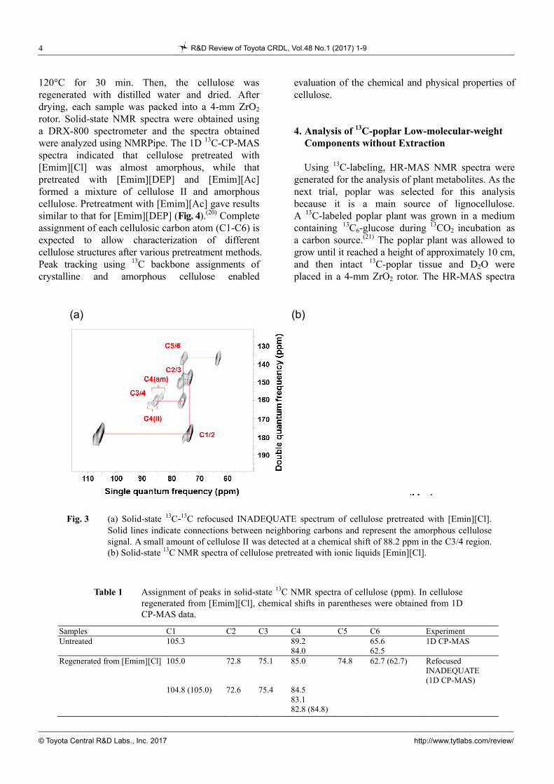
To determine the assignments of each carbon atom (C1-C6) within cellulose, 2D 13C-13C refocused INADEQUATE spectroscopy was attempted (Fig. 3).(20) The 13C-labeling provided clear identification of cross correlations with an increased signal-to-noise ratio. Starting with the C1 chemical shifts obtained from a previous study, all chemical shifts could be determined using the same overlapping positions for each cross peak (Table 1). The C2 and C3 chemical shifts were expected to result from intermolecular hydrogen bonding and the interactions between cellulose chains. Hydrogen atoms linked to C2 and C3 were involved in intermolecular hydrogen bonding. Differentiating C2 and C3 atoms enables analysis of the interactions among cellulose chains that are important in cellulose degradation. In addition, assignment of C4 chemical shifts revealed
differences in the carbon atoms of different cellulose structures, such as cellulose I, cellulose II, and amorphous cellulose. Cellulose II appears to contain molecular chains in antiparallel alignment against those in cellulose I. Untreated bacterial cellulose could be differentiated from cellulose I and amorphous cellulose, because, upon pretreatment with [Emin][Cl], it formed amorphous cellulose.
3. Assignment of NMR Spectra to Obtain the Structure of Pretreated Cellulose The C4 chemical shifts were used to help
determine the cellulose structure. Ratios of cellulose structural states after various pretreatment conditions were examined using solid-state NMR. To prepare samples of cellulose with various structures, two types of ionic liquids were used, 1-ethyl-3-methylimidazolium acetate ([Emim][Ac]) and 1-ethyl-3-methylimidazolium diethylphosphate ([Emim][DEP]). The pretreated 13C-labeled cellulose was dissolved in each ionic liquid and incubated at
13C-labelled poplar
13C-labelled cellulose
Solution-state
Solid-state
Cellulose solubilizationin ionic liquid
Preparation Multidimensional NMR
Lignocellulose solubilizationin DMSO/pyridine
HR-MAS
Amorphouscellulose
1H
13C
1H
13C
13C
13C-13C
13C 13C 13C
H
H H
H H
H
Biomass
Fig. 2 Overview of NMR analysis used in this study: Solubilization of lignocellulose andmulti-dimensional NMR was applied for analyzing lignocellulose components; the preparationof 13C-amorphous cellulose via regeneration from an ionic liquid; metabolites of intact tissues of13C-poplar were analyzed by high-resolution magic angle spinning (HR-MAS).
4 R&D Review of Toyota CRDL, Vol.48 No.1 (2017) 1-9
© Toyota Central R&D Labs., Inc. 2017 http://www.tytlabs.com/review/
120°C for 30 min. Then, the cellulose was regenerated with distilled water and dried. After drying, each sample was packed into a 4-mm ZrO2 rotor. Solid-state NMR spectra were obtained using a DRX-800 spectrometer and the spectra obtained were analyzed using NMRPipe. The 1D 13C-CP-MAS spectra indicated that cellulose pretreated with [Emim][Cl] was almost amorphous, while that pretreated with [Emim][DEP] and [Emim][Ac] formed a mixture of cellulose II and amorphous cellulose. Pretreatment with [Emim][Ac] gave results similar to that for [Emim][DEP] (Fig. 4).(20) Complete assignment of each cellulosic carbon atom (C1-C6) is expected to allow characterization of different cellulose structures after various pretreatment methods. Peak tracking using 13C backbone assignments of crystalline and amorphous cellulose enabled
evaluation of the chemical and physical properties of cellulose.
4. Analysis of 13C-poplar Low-molecular-weight Components without Extraction Using 13C-labeling, HR-MAS NMR spectra were
generated for the analysis of plant metabolites. As the next trial, poplar was selected for this analysis because it is a main source of lignocellulose. A 13C-labeled poplar plant was grown in a medium containing 13C6-glucose during 13CO2
incubation as a carbon source.(21) The poplar plant was allowed to grow until it reached a height of approximately 10 cm, and then intact 13C-poplar tissue and D2O were placed in a 4-mm ZrO2 rotor. The HR-MAS spectra
Samples C1 C2 C3 C4 C5 C6 Experiment Untreated 105.3 89.2
84.0 65.6
62.5 1D CP-MAS
Regenerated from [Emim][Cl]
105.0 104.8 (105.0)
72.8 72.6
75.1 75.4
85.0 84.5 83.1 82.8 (84.8)
74.8 62.7 (62.7) Refocused INADEQUATE (1D CP-MAS)
Table 1 Assignment of peaks in solid-state 13C NMR spectra of cellulose (ppm). In cellulose regenerated from [Emim][Cl], chemical shifts in parentheses were obtained from 1DCP-MAS data.
Fig. 3 (a) Solid-state 13C-13C refocused INADEQUATE spectrum of cellulose pretreated with [Emin][Cl].Solid lines indicate connections between neighboring carbons and represent the amorphous cellulosesignal. A small amount of cellulose II was detected at a chemical shift of 88.2 ppm in the C3/4 region.(b) Solid-state 13C NMR spectra of cellulose pretreated with ionic liquids [Emin][Cl].
(a) (b)

R&D Review of Toyota CRDL, Vol.48 No.1 (2017) 1-9 5
© Toyota Central R&D Labs., Inc. 2017 http://www.tytlabs.com/review/
for 13C-poplar without extraction were obtained using DRX-400 and DRX-500 spectrometers equipped with Z-axis high-resolution magic angle spinning probes. The MAS rotational speed was maintained at a constant 4000 Hz. The 2D 1H-13C-HSQC and 3D HCCH-COSY spectra were obtained using a DRX-500 spectrometer and DRX-400 spectrometer, respectively. Each NMR spectrum was processed using NMRPipe. Signals from each metabolite were assigned using the 1H and 13C chemical shift database SpinAssign.(22-24)
The HR-MAS 2D 1H-13C-HSQC spectrum is shown in Fig. 5.(25) Although each signal was broadened in the 1H dimension by residual 1H-1H dipolar interactions, chemical shift dispersion could be resolved in intact tissues. Using the SpinAssign database,(22-24) a number of peaks could be assigned to particular metabolites, such as amino acids, ethanol, malate, choline, ethanolamine, and glucose. Multiple candidate metabolites also were observed in the database during the process of matching chemical shift data for each signal. In general, solution-state NMR requires extraction of plants, and depends on the solvents used for extraction. However, the
HR-MAS technique demonstrated here did not have this limitation and was used to analyze intact tissue to gain information about metabolites such as lipids, leucine, ethanol, and isoleucine.
5. Polysaccharide Analysis of 13C-poplar Dissolved in DMSO/pyridine Solvent Polysaccharides are polymeric carbohydrates in
which monosaccharides are connected through glyosidic linkages. Freeze-dried 13C poplar was pretreated according to an above method, and the crushed sample was ball-milled. The milled sample was extracted with ethanol and distilled water, and then dissolved in DMSO-d6/pyridine-d5 (4:1), shaken, and centrifuged. Solution NMR spectra were obtained from the soluble matter in the sample using a DRU-700 spectrometer. Figure 6 shows the anomeric and aliphatic regions assigned using solution-state 2D 1H-13C-HSQC NMR (each peak corresponds to a value in Table 2).(25) The signals detected in the polysaccharide anomeric region of the 1H-13C-HSQC spectrum were assigned. However, only
Fig. 4 Solid-state 13C NMR spectra of (a) untreated cellulose, and cellulose pretreated with(b) [Emim][Cl], (c) [Emim][DEP], and (d) [Emim][Ac] at 120°C for 30 min. C1-C6represent the carbon backbone of cellulose.
(a) (b)
(c) (d)

6 R&D Review of Toyota CRDL, Vol.48 No.1 (2017) 1-9
© Toyota Central R&D Labs., Inc. 2017 http://www.tytlabs.com/review/
Fig. 5 HR-MAS 1H-13C-HSQC spectrum of 13C-poplar, measured without sample extraction. Peaks in the spectrum corresponding to metabolites were assigned by 3D HCCH-COSY experiments and matched by standard metabolites and SpinAssign. Ala, Alanine; Glu, Glutamic acid; Phe,Phenylalanine; Gly, Glycine; Ile, Isoleucine; Lys, Lysine; Leu, Leucine; Met, Methionine; Asn,Asparagine; Gln, Glutamine; Arg, Arginine; Ser, Serine; Thr, Threonine; Val, Valine; GABA,-amino butyric acid.
1H chemical shift (ppm)
13C
ch
em
ica
l sh
ift
(pp
m)
Fig. 6 Solution-state 1H-13C HSQC spectrum of 13C-poplar extracted in DMSO/pyridine. (a) Anomeric region. (b) Aliphatic region. Peaks were assigned by 3D HCCH-TOCSY experiments and matched on the basis of the previous reports. Peak numbers in the figure correspond with those listed in Table 2.
13 C
ch
emic
al s
hif
t (p
pm
)
1H chemical shift (ppm)
(a) (b)

R&D Review of Toyota CRDL, Vol.48 No.1 (2017) 1-9 7
© Toyota Central R&D Labs., Inc. 2017 http://www.tytlabs.com/review/
anomeric carbons could be identified using this approach. To identify other carbon signals, 3D NMR experiments were combined with 2D experiments, similar to HR-MAS. The peaks detected in the anomeric and aliphatic regions were assigned based
on 3D HCCH-TOCSY data. Figure 7 shows 1H-1H planes from the 3D 1H-1H-13C spectrum sliced along the 13C axis of the chemical shifts of (1,4)--D-glucopyranoside.(25) Based on the connections among C1-C6, the signals for
No Saccharide Chemical shift (ppm)
C1/H1 C2/H2 C3/H3 C4/H4 C5/H5 C6/H6
1 (1, 4)--D-Glcp 102.6/4.45 74.6/3.48 74.6/3/48 79.9/3.48 74.6/3.48 60.1/3.90, 60.1/3.72
2 Unknown
polysaccharide G 103.1/4.27 72.8/3.01 ND ND ND ND
3 Unknown
polysaccharide X 101.6/4.39 72.6/3.17 73.6/3.34 76.6/3.55 62.5/3.93, 62.9/3.14
4 2-O-Ac--D-Xylp 99.3/4.58 73.2/4.64 71.6/3.59 75.4/3.67 62.9/4.02, 62.9/3.32
5 2-O-Ac-Manp 98.9/4.76 72.4/5.10 74.8/3.84 ND ND ND
6 4-O-MeGlcA 97.2/5.31 71.4/3.35 ND ND ND ND
7 (1, 4)--D-Galp 105.5/4.36 71.0/3.55 ND ND ND ND
8 -L-Araf 107.9/4.90 81.8/3.98 ND ND ND
Table 2 1H and 13C chemical shift assignments for polysaccharide components from13C-poplar, based on a combination of the 3D HCCH-TOCSY.
ND: not determined
Fig. 7 Analysis of (1, 4)-β-D-Glcp using solution-state 2D HSQC and 3D HCCH-TOCSY spectra of 13C-poplar extracted in DMSO/pyridine. (a) 2D 1H-13C HSQC spectrum. Crossed marks show C1-6 signals of (1, 4)-β-D-Glcp assigned by 3D HCCH-TOCSY. (b) 2D 1H-1H planes at 60.2, 74.6, 79.9, 74.6, 72.7 and 62.7 (folded spectrum; 102.7) ppm of 13C, which correspond to the C6, C5, C4, C3, C2, and C1 of(1, 4)-β-D-Glcp, slicing the 3D 1H-1H-13C spectrum. Red transverse lines connect 1H-13C-13C-1H cross peaks and vertical dashed lines connect corresponding signals between 3D and 2D spectra.
13C
ch
emic
al s
hif
t (p
pm
) 1H
ch
emic
al s
hif
t (p
pm
)
1H chemical shift (ppm)
(a)
(b)

8 R&D Review of Toyota CRDL, Vol.48 No.1 (2017) 1-9
© Toyota Central R&D Labs., Inc. 2017 http://www.tytlabs.com/review/
(1,4)--D-Glcp, 2-O-acetyl--D-xylopyranoside, and 2-O-Ac--D-Xylp were completely assigned. In addition, (1,6)--D-glucopyranoside, 3-O-acetyl--D- xylopyranoside, 2-O-acetyl--D-mannopyranoside, 4-O-methyl--D-glucuronic acid, -D-galactopyranoside (-D-Galp), and -L-arabinofuranoside could be detected (Table 2). These results demonstrate that a significant number of peaks corresponding to polysaccharide could be identified, especially by combining 2D and 3D NMR spectral data. Although some polysaccharides had only low-intensity signals that were assigned, the results of this study provide an approach that could improve signal sensitivity from small amounts of polymeric components.
6. Conclusions To gain insights into bio-refining, the
supramolecular structure of lignocellulose must be elucidated, even though it is not well understood due to the complex mixture of cellulose, hemicellulose, and lignin it contains. Analyzing solid-state and solution-state NMR data provided an effective approach for identifying biomolecular compounds. By enhancing the NMR signals with 13C-labeling techniques, polymeric components and metabolites of polysaccharides could be assigned completely. Since chemical shifts reflect structural characteristics, the database of chemical shifts accumulated for lignocellulose components allowed construction of a biomass profile. NMR-based technologies can help provide a more comprehensive understanding of cell wall dynamics. In the future, effective utilization of biomass may be enhanced by information about the lignocellulose structure, which can contribute to a more sustainable society.
Acknowledgements
The authors thank Dr. E. Chikayama, Y. Tsuboi, and Dr. S. Moriya at the RIKEN Center for Sustainable Resource Science for technical support and valuable discussions.
References
(1) Sticklen, M. B., “Plant Genetic Engineering for Biofuel Production: Towards Affordable Cellulosic Ethanol”, Nature Reviews Genetics, Vol. 9 (2008), pp. 433-443.
(2) Rubin, E. M., “Genomics of Cellulosic Bbiofuels”, Nature, Vol. 454 (2008), pp. 841-845.
(3) Meyer, K. H. and Mark, H., “The Structure of the Crystallised Components of Cellulose”, Berichte der Deutschen Chemica Gesellschaft, Vol. 61 (1928), pp. 593-614.
(4) Sills, D. L. and Gossett, J. M., “Using FTIR to Predict Saccharification from Enzymatic Hydrolysis of Alkali-pretreated Biomasses”, Biotechnology and Bioengineering, Vol. 109 (2012), pp. 353-362.
(5) Singh, S. et al., “Visualization of Biomass Solubilization and Cellulose Regeneration during Ionic Liquid Pretreatment of Switchgrass”, Biotechnology and Bioengineering, Vol. 104 (2009), pp. 68-75.
(6) Tuskan, G. et al., “Two High-throughput Techniques for Determining Wood Properties as Part of a Molecular Genetics Analysis of Hybrid Poplar and Loblolly Pine”, Applied Biochemistry and Biotechnology, Vol. 77-79 (1999), pp. 55-65.
(7) Ke, J. et al., “Biodegradation of Hardwood Lignocellulosics by the Western Poplar Clearwing Borer, Paranthrenerobiniae (Hy. Edwards)”, Biomacromolecules, Vol. 12 (2011), pp. 1610-1620.
(8) Sekiyama, Y. et al., “Evaluation of a Semi Polar Solvent System as a Step toward Heteronuclear Multidimensional NMR-based Metabolomics for 13C-labeled Bacteria, Plants, and Animals”, Analytical Chemistry, Vol. 83 (2011), pp. 719-726.
(9) Maunu, S. L., “NMR Studies of Wood and Wood Products”, Progress in Nuclear Magnetic Resonance Spectroscopy, Vol. 40 (2002), pp. 151-174.
(10) Matulova, M. et al., “Degradation of Wheat Straw by Fibrobacter succinogenes S85: a Liquid- and Solid-state Nuclear Magnetic Resonance Study”, Applied and Environmental Microbiology, Vol. 71 (2005), pp. 1247-1253.
(11) Samuel, R. et al., “HSQC (Heteronuclear Single Quantum Coherence) 13C-1H Correlation Spectra of Whole Biomass in Perdeuterated Pyridinium Chloride-DMSO System: An Effective Tool for Evaluating Pretreatment”, Fuel, Vol. 90 (2011), pp. 2836-2842.
(12) Kim, H. et al., “Solution-state 2D NMR of Ball-milled Plant Cell Wall Gels in DMSO-d (6)”, BioEnergy Research, Vol. 1 (2008), pp. 56-66.
(13) Kim, H. and Ralph, J., “Solution-state 2D NMR of Ball-milled Plant Cell Wall Gels in DMSO-d (6)/ pyridine-d (5)”, Organic and Biomolecular Chemistry, Vol. 8 (2010), pp. 576-591.
(14) Date, Y. et al., “New Monitoring Approach for Metabolic Dynamics in Microbial Ecosystems Using Stable-isotope-labeling Technologies”, Journal of Bioscience and Bioengineering, Vol. 110 (2010), pp. 87-93.

R&D Review of Toyota CRDL, Vol.48 No.1 (2017) 1-9 9
© Toyota Central R&D Labs., Inc. 2017 http://www.tytlabs.com/review/
(15) Chikayama, E. et al., “Systematic NMR Analysis of Stable Isotope Labeled Metabolite Mixtures in Plant and Animal Systems: Coarse Grained Views of Metabolic Pathways”, PLoS One, Vol. 3 (2008), e3805 .
(16) Bodenhausen, G. and Ruben, D. J., “Natural Abundance N-15 NMR by Enhanced Heteronuclear Spectroscopy”, Chemical Physics Letters, Vol. 69 (1980), pp. 185-189.
(17) Kay, L. et al., “Proton-Proton Correlation via Carbon-carbon Couplings - a 3-dimensional NMR Approach for the Assignment of Aliphatic Resonances in Proteins Labeled with C-13”, Journal of the American Chemical Society, Vol. 112 (1990), pp. 888-889.
(18) Swatloski, R. P. et al., “Dissolution of Cellulose with Ionic Liquids”, Journal of the American Chemical Society, Vol. 124 (2002), pp. 4974-4975.
(19) Kuga, S. and Brown, R. M., “Silver Labeling of the Reducing Ends of Bacterial Cellulose”, Carbohydrate Research, Vol. 180 (1988), pp. 345-350.
(20) Mori, T. et al., “Exploring the Conformational Space of Amorphous Cellulose Using NMR Chemical Shifts”, Carbohydrate Polymers, Vol. 90 (2012), pp. 1197-1203.
(21) Sekiyama, Y. and Kikuchi, J., “Towards Dynamic Metabolic Network Measurements by Multi-dimensional NMR-based Fluxomics”, Phytochemistry, Vol. 68 (2007), pp. 2320-2329.
(22) Kono, H. et al., “13C and 1H Resonance Assignment of Mercerized Cellulose II by Two-dimensional MAS NMR Spectroscopies”, Macromolecules, Vol. 37 (2004), pp. 5310-5316.
(23) Akiyama, K. et al., “PRIMe: A Web Site that Assembles Tools for Metabolomics and Transcriptomics”, In Silico Biology, Vol. 8 (2008), pp. 339-345.
(24) Chikayama, E. et al., “Statistical Indices for Simultaneous Large-scale Metabolite Detections for a Single NMR Spectrum”, Analytical Chemistry, Vol. 82 (2010), pp. 1653-1658.
(25) Mori, T. et al., “Multidimensional High-resolution Magic Angle Spinning and Solution-state NMR Characterization of 13C-labeled Plant Metabolites and Lignocellulose”, Scientific Reports, Vol. 5 (2015), 11848.
Figs. 3, 4 and Table 1 Reprinted from Carbohydrate Polymers, Vol. 90 (2012), pp. 1197-1203, Mori, T. et al., Exploring the Conformational Space of Amorphous Cellulose Using NMR Chemical Shifts, © 2012 Elsevier.
Figs. 5-7 and Table 2 Reprinted from Scientific Reports, Vol. 5 (2015), 11848, Mori, T. et al., Multidimensional High-resolution Magic Angle Spinning and Solution-state NMR Characterization of 13C-labeled Plant Metabolites and Lignocellulose, © 2015 Nature Publishing Group.
Nobuhiro Ishida Research Fields:
- Applied Microbiology - Biochemical Engineering - Chemical Biology
Academic Degree: Dr.Agr. Academic Societies:
- The Society for Biotechnology, Japan - The Society of Chemical Engineers, Japan - The Chemical Society of Japan
Award: - Excellent Paper Award, The Society for Biotechnology,
Japan, 2007 and 2014
Tetsuya Mori Research Fields:
- Plant Science - Biology & Biochemistry - Analytical Chemistry of Plant
Academic Degree: Dr.Agr. Present Affiliation:
- RIKEN, Center for Sustainable Resource Science
Jun Kikuchi* Research Field:
- Technological Advancement of Various Spectrometric Measurements for Complex Biomolecular Mixtures and Microbiota
Academic Degree: Ph.D. Academic Societies:
- The Nuclear Magnetic Resonance Society of Japan - The Japan Society for Bioscience, Biotechnology and
Agrochemistry - The Society for Biotechnology, Japan
Awards: - The Award of Poster Presentation, The Nuclear
Magnetic Resonance Society of Japan, 2003 - Excellent Paper Award, The Society for Biotechnology,
Japan, 2011 - Encouragement Award (Saito Award), The Society for
Biotechnology, Japan, 2013
*RIKEN, Center for Sustainable Resource Science