Embed Size (px)
Citation preview
3/2/2016
1
Ryan J. Lynch, DO, MSEdGeneral Adult Neurology/Neurorehabilitation
Medical Director of the Stroke Program
LECOM Health
Stroke Prognosis and Treatment in Elderly Patients
Disclosures I have nothing to disclose.
3/2/2016
2
References1. Statistical data taken from the American Heart and Stroke Association web sites, CDC web site, NIH web site
2. “Stroke Outcome in Those Over 80, A Multicenter Cohort Study Across Canada,” Stroke. 2008; 39: 2310‐2317
3. “Rehabilitation following stroke in patients aged 85 and above,” Journal of Rehabilitation Research and Development. January/February 2005; Volume 42, Number 1, Pages 47–54
4. ”Short‐ and long‐term prognosis for very old stroke patients. The Copenhagen Stroke Study,” Age and Ageing. 2004; 33: 149–154
5. “Ischemic Stroke Prognosis in Adults,” www.uptodate.com. December 28, 2015
References
6. “Long‐Term Outcomes of Acute Ischemic Stroke in Patients Aged 80 Years and Older,” Yonsei Med J. 49(3):400 ‐ 404, 2008
7. ”Definition and Evaluation of Transient Ischemic Attack: A Scientific Statement Healthcare Professionals From the American Heart Association/American Stroke Association Stroke Council; Council on Cardiovascular Surgery and Anesthesia,” Stroke. 2009;40:2276‐2293; originally published online May 7, 2009;
3/2/2016
3
Objectives
At the end of this course, one should have:
A basic understanding of the stroke definition
A basic understanding of stroke types
A basic understanding of stroke work up and treatment
A basic understanding of TIA work up and treatment
A basic understanding of hemorrhagic stroke types
A basic knowledge of stroke statistics
A basic understanding of neurorehabilitation of a stroke
A basic understanding of stroke rehab potential and prognosis
Definition of Stroke
“Any clinical disorder produced by a derangement of the cerebral
circulation”Non-Specific
All-Inclusive
3/2/2016
4
Obsolete and incorrect Terminology
“Nothing is constant except change”Heraclitus
Cerebrovascular Accident (CVA)
Reversible Ischemic Neurologic Deficit (RIND)
“Completed” Stroke & Stroke “In Evolution”
Mini Stroke
Nomenclature: General Classification
87% Ischemic Stroke:Cerebral Infarction
Transient Ischemic Attack (minutes!!!)
13% Hemorrhagic Stroke: Intracerebral Hemorrhage
Subarachnoid Hemorrhage
3/2/2016
5
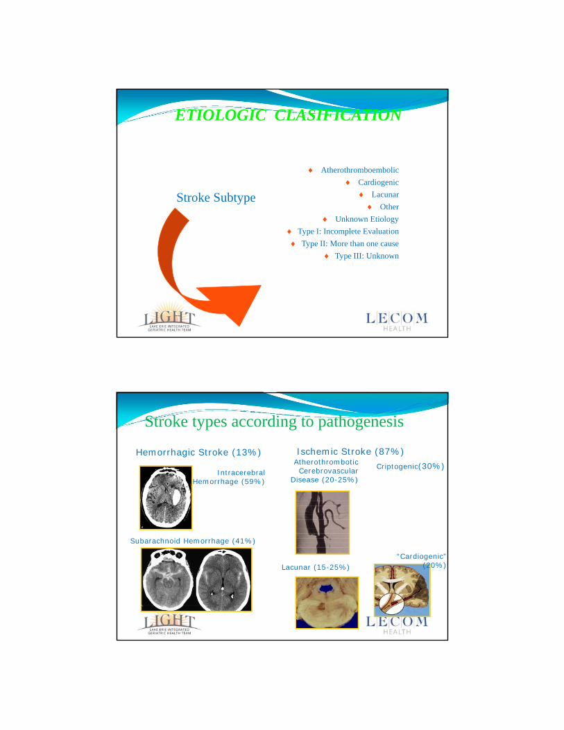
ETIOLOGIC CLASIFICATION
Stroke Subtype
Secondary Prevention
Atherothromboembolic
Cardiogenic
Lacunar
Other
Unknown Etiology
Type I: Incomplete Evaluation
Type II: More than one cause
Type III: Unknown
Ischemic Stroke (87%)Hemorrhagic Stroke (13%)AtherothromboticCerebrovascular
Disease (20-25%)
“Cardiogenic” (20%)Lacunar (15-25%)
IntracerebralHemorrhage (59%)
Subarachnoid Hemorrhage (41%)
Stroke types according to pathogenesis
Criptogenic(30%)
3/2/2016
6
Stroke Fundamentals and Beyond
“Lacunes, Lacunar Infarcts and Small vessel disease”
Definition of Lacunar Infarct
Occlusion of the deep penetrating small arterioles
Epidemiology Background: Stroke Worldwide
Second most common cause of death (6.7 million deaths annually)
Severe disabilities among the survivors. (30 million).
3/2/2016
7
Epidemiology Background: Stroke in the U.S.
Fifth leading cause of death
800,000 new or recurrent strokes per year
130,000 deaths per year
Epidemiology Background: Stroke in the U.S.
An estimate 6.8 million people are living in the U.S. following a stroke.
Leading cause of serious, long-term disability
3/2/2016
8
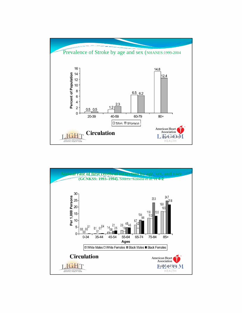
Copyright ©2007 American Heart Association
Rosamond, W. et al. Circulation 2007;115:e69-e171
Prevalence of Stroke by age and sex (NHANES:1999-2004)
Copyright ©2007 American Heart Association
Rosamond, W. et al. Circulation 2007;115:e69-e171
Annual rate of first cerebral infarction by age, sex, and race (GCNKSS: 1993–1994). Source: Kissela et al.1rt 4-2
3/2/2016
9
Epidemiology Background: Economic Impact
The current cost of stroke in the US is $34 Billion
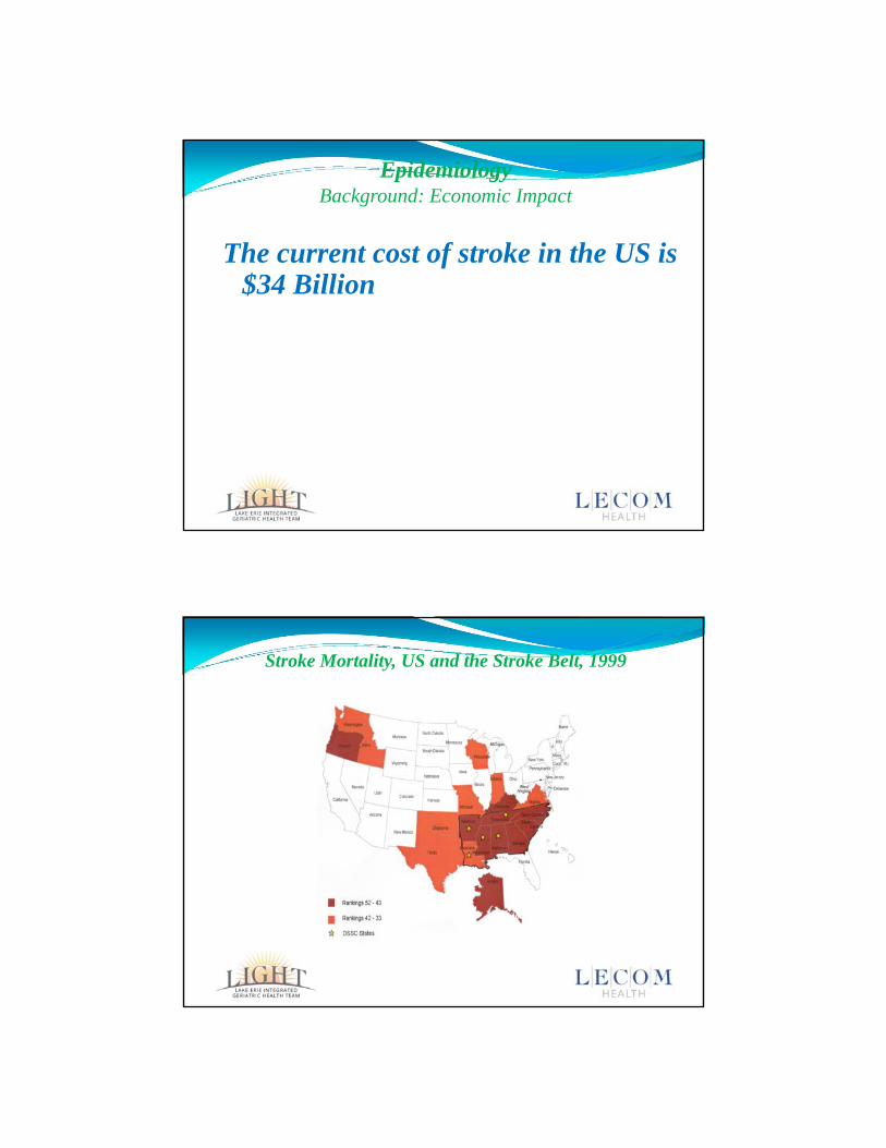
Stroke Mortality, US and the Stroke Belt, 1999
Delta Stroke Consortium States
Top 10 mortality rates
2nd 10 mortality rates
Source: American Heart Association, 2003
3/2/2016
10
Warning Signs of Stroke
Sudden Weakness of Arm , Leg, face
Sudden Sensory Loss
Sudden Speech Abnormalities
Sudden and Unusual Headache
Sudden Dizziness or Loss of Balance
Sudden Loss of Vision or Double Vision
Warning Signs of Stroke
Limb shaking TIA’s
Drop Attacks
Amauroxis fugax
Anton’s syndrome (denial of cortical blindness)
Posterior circulation
Abulia
3/2/2016
11
Stroke Mimics
Tumors “ Pseudo stroke”
Disorders of metabolism Glucose disorders
Dehydration
Migraine attack
Seizures
Conversion
Infections
Recurrent symptoms of prior infarction
Questions to ask when evaluating a stroke patient
Does the patient have a stroke?
Hemorrhage v/s ischemia?
Localization? Small vessel v/s large vessels
Cortical v/s sub cortical
Anterior v/s posterior
What is the likelihood of severe disability or mortality ?
What is the likelihood of clinical deterioration and co morbidities?
3/2/2016
12
Stroke Localization - Cortical Symptoms of Stroke
“Small vessel v/s large vessels”
“Cortical versus subcortical”Aphasia ( can the patient repeat )
Neglect
Extinction
Spatial disorientation / acalculia
Face arm v/s face arm and leg
Graphystesia, tow point discrimination
Horizontal gaze preference
Hemianopsia
Stroke SyndromesLarge vesselsMCA
PCA
ACA / RAH
BA syndrome
Small vesselsPurely motor
Purely sensory
Dysarthria clumsy hand
III nerve +
Ataxic hemiparesis
3/2/2016
13
Xmiclotr.mpg
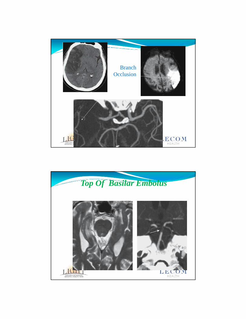
BranchOcclusion
Top Of Basilar Embolus
Xmiclotr.mpg
3/2/2016
14
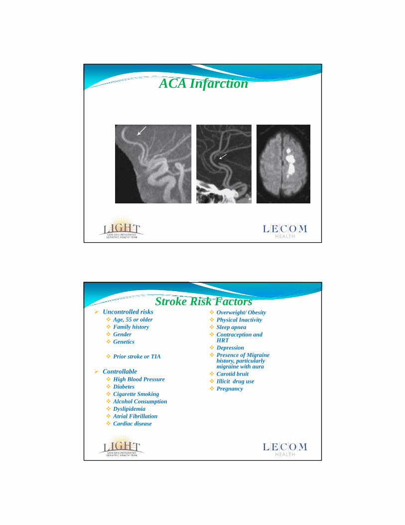
ACA Infarction
Stroke Risk Factors Uncontrolled risks
Age, 55 or older Family history Gender Genetics
Prior stroke or TIA
Controllable High Blood Pressure Diabetes Cigarette Smoking Alcohol Consumption Dyslipidemia Atrial Fibrillation Cardiac disease
Overweight/ Obesity Physical Inactivity Sleep apnea Contraception and
HRT Depression Presence of Migraine
history, particularly migraine with aura
Carotid bruit Illicit drug use Pregnancy
3/2/2016
15
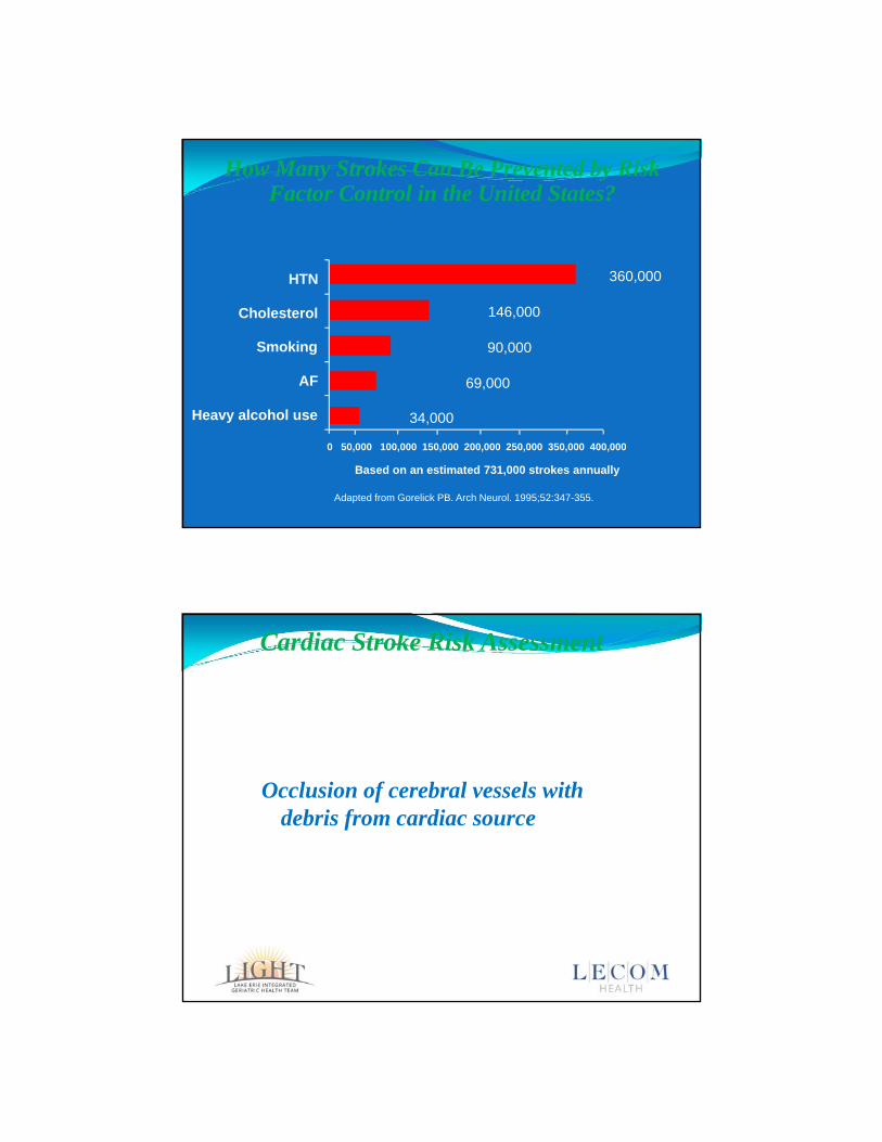
How Many Strokes Can Be Prevented by Risk Factor Control in the United States?
0 50,000 100,000 150,000 200,000 250,000 350,000 400,000
Based on an estimated 731,000 strokes annually
Heavy alcohol use
AF
Smoking
Cholesterol
HTN 360,000
146,000
90,000
69,000
34,000
Adapted from Gorelick PB. Arch Neurol. 1995;52:347-355.
Cardiac Stroke Risk Assessment
Occlusion of cerebral vessels with debris from cardiac source
3/2/2016
16
Cardiac Stroke Risk Assessment
Debris;Platelet aggregates
Thrombus
Platelet-thrombi
Cholesterol
Calcium
Bacterial
Neoplastic cells form Mixomatous material
Cardiac Stroke Risk Assessment
20 % of cardiac embolus goes to brain
80 % of cardiac embolus involved anterior circulation
20 % vertebro –basilar system
3/2/2016
17
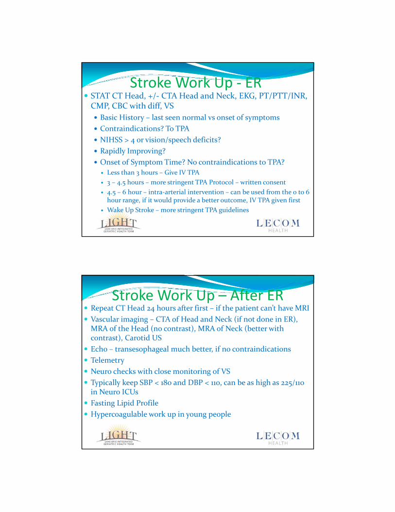
Stroke Work Up ‐ ER STAT CT Head, +/‐ CTA Head and Neck, EKG, PT/PTT/INR, CMP, CBC with diff, VS
Basic History – last seen normal vs onset of symptoms
Contraindications? To TPA
NIHSS > 4 or vision/speech deficits?
Rapidly Improving?
Onset of Symptom Time? No contraindications to TPA?
Less than 3 hours – Give IV TPA
3 – 4.5 hours – more stringent TPA Protocol – written consent
4.5 – 6 hour – intra‐arterial intervention – can be used from the 0 to 6 hour range, if it would provide a better outcome, IV TPA given first
Wake Up Stroke – more stringent TPA guidelines
Stroke Work Up – After ER Repeat CT Head 24 hours after first – if the patient can’t have MRI
Vascular imaging – CTA of Head and Neck (if not done in ER), MRA of the Head (no contrast), MRA of Neck (better with contrast), Carotid US
Echo – transesophageal much better, if no contraindications
Telemetry
Neuro checks with close monitoring of VS
Typically keep SBP < 180 and DBP < 110, can be as high as 225/110 in Neuro ICUs
Fasting Lipid Profile
Hypercoagulable work up in young people
3/2/2016
18
Stroke Work Up – After ER
30 day cardiac monitor as an outpatient – quickly going to become a standard of care – a lot of missed Afib
Stroke Treatment No ASA or Anticoagulation for 24 hours following TPA administration –
prevents hemorrhagic conversion
Initiating or changing antiplatelet therapy
ASA 81 mg Qdaily is indicated – doesn’t need to be higher, studies were done on 50 mg dosing
Switching to Clopidorgrel or aspirin/extended‐release dipyrdamole
aspirin/extended‐release dipyrdamole not used much any more due to it being difficult to tolerate and some studies show it may be better for small vessel disease
Anticoagulation – Warfarin, Heparin, Lovenox, vs new ones – dabigatran etexilate, rivaroxaban, apixaban, edoxaban
Used under certain circumstances, stroke size is factor
3/2/2016
19
Stroke Treatment Initiate statins for hyperlipidemia, mainly LDL > 99, show reduction in stroke
risk
Initiation or changing of BP meds, slowly normalize back to normal
ACE inhibitors show reduction in stroke risk
Avoidance on clonidine – can have profound rebound HTN
Nicardipine GTT – can be used to control malignant HTN well
Keep blood glucose close to normal
Hydrate – NS works best
Avoid opiates and sedatives in the first 72 hours of stroke to avoid side effects that mimic stroke worsening
Good Nutrition – favor PEG over NG tube
Stroke Treatment
Treatment of any infections – studies show that infections can cause enough inflammation in the body to potentially cause strokes
Some institutions don’t vaccinate during stroke admissions for this reason
Treatment of underlying medical problems – in order to maximize rehab potential
Treatment of PFOs, vascular problems
Treat stroke induced depression – improves rehab potential – may need psychiatry and neuropsychology
Explain to family, goals of treatment is to prevent the next stroke, we can only “support” the active stroke, if the patient was not given TPA or had an intra‐arterial intervention
3/2/2016
20
Stroke Treatment Initiate rehab – PT, OT, and ST
Watch for endurance with therapies and monitor therapists recommendations for rehab need and level following inpatient discharge
Does rehab suggest assistive devices such as: canes, walkers, wheelchairs, orthotics, splints, etc
Assess for spasticity vs flaccidity – will the patient need spasticity reduction with: oral meds, botulinum toxin, or baclofen pump vs will they need splinting for flaccidity?
Monitor the patient’s mood and apathy – do they want to get better? Do they want therapy? Do they want to give up and die?
TIAOld Definition Time base
1960
Misleading
New definitionTissue base
Sense of urgency
Optimal risk assessment
2009 definition
3/2/2016
21
2009 Definition of TIA “TIAs are brief episodes of neurological dysfunction resulting from focal cerebral ischemia not associated with a permanent cerebral infarction.”
Definition and Evaluation of Transient Ischemic Attack: A Scientific Statement Healthcare Professionals From the American Heart Association/American Stroke Association Stroke Council; Council on Cardiovascular Surgery and Anesthesia ‐ Stroke. 2009;40:2276‐2293; originally published online May 7, 2009;
ACUTE VASCULAR SYNDROME
Any acute onset of focal neurological deficit from brain, cord or retina of presume vascular origin which is undefined as either a TIA or Stroke due to pending evaluation
3/2/2016
22
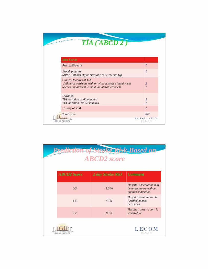
TIA ( ABCD 2 )
Risk Factor
Age > 60 years 1
Blood pressure SBP > 140 mm Hg or Diastolic BP > 90 mm Hg
1
Clinical features of TIA Unilateral weakness with or without speech impairment Speech impairment without unilateral weakness
21
Duration TIA duration > 60 minutesTIA duration 10- 59 minutes
21
History of DM 1
Total score 0-7
Prediction of Stroke Risk Based on ABCD2 score
ABCD2 Score 2 day Stroke Risk Comment
0-3 1.0 % Hospital observation may be unnecessary without another indication
4-5 4.1% Hospital observation is justified in most occasions
6-7 8.1% Hospital observation is worthwhile
Lancet , 369:283-292,2007
3/2/2016
23
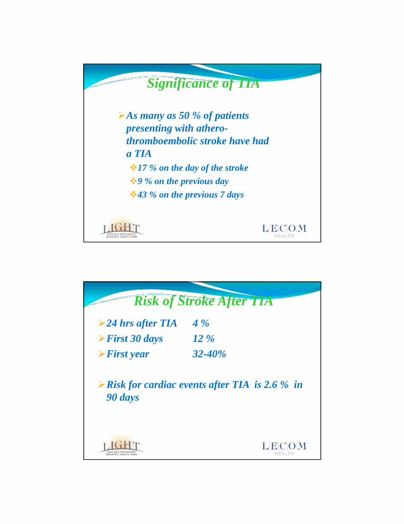
Significance of TIA
As many as 50 % of patients presenting with athero-thromboembolic stroke have had a TIA 17 % on the day of the stroke
9 % on the previous day
43 % on the previous 7 days
Risk of Stroke After TIA
24 hrs after TIA 4 %
First 30 days 12 %
First year 32-40%
Risk for cardiac events after TIA is 2.6 % in 90 days
3/2/2016
24
Management of TIA
Imaging evaluation within 24 hrs
Electrocardiography ( ASAP )
Prolonged cardiac monitoring ( ASAP)
Echocardiography (ASAP )
Admit to hospital if:If symptoms < 72 hrs and ABCD > 3
Evaluation can’t be obtained in a timely manner
Work up and treatment for TIA is very similar to a stroke, but with a TIA, the patient’s deficits have resolved, so typically, rehab is not necessary for the “new symptoms.”
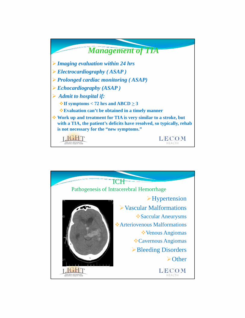
ICHPathogenesis of Intracerebral Hemorrhage
Hypertension
Vascular MalformationsSaccular Aneurysms
Arteriovenous Malformations
Venous Angiomas
Cavernous Angiomas
Bleeding Disorders
Other
3/2/2016
25
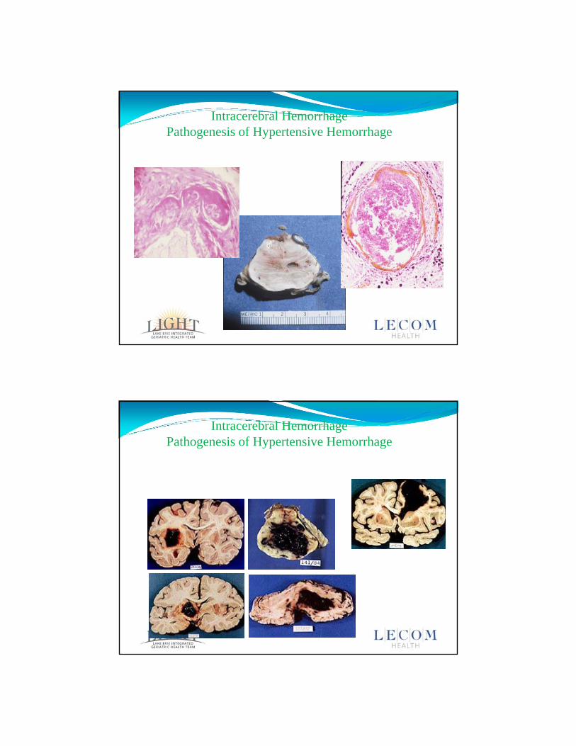
Intracerebral HemorrhagePathogenesis of Hypertensive Hemorrhage
Intracerebral HemorrhagePathogenesis of Hypertensive Hemorrhage
3/2/2016
26
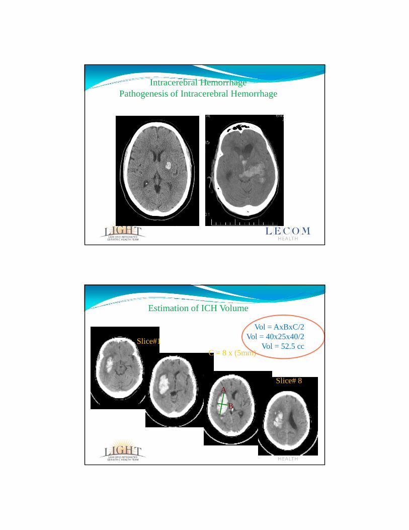
Intracerebral HemorrhagePathogenesis of Intracerebral Hemorrhage
C = 8 x (5mm)
Slice#1
Slice# 8A
B
Vol = AxBxC/2Vol = 40x25x40/2
Vol = 52.5 cc
Estimation of ICH Volume
3/2/2016
27
SAH
.
SAHEpidemiology
5 % of all strokes Incidence increases with age, median (50-60yrs) Women/men 3:2 in> 40 yrs old. Man > woman
< 40 yrs African Americans 10,000 pts die prior to hospital arrival Of patient admitted to 50 % death or severe
disability Usually caused by a ruptured aneurysm
3/2/2016
28
SAHNon Aneurysmal Causes
Trauma Arterial dissection Mycotic Aneurysm (Infected) Arteriovenous malformation Cocaine and amphetamine use Moyamoya disease Central venous system vasculitis Idiopathic perimensencephalic nonaneurysmal
subarachnoid hemorrhage (PM-NASAH) – studies show that 15-20%of SAH do not have a vascular lesion in a 4 vessel cerebral angiogram (seen in increased ICP)
Aneurysmal location 80 % in anterior cerebral circulation
3/2/2016
29
SAHRisk factors
Alcohol
Transient hypertension
Hypertension
Smoking
Hormonal
Postmenopausal woman,decreased risk in patients of HRT
.
Risk factors Non modifieble
Family history ( 4 fold ) AD polycystic kidney disease Marfan’s Syndrome Ehler- Danlos syndrome Fibromuscular displasia Pseudoxanthoma elasticum Coartation of Aorta
Patients with at least two first defgree family menbers or AD PKD sould undergo screeaning
3/2/2016
30
Treatment of ICH Stabilize ABCs and VS
Reversal of offending agent, if present, FFP, Vitamin K, idarucizumab (if available in your institution), etc
Transfer of patient to a neurosurgical institution for potential evacuation, repair of trauma and transfer to neuro ICU, if indicated
Decrease BP if elevated
Monitor for vasospasm
Discover cause, if not known, to prevent a repeat bleed
Treat other medical problems if present
Once stable, start rehab, PT, OT, ST
Imaging Techniques
3/2/2016
31
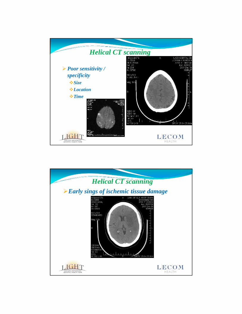
Helical CT scanning
Poor sensitivity / specificitySize
Location
Time
Helical CT scanningEarly sings of ischemic tissue damage
3/2/2016
32
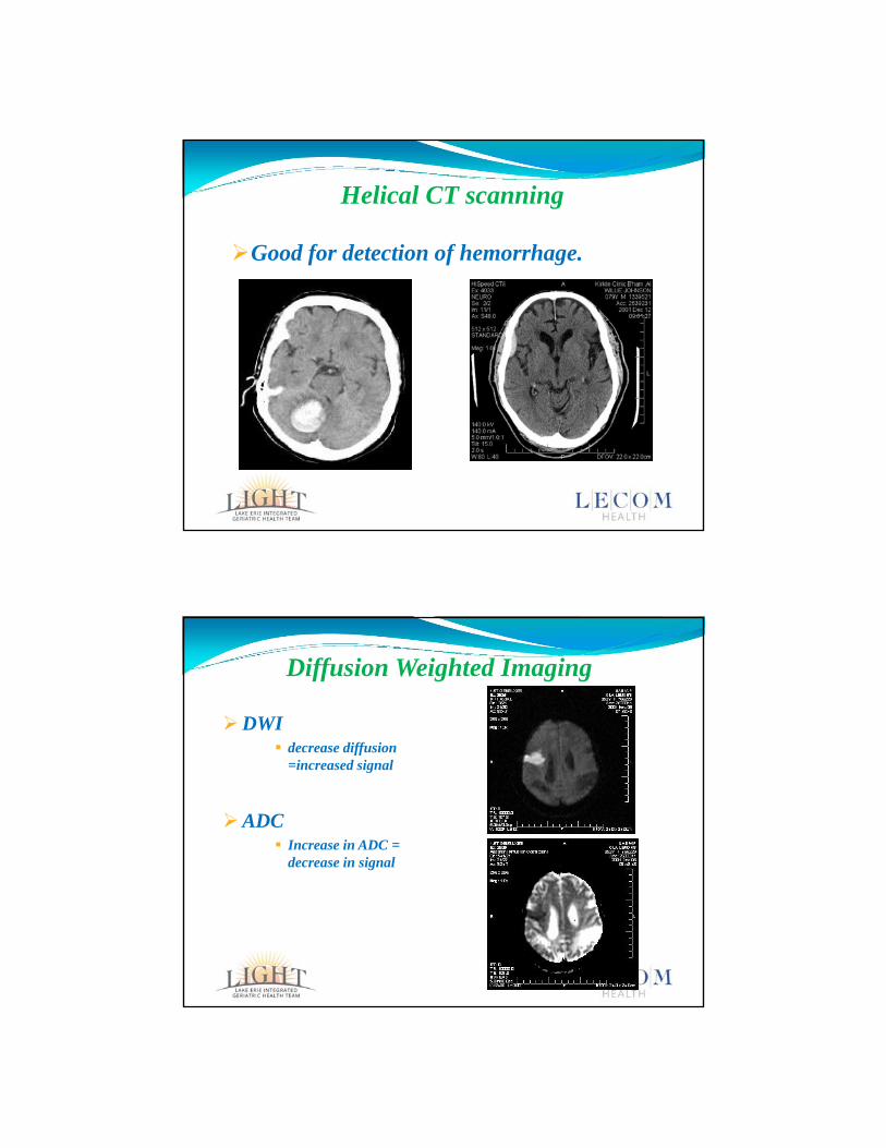
Helical CT scanning
Good for detection of hemorrhage.
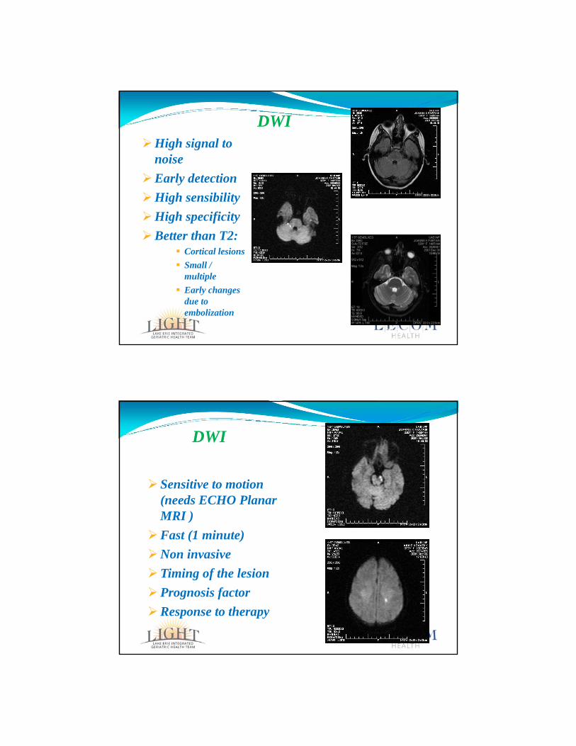
Diffusion Weighted Imaging
DWI decrease diffusion
=increased signal
ADC Increase in ADC =
decrease in signal
3/2/2016
33
DWIHigh signal to
noise
Early detection
High sensibility
High specificity
Better than T2: Cortical lesions
Small / multiple
Early changes due to embolization
DWI
FLAIR
T2
DWI
Sensitive to motion (needs ECHO Planar MRI )
Fast (1 minute)
Non invasive
Timing of the lesion
Prognosis factor
Response to therapy
3/2/2016
34
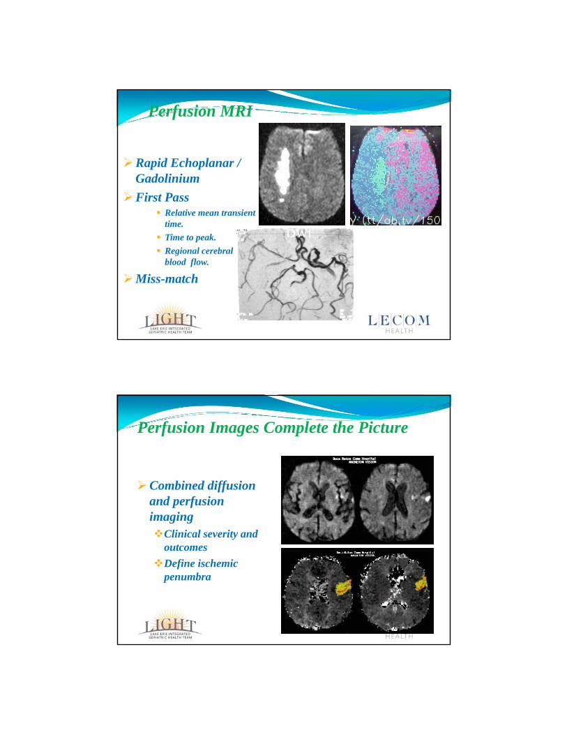
Perfusion MRI
Rapid Echoplanar / Gadolinium
First Pass Relative mean transient
time.
Time to peak.
Regional cerebral blood flow.
Miss-match
MRA
PIDWI
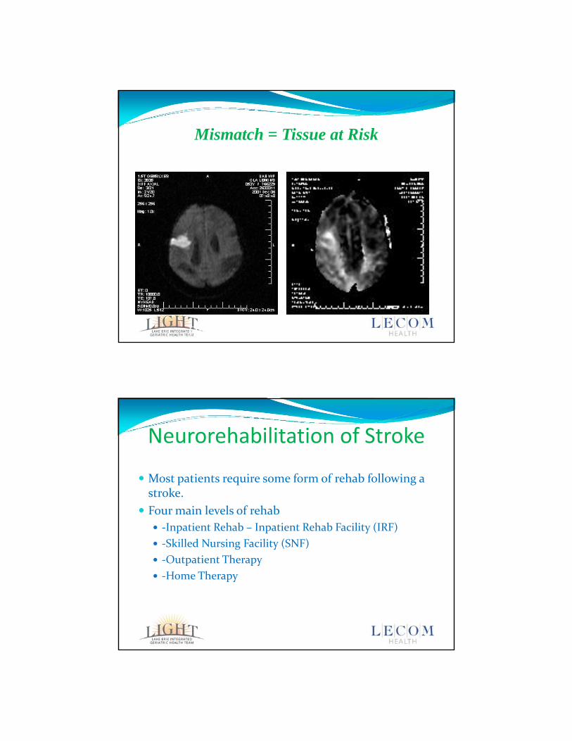
Perfusion Images Complete the Picture
Combined diffusion and perfusion imagingClinical severity and
outcomes
Define ischemic penumbra
3/2/2016
35
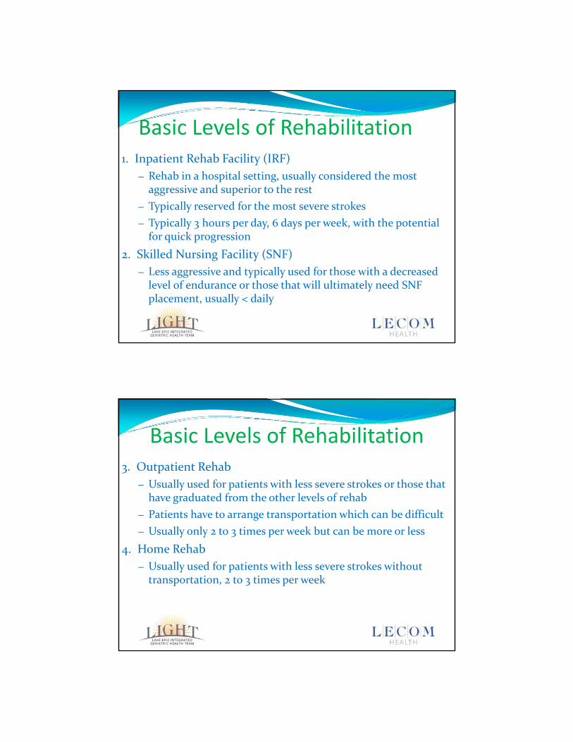
Mismatch = Tissue at Risk
DWI PI
Neurorehabilitation of Stroke
Most patients require some form of rehab following a stroke.
Four main levels of rehab
‐Inpatient Rehab – Inpatient Rehab Facility (IRF)
‐Skilled Nursing Facility (SNF)
‐Outpatient Therapy
‐Home Therapy
3/2/2016
36
Basic Levels of Rehabilitation1. Inpatient Rehab Facility (IRF)
– Rehab in a hospital setting, usually considered the most aggressive and superior to the rest
– Typically reserved for the most severe strokes
– Typically 3 hours per day, 6 days per week, with the potential for quick progression
2. Skilled Nursing Facility (SNF)
– Less aggressive and typically used for those with a decreased level of endurance or those that will ultimately need SNF placement, usually < daily
Basic Levels of Rehabilitation3. Outpatient Rehab
– Usually used for patients with less severe strokes or those that have graduated from the other levels of rehab
– Patients have to arrange transportation which can be difficult
– Usually only 2 to 3 times per week but can be more or less
4. Home Rehab
– Usually used for patients with less severe strokes without transportation, 2 to 3 times per week
3/2/2016
37
Predictors of Rehab Potential In the rehab world these are called “barriers” to rehab
Some patients are great candidates for rehab and others are not.
• There are many limitations/barriers for neurorehabilitation potential in patients with stroke and other neurological diseases
– It's important to consider these when ordering rehab for our patients
Knowing and recognizing all of a patient’s rehab barriers, will allow you to predict a patient’s rehab potential and ultimately their prognosis.
Barriers to Neurorehabilitation
The number one barrier to rehab, for any disease, is the patient’s desire to participate in rehab
In the elderly, if the patient does want therapy and their kids do, this patient’s rehab potential and prognosis will be very poor.
It’s also important to choose rehab close to family, loneliness from friends and family can be a barrier
Convenience to home can be a barrier
3/2/2016
38
Barriers to Neurorehabilitation• Age can be large limitation/barrier to rehab, it can not be
modified, of course, however, it should be recognized
• Most studies show that older patients have a poorer prognosis for recovery from stroke when compared to younger patients
• One of the biggest limiting factors for patients is endurance.
– Following a stroke, patients typically need PT, OT, and ST
– For inpatient therapy, plan 1 hour for each type of therapy, thus 3 hours per day
Barriers to Neurorehabilitation
• Remember, 3 hours a day of exercise can be difficult for “normal people” let alone stroke patients, think about this when ordering inpatient therapy
• Next limitation is co‐morbidities:
– COPD, CAD/MI, Seizures, PD, HTN, Depression, Anxiety, OSA, DM, DVTs, Infections, smoking/addictions, etc
– When under control these are OK with rehab but therapies can be interrupted greatly when they are not
3/2/2016
39
Barriers to Neurorehabilitation
• Another limitation is nutrition, patients need adequate nutrition to rehab effectively, if dysphagic, we recommend PEG tube over NG and TPN for much better nutrition, poor nutrition usually means poor rehab
Barriers to Neurorehabilitation• Depression can limit rehab greatly, and strokes typically
cause organic depression, if treated, patients typically rehab much better
• Mental status can limit rehab greatly too, obviously, a severely sedated person won’t be able to participate in rehab but a perfectly healthy demented patient, who can’t follow any commands, will not rehab well either, if at all
3/2/2016
40
Barriers to Neurorehabilitation• Other psychiatric issues can play a huge role in how well a
patient rehabs
– Typically we consult a neuropsychologist to help in assessing for these problems, which occur more often than you think and were never diagnosed before
• Pure sedation from a nondominant (usually right hemispheric stroke) can be a major limitation to rehab and we can give alerting medications for this
Barriers to Neurorehabilitation• Sleep is a major limitation in therapy following a stroke, we
need to start checking for more OSA following strokes
– Day‐Night Confusion falls into this category too
• Finally, some medications can be limiting factors in patients, mainly side effects
– Usually we gear med changes around keeping the patient awake and able to participate in therapy
– Abused drugs also fall into this category, ie cocaine, meth, ETOH
3/2/2016
41
Barriers to Neurorehabilitation• It’s important to realize that inpatient rehab is not for
everyone, especially if we can’t change or treat these limitations
• Also realize that patients who can’t tolerate inpatient rehab initially, may tolerate other forms first, and once they are “strong enough,” we can admit to the inpatient unit.
• If these limitations are addressed prior to admission to the inpatient unit, the patient will rehab better and faster
Barriers to NeurorehabilitationWho is a neurorehabilitation candidate?
Anyone with a persistent neurological deficit who wants it to resolve
This is true for strokes or any other type of neurological diseases
It also helps to choose patients who want to get better, if they don't, they won't get better
Some of the worst rehab candidates are the one's who have their families choose for them
3/2/2016
42
Barriers to Neurorehabilitation
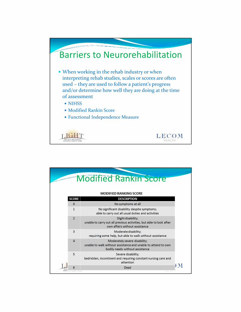
When working in the rehab industry or when interpreting rehab studies, scales or scores are often used – they are used to follow a patient’s progress and/or determine how well they are doing at the time of assessment
NIHSS
Modified Rankin Score
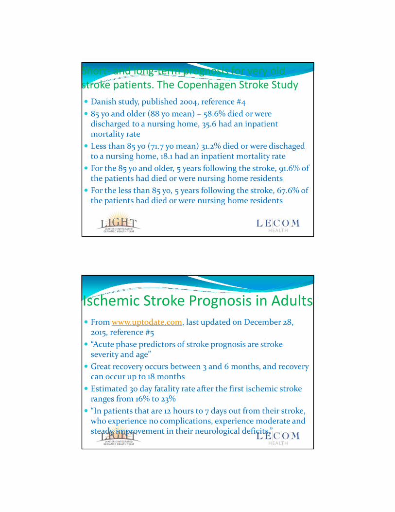
Functional Independence Measure
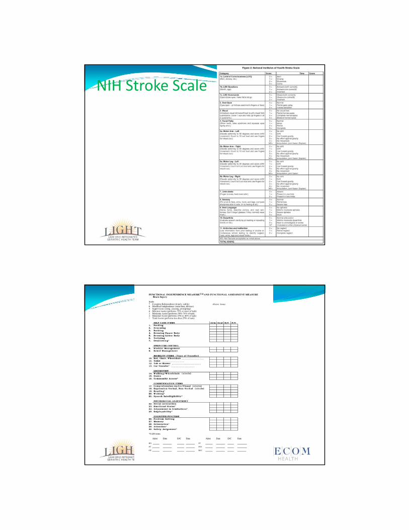
Modified Rankin Score
3/2/2016
43
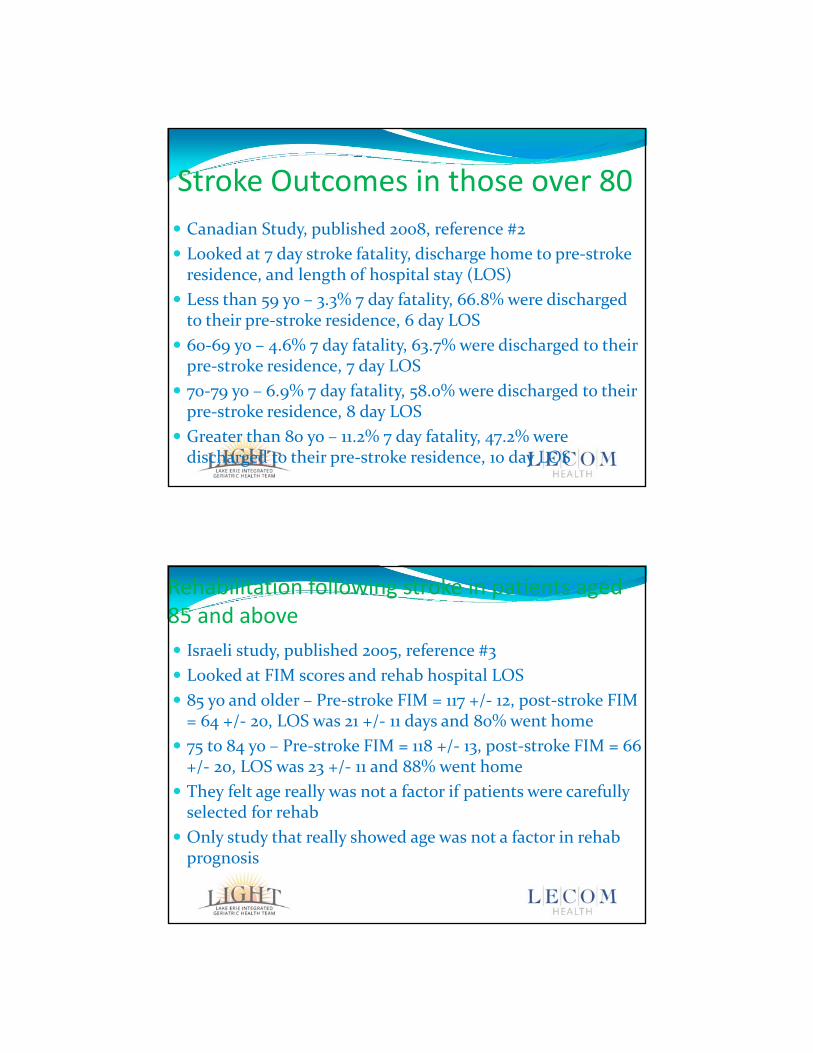
NIH Stroke Scale
3/2/2016
44
Stroke Outcomes in those over 80
Canadian Study, published 2008, reference #2
Looked at 7 day stroke fatality, discharge home to pre‐stroke residence, and length of hospital stay (LOS)
Less than 59 yo – 3.3% 7 day fatality, 66.8% were discharged to their pre‐stroke residence, 6 day LOS
60‐69 yo – 4.6% 7 day fatality, 63.7% were discharged to their pre‐stroke residence, 7 day LOS
70‐79 yo – 6.9% 7 day fatality, 58.0% were discharged to their pre‐stroke residence, 8 day LOS
Greater than 80 yo – 11.2% 7 day fatality, 47.2% were discharged to their pre‐stroke residence, 10 day LOS
Rehabilitation following stroke in patients aged 85 and above
Israeli study, published 2005, reference #3
Looked at FIM scores and rehab hospital LOS
85 yo and older – Pre‐stroke FIM = 117 +/‐ 12, post‐stroke FIM = 64 +/‐ 20, LOS was 21 +/‐ 11 days and 80% went home
75 to 84 yo – Pre‐stroke FIM = 118 +/‐ 13, post‐stroke FIM = 66 +/‐ 20, LOS was 23 +/‐ 11 and 88% went home
They felt age really was not a factor if patients were carefully selected for rehab
Only study that really showed age was not a factor in rehab prognosis
3/2/2016
45
Short‐ and long‐term prognosis for very old stroke patients. The Copenhagen Stroke Study
Danish study, published 2004, reference #4
85 yo and older (88 yo mean) – 58.6% died or were discharged to a nursing home, 35.6 had an inpatient mortality rate
Less than 85 yo (71.7 yo mean) 31.2% died or were dischaged to a nursing home, 18.1 had an inpatient mortality rate
For the 85 yo and older, 5 years following the stroke, 91.6% of the patients had died or were nursing home residents
For the less than 85 yo, 5 years following the stroke, 67.6% of the patients had died or were nursing home residents
Ischemic Stroke Prognosis in Adults From www.uptodate.com, last updated on December 28, 2015, reference #5
“Acute phase predictors of stroke prognosis are stroke severity and age”
Great recovery occurs between 3 and 6 months, and recovery can occur up to 18 months
Estimated 30 day fatality rate after the first ischemic stroke ranges from 16% to 23%
“In patients that are 12 hours to 7 days out from their stroke, who experience no complications, experience moderate and steady improvement in their neurological deficits.”
3/2/2016
46
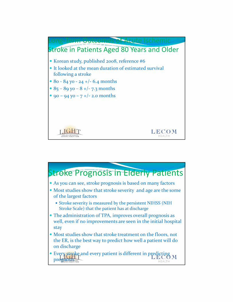
Long‐Term Outcomes of Acute Ischemic Stroke in Patients Aged 80 Years and Older
Korean study, published 2008, reference #6
It looked at the mean duration of estimated survival following a stroke
80 ‐ 84 yo ‐ 24 +/‐ 6.4 months
85 – 89 yo – 8 +/‐ 7.3 months
90 – 94 yo – 7 +/‐ 2.0 months
Stroke Prognosis in Elderly Patients As you can see, stroke prognosis is based on many factors
Most studies show that stroke severity and age are the some of the largest factors
Stroke severity is measured by the persistent NIHSS (NIH Stroke Scale) that the patient has at discharge
The administration of TPA, improves overall prognosis as well, even if no improvements are seen in the initial hospital stay
Most studies show that stroke treatment on the floors, not the ER, is the best way to predict how well a patient will do on discharge
Every stroke and every patient is different in predicting prognosis

3/2/2016
47
Questions?