Embed Size (px)
Citation preview
Strip of the Month: December 2016Lena Braginsky, MD,* Beth A. Plunkett, MD*
*Department of Obstetrics and Gynecology, NorthShore University HealthSystem, Evanston Hospital,
Evanston, IL.
ELECTRONIC FETAL MONITORING CASE REVIEW SERIES
Electronic fetal monitoring (EFM) is a popular technology used to establish fetal
well-being. Despite its widespread use, the terminology used to describe patterns
seen on the monitor has not been consistent until recently. In 1997, the National
Institute of Child Health and Human Development (NICHD) Research Plan-
ning Workshop published guidelines for interpretation of fetal tracings. This
publication was the culmination of 2 years of work by a panel of experts in the
field of fetal monitoring, and was endorsed in 2005 by both the American
College of Obstetricians and Gynecologists (ACOG) and the Association of
Women’s Health, Obstetric and Neonatal Nurses (AWHONN). In 2008, ACOG,
NICHD, and the Society for Maternal-Fetal Medicine reviewed and updated
the definitions for fetal heart rate (FHR) patterns, interpretation, and research
recommendations. Following is a summary of the terminology definitions and
assumptions found in the 2008 NICHD workshop report. Normal values for
arterial umbilical cord gas values and indications of acidosis are defined in
Table 1.
Assumptions from the NICHD Workshop• Definitions are developed for visual interpretation, assuming that both the FHR
and uterine activity recordings are of adequate quality• Definitions apply to tracings generated by internal or external monitoring
devices• Periodic patterns are differentiated based on waveform, abrupt or gradual (eg,
late decelerations have a gradual onset and variable decelerations have an
abrupt onset)
• Long- and short-term variability are evaluated visually as a unit
• Gestational age of the fetus is considered when evaluating patterns• Components of FHR do not occur alone and generally evolve over time
DEFINITIONS
Baseline Fetal Heart Rate• Approximate mean FHR rounded to increments of 5 beats per minute in a 10-
minute segment of tracing, excluding accelerations and decelerations, periods
of marked variability, and segments of baseline that differ by >25 beats per
minute
• In the 10-minute segment, the minimum baseline duration must be at least
2 minutes (not necessarily contiguous) or the baseline for that segment is
indeterminate
• Bradycardia is a baseline of <110 beats per minute; tachycardia is a baseline of
>160 beats per minute
AUTHOR DISCLOSURE Drs Braginskyand Plunkett have disclosed no financialrelationships relevant to this article. Thiscommentary does not contain a discussion ofan unapproved/investigative use of acommercial product/device.
Vol. 17 No. 12 DECEMBER 2016 e753
Strip of the Month
by 165225 on October 21, 2018http://neoreviews.aappublications.org/Downloaded from
• Sinusoidal baseline has a smooth sine wave–like undu-
lating pattern, with waves having regular frequency and
amplitude
Baseline Variability• Fluctuations in the baseline FHR of ‡2 cycles per minute,
fluctuations are irregular in amplitude and frequency,
fluctuations are visually quantitated as the amplitude of
the peak to trough in beats per minute• Classification of variability:
Absent: Amplitude range is undetectable
Minimal: Amplitude range is greater than undetectable to
5 beats per minute
Moderate: Amplitude range is 6–25 beats per minute
Marked: Amplitude range is >25 beats per minute
Accelerations• Abrupt increase in FHR above the most recently deter-
mined baseline• Onset to peak of acceleration is <30 seconds, acme is ‡15beats per minute above the most recently determined
baseline and lasts ‡15 seconds but <2 minutes
• Before 32 weeks’ gestation, accelerations are defined by
an acme ‡10 beats per minute above the most recently
determined baseline for ‡10 seconds• Prolonged acceleration lasts ‡2minutes but<10minutes
Late Decelerations• Gradual decrease in FHR (onset to nadir ‡30 seconds)
below the most recently determined baseline, with nadir
occurring after the peak of uterine contractions• Considered a periodic pattern because it occurs with
uterine contractions
Early Decelerations• Gradual decrease in FHR (onset to nadir ‡30 seconds)
below the most recently determined baseline, with nadir
occurring coincident with uterine contraction
• Also considered a periodic pattern
Variable Decelerations• Abrupt decrease in FHR (onset to nadir <30 seconds)
• Decrease is ‡15 beats per minute below the most recently
determined baseline lasting ‡15 seconds but <2 minutes
• May be episodic (occurs without a contraction) or
periodic
Prolonged Decelerations• Decrease in the FHR ‡15 beats perminute below themost
recently determined baseline lasting ‡2 minutes but <10
minutes from onset to return to baseline
∘ Decelerations are tentatively called recurrent if they
occur with ‡50% of uterine contractions in a 20-minute
period
∘ Decelerations occurring with <50% of uterine con-
tractions in a 20-minute segment are intermittent
Sinusoidal Fetal Heart Rate Pattern• Visually apparent, smooth sine wave–like undulating
pattern in the baseline with a cycle frequency of 3 to 5 per
minute that persists for ‡20 minutes
Uterine Contractions• Quantified as the number of contractions in a 10-minute
window, averaged over 30 minutes
∘ Normal: £5 contractions in 10 minutes
∘ Tachysystole: >5 contractions in 10 minutes
INTERPRETATION
A 3-tier FHR Interpretation system has been recommended
as follows:
• Category I FHR tracings: Normal, strongly predictive of
normal fetal acid-base status and require routine care.
These tracings include all of the following:
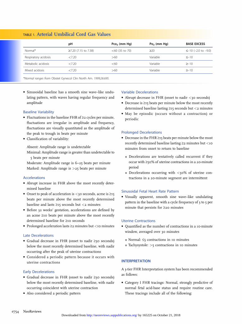
TABLE 1. Arterial Umbilical Cord Gas Values
pH Pco2 (mm Hg) Po2 (mm Hg) BASE EXCESS
Normal* ‡7.20 (7.15 to 7.38) <60 (35 to 70) ‡20 £–10 (–2.0 to –9.0)
Respiratory acidosis <7.20 >60 Variable £–10
Metabolic acidosis <7.20 <60 Variable ‡–10
Mixed acidosis <7.20 >60 Variable ‡–10
*Normal ranges from Obstet Gynecol Clin North Am. 1999;26:695.
e754 NeoReviews by 165225 on October 21, 2018http://neoreviews.aappublications.org/Downloaded from
� Baseline rate: 110 to 160 beats per minute
� Baseline FHR variability: Moderate
� Late or variable decelerations: Absent
� Early decelerations: Present or absent
� Accelerations: Present or absent
• Category II FHR tracings: Indeterminate, require eval-
uation and continued surveillance and reevaluation.
Examples of these tracings include any of the following:
� Bradycardia not accompanied by absent variability
� Tachycardia
� Minimal or marked baseline variability
� Absent variability without recurrent decelerations
� Absence of induced accelerations after fetal stimulation
� Recurrent variable decelerations with minimal or
moderate variability
� Prolonged decelerations
� Recurrent late decelerations with moderate variability
� Variable decelerations with other characteristics, such
as slow return to baseline
• Category III FHR tracings: Abnormal, predictive of ab-
normal fetal acid-base status and require prompt inter-
vention. These tracings include:
� Absent variability with any of the following:
n Recurrent late decelerations
n Recurrent variable decelerations
n Bradycardia
� Sinusoidal pattern
Data fromMaconesGA,HankinsGDV, SpongCY,Hauth
J, Moore T. The 2008 National Institute of Child Health
and Human Development workshop report on electronic
fetal monitoring. Obstet Gynecocol. 2008;112:661–666 and
American College of Obstetricians and Gynecologists.
Intrapartum fetal heart rate monitoring: nomenclature, in-
terpretation, and general management principles. ACOG
Practice Bulletin No. 106. Washington, DC: American College
of Obstetricians and Gynecologists; 2009.
We encourage readers to examine each strip in the case
presentation and make a personal interpretation of the
findings before advancing to the expert interpretation
provided.
PRESENTATION
HistoryA 37-year-old gravida 5, para 3-0-1-3 at 39 weeks and 0 days’
gestation presented to the labor and delivery department
for scheduled elective induction of labor. She denied any
bleeding, contractions, loss of fluid, or other complaints.
The patient’s pregnancy was complicated by advanced
maternal age. She underwent cell-free fetal DNA screen-
ing, which found her to be at low risk for aneuploidy. She
underwent level II ultrasonography at 19 weeks and 5 days’
gestation, which demonstrated normal fetal anatomy. A
posterior placenta was documented without notable pla-
cental abnormalities. The placental cord insertion was
not well visualized. She had no other prenatal issues. Her
obstetric history was notable for 3 prior full-term normal
spontaneous vaginal deliveries without complications. She
had no significant medical, surgical, social, or family history.
On admission, her blood pressure was 115/62 mmHg,
heart rate 68 beats per minute, and temperature 98.2°F.
A vaginal examination revealed her cervix to be 1-cm di-
lated, 25% effaced, and -3 station. Bedside ultrasonog-
raphy confirmed vertex presentation. Her induction of
labor began using an intracervical Foley catheter with
extra-amniotic saline infusion, concurrent with oxyto-
cin administration as per institutional protocol. The initial
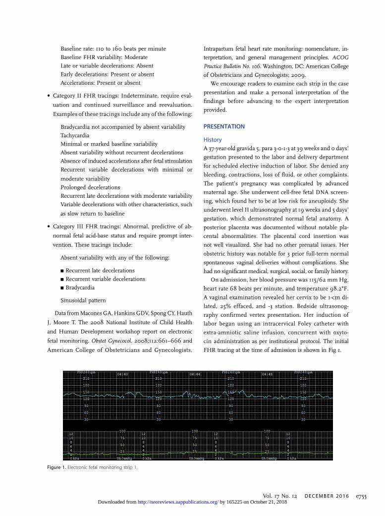
FHR tracing at the time of admission is shown in Fig 1.
Figure 1. Electronic fetal monitoring strip 1.
Vol. 17 No. 12 DECEMBER 2016 e755 by 165225 on October 21, 2018http://neoreviews.aappublications.org/Downloaded from
Findings from EFM strip 1:
• Variability: Moderate
• Baseline rate: 130 beats per minute• Episodic patterns: Accelerations
• Periodic patterns: None• Uterine contractions: None
• Interpretation: Category I• Differential diagnosis: Normally oxygenated infant
• Action: Proceed with induction of labor
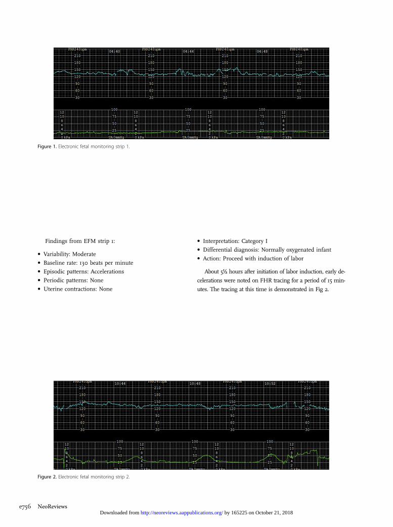
About 5½ hours after initiation of labor induction, early de-
celerations were noted on FHR tracing for a period of 15 min-
utes. The tracing at this time is demonstrated in Fig 2.
Figure 2. Electronic fetal monitoring strip 2.
Figure 1. Electronic fetal monitoring strip 1.
e756 NeoReviews by 165225 on October 21, 2018http://neoreviews.aappublications.org/Downloaded from
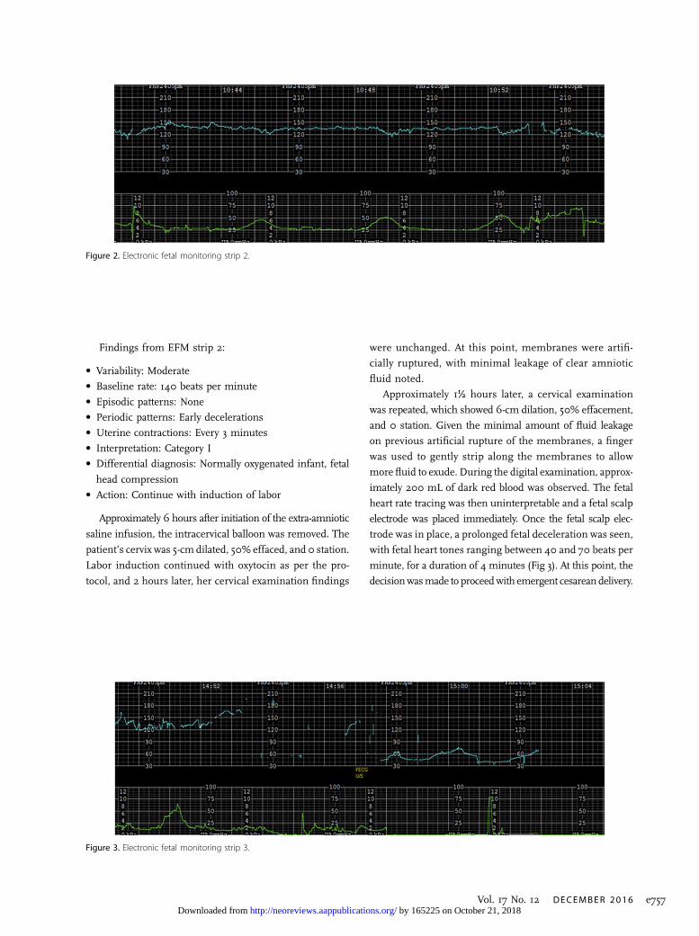
Findings from EFM strip 2:
• Variability: Moderate• Baseline rate: 140 beats per minute
• Episodic patterns: None• Periodic patterns: Early decelerations• Uterine contractions: Every 3 minutes
• Interpretation: Category I• Differential diagnosis: Normally oxygenated infant, fetal
head compression• Action: Continue with induction of labor
Approximately 6 hours after initiation of the extra-amniotic
saline infusion, the intracervical balloon was removed. The
patient’s cervix was 5-cm dilated, 50% effaced, and 0 station.
Labor induction continued with oxytocin as per the pro-
tocol, and 2 hours later, her cervical examination findings
were unchanged. At this point, membranes were artifi-
cially ruptured, with minimal leakage of clear amniotic
fluid noted.
Approximately 1½ hours later, a cervical examination
was repeated, which showed 6-cm dilation, 50% effacement,
and 0 station. Given the minimal amount of fluid leakage
on previous artificial rupture of the membranes, a finger
was used to gently strip along the membranes to allow
more fluid to exude. During the digital examination, approx-
imately 200 mL of dark red blood was observed. The fetal
heart rate tracing was then uninterpretable and a fetal scalp
electrode was placed immediately. Once the fetal scalp elec-
trode was in place, a prolonged fetal deceleration was seen,
with fetal heart tones ranging between 40 and 70 beats per
minute, for a duration of 4 minutes (Fig 3). At this point, the
decisionwasmade to proceedwith emergent cesarean delivery.
Figure 3. Electronic fetal monitoring strip 3.
Figure 2. Electronic fetal monitoring strip 2.
Vol. 17 No. 12 DECEMBER 2016 e757 by 165225 on October 21, 2018http://neoreviews.aappublications.org/Downloaded from
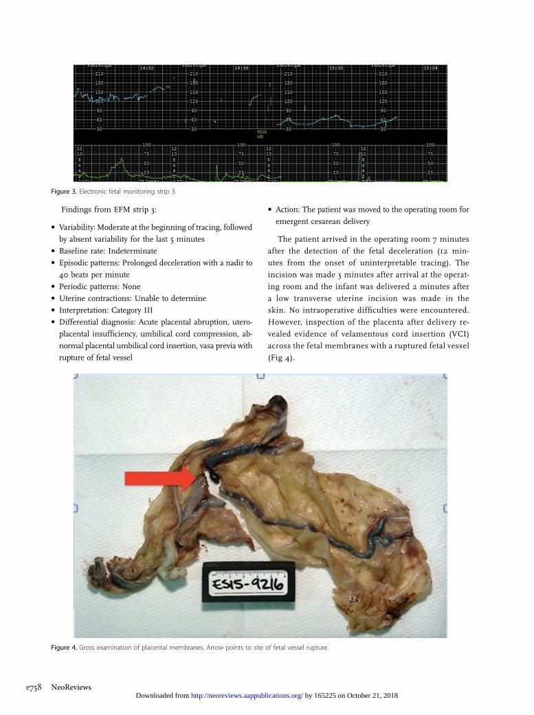
Findings from EFM strip 3:
• Variability: Moderate at the beginning of tracing, followed
by absent variability for the last 5 minutes
• Baseline rate: Indeterminate• Episodic patterns: Prolonged deceleration with a nadir to
40 beats per minute
• Periodic patterns: None• Uterine contractions: Unable to determine
• Interpretation: Category III• Differential diagnosis: Acute placental abruption, utero-
placental insufficiency, umbilical cord compression, ab-
normal placental umbilical cord insertion, vasa previa with
rupture of fetal vessel
• Action: The patient was moved to the operating room for
emergent cesarean delivery
The patient arrived in the operating room 7 minutes
after the detection of the fetal deceleration (12 min-
utes from the onset of uninterpretable tracing). The
incision was made 3 minutes after arrival at the operat-
ing room and the infant was delivered 2 minutes after
a low transverse uterine incision was made in the
skin. No intraoperative difficulties were encountered.
However, inspection of the placenta after delivery re-
vealed evidence of velamentous cord insertion (VCI)
across the fetal membranes with a ruptured fetal vessel
(Fig 4).
Figure 3. Electronic fetal monitoring strip 3.
Figure 4. Gross examination of placental membranes. Arrow points to site of fetal vessel rupture.
e758 NeoReviews by 165225 on October 21, 2018http://neoreviews.aappublications.org/Downloaded from
OutcomeA viable male infant weighing 3,070 g (6 lb 12 oz) was de-
livered via emergent cesarean delivery. The infant was limp
and pale, and had apnea initially, with a heart rate of 40 to
60 beats per minute. The heart rate became undetectable
1 minute after birth. Cardiac compressions were initiated
with positive pressure ventilation. The infant underwent
mechanical intubation and received epinephrine with-
out significant improvement in heart rate. At 7 minutes
after birth, an umbilical artery catheter was placed and
epinephrine was administered through the catheter, with-
out improvement. Slow improvement was seen with fluid
resuscitation. The heart rate was higher than 100 beats per
minute by 11 minutes after birth, and spontaneous respira-
tions occurred by 12 minutes after birth. The infant’s Apgar
scores were 0 and 0 at 1 and 5 minutes, respectively. After
resuscitation, the Apgar scores improved to 2 and 6 at 10
and 15 minutes, respectively. The umbilical cord blood gases
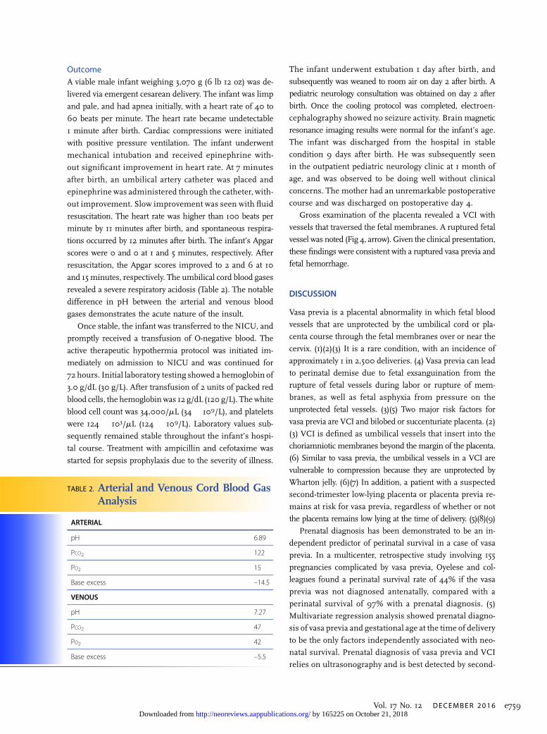
revealed a severe respiratory acidosis (Table 2). The notable
difference in pH between the arterial and venous blood
gases demonstrates the acute nature of the insult.
Once stable, the infant was transferred to the NICU, and
promptly received a transfusion of O-negative blood. The
active therapeutic hypothermia protocol was initiated im-
mediately on admission to NICU and was continued for
72 hours. Initial laboratory testing showed a hemoglobin of
3.0 g/dL (30 g/L). After transfusion of 2 units of packed red
blood cells, the hemoglobinwas 12 g/dL (120 g/L). Thewhite
blood cell count was 34,000/mL (34 � 109/L), and platelets
were 124 � 103/mL (124 � 109/L). Laboratory values sub-
sequently remained stable throughout the infant’s hospi-
tal course. Treatment with ampicillin and cefotaxime was
started for sepsis prophylaxis due to the severity of illness.
The infant underwent extubation 1 day after birth, and
subsequently was weaned to room air on day 2 after birth. A
pediatric neurology consultation was obtained on day 2 after
birth. Once the cooling protocol was completed, electroen-
cephalography showed no seizure activity. Brain magnetic
resonance imaging results were normal for the infant’s age.
The infant was discharged from the hospital in stable
condition 9 days after birth. He was subsequently seen
in the outpatient pediatric neurology clinic at 1 month of
age, and was observed to be doing well without clinical
concerns. The mother had an unremarkable postoperative
course and was discharged on postoperative day 4.
Gross examination of the placenta revealed a VCI with
vessels that traversed the fetal membranes. A ruptured fetal
vessel was noted (Fig 4, arrow). Given the clinical presentation,
these findings were consistent with a ruptured vasa previa and
fetal hemorrhage.
DISCUSSION
Vasa previa is a placental abnormality in which fetal blood
vessels that are unprotected by the umbilical cord or pla-
centa course through the fetal membranes over or near the
cervix. (1)(2)(3) It is a rare condition, with an incidence of
approximately 1 in 2,500 deliveries. (4) Vasa previa can lead
to perinatal demise due to fetal exsanguination from the
rupture of fetal vessels during labor or rupture of mem-
branes, as well as fetal asphyxia from pressure on the
unprotected fetal vessels. (3)(5) Two major risk factors for
vasa previa are VCI and bilobed or succenturiate placenta. (2)
(3) VCI is defined as umbilical vessels that insert into the
choriamniotic membranes beyond the margin of the placenta.
(6) Similar to vasa previa, the umbilical vessels in a VCI are
vulnerable to compression because they are unprotected by
Wharton jelly. (6)(7) In addition, a patient with a suspected
second-trimester low-lying placenta or placenta previa re-
mains at risk for vasa previa, regardless of whether or not
the placenta remains low lying at the time of delivery. (5)(8)(9)
Prenatal diagnosis has been demonstrated to be an in-
dependent predictor of perinatal survival in a case of vasa
previa. In a multicenter, retrospective study involving 155
pregnancies complicated by vasa previa, Oyelese and col-
leagues found a perinatal survival rate of 44% if the vasa
previa was not diagnosed antenatally, compared with a
perinatal survival of 97% with a prenatal diagnosis. (5)
Multivariate regression analysis showed prenatal diagno-
sis of vasa previa and gestational age at the time of delivery
to be the only factors independently associated with neo-
natal survival. Prenatal diagnosis of vasa previa and VCI
relies on ultrasonography and is best detected by second-
TABLE 2. Arterial and Venous Cord Blood GasAnalysis
ARTERIAL
pH 6.89
PCO2 122
PO2 15
Base excess –14.5
VENOUS
pH 7.27
PCO2 47
PO2 42
Base excess –5.5
Vol. 17 No. 12 DECEMBER 2016 e759 by 165225 on October 21, 2018http://neoreviews.aappublications.org/Downloaded from
trimester transvaginal ultrasonography, in combination
with pulsed-wave and color Doppler. (3)(4) Ultrasonogra-
phy findings suggestive of a vasa previa include a linear,
tubular, echolucent structure overlying the cervix or near
the cervix in the lower uterine segment. (2)(4)(5) Simi-
larly, a diagnosis of VCI can be made with color Doppler
ultrasonography, by visualizing at least 1 cm of umbilical
vessels coursing through the fetal membranes beyond the
placental edge. (6)(7) Once a diagnosis of VCI is made, it
is important to look for signs of vasa previa, because
approximately 3% to 4% of women with a VCI may also
have a vasa previa. (9) According to a systematic review,
ultrasonography is associated with a 93% sensitivity and
99% specificity for vasa previa. (10)
In the present case, gross examination of the placenta
showed a VCI, and a vasa previa was suggested by the acute
onset of bleeding and fetal distress during digital examina-
tion. If a prenatal diagnosis of vasa previa had been established,
a scheduled cesareandelivery at 34 to 35weeks’ gestationwould
have been recommended to avoid a catastrophic fetal hemor-
rhage from the rupture of a fetal vessel. (5)(11)
References1. Catanzarite V, Oyelese Y. Diagnosis andmanagement of vasa previa.Am J Obstet Gynecol. 2016;214(6):764
2. Silver RM. Abnormal placentation: placenta previa, vasa previa, andplacenta accreta. Obstet Gynecol. 2015;126(3):654–668
3. Society for Maternal Fetal Medicine (SMFM) PublicationsCommittee; Sinkey RG, Odibo AO, Dashe JS. # 37: diagnosis andmanagement of vasa previa. Am J Obstet Gynecol. 2015;213(5):615–619
4. Oyelese KO, Turner M, Lees C, Campbell S. Vasa previa: anavoidable obstetric tragedy. Obstet Gynecol Surv. 1999;54(2):138–145
5. Oyelese Y, Catanzarite V, Prefumo F, et al. Vasa previa: the impact ofprenatal diagnosis on outcomes. Obstet Gynecol. 2004;103(5 Pt1):937–942
6. Creasy RK, Resnik R, Iams JD, Lockwood CJ, Moore TR,Greene MF. Creasy and Resnik’s Maternal-Fetal Medicine:Principles and Practice. 7th ed. Philadelphia, PA: Saunders/Elsevier; 2014
7. Vintzileos AM, Ananth CV, Smulian JC. Using ultrasound in theclinical management of placental implantation abnormalities. Am JObstet Gynecol. 2015;213(4 Suppl):S70–S77
8. Bronsteen R, Whitten A, Balasubramanian M, et al. Vasa previa:clinical presentations, outcomes, and implications formanagement.Obstet Gynecol. 2013;122(2 Pt 1):352–357
9. Eddleman KA, Lockwood CJ, Berkowitz GS, Lapinski RH,Berkowitz RL. Clinical significance and sonographic diagnosis ofvelamentous umbilical cord insertion. Am J Perinatol. 1992;9(2):123–126
10. Ruiter L, Kok N, Limpens J, et al. Systematic review of accuracy ofultrasound in the diagnosis of vasa previa.Ultrasound Obstet Gynecol.2015;45(5):516–522
11. Robinson BK, Grobman WA. Effectiveness of timing strategies fordelivery of individuals with vasa previa. Obstet Gynecol. 2011;117(3):542–549
American Board of PediatricsNeonatal–Perinatal ContentSpecification• Know the diagnosis and management of maternal/fetal bloodloss such as placenta previa, placenta abruption, vasa previa, andmaternal-fetal hemorrhage
e760 NeoReviews by 165225 on October 21, 2018http://neoreviews.aappublications.org/Downloaded from

DOI: 10.1542/neo.17-12-e7532016;17;e753NeoReviews
Lena Braginsky and Beth A. PlunkettStrip of the Month: December 2016
ServicesUpdated Information &
http://neoreviews.aappublications.org/content/17/12/e753including high resolution figures, can be found at:
References
t-1http://neoreviews.aappublications.org/content/17/12/e753.full#ref-lisThis article cites 10 articles, 0 of which you can access for free at:
Subspecialty Collections
_drug_labeling_updatehttp://classic.neoreviews.aappublications.org/cgi/collection/pediatricPediatric Drug Labeling Updatefollowing collection(s): This article, along with others on similar topics, appears in the
Permissions & Licensing
https://shop.aap.org/licensing-permissions/in its entirety can be found online at: Information about reproducing this article in parts (figures, tables) or
Reprintshttp://classic.neoreviews.aappublications.org/content/reprintsInformation about ordering reprints can be found online:
by 165225 on October 21, 2018http://neoreviews.aappublications.org/Downloaded from
DOI: 10.1542/neo.17-12-e7532016;17;e753NeoReviews
Lena Braginsky and Beth A. PlunkettStrip of the Month: December 2016
http://neoreviews.aappublications.org/content/17/12/e753located on the World Wide Web at:
The online version of this article, along with updated information and services, is
Online ISSN: 1526-9906. Illinois, 60007. Copyright © 2016 by the American Academy of Pediatrics. All rights reserved. by the American Academy of Pediatrics, 141 Northwest Point Boulevard, Elk Grove Village,it has been published continuously since 2000. Neoreviews is owned, published, and trademarked Neoreviews is the official journal of the American Academy of Pediatrics. A monthly publication,
by 165225 on October 21, 2018http://neoreviews.aappublications.org/Downloaded from