Embed Size (px)
Citation preview
Stretch Activates Human Myometrium via ERK, Caldesmon and Focal Adhesion Signaling
CitationLi, Yunping, Maya Reznichenko, Rachel M. Tribe, Philip E. Hess, Michael Taggart, HakRim Kim, Jon P. DeGnore, Samudra Gangopadhyay, and Kathleen G. Morgan. 2009. Stretch activates human myometrium via ERK, caldesmon and focal adhesion signaling. PLoS ONE 4(10): e7489.
Published Versiondoi:10.1371/journal.pone.0007489
Permanent linkhttp://nrs.harvard.edu/urn-3:HUL.InstRepos:4728748
Terms of UseThis article was downloaded from Harvard University’s DASH repository, and is made available under the terms and conditions applicable to Other Posted Material, as set forth at http://nrs.harvard.edu/urn-3:HUL.InstRepos:dash.current.terms-of-use#LAA
Share Your StoryThe Harvard community has made this article openly available.Please share how this access benefits you. Submit a story .
Accessibility
Stretch Activates Human Myometrium via ERK,Caldesmon and Focal Adhesion SignalingYunping Li1,2*, Maya Reznichenko1, Rachel M. Tribe3, Philip E. Hess1, Michael Taggart4, HakRim Kim5,
Jon P. DeGnore6, Samudra Gangopadhyay5, Kathleen G. Morgan2,5
1 Department of Anesthesia, Critical Care and Pain Medicine, Beth Israel Deaconess Medical Center, Harvard Medical School, Boston, Massachusetts, United States of
America, 2 Boston Biomedical Research Institute, Watertown, Massachusetts, United States of America, 3 Division of Reproduction and Endocrinology, King’s College
London, London, United Kingdom, 4 Institute of Cellular Medicine, Newcastle University, Newcastle upon Tyne, United Kingdom, 5 Health Sciences Department, Boston
University, Boston, Massachusetts, United States of America, 6 Department of Physiology, Tufts University School of Medicine, Boston, Massachusetts, United States of
America
Abstract
An incomplete understanding of the molecular mechanisms responsible for myometrial activation from the quiescentpregnant state to the active contractile state during labor has hindered the development of effective therapies for pretermlabor. Myometrial stretch has been implicated clinically in the initiation of labor and the etiology of preterm labor, but themolecular mechanisms involved in the human have not been determined. We investigated the mechanisms by whichgestation-dependent stretch contributes to myometrial activation, by using human uterine samples from gynecologichysterectomies and Cesarean sections. Here we demonstrate that the Ca requirement for activation of the contractilefilaments in human myometrium increases with caldesmon protein content during gestation and that an increase incaldesmon phosphorylation can reverse this inhibitory effect during labor. By using phosphotyrosine screening and massspectrometry of stretched human myometrial samples, we identify 3 stretch-activated focal adhesion proteins, FAK,p130Cas, and alpha actinin. FAK-Y397, which signals integrin engagement, is constitutively phosphorylated in term humanmyometrium whereas FAK-Y925, which signals downstream ERK activation, is phosphorylated during stretch. We haverecently identified smooth muscle Archvillin (SmAV) as an ERK regulator. A newly produced SmAV-specific antibodydemonstrates gestation-specific increases in SmAV protein levels and stretch-specific increases in SmAV association withfocal adhesion proteins. Thus, whereas increases in caldesmon levels suppress human myometrium contractility duringpregnancy, stretch-dependent focal adhesion signaling, facilitated by the ERK activator SmAV, can contribute to myometrialactivation. These results suggest that focal adhesion proteins may present new targets for drug discovery programs aimedat regulation of uterine contractility.
Citation: Li Y, Reznichenko M, Tribe RM, Hess PE, Taggart M, et al. (2009) Stretch Activates Human Myometrium via ERK, Caldesmon and Focal AdhesionSignaling. PLoS ONE 4(10): e7489. doi:10.1371/journal.pone.0007489
Editor: Wen-Liang Zhou, Sun Yat-Sen University, China
Received June 1, 2009; Accepted September 24, 2009; Published October 16, 2009
Copyright: � 2009 Li et al. This is an open-access article distributed under the terms of the Creative Commons Attribution License, which permits unrestricteduse, distribution, and reproduction in any medium, provided the original author and source are credited.
Funding: Grant Sponsors: National Heart, Lung & Blood Institute, National Institute of Child Health & Human Development, USA; grant numbers: HD043054,HL80003 and HL86655 to KGM. Grants from Foundation of Anesthesia for Education and Research and Eleanor and Miles Shore Fellowships for Scholars inMedicine, Harvard Medical School to YL. Grants from the Wellcome Trust (UK) to MJT. The funders had no role in study design, data collection and analysis,decision to publish, or preparation of the manuscript.
Competing Interests: The authors declare that no competing interests exist.
* E-mail: [email protected]
Introduction
In late pregnancy increasing fetal growth significantly increases
uterine wall tension. Compared to the nonpregnant uterus, human
uterine weight increases from 70 grams to about 1100 grams at
term pregnancy. Its total volume averages about 5000 ml, an
expansion in size of approximately 250 fold [1]. No other smooth
muscle organ in the human is able to stretch as much as the uterus.
Myometrial stretch has been implicated, clinically, in the
activation of the myometrium for labor, but the mechanisms
involved are unclear. For example, it is known that multiple
gestation pregnancies and polyhydramnios, conditions associated
with increased tension/stretch on the uterine wall, cause an
increased incidence of premature labor. Understanding the
molecular basis of uterine contraction will aid the better control
and manipulation of uterine contractile function in preterm and
dysfunctional labor.
Focal adhesion complexes (also called dense plaques in smooth
muscle) connect the intracellular cytoskeleton to the extracellular
matrix and are recognized sites of mechanotransduction [2].
Previous studies [3,4] in rodent myometrium have demonstrated
that focal adhesion signaling is activated at late pregnancy. The
authors suggested that neuronal and hormonal pathways alone
may not be sufficient to bring about myometrial activation for
labor and that a synergy of neuronal-hormonal pathways and
mechanotransduction pathways could play an important role in
parturition. Current knowledge of the roles of mechanical stretch
in uterine regulation is based on data from animal models, and
little information is available as to how this system works in human
myometrium. Furthermore, the identity of signaling molecules
involved in mechanotransduction pathways in human myometri-
um is little studied [5].
We have recently reported in the timed pregnant rat model that
mechanical stretch of pregnant uterine smooth muscle activates
PLoS ONE | www.plosone.org 1 October 2009 | Volume 4 | Issue 10 | e7489
ERK via focal adhesion signaling. In the rat, we have shown that
in addition to classical GPCR-mediated pathways, this ERK
pathway, in a cause-and-effect manner, facilitates myometrial
contraction, and plays a distinct role in the switch from the
quiescent phase of pregnancy to a more contractile phenotype at
the end of pregnancy [6,7].
Smooth muscle archvillin (SmAV) is a regulator of ERK
pathways newly identified by our group [8,9]. It is a member of
the supervillin family that is preferentially expressed in smooth
muscle and was first identified as an interactor of the smooth
muscle differentiation marker, h-1 calponin in a 2-hybrid assay. Its
function in myometrium has not been previously studied.
In the present study, we tested the hypothesis that the stretch-
mediated activation of focal adhesion signaling molecules occurs
during human pregnancies and describe, for the first time, an up-
regulation during gestation and association with focal adhesion
complexes of the ERK regulator, SmAV, in human myometrium.
Materials and Methods
Human Myometrial Tissue CollectionEthics Statement: The consent forms for human myometrial
tissue collection were approved by the Committee on Clinical
Investigations at Beth Israel Deaconess Medical Center and
Central Manchester Healthcare Trust LREC (UK). The human
pregnant myometrial samples were taken from the upper edges of
the lower segment uterine incision during Cesarean sections
(n = 44) or ex-utero intrapartum treatment (EXIT) procedures
(n = 3). The nonpregnant human myometrial samples (n = 8) were
collected from pre-menopausal patients undergoing hysterectomy
to remove the uterus for benign gynecological conditions and
samples were obtained at the uterine lower segment. Exclusions
for sample collection were patients with a history of hypertension
or pregnancy-induced hypertension who are on antihypertensive
medication, those with preterm labor who are currently on
medication treatment, or patients undergoing emergent cesarean
section from whom unable to obtain a formal consent.
Rat Myometrial Tissue CollectionAll procedures were approved by the Boston Biomedical Research
Institute Animal Care and Use Committee and complied with he
American Physiological Society ‘‘Guiding Principles for Research
Involving Animals and Human Beings’’. Sprague-Dawley nonpreg-
nant and timed-pregnant rats (Taconic, Germantown, NY) were
euthanized by carbon dioxide inhalation. Delivery was observed to
occur on the 22nd or 23rd gestational day. For the collection of in-
labor uterine smooth muscle samples, the rat was closely observed
and the delivery of the first pup was used as the indication of labor.
Excised uteri were immersed immediately into oxygenated Krebs
solution at room temperature. The details of tissue handling and
protein extraction have been published previously [6,10].
Tissue Preparation and Force RecordingThe whole-thickness human uterine smooth muscle strips were
microdissected to a size of approximately 106363 mm, and
oriented parallel to the longitudinal axis of muscle bundles. Four
myometrial strips were dissected from each sample for in vitro
stretch experiments. The satisfactory condition of each uterine
smooth muscle strip was confirmed before the experiment by the
presence of active, spontaneous contractions with or without
51 mM KCl treatment. Isometric force was recorded at 37uC as
previously described [10]. For in vitro stretch experiments, the
strips were stretched to 26 slack length. Tension was applied
gradually over 20–30 seconds and maintained for the duration of
experiment. The contractile activity was digitalized with MacLab/
8e, Chart v3.5.4 (AD Instrument, Castle Hill, Australia).
All the samples used for in vitro stretch experiments were term,
not in labor human pregnant myometrium. The justification for
stretch of 26 slack length is based on the optimal length of term
pregnant uterine smooth muscle strips in the rat model [7,10] and
the pilot stretch experiments on human myometrium. Also, this
optimal length is same as in vivo length as measured when the
uterine wall is stretched by the fetus in the rat model. Clinically, it
is impossible to measure the in vivo length of the uterine smooth
muscle that will be sampled later. In general, in vitro stretch to 26slack length generates force in the range of 10–15 grams. This is
about a 20-fold increase in force compared to baseline tension of
term, not-in-labor uterine strips in the organ bath. The area under
the curve (AUC) was obtained by integrating the force signal (in
gram) over indicated time period (in second).
ImmunoblottingStrips were quick-frozen at different time points after a
mechanical stretch in a dry ice/trichloroacetic acid/acetone slurry
containing 10 mM DTT. The frozen samples were homogenized
as previously described [6]. Protein concentrations were quantified
by modified Lowry protein assay (DC Protein Assay Kit, Bio-Rad).
Protein-matched samples were boiled, separated by SDS-PAGE,
transferred to a PVDF membrane and analyzed by immunoblot-
ting with appropriate antibodies.
For caldesmon (CaD) and rat SmAV protein content quanti-
fication, the blots were visualized with a SuperSignal West Pico
peroxide solution (Pierce, Rockford, IL). The images were
detected with a chemiluminescence screen and quantified with a
Bio-Rad Phospho Imager and Multi-Analyst software. In rest
experiments, the intensity of Western blot signals was quantitated
by using an Odyssey Infrared Imaging System (LI-COR
Bioscience, Lincoln, Nebraska). To minimize the variation of the
signal intensity between different Western blotting experiments,
we developed blots to similar intensities or used a reference sample
in each blot for normalization if multiple samples were used from
different pregnant women.
Calcium sensitivity in alpha-toxin permeabilized humanmyometrial strips
The method was identical to the method previously published
[10]. The force of contractions generated at 1027 M free calcium
concentration were recorded and expressed as a percentage of
maximal contraction at pCa 5. This measurement was used as an
index of calcium sensitivity.
ImmunoprecipitationThe myometrial smooth muscle strips were frozen in a dry ice/
acetone slurry containing DTT. The tissue was pulverized with a
mortar and pestle cooled with liquid nitrogen. The tissue powder
was dissolved in a buffer containing 50 mM Tris (pH 7.4), 5 mM
EGTA, 140 mM NaCl, 1% NP40, 1% Na deoxycholate, 20KIU
aprotinin, 5 mM leupeptin, 5 mM pepstatin A, 2 mM Na3VO4,
and 1 mM NaF and centrifuged at 12,000 rpm at 4uC for 15 min.
The supernatant was precleared with protein A agarose and then
incubated with anti-phospho tyrosine or anti-ERK conjugated
agarose beads overnight at 4uC with gentle rotation. The immune
complex beads were washed, resuspended in a sample buffer
(25 mM Tris (pH 6.8), 4% SDS, 20% glycerol, 5% b-mercapto-
ethanol, 1 mM EDTA and 0.001% bromophenol blue), and
boiled at 100uC for 5 min. The proteins of interest were detected
by western blotting with specific antibodies.
Myometrial Stretch Activation
PLoS ONE | www.plosone.org 2 October 2009 | Volume 4 | Issue 10 | e7489
MaterialsAn anti-SmAV antibody was produced from a bacterially
expressed, His-tagged, fragment containing the first 250 N-
terminal residues of ferret SmAV. The purified peptide was
injected into rabbits and polyclonal antiserum was developed by
Capralogics, Hardwick, MA. The antibody was affinity purified
from the serum (AminoLink Kit, Pierce Biotechnology) and the
purified antibody recognizes the antigen and also recognizes a
single protein band of appropriate molecular weight and no other
bands in aorta tissue homogenates. This antibody was used for
immunoblot and imaging studies.
The CaD polyclonal antibody (1:30,000) was raised against full-
length human myometrial CaD and was a gift from Dr. K.
Mabuchi (Boston Biomedical Research Institute). The phospho-
CaD antibody (1:500 Upstate, Lake Placid, NY) was produced
against a phosphopeptide containing the CaD sequencing
surrounding the Ser789 ERK phosphorylation site. The p44/42
MAP kinase1/2 (1:1000), phospho p44/42 MAP1/2 kinase
antibody (1:1500), phospho-FAK (Y925, 1:500) and phospho-
p130Cas (1:500) were purchased from Cell Signaling Technology
(Beverly, MA). Phospho-tyrosine monoclonal antibody, clone
4G10 (1:500–1500), anti-phospho-tyrosine and anti-MAP kinase
ERK1/2 agarose-conjugated beads were the products of Upstate.
P130Cas and Phospho-FAK (Y397, 1:1000) antibodies were
purchased from BD Transduction Laboratories (San Diego, CA).
FAK (1:300) was a polyclonal antibody from Santa Cruz
Biotechnology Inc. (Santa Cruz, CA). Monoclonal alpha-actinin
(1:1000) antibody was purchased from Sigma.
Mass SpectrometryThe protein band of interest was excised from one-dimensional,
Coomassie blue stained PAGE gels. Mass spectrometry for protein
identification was performed by the Tufts University Core Facility
(Boston, Massachusetts). Excised bands were subjected to in-gel
reduction, alkylation, and enzymatic digestion with trypsin (Roche
Applied Science, Indianapolis, IN). LC/MS/MS analysis was
performed on the in-gel digest extracts using an Agilent (Santa
Clara, CA) 1100 binary pump directly coupled to a mass
spectrometer. Nanobore electrospray columns were constructed
from 360 mm o.d., 75 mm i.d. fused silica capillary with the column
tip tapered to a 15 mm opening (New Objective, Woburn, MA)
and were packed with 200 A 5 mm C18 beads (Michrom
BioResources. Auburn, CA.) to a length of 10 cm. The flow
through the column was split pre-column to achieve a flow rate of
300 nL/min. The mobile phase used for gradient elution consisted
of (A) 0.3% acetic acid 99.7% water and (B) 0.3% acetic acid
99.7% Acetonitrile. Tandem mass spectra (LC/MS/MS) were
acquired on a Thermo LTQ ion trap mass spectrometer (Thermo
Corp., San Jose, CA). The MS/MS spectra were searched against
the NCBI non-redundant protein sequence database using the
SEQUEST computer algorithm [11] to produce a list of proteins
identified in each sample. Confident protein identification was
determined by the Proteomics Browser software (Thermo Corp.,
San Jose, CA) and was defined as at least 4 unique peptides
matching for a given protein.
ImmunohistochemistryThe whole-thickness, term, not-in-labor, human uterine smooth
muscle strips were microdissected and stretched to 2 fold slack
length for 7 minutes. Stretched muscles were fixed by immersion
in 4% paraformaldehyde for 2 hours at 4uC and embedded with
optimal cutting temperature compound (OCT, Tissue-Tek).
Frozen samples were sliced 10 micrometers thick and stained
with rabbit anti-SmAV and mouse anti-vinculin (Sigma). Alexa
568-conjugated goat anti-rabbit (red) and Alexa 488 goat anti-
mouse (green) secondary antibodies (Molecular Probe Inc.) were
used for staining. DAPI was used as a nuclei stain. Images of
immunostained myometrium were taken with a Nikon Eclipse TE
2000-E inverted microscope equipped with a Nikon Plan
Apochromat 60XA (NA 1.4) oil immersion objective. Images
were recorded by a high-resolution fluorescence CCD camera
(CoolSNAPTM HQ2, PhotometricsH) with NIS-Elements Ad-
vanced Research (Nikon) software. In all cases, it was confirmed
that there was no detectable background fluorescence by staining
cells in the absence of a primary antibody. In addition, for all
colabeling experiments, it was confirmed that there was no
detectable cross talk between fluorescent labels by exchanging
excitation/emission filters on single labeled samples. Resolution
was optimized by deconvolution microscopy software. The
algorithm used image information from different Z-slices
(Richardson-Lucy algorithm, constrained iterative-maximum like-
lihood estimation algorithm).
StatisticsData were expressed as mean6 SE. Significance of difference
between two individual sets of means was taken at p,0.05 by an
unpaired Student’s t test unless indicated otherwise. In stretch
experiments, a one-way ANOVA test was used for the time course
study. The probability values of p,0.05 was considered
significant.
Results
Human myometrial CaD protein content and the [Ca2+]requirement for contractile activation increase duringpregnancy
h-Caldesmon (CaD) is a smooth muscle-specific actin binding
protein that interferes with acto-myosin interactions [12]. In
myometrial samples collected from pregnant women who under-
went Cesarean sections or non-pregnant women who underwent
gynecological procedures, we confirmed the earlier reports of Word
et al [13] and Riley et al [14] that h-CaD protein expression levels
increase significantly during pregnancy (2690763535 in $37 weeks
group vs. 1365663752 in NP group)(Fig. 1A).
Since h-CaD is known to inhibit acto-myosin interactions in vitro
[15], we tested the relative Ca sensitivity of activation of the
contractile filaments of human myometrial preparations permeabi-
lized with alpha toxin [10]. Shown in Figure 1B are the average
contractile force responses of myometrial preparations from
nonpregnant women, women pregnant for ,37 weeks (not in
labor) and those pregnant for $37 weeks (not in labor) to a [Ca2+] of
1027 M, expressed as a percentage of the maximal force obtained at
1025 M [Ca2+]. Calcium responsiveness of the contractile appara-
tus is significantly decreased in the late pregnancy samples
compared to that of samples from non-pregnant women. Our data
are consistent with the hypothesis that the observed increased
inhibitory caldesmon protein content in human myometrium leads
to a decreased contractile responsiveness to Ca during pregnancy.
During a normal pregnancy the uterus is known to clinically display
a ‘‘myometrial quiescence’’ before labor. This decreased Ca
responsiveness of the contractile apparatus is one factor that could
explain this quiescence during pregnancy.
Gestation- dependent increases in h-CaDphosphorylation in human myometrium
In the timed pregnant rat model, we have shown that as labor
begins, an ERK pathway is activated that phosphorylates CaD [10]
Myometrial Stretch Activation
PLoS ONE | www.plosone.org 3 October 2009 | Volume 4 | Issue 10 | e7489
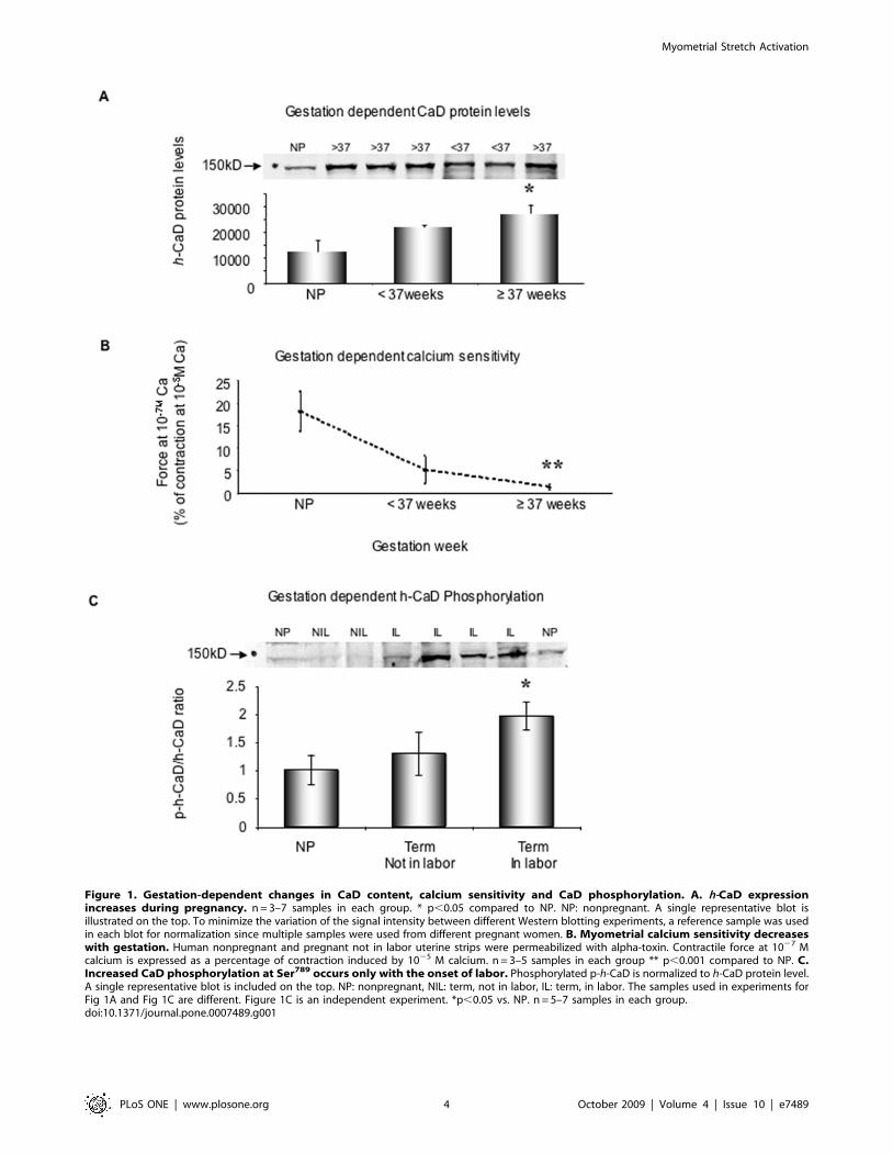
Figure 1. Gestation-dependent changes in CaD content, calcium sensitivity and CaD phosphorylation. A. h-CaD expressionincreases during pregnancy. n = 3–7 samples in each group. * p,0.05 compared to NP. NP: nonpregnant. A single representative blot isillustrated on the top. To minimize the variation of the signal intensity between different Western blotting experiments, a reference sample was usedin each blot for normalization since multiple samples were used from different pregnant women. B. Myometrial calcium sensitivity decreaseswith gestation. Human nonpregnant and pregnant not in labor uterine strips were permeabilized with alpha-toxin. Contractile force at 1027 Mcalcium is expressed as a percentage of contraction induced by 1025 M calcium. n = 3–5 samples in each group ** p,0.001 compared to NP. C.Increased CaD phosphorylation at Ser789 occurs only with the onset of labor. Phosphorylated p-h-CaD is normalized to h-CaD protein level.A single representative blot is included on the top. NP: nonpregnant, NIL: term, not in labor, IL: term, in labor. The samples used in experiments forFig 1A and Fig 1C are different. Figure 1C is an independent experiment. *p,0.05 vs. NP. n = 5–7 samples in each group.doi:10.1371/journal.pone.0007489.g001
Myometrial Stretch Activation
PLoS ONE | www.plosone.org 4 October 2009 | Volume 4 | Issue 10 | e7489
and removes the inhibitory influences of CaD on actomyosin
interactions [15] and, as a result contributes to the onset of labor [7].
Whether this ERK-mediated phosphorylation of h-CaD during
labor occurs in the human has not been reported. We probed
quickly frozen human myometrial samples from nonpregnant
women, term pregnant women, but not in labor, and term pregnant
women and in labor. We found (Fig. 1C) that h-CaD phosphor-
ylation at an ERK specific site (Ser 789) increases significantly in
human myometrial tissue from pregnant women in labor
(1.9760.24 vs. 1.0160.27 in NP group) but not from those not in
labor (1.360.39). These results are consistent with phosphorylation
of CaD being a contributing factor for labor onset.
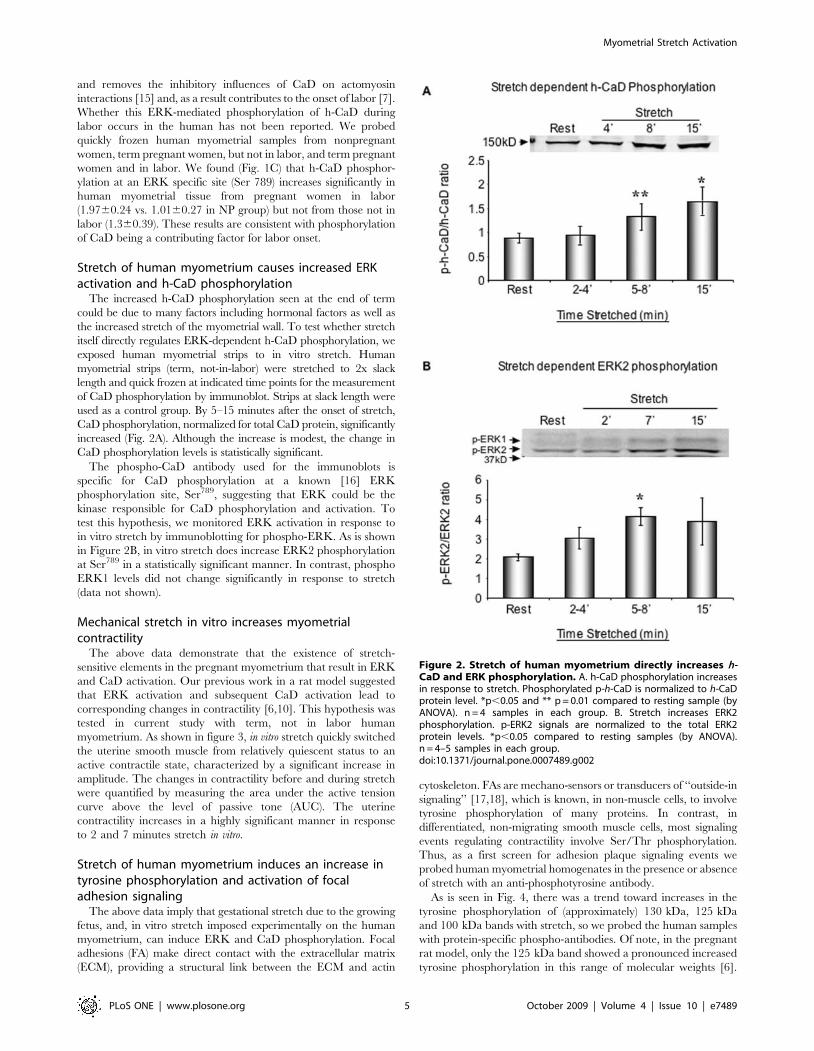
Stretch of human myometrium causes increased ERKactivation and h-CaD phosphorylation
The increased h-CaD phosphorylation seen at the end of term
could be due to many factors including hormonal factors as well as
the increased stretch of the myometrial wall. To test whether stretch
itself directly regulates ERK-dependent h-CaD phosphorylation, we
exposed human myometrial strips to in vitro stretch. Human
myometrial strips (term, not-in-labor) were stretched to 2x slack
length and quick frozen at indicated time points for the measurement
of CaD phosphorylation by immunoblot. Strips at slack length were
used as a control group. By 5–15 minutes after the onset of stretch,
CaD phosphorylation, normalized for total CaD protein, significantly
increased (Fig. 2A). Although the increase is modest, the change in
CaD phosphorylation levels is statistically significant.
The phospho-CaD antibody used for the immunoblots is
specific for CaD phosphorylation at a known [16] ERK
phosphorylation site, Ser789, suggesting that ERK could be the
kinase responsible for CaD phosphorylation and activation. To
test this hypothesis, we monitored ERK activation in response to
in vitro stretch by immunoblotting for phospho-ERK. As is shown
in Figure 2B, in vitro stretch does increase ERK2 phosphorylation
at Ser789 in a statistically significant manner. In contrast, phospho
ERK1 levels did not change significantly in response to stretch
(data not shown).
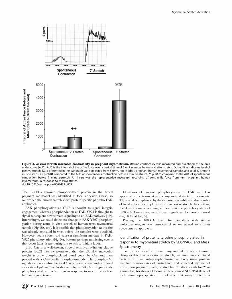
Mechanical stretch in vitro increases myometrialcontractility
The above data demonstrate that the existence of stretch-
sensitive elements in the pregnant myometrium that result in ERK
and CaD activation. Our previous work in a rat model suggested
that ERK activation and subsequent CaD activation lead to
corresponding changes in contractility [6,10]. This hypothesis was
tested in current study with term, not in labor human
myometrium. As shown in figure 3, in vitro stretch quickly switched
the uterine smooth muscle from relatively quiescent status to an
active contractile state, characterized by a significant increase in
amplitude. The changes in contractility before and during stretch
were quantified by measuring the area under the active tension
curve above the level of passive tone (AUC). The uterine
contractility increases in a highly significant manner in response
to 2 and 7 minutes stretch in vitro.
Stretch of human myometrium induces an increase intyrosine phosphorylation and activation of focaladhesion signaling
The above data imply that gestational stretch due to the growing
fetus, and, in vitro stretch imposed experimentally on the human
myometrium, can induce ERK and CaD phosphorylation. Focal
adhesions (FA) make direct contact with the extracellular matrix
(ECM), providing a structural link between the ECM and actin
cytoskeleton. FAs are mechano-sensors or transducers of ‘‘outside-in
signaling’’ [17,18], which is known, in non-muscle cells, to involve
tyrosine phosphorylation of many proteins. In contrast, in
differentiated, non-migrating smooth muscle cells, most signaling
events regulating contractility involve Ser/Thr phosphorylation.
Thus, as a first screen for adhesion plaque signaling events we
probed human myometrial homogenates in the presence or absence
of stretch with an anti-phosphotyrosine antibody.
As is seen in Fig. 4, there was a trend toward increases in the
tyrosine phosphorylation of (approximately) 130 kDa, 125 kDa
and 100 kDa bands with stretch, so we probed the human samples
with protein-specific phospho-antibodies. Of note, in the pregnant
rat model, only the 125 kDa band showed a pronounced increased
tyrosine phosphorylation in this range of molecular weights [6].
Figure 2. Stretch of human myometrium directly increases h-CaD and ERK phosphorylation. A. h-CaD phosphorylation increasesin response to stretch. Phosphorylated p-h-CaD is normalized to h-CaDprotein level. *p,0.05 and ** p = 0.01 compared to resting sample (byANOVA). n = 4 samples in each group. B. Stretch increases ERK2phosphorylation. p-ERK2 signals are normalized to the total ERK2protein levels. *p,0.05 compared to resting samples (by ANOVA).n = 4–5 samples in each group.doi:10.1371/journal.pone.0007489.g002
Myometrial Stretch Activation
PLoS ONE | www.plosone.org 5 October 2009 | Volume 4 | Issue 10 | e7489
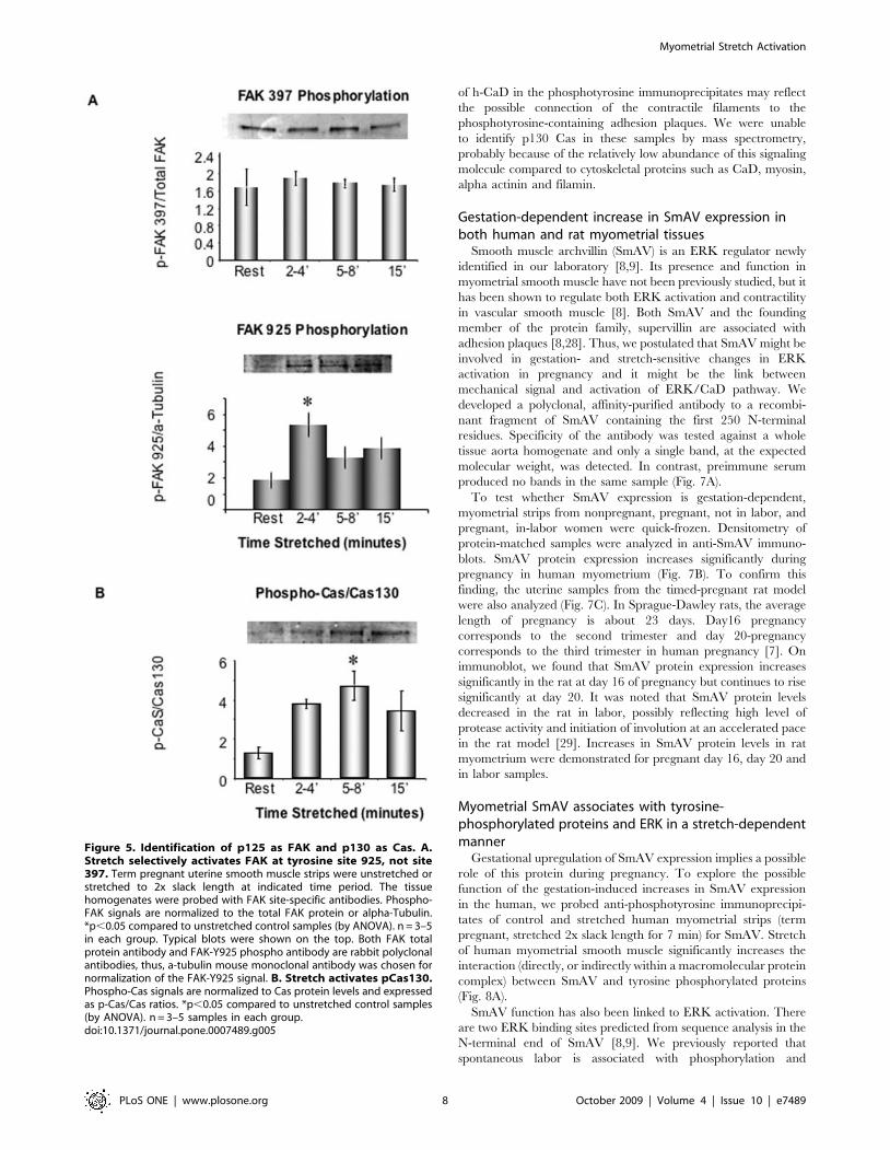
The 125 kDa tyrosine phosphorylated protein in the timed
pregnant rat model was identified as focal adhesion kinase, so
we probed the human samples with protein-specific phospho-FAK
antibodies.
FAK phosphorylation at Y397 is thought to signal integrin
engagement whereas phosphorylation at FAK-Y925 is thought to
signal subsequent downstream signaling to an ERK pathway [19].
Interestingly, we could detect no change in FAK-Y397 phosphor-
ylation during acute in vitro stretch of human term myometrial
samples (Fig. 5A, top). It is possible that phosphorylation at this site
was already activated in vivo, before the samples were obtained.
However, acute stretch did cause a significant increase in FAK-
Y925 phosphorylation (Fig. 5A, bottom) perhaps mimicking events
that occur later in vivo during the switch to initiate labor.
p130 Cas is a well-known, stretch sensitive, adhesion plaque
protein [20,21], so we postulated that the 130 kDa molecular
weight tyrosine phosphorylated band could be Cas and then
probed with a Cas-specific phospho-antibody. The phospho-Cas
signals were normalized to total Cas protein content and expressed
as a ratio of p-Cas/Cas. As shown in figure 5B, Cas is significantly
phosphorylated within 5–8 min in response to in vitro stretch in
human myometrium.
Elevations of tyrosine phosphorylation of FAK and Cas
appeared to be transient in the myometrial stretch experiments.
This could be explained by the dynamic assembly and disassembly
of focal adhesion complexes as a function of stretch. In contrast,
the downstream of resulting serine/threonine phosphorylation of
ERK/CaD may integrate upstream signals and be more sustained
(Fig. 1C and Fig. 2).
Probing the 100 kDa band for candidates with similar
molecular weights was unsuccessful so we turned to a mass
spectrometry approach.
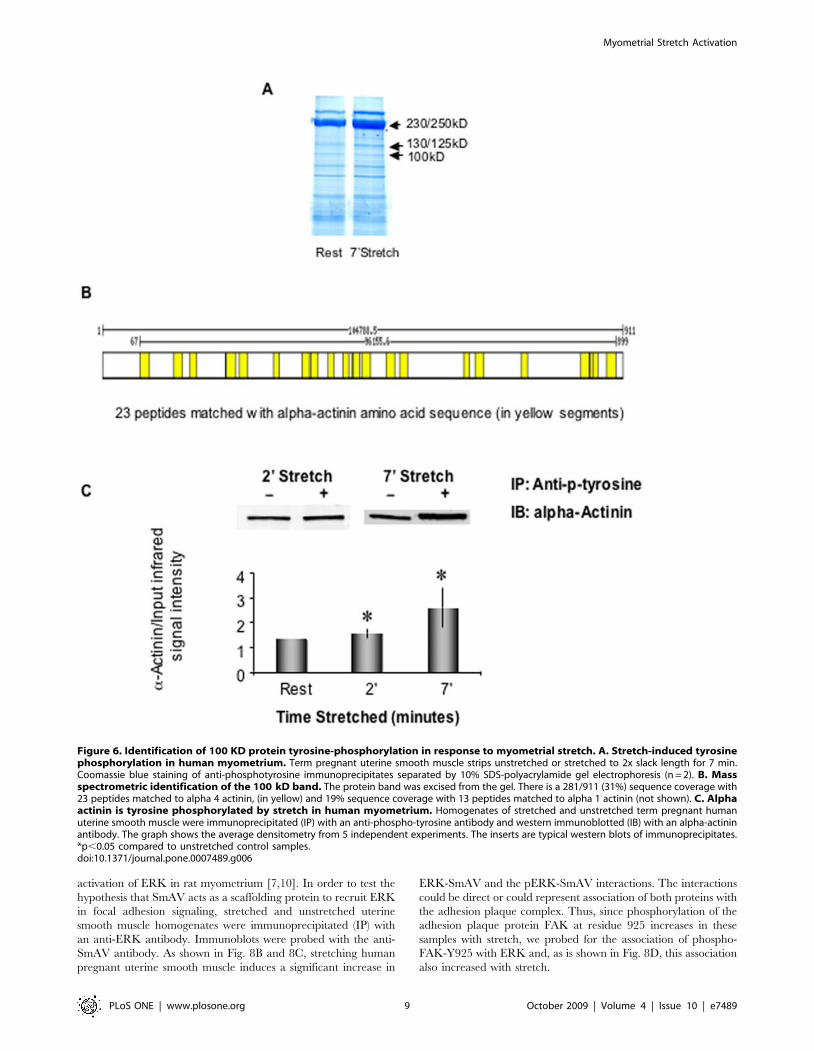
Identification of proteins tyrosine phosphorylated inresponse to myometrial stretch by SDS/PAGE and MassSpectrometry
To further identify human myometrial proteins tyrosine
phosphorylated in response to stretch, we immunoprecipitated
proteins with an anti-phosphotyrosine antibody using protein-
matched homogenates of unstretched and stretched myometrial
strips (term pregnant, slack, or stretched 2x slack length for 29 or
7 min). Fig. 6A shows a Coomassie blue stained SDS/PAGE gel of
such immunoprecipitates. It is of note that many proteins in
Figure 3. In vitro stretch increases contractility in pregnant myometrium. Uterine contractility was measured and quantified as the areaunder curve (AUC). AUC is the integral of the active force over a period time of 2 or 7 minutes before and after stretch. Dotted line indicates level ofpassive stretch. Data presented in the bar graph were collected from 8 term, not in labor, pregnant human myometrial samples and total 17 smoothmuscle strips. ++ p,0.01 compared to the AUC of spontaneous contraction before 2 minute-stretch. ** p,0.01 compared to the AUC of spontaneouscontraction before 7 minute-stretch. An insert was the representative myograph recording of contractile force from term pregnant humanmyometrium in response to in vitro stretch.doi:10.1371/journal.pone.0007489.g003
Myometrial Stretch Activation
PLoS ONE | www.plosone.org 6 October 2009 | Volume 4 | Issue 10 | e7489
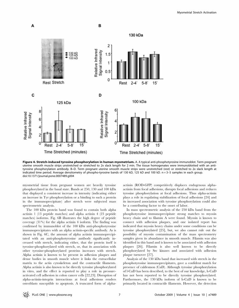
myometrial tissue from pregnant women are heavily tyrosine
phosphorylated in the basal state. Bands at 250, 130 and 100 kDa
that displayed a consistent increase in intensity (indicating either
an increase in Tyr phosphorylation or a binding to such a protein
in the immunoprecipitate) after stretch were subjected mass
spectrometric analysis.
The 100 kDa protein band was found to contain both alpha
actinin 1 (13 peptide matches) and alpha actinin 4 (23 peptide
matches) isoforms. Fig. 6B illustrates the high degree of peptide
coverage (31%) for the alpha actinin 4 isoform. The finding was
confirmed by immunoblot of the 100 kDa anti-phosphotyrosine
immunoprecipitates with an alpha actinin-specific antibody. As is
shown in Fig. 6C, the amount of alpha actinin immunoprecipi-
tated with an anti-phosphotyrosine antibody significantly in-
creased with stretch, indicating either, that the protein itself is
tyrosine-phosphorylated with stretch, or, that its association with
other tyrosine-phosphorylated proteins increases with stretch.
Alpha actinin is known to be present in adhesion plaques and
dense bodies in smooth muscle where it links the extracellular
matrix to the actin cytoskeleton and the contractile filaments.
Alpha actinin is also known to be directly tyrosine phosphorylated
in vitro, and the effect is reported to play a role in pressure-
activated cell adhesion in colon cancer cells [22,23]. Disruption of
alpha-actinin-integrin interactions at focal adhesions renders
osteoblasts susceptible to apoptosis. A truncated form of alpha-
actinin (ROD-GFP) competitively displaces endogenous alpha-
actinin from focal adhesions, disrupts focal adhesions and reduces
tyrosine phosphorylation at focal adhesions. Thus alpha-actinin
plays a role in regulating stabilization of focal adhesions [24] and
its increased association with tyrosine phosphorylation could also
be a contributing factor to the onset of labor.
In mass spectrometric analysis of the 250 kDa band from the
phosphotyrosine immunoprecipitate strong matches to myosin
heavy chain and to filamin A were found. Myosin is known to
connect with adhesion plaques, and one isolated report has
indicated that myosin heavy chains under some conditions can be
tyrosine phosphorylated [25], but, we also cannot rule out the
possibility of myosin contamination of the mass spectrometry
samples given its abundance in smooth muscle. Filamin was also
identified in this band and is known to be associated with adhesion
plaques [26]. Filamin is also well known to be directly
phosphorylated by Src kinases and associated with adhesion
plaque turnover [27].
Analysis of the 130 kDa band that increased with stretch in the
phosphotyrosine immunoprecipitates, gave a confident match for
presence of caldesmon (CaD). Although tyrosine phosphorylation
of l-CaD has been described, to the best of our knowledge, h-CaD
has not been reported to be directly tyrosine phosphorylated.
Furthermore, the 130 kDa isoform of h-CaD is known to be
primarily located in contractile filaments. However, the detection
Figure 4. Stretch-induced tyrosine phosphorylation in human myometrium. A. A typical anti-phosphotyrosine immunoblot. Term pregnantuterine smooth muscle strips unstretched or stretched to 2x slack length for 2 min. The tissue homogenates were immunoblotted with an anti-tyrosine phosphorylation antibody. B–D. Term pregnant uterine smooth muscle strips were unstretched (rest) or stretched to 2x slack length atindicated time period. Average densitometry of phospho-tyrosine bands of 130 kD, 125 kD and 100 kD. n = 3–5 samples in each group.doi:10.1371/journal.pone.0007489.g004
Myometrial Stretch Activation
PLoS ONE | www.plosone.org 7 October 2009 | Volume 4 | Issue 10 | e7489
of h-CaD in the phosphotyrosine immunoprecipitates may reflect
the possible connection of the contractile filaments to the
phosphotyrosine-containing adhesion plaques. We were unable
to identify p130 Cas in these samples by mass spectrometry,
probably because of the relatively low abundance of this signaling
molecule compared to cytoskeletal proteins such as CaD, myosin,
alpha actinin and filamin.
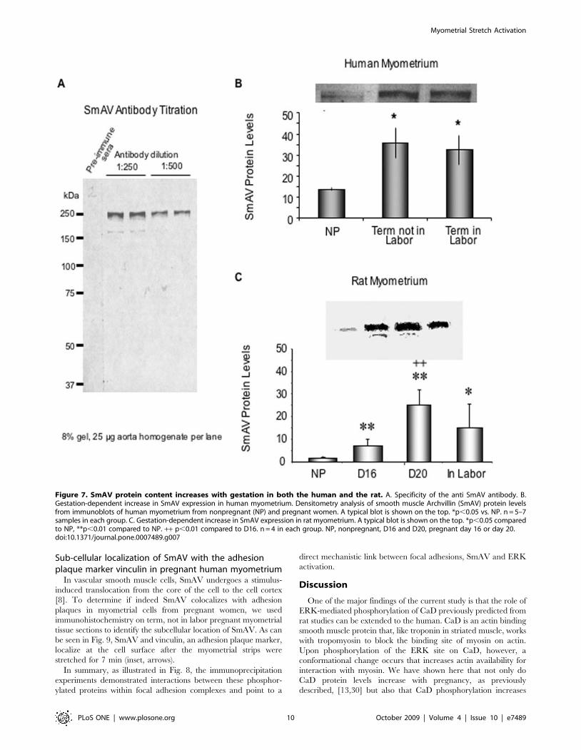
Gestation-dependent increase in SmAV expression inboth human and rat myometrial tissues
Smooth muscle archvillin (SmAV) is an ERK regulator newly
identified in our laboratory [8,9]. Its presence and function in
myometrial smooth muscle have not been previously studied, but it
has been shown to regulate both ERK activation and contractility
in vascular smooth muscle [8]. Both SmAV and the founding
member of the protein family, supervillin are associated with
adhesion plaques [8,28]. Thus, we postulated that SmAV might be
involved in gestation- and stretch-sensitive changes in ERK
activation in pregnancy and it might be the link between
mechanical signal and activation of ERK/CaD pathway. We
developed a polyclonal, affinity-purified antibody to a recombi-
nant fragment of SmAV containing the first 250 N-terminal
residues. Specificity of the antibody was tested against a whole
tissue aorta homogenate and only a single band, at the expected
molecular weight, was detected. In contrast, preimmune serum
produced no bands in the same sample (Fig. 7A).
To test whether SmAV expression is gestation-dependent,
myometrial strips from nonpregnant, pregnant, not in labor, and
pregnant, in-labor women were quick-frozen. Densitometry of
protein-matched samples were analyzed in anti-SmAV immuno-
blots. SmAV protein expression increases significantly during
pregnancy in human myometrium (Fig. 7B). To confirm this
finding, the uterine samples from the timed-pregnant rat model
were also analyzed (Fig. 7C). In Sprague-Dawley rats, the average
length of pregnancy is about 23 days. Day16 pregnancy
corresponds to the second trimester and day 20-pregnancy
corresponds to the third trimester in human pregnancy [7]. On
immunoblot, we found that SmAV protein expression increases
significantly in the rat at day 16 of pregnancy but continues to rise
significantly at day 20. It was noted that SmAV protein levels
decreased in the rat in labor, possibly reflecting high level of
protease activity and initiation of involution at an accelerated pace
in the rat model [29]. Increases in SmAV protein levels in rat
myometrium were demonstrated for pregnant day 16, day 20 and
in labor samples.
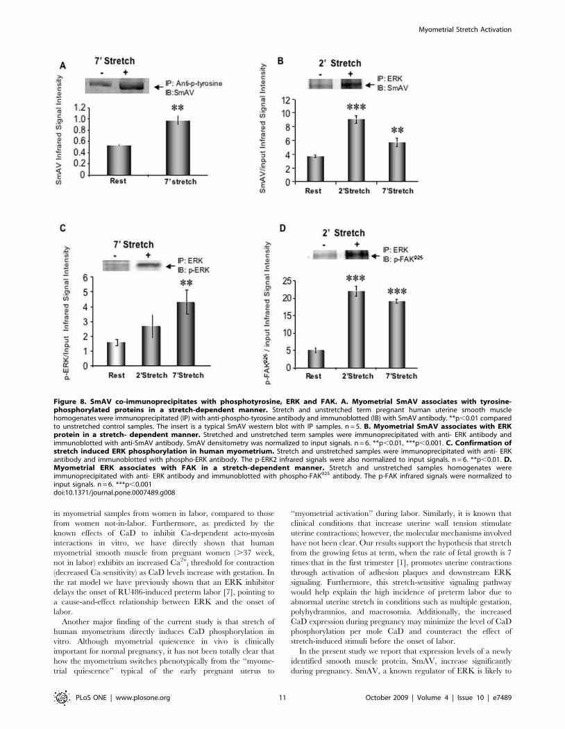
Myometrial SmAV associates with tyrosine-phosphorylated proteins and ERK in a stretch-dependentmanner
Gestational upregulation of SmAV expression implies a possible
role of this protein during pregnancy. To explore the possible
function of the gestation-induced increases in SmAV expression
in the human, we probed anti-phosphotyrosine immunoprecipi-
tates of control and stretched human myometrial strips (term
pregnant, stretched 2x slack length for 7 min) for SmAV. Stretch
of human myometrial smooth muscle significantly increases the
interaction (directly, or indirectly within a macromolecular protein
complex) between SmAV and tyrosine phosphorylated proteins
(Fig. 8A).
SmAV function has also been linked to ERK activation. There
are two ERK binding sites predicted from sequence analysis in the
N-terminal end of SmAV [8,9]. We previously reported that
spontaneous labor is associated with phosphorylation and
Figure 5. Identification of p125 as FAK and p130 as Cas. A.Stretch selectively activates FAK at tyrosine site 925, not site397. Term pregnant uterine smooth muscle strips were unstretched orstretched to 2x slack length at indicated time period. The tissuehomogenates were probed with FAK site-specific antibodies. Phospho-FAK signals are normalized to the total FAK protein or alpha-Tubulin.*p,0.05 compared to unstretched control samples (by ANOVA). n = 3–5in each group. Typical blots were shown on the top. Both FAK totalprotein antibody and FAK-Y925 phospho antibody are rabbit polyclonalantibodies, thus, a-tubulin mouse monoclonal antibody was chosen fornormalization of the FAK-Y925 signal. B. Stretch activates pCas130.Phospho-Cas signals are normalized to Cas protein levels and expressedas p-Cas/Cas ratios. *p,0.05 compared to unstretched control samples(by ANOVA). n = 3–5 samples in each group.doi:10.1371/journal.pone.0007489.g005
Myometrial Stretch Activation
PLoS ONE | www.plosone.org 8 October 2009 | Volume 4 | Issue 10 | e7489
activation of ERK in rat myometrium [7,10]. In order to test the
hypothesis that SmAV acts as a scaffolding protein to recruit ERK
in focal adhesion signaling, stretched and unstretched uterine
smooth muscle homogenates were immunoprecipitated (IP) with
an anti-ERK antibody. Immunoblots were probed with the anti-
SmAV antibody. As shown in Fig. 8B and 8C, stretching human
pregnant uterine smooth muscle induces a significant increase in
ERK-SmAV and the pERK-SmAV interactions. The interactions
could be direct or could represent association of both proteins with
the adhesion plaque complex. Thus, since phosphorylation of the
adhesion plaque protein FAK at residue 925 increases in these
samples with stretch, we probed for the association of phospho-
FAK-Y925 with ERK and, as is shown in Fig. 8D, this association
also increased with stretch.
Figure 6. Identification of 100 KD protein tyrosine-phosphorylation in response to myometrial stretch. A. Stretch-induced tyrosinephosphorylation in human myometrium. Term pregnant uterine smooth muscle strips unstretched or stretched to 2x slack length for 7 min.Coomassie blue staining of anti-phosphotyrosine immunoprecipitates separated by 10% SDS-polyacrylamide gel electrophoresis (n = 2). B. Massspectrometric identification of the 100 kD band. The protein band was excised from the gel. There is a 281/911 (31%) sequence coverage with23 peptides matched to alpha 4 actinin, (in yellow) and 19% sequence coverage with 13 peptides matched to alpha 1 actinin (not shown). C. Alphaactinin is tyrosine phosphorylated by stretch in human myometrium. Homogenates of stretched and unstretched term pregnant humanuterine smooth muscle were immunoprecipitated (IP) with an anti-phospho-tyrosine antibody and western immunoblotted (IB) with an alpha-actininantibody. The graph shows the average densitometry from 5 independent experiments. The inserts are typical western blots of immunoprecipitates.*p,0.05 compared to unstretched control samples.doi:10.1371/journal.pone.0007489.g006
Myometrial Stretch Activation
PLoS ONE | www.plosone.org 9 October 2009 | Volume 4 | Issue 10 | e7489
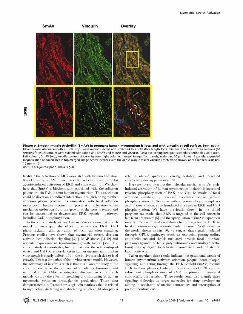
Sub-cellular localization of SmAV with the adhesionplaque marker vinculin in pregnant human myometrium
In vascular smooth muscle cells, SmAV undergoes a stimulus-
induced translocation from the core of the cell to the cell cortex
[8]. To determine if indeed SmAV colocalizes with adhesion
plaques in myometrial cells from pregnant women, we used
immunohistochemistry on term, not in labor pregnant myometrial
tissue sections to identify the subcellular location of SmAV. As can
be seen in Fig. 9, SmAV and vinculin, an adhesion plaque marker,
localize at the cell surface after the myometrial strips were
stretched for 7 min (inset, arrows).
In summary, as illustrated in Fig. 8, the immunoprecipitation
experiments demonstrated interactions between these phosphor-
ylated proteins within focal adhesion complexes and point to a
direct mechanistic link between focal adhesions, SmAV and ERK
activation.
Discussion
One of the major findings of the current study is that the role of
ERK-mediated phosphorylation of CaD previously predicted from
rat studies can be extended to the human. CaD is an actin binding
smooth muscle protein that, like troponin in striated muscle, works
with tropomyosin to block the binding site of myosin on actin.
Upon phosphorylation of the ERK site on CaD, however, a
conformational change occurs that increases actin availability for
interaction with myosin. We have shown here that not only do
CaD protein levels increase with pregnancy, as previously
described, [13,30] but also that CaD phosphorylation increases
Figure 7. SmAV protein content increases with gestation in both the human and the rat. A. Specificity of the anti SmAV antibody. B.Gestation-dependent increase in SmAV expression in human myometrium. Densitometry analysis of smooth muscle Archvillin (SmAV) protein levelsfrom immunoblots of human myometrium from nonpregnant (NP) and pregnant women. A typical blot is shown on the top. *p,0.05 vs. NP. n = 5–7samples in each group. C. Gestation-dependent increase in SmAV expression in rat myometrium. A typical blot is shown on the top. *p,0.05 comparedto NP, **p,0.01 compared to NP. ++ p,0.01 compared to D16. n = 4 in each group. NP, nonpregnant, D16 and D20, pregnant day 16 or day 20.doi:10.1371/journal.pone.0007489.g007
Myometrial Stretch Activation
PLoS ONE | www.plosone.org 10 October 2009 | Volume 4 | Issue 10 | e7489
in myometrial samples from women in labor, compared to those
from women not-in-labor. Furthermore, as predicted by the
known effects of CaD to inhibit Ca-dependent acto-myosin
interactions in vitro, we have directly shown that human
myometrial smooth muscle from pregnant women (.37 week,
not in labor) exhibits an increased Ca2+i threshold for contraction
(decreased Ca sensitivity) as CaD levels increase with gestation. In
the rat model we have previously shown that an ERK inhibitor
delays the onset of RU486-induced preterm labor [7], pointing to
a cause-and-effect relationship between ERK and the onset of
labor.
Another major finding of the current study is that stretch of
human myometrium directly induces CaD phosphorylation in
vitro. Although myometrial quiescence in vivo is clinically
important for normal pregnancy, it has not been totally clear that
how the myometrium switches phenotypically from the ‘‘myome-
trial quiescence’’ typical of the early pregnant uterus to
‘‘myometrial activation’’ during labor. Similarly, it is known that
clinical conditions that increase uterine wall tension stimulate
uterine contractions; however, the molecular mechanisms involved
have not been clear. Our results support the hypothesis that stretch
from the growing fetus at term, when the rate of fetal growth is 7
times that in the first trimester [1], promotes uterine contractions
through activation of adhesion plaques and downstream ERK
signaling. Furthermore, this stretch-sensitive signaling pathway
would help explain the high incidence of preterm labor due to
abnormal uterine stretch in conditions such as multiple gestation,
polyhydramnios, and macrosomia. Additionally, the increased
CaD expression during pregnancy may minimize the level of CaD
phosphorylation per mole CaD and counteract the effect of
stretch-induced stimuli before the onset of labor.
In the present study we report that expression levels of a newly
identified smooth muscle protein, SmAV, increase significantly
during pregnancy. SmAV, a known regulator of ERK is likely to
Figure 8. SmAV co-immunoprecipitates with phosphotyrosine, ERK and FAK. A. Myometrial SmAV associates with tyrosine-phosphorylated proteins in a stretch-dependent manner. Stretch and unstretched term pregnant human uterine smooth musclehomogenates were immunoprecipitated (IP) with anti-phospho-tyrosine antibody and immunoblotted (IB) with SmAV antibody. **p,0.01 comparedto unstretched control samples. The insert is a typical SmAV western blot with IP samples. n = 5. B. Myometrial SmAV associates with ERKprotein in a stretch- dependent manner. Stretched and unstretched term samples were immunoprecipitated with anti- ERK antibody andimmunoblotted with anti-SmAV antibody. SmAV densitometry was normalized to input signals. n = 6. **p,0.01, ***p,0.001. C. Confirmation ofstretch induced ERK phosphorylation in human myometrium. Stretch and unstretched samples were immunoprecipitated with anti- ERKantibody and immunoblotted with phospho-ERK antibody. The p-ERK2 infrared signals were also normalized to input signals. n = 6. **p,0.01. D.Myometrial ERK associates with FAK in a stretch-dependent manner. Stretch and unstretched samples homogenates wereimmunoprecipitated with anti- ERK antibody and immunoblotted with phospho-FAK925 antibody. The p-FAK infrared signals were normalized toinput signals. n = 6. ***p,0.001doi:10.1371/journal.pone.0007489.g008
Myometrial Stretch Activation
PLoS ONE | www.plosone.org 11 October 2009 | Volume 4 | Issue 10 | e7489
facilitate the activation of ERK associated with the onset of labor.
Knockdown of SmAV in vascular cells has been shown to inhibit
agonist-induced activation of ERK and contraction [8]. We show
here that SmAV is biochemically associated with, the adhesion
plaque protein FAK in term human myometrium. This association
could be direct or, an indirect interaction through binding to other
adhesion plaque proteins. Its association with focal adhesion
molecules in human myometrium places it in a location where
mechanotransduction from the growth of the fetus is sensed and
can be transmitted to downstream ERK-dependent pathways
including CaD phosphorylation.
In the current study we used an in vitro experimental stretch
model to investigate the effect of stretch on ERK, CaD
phosphorylation and activation of focal adhesion signaling.
Previous studies have shown that myometrial stretch also can
activate focal adhesion signaling [3,4], MAP kinase [31,32] and
regulate expression of transforming growth factor [33]. The
current study demonstrates, for the first time the relationship of
stretch and CaD phosphorylation in human myometrium. Brief in
vitro stretch is clearly different from the in vivo stretch due to fetal
growth. This is a limitation of the in vitro stretch model. However,
the advantage of in vitro stretch is that it is allows the study of the
effect of stretch in the absence of circulating hormones and
neuronal inputs. Other investigators also used in vitro stretch
models to study the effect of stretching and shortening of human
myometrial strips on prostaglandin production. Those data
demonstrated a differential prostaglandin synthesis that is related
to myometrial stretching and shortening which could also play a
role in uterine quiescence during gestation and increased
contractility during parturition [34].
Here we have shown that the molecular mechanisms of stretch-
induced activation of human myometrium include (1) increased
tyrosine phosphorylation of FAK, and Cas, hallmarks of focal
adhesion signaling, (2) increased association of, or tyrosine
phosphorylation of, a-actinin with adhesion plaque complexes
and (3) downstream, stretch-induced increases in ERK and CaD
phosphorylation. We have previously shown in the timed
pregnant rat model that ERK is targeted to the cell cortex in
late term pregnancy [6] and the upregulation of SmAV expression
may be one factor that contributes to the targeting of ERK to
focal adhesions in a gestation-dependent manner. As illustrated in
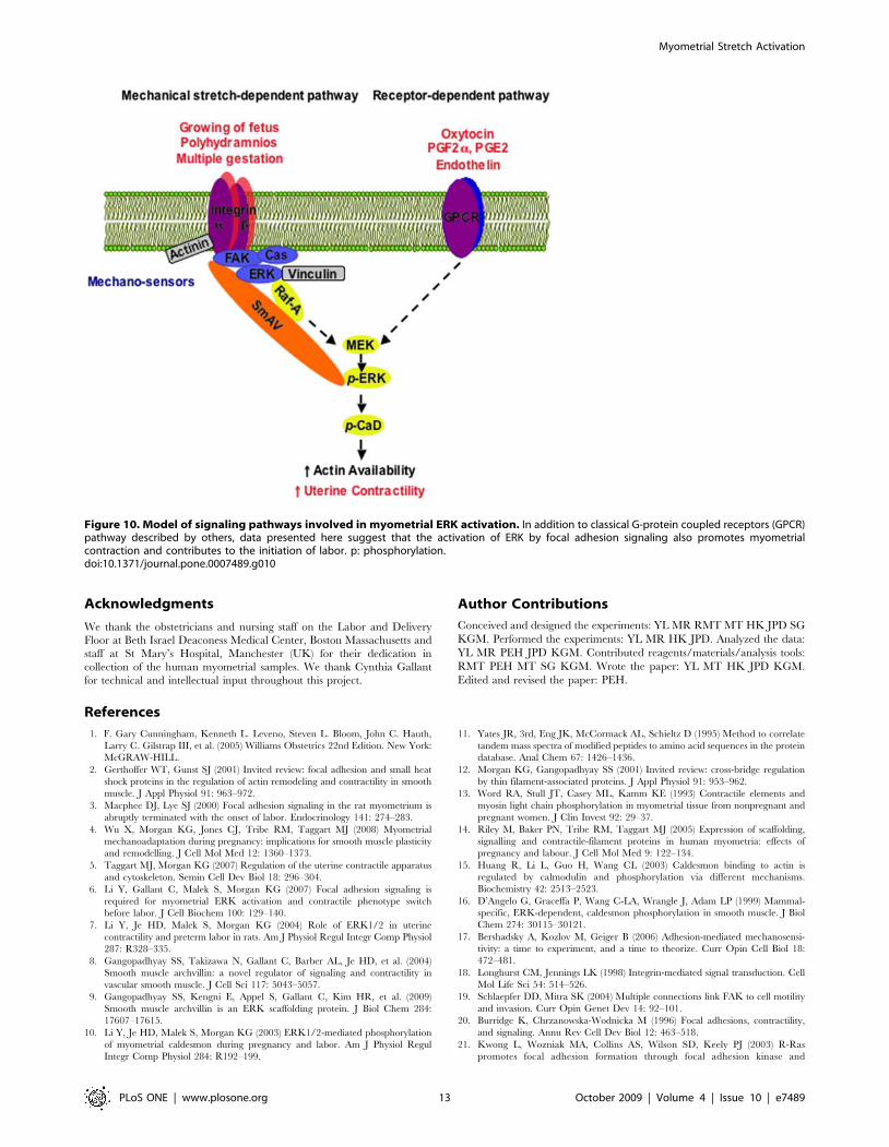
the model shown in Fig. 10, we suggest that signals mediated
through GPCR pathways (such as oxytocin, prostaglandins,
endothelin etc) and signals mediated through focal adhesions
pathways (growth of fetus, polyhydramnios and multiple gesta-
tions) may synergize to activate myometrium and initiate the
labor contractions.
Taken together, these results indicate that gestational stretch of
human myometrium activates adhesion plaque (dense plaque)
signaling, and acting through the ERK scaffold SmAV, recruits
ERK to dense plaques, leading to the activation of ERK and the
subsequent phosphorylation of CaD to promote myometrial
contractility during labor. These results could also identify these
signaling molecules as target molecules for drug development
aiming in regulation of uterine contractility and interruption of
preterm contractions.
Figure 9. Smooth muscle Archvillin (SmAV) in pregnant human myometrium is localized with vinculin at cell surface. Term, not-in-labor, human uterine smooth muscle strips were microdissected and stretched to 2 fold slack length for 7 minutes. The fresh frozen sections (10sections for each sample) were stained with rabbit anti-SmAV and mouse anti-vinculin. Alexa-dye-conjugated goat secondary antibodies were used.Left column, SmAV (red); middle column vinculin (green); right column, merged image. Top panels, scale bar, 20 mm. Lower 3 panels, expandedmagnification of boxed area in top merged image. SmAV localizes with the dense plaque maker vinculin (inset, white arrows) at cell surface. Scale bar,10 mm, n = 3.doi:10.1371/journal.pone.0007489.g009
Myometrial Stretch Activation
PLoS ONE | www.plosone.org 12 October 2009 | Volume 4 | Issue 10 | e7489
Acknowledgments
We thank the obstetricians and nursing staff on the Labor and Delivery
Floor at Beth Israel Deaconess Medical Center, Boston Massachusetts and
staff at St Mary’s Hospital, Manchester (UK) for their dedication in
collection of the human myometrial samples. We thank Cynthia Gallant
for technical and intellectual input throughout this project.
Author Contributions
Conceived and designed the experiments: YL MR RMT MT HK JPD SG
KGM. Performed the experiments: YL MR HK JPD. Analyzed the data:
YL MR PEH JPD KGM. Contributed reagents/materials/analysis tools:
RMT PEH MT SG KGM. Wrote the paper: YL MT HK JPD KGM.
Edited and revised the paper: PEH.
References
1. F. Gary Cunningham, Kenneth L. Leveno, Steven L. Bloom, John C. Hauth,
Larry C. Gilstrap III, et al. (2005) Williams Obstetrics 22nd Edition. New York:
McGRAW-HILL.
2. Gerthoffer WT, Gunst SJ (2001) Invited review: focal adhesion and small heat
shock proteins in the regulation of actin remodeling and contractility in smooth
muscle. J Appl Physiol 91: 963–972.
3. Macphee DJ, Lye SJ (2000) Focal adhesion signaling in the rat myometrium is
abruptly terminated with the onset of labor. Endocrinology 141: 274–283.
4. Wu X, Morgan KG, Jones CJ, Tribe RM, Taggart MJ (2008) Myometrial
mechanoadaptation during pregnancy: implications for smooth muscle plasticity
and remodelling. J Cell Mol Med 12: 1360–1373.
5. Taggart MJ, Morgan KG (2007) Regulation of the uterine contractile apparatus
and cytoskeleton. Semin Cell Dev Biol 18: 296–304.
6. Li Y, Gallant C, Malek S, Morgan KG (2007) Focal adhesion signaling is
required for myometrial ERK activation and contractile phenotype switch
before labor. J Cell Biochem 100: 129–140.
7. Li Y, Je HD, Malek S, Morgan KG (2004) Role of ERK1/2 in uterine
contractility and preterm labor in rats. Am J Physiol Regul Integr Comp Physiol
287: R328–335.
8. Gangopadhyay SS, Takizawa N, Gallant C, Barber AL, Je HD, et al. (2004)
Smooth muscle archvillin: a novel regulator of signaling and contractility in
vascular smooth muscle. J Cell Sci 117: 5043–5057.
9. Gangopadhyay SS, Kengni E, Appel S, Gallant C, Kim HR, et al. (2009)
Smooth muscle archvillin is an ERK scaffolding protein. J Biol Chem 284:
17607–17615.
10. Li Y, Je HD, Malek S, Morgan KG (2003) ERK1/2-mediated phosphorylation
of myometrial caldesmon during pregnancy and labor. Am J Physiol Regul
Integr Comp Physiol 284: R192–199.
11. Yates JR, 3rd, Eng JK, McCormack AL, Schieltz D (1995) Method to correlate
tandem mass spectra of modified peptides to amino acid sequences in the protein
database. Anal Chem 67: 1426–1436.
12. Morgan KG, Gangopadhyay SS (2001) Invited review: cross-bridge regulation
by thin filament-associated proteins. J Appl Physiol 91: 953–962.
13. Word RA, Stull JT, Casey ML, Kamm KE (1993) Contractile elements and
myosin light chain phosphorylation in myometrial tissue from nonpregnant and
pregnant women. J Clin Invest 92: 29–37.
14. Riley M, Baker PN, Tribe RM, Taggart MJ (2005) Expression of scaffolding,
signalling and contractile-filament proteins in human myometria: effects of
pregnancy and labour. J Cell Mol Med 9: 122–134.
15. Huang R, Li L, Guo H, Wang CL (2003) Caldesmon binding to actin is
regulated by calmodulin and phosphorylation via different mechanisms.
Biochemistry 42: 2513–2523.
16. D’Angelo G, Graceffa P, Wang C-LA, Wrangle J, Adam LP (1999) Mammal-
specific, ERK-dependent, caldesmon phosphorylation in smooth muscle. J Biol
Chem 274: 30115–30121.
17. Bershadsky A, Kozlov M, Geiger B (2006) Adhesion-mediated mechanosensi-
tivity: a time to experiment, and a time to theorize. Curr Opin Cell Biol 18:
472–481.
18. Longhurst CM, Jennings LK (1998) Integrin-mediated signal transduction. Cell
Mol Life Sci 54: 514–526.
19. Schlaepfer DD, Mitra SK (2004) Multiple connections link FAK to cell motility
and invasion. Curr Opin Genet Dev 14: 92–101.
20. Burridge K, Chrzanowska-Wodnicka M (1996) Focal adhesions, contractility,
and signaling. Annu Rev Cell Dev Biol 12: 463–518.
21. Kwong L, Wozniak MA, Collins AS, Wilson SD, Keely PJ (2003) R-Ras
promotes focal adhesion formation through focal adhesion kinase and
Figure 10. Model of signaling pathways involved in myometrial ERK activation. In addition to classical G-protein coupled receptors (GPCR)pathway described by others, data presented here suggest that the activation of ERK by focal adhesion signaling also promotes myometrialcontraction and contributes to the initiation of labor. p: phosphorylation.doi:10.1371/journal.pone.0007489.g010
Myometrial Stretch Activation
PLoS ONE | www.plosone.org 13 October 2009 | Volume 4 | Issue 10 | e7489
p130(Cas) by a novel mechanism that differs from integrins. Mol Cell Biol 23:
933–949.
22. Craig DH, Haimovich B, Basson MD (2007) Alpha-actinin-1 phosphorylation
modulates pressure-induced colon cancer cell adhesion through regulation of
focal adhesion kinase-Src interaction. Am J Physiol Cell Physiol 293:
C1862–1874.
23. Zhang Z, Lin SY, Neel BG, Haimovich B (2006) Phosphorylated alpha-actinin
and protein-tyrosine phosphatase 1B coregulate the disassembly of the focal
adhesion kinase x Src complex and promote cell migration. J Biol Chem 281:
1746–1754.
24. Triplett JW, Pavalko FM (2006) Disruption of alpha-actinin-integrin interactions
at focal adhesions renders osteoblasts susceptible to apoptosis. Am J Physiol Cell
Physiol 291: C909–921.
25. Goel HL, Dey CS (2002) Insulin-mediated tyrosine phosphorylation of myosin
heavy chain and concomitant enhanced association of C-terminal SRC kinase
during skeletal muscle differentiation. Cell Biol Int 26: 557–561.
26. Calderwood DA, Huttenlocher A, Kiosses WB, Rose DM, Woodside DG, et al.
(2001) Increased filamin binding to beta-integrin cytoplasmic domains inhibits
cell migration. Nat Cell Biol 3: 1060–1068.
27. Pal Sharma C, Goldmann WH (2004) Phosphorylation of actin-binding protein
(ABP-280; filamin) by tyrosine kinase p56lck modulates actin filament cross-
linking. Cell Biol Int 28: 935–941.
28. Takizawa N, Smith TC, Nebl T, Crowley JL, Palmieri SJ, et al. (2006)
Supervillin modulation of focal adhesions involving TRIP6/ZRP-1. J Cell Biol174: 447–458.
29. Afting EG, Becker ML, Elce JS (1979) Proteinase and proteinase-inhibitor
activities of rat uterine myometrium during pregnancy and involution. Biochem J177: 99–106.
30. Laporte R, Cohen BF, Herzlinger SF, Ansley DR, Wang C-LA, et al. (1998)Caldesmon, a putative inhibitor of smooth muscle contraction, is greatly
increased in pregnant human myometrium [abstract]. Am J Obstet Gynecol
180: S133.31. Sooranna SR, Engineer N, Loudon JA, Terzidou V, Bennett PR, et al. (2005)
The mitogen-activated protein kinase dependent expression of prostaglandin Hsynthase-2 and interleukin-8 messenger ribonucleic acid by myometrial cells: the
differential effect of stretch and interleukin-1{beta}. J Clin Endocrinol Metab90: 3517–3527.
32. Oldenhof AD, Shynlova OP, Liu M, Langille BL, Lye SJ (2002) Mitogen-
activated protein kinases mediate stretch-induced c-fos mRNA expression inmyometrial smooth muscle cells. Am J Physiol Cell Physiol 283: C1530–1539.
33. Shynlova O, Tsui P, Dorogin A, Langille BL, Lye SJ (2007) The expression oftransforming growth factor beta in pregnant rat myometrium is hormone and
stretch dependent. Reproduction 134: 503–511.
34. Hurd WW, Gibbs SG, Rudinsky KA (2008) Differential regulation ofmyometrial prostaglandin production by changes in length. Am J Obstet
Gynecol 198: 225 e221–224.
Myometrial Stretch Activation
PLoS ONE | www.plosone.org 14 October 2009 | Volume 4 | Issue 10 | e7489