Embed Size (px)
Citation preview
Stress response in the cyanobacterium
Synechocystis sp. PCC 6803
Helder Miranda
Department of Chemistry
Umeå University, Sweden
Doctoral Thesis 2011

Copyright © Helder Miranda, 2011
ISBN: 978-91-7459-201-6
Printed by: VMC-KBC, Umeå University
Umeå, Sweden 2011

To my dear family and friends

5
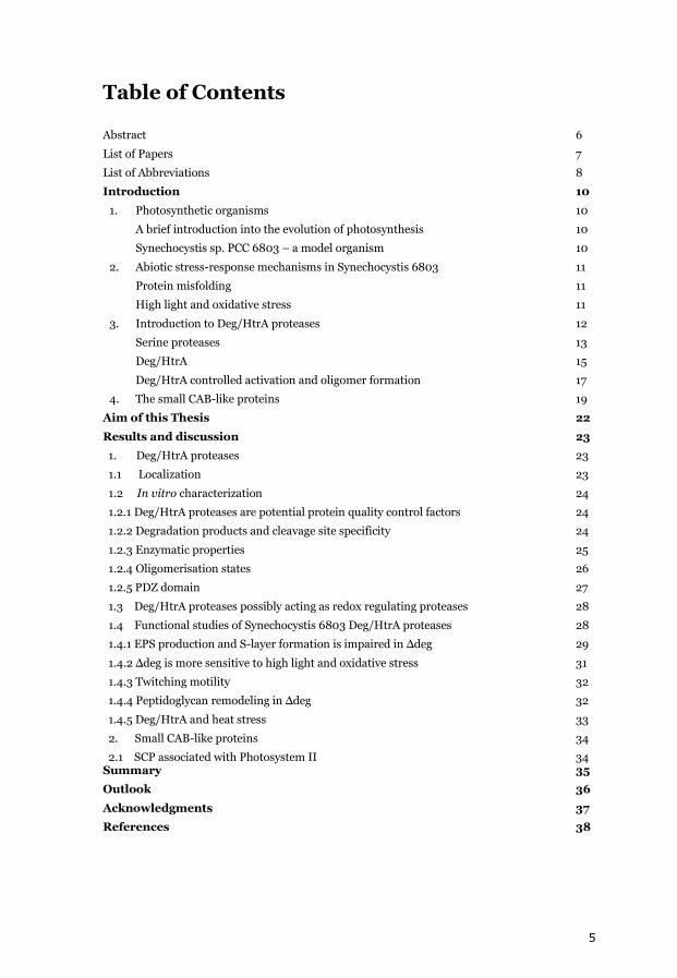
Table of Contents
Abstract 6
List of Papers 7
List of Abbreviations 8
Introduction 10
1. Photosynthetic organisms 10
A brief introduction into the evolution of photosynthesis 10
Synechocystis sp. PCC 6803 – a model organism 10
2. Abiotic stress-response mechanisms in Synechocystis 6803 11
Protein misfolding 11
High light and oxidative stress 11
3. Introduction to Deg/HtrA proteases 12
Serine proteases 13
Deg/HtrA 15
Deg/HtrA controlled activation and oligomer formation 17
4. The small CAB-like proteins 19
Aim of this Thesis 22
Results and discussion 23
1. Deg/HtrA proteases 23
1.1 Localization 23
1.2 In vitro characterization 24
1.2.1 Deg/HtrA proteases are potential protein quality control factors 24
1.2.2 Degradation products and cleavage site specificity 24
1.2.3 Enzymatic properties 25
1.2.4 Oligomerisation states 26
1.2.5 PDZ domain 27
1.3 Deg/HtrA proteases possibly acting as redox regulating proteases 28
1.4 Functional studies of Synechocystis 6803 Deg/HtrA proteases 28
1.4.1 EPS production and S-layer formation is impaired in Δdeg 29
1.4.2 Δdeg is more sensitive to high light and oxidative stress 31
1.4.3 Twitching motility 32
1.4.4 Peptidoglycan remodeling in Δdeg 32
1.4.5 Deg/HtrA and heat stress 33
2. Small CAB-like proteins 34
2.1 SCP associated with Photosystem II 34 Summary 35
Outlook 36
Acknowledgments 37
References 38
6
Abstract
Adaptation to environmental changes is important for the survival of living organisms. Under extreme abiotic conditions, organic molecules (such as lipids, proteins and nucleic acids) are prone to damage. Under these conditions stress response mechanisms are activated, either to prevent the source of damage or to promote the rapid turnover of damaged molecules. Like all photoautotrophic organisms, cyanobacteria are sensitive to high light intensity and oxidative stress, which induces damage to the photosynthetic apparatus. My thesis is divided in two subjects related to particular stress responses in the cyanobacterium Synechocystis sp. PCC 6803: 1) the role of Deg/HtrA proteases and 2) investigations on the small CAB-like proteins. Deg/HtrA proteases are ATP-independent serine endopeptidases with a characteristic C-terminal PDZ domain. These proteases are largely dispersed among living organisms, with many different functions, mostly involved in protein quality control. The genome of Synechocystis sp. PCC 6803 contains three genes coding for Deg/HtrA proteases: HtrA, HhoA and HhoB. These proteases are essential for survival under high light and heat stress, and may overlap in their functions (1). During my Ph.D. studies I focused on the identification of the precise localization of the Deg/HtrA proteases in the cyanobacterial cell, analyzed the biochemical properties of recombinant Synechocystis Deg/HtrA proteases in vitro and adopted proteomic and metabolomic approaches to study the physiological importance of these proteases. My data show that Deg/HtrA proteases are not only important in stress response mechanisms under adverse conditions, but are also involved in the stabilization of important physiological processes, such as polysaccharides biosynthesis and peptidoglycan turnover. The small CAB-like proteins (SCPs) belong to the light harvesting-like family of stress induced proteins and are thought to be involved in the photoprotection of the photosynthetic apparatus. Five small CAB-like proteins where identified in Synechocystis sp. PCC 6803 (ScpA-E). In my studies I identified another relative to the SCPs, LilA, which I found to be co-transcribed with ScpD. I also focused on the subcellular localization and identification of potential interaction partners of the SCPs.
Keywords High light, heat stress, ROS, Photosynthesis, Photosystem II, Deg/HtrA proteases, small CAB-like proteins, Refraction-2D™, MALDI-TOF, 2D-gel, GC-MS, PCA, OPLS-DA, EPS, S-layer
7
List of Papers This thesis is based on the following papers (referred to in the text by Roman numeral):
I. Recombinant Deg/HtrA proteases from Synechocystis sp. PCC 6803 differ in substrate specificity, biochemical characteristics and mechanism. Huesgen, P.F.*, Miranda, H.*, Lam, L.X., Perthold, M., Schuhmann, H., Adamska, I. and Funk, C. Biochemical Journal (2011) Immediate Publication, doi:10.1042/BJ20102131
II. Redox-dependent degradation of Photosystem II PsbO protein is
performed by Deg proteases. Roberts, I.N., Miranda, H., Lam, L.X., Kieselbach, T. and Funk, C. Submitted
III. The Deg proteases of the cyanobacterium Synechocystis sp. PCC 6803 are involved in the biosynthesis of the exopolysaccharide production and S-layer formation. Miranda, H., Sveshnikov, D., Lundgren, K., Moritz, T. and Funk, C. Submitted
IV. Proteomic comparison of a Synechocystis sp. PCC 6803 triple-Deg-deletion mutant and wild type cells under high light stress and heat shock. Miranda, H. and Funk, C. Manuscript
V. Association of small CAB-like proteins (SCPs) of Synechocystis sp. PCC 6803 with Photosystem II. Kufryk, G.*, Hernandez-Prieto, M.*, Kieselbach, T., Miranda, H. and Funk, C. Photosynthesis Research (2008) 95:135–145
*Shared first authorship

8
List of Abbreviations HtrA, high temperature requirement;
Deg, degradation of periplasmic proteins;
PDZ domain, domain originally identified in PSD95, DlgA and Zo-1 proteins;
SCP, small CAB-like protein;
CAB, chlorophyll a/b binding;
Lhc, light harvesting complex;
TMH, transmembrane helices;
ELIP, early light induced protein;
OHP, one helix proteins;
HLIP, high light induced protein;
PBS, phycobilisomes;
Lil, light harvesting-like;
Hsp, heat shock proteins;
PSII, Photosystem II;
PSI, Photosystem I;
NADPH, nicotinamide adenine dinucleotide phosphate;
ATP, adenosine triphosphate;
GDP, guanosine diphosphate;
G6P, glucose-6-phosphate;
F6P, fructose-6-phosphate;
3-PGA, phosphoglycerate;
PQ, plastoquinone;
OEC, oxygen evolving complex;
ROS, reactive oxygen species;
Δdeg, triple Deg/HtrA deletion mutant;
WT, wilde type;
IEF, isoelectric focusing;
DTT, dithiothreitol;
2D, two dimentional;
SDS PAGE, sodium dodecyl sulfate polyacrylamide gel electrophoresis;
MALDI-TOF, Matrix-assisted laser desorption/ionization-time of flight;
9
CHCA, α-Cyano-4-hydroxycinnamic acid;
CBB, Coomassie Brilliant Blue;
MS, mass spectrometry;
GC-MS, gas chromatography-mass spectrometry;
PCA, principal component analysis;
OPLS-DA, orthogonal projection to latent structures discriminant analysis;
EPS, exopolysaccharides;
TEM, transmission electron microscope;
TCA, citric acid cycle;
GST, glutathione-S-transferase;
SDR, short-chain dehydrogenases/reductases;
PRX, peroxiredoxin;
NDH-1, NAD(P)H:quinone oxidoreductase;
THF, tetrahydrofolate;
SAM, S-Adenosyl methionine;
T4P, type IV pili
10
Introduction 1. Photosynthetic organisms
A brief introduction into the evolution of photosynthesis Around 2500 million years ago, on planet Earth an extraordinary event occurred in the still young life’s history, something that would change all life permanently: the rise of the first organisms performing oxygenic photosynthesis. Early photosynthetic organisms - green sulfur bacteria, Chloroflexi (Chlorobacteria) and purple bacteria – used molecules different from water as electron donors: The green sulfur and purple bacteria used hydrogen and sulfur molecules (2); the Chloroflexi used organic acids instead (2). These organisms prospered in the highly reduced environment at the time. The first organisms performing oxygenic photosynthesis, the cyanobacteria, had the evolutionary advantage of being able to use a highly abundant molecule as electron donor, water. Using the energy of sun light to split water to gain reducing power and releasing O2 as byproduct into the atmosphere, was a breakthrough for a new era in life. Water splitting organisms appeared all over the planet and within 900 million years their massive release of oxygen changed the Earth’s atmosphere from highly reducing to an oxidizing one. The Great Oxygenation Event (3), a catastrophe with no parallel in Earth’s history, wiped out a great portion of the planet’s anaerobic organisms at the time. Life changed drastically. A multitude of organisms evolved. Capable of using oxygen – a strong electron acceptor – as the last element in the respiratory electron chain, these organisms were able to convert energy more efficiently, which opened the possibility to evolve into more complex organisms, the eukaryotes. Around 1000 million years ago the first unicellular algae evolved through symbioses between eukaryotic heterotrophic cells and cyanobacteria. Endosymbiosis led to all algae and plants today, with chloroplasts being the direct descendants of primitive cyanobacteria. Life as we know it today would be impossible without photosynthesis, therefore understanding this process is of extreme importance. Cyanobacteria, being on the one hand quite simple prokaryotes and on the other hand the ancestor of plant chloroplasts, are very well suited to study the photosynthetic process, its mechanisms and regulations.
Synechocystis sp. PCC 6803 – a model organism The unicellular cyanobacterium Synechocystis sp. PCC 6803 (hereafter Synechocystis 6803) was first isolated from a freshwater lake in 1968. It can grow photoautotrophically as well as heterotrophically in the absence of light, making it very suitable for photosynthetic studies and mutant generation. It is a naturally competent strain - able to uptake and integrate exogenous DNA into its genome – and was the first photosynthetic organism to have its entire genome sequenced (4).
11
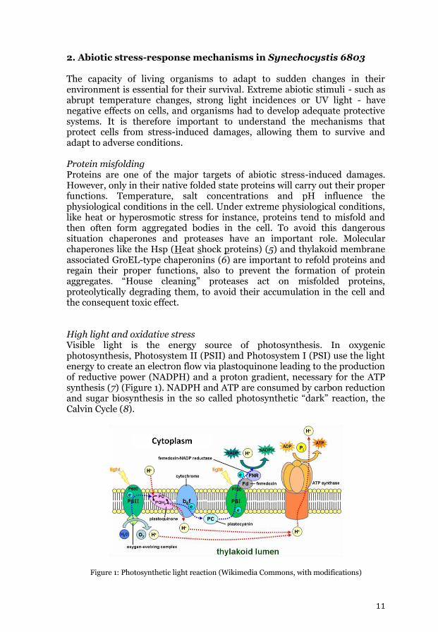
2. Abiotic stress-response mechanisms in Synechocystis 6803 The capacity of living organisms to adapt to sudden changes in their environment is essential for their survival. Extreme abiotic stimuli - such as abrupt temperature changes, strong light incidences or UV light - have negative effects on cells, and organisms had to develop adequate protective systems. It is therefore important to understand the mechanisms that protect cells from stress-induced damages, allowing them to survive and adapt to adverse conditions. Protein misfolding Proteins are one of the major targets of abiotic stress-induced damages. However, only in their native folded state proteins will carry out their proper functions. Temperature, salt concentrations and pH influence the physiological conditions in the cell. Under extreme physiological conditions, like heat or hyperosmotic stress for instance, proteins tend to misfold and then often form aggregated bodies in the cell. To avoid this dangerous situation chaperones and proteases have an important role. Molecular chaperones like the Hsp (Heat shock proteins) (5) and thylakoid membrane associated GroEL-type chaperonins (6) are important to refold proteins and regain their proper functions, also to prevent the formation of protein aggregates. “House cleaning” proteases act on misfolded proteins, proteolytically degrading them, to avoid their accumulation in the cell and the consequent toxic effect. High light and oxidative stress Visible light is the energy source of photosynthesis. In oxygenic photosynthesis, Photosystem II (PSII) and Photosystem I (PSI) use the light energy to create an electron flow via plastoquinone leading to the production of reductive power (NADPH) and a proton gradient, necessary for the ATP synthesis (7) (Figure 1). NADPH and ATP are consumed by carbon reduction and sugar biosynthesis in the so called photosynthetic “dark” reaction, the Calvin Cycle (8).
Figure 1: Photosynthetic light reaction (Wikimedia Commons, with modifications)
12
PSII is the oxygen-evolving photosystem, where light energy is used to split water into hydrogen (4H+) and oxygen (O2). Plastoquinone (PQ) molecules transport these electrons through the lipid-bilayer of the thylakoid membrane. With the incidence of light, PQ is reduced by electrons from PSII and then will be oxidized by PSI via the cytochrome b6/f complex. High light intensities promote an increase of the PQH2 pool (9, 10). The double protonated PQ does not function in electron transport, but inhibits PSII and increases the rate of triplet chlorophyll (P680) formation (10). Reacting with oxygen, triplet P680 produces singlet oxygen (1O2), highly reactive oxygen specie (ROS). ROS are extremely reactive molecules that contain oxygen with unpaired valence electrons, consisting of free radicals (oxygen ions) or non-radicals (H2O2 and 1O2). They tend to react with non-radical compounds, converting them into radicals (11), inducing damage in membrane lipids, nuclear acids and proteins. Singlet oxygen interacts with integral proteins of PSII, such as the reaction center protein D1 (12), which binds cofactors involved in the primary and secondary photosynthetic electron flow and is therefore prone to irreversible oxidative damage (12). D1 turnover is an essential process to maintain PSII activity and the photosynthetic electron flow (13). Photoinhibition occurs when the excess light energy is not used by the photosynthetic electron flow, but instead is responsible for the increasing rate of singlet oxygen formation, which overcomes the D1 turnover rate, impairing PSII activity (13, 14). Oxidative stress is the result of an imbalance between the formation of ROS and the ability of an organism to remove/detoxify the reactive intermediates or damage reparation. Oxidative stress can be the result of an increase in ROS formation, antioxidant protection decrease or less effective ROS induced oxidative damage repair. For protection against high light intensities, plants and algae developed various mechanisms, one of them being the non-photochemical quenching mechanism, allowing energy dissipation through heat and therefore preventing the formation of triplet P680.
3. Introduction to Deg/HtrA proteases Proteases are enzymes that mediate protein degradation - proteolysis - by hydrolyzing the peptide bond of the polypeptide chain. Therefore proteases are extremely important enzymes involved in a wide range of physiological reactions, generally related to protein digestion, degradation of misfolded proteins, protein maturation and highly regulated signaling cascades. Research on proteases therefore is extremely relevant, covering close to 9000 research publications each year. Proteases are classified either according to their optimal pH for activity into acidic, neutral or basic proteases, or according to the properties of their catalytic site and mechanism of action. In the later classification six protease groups are nominated:
13
Aspartate proteases
Glutamic acid proteases
Cysteine proteases
Threonine proteases
Metallo proteases
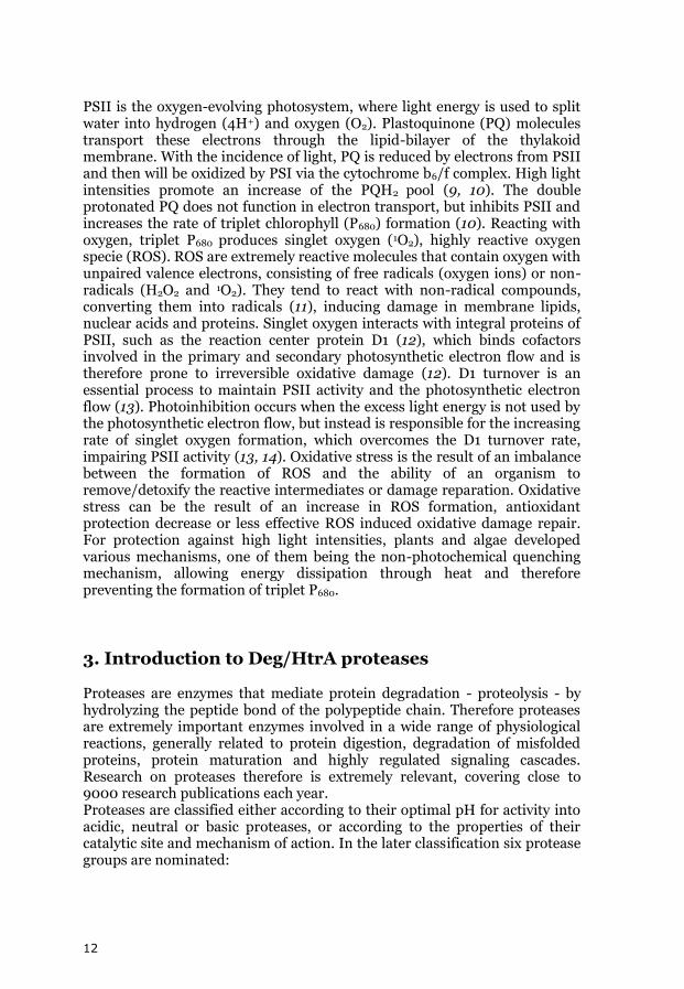
Serine proteases Serine proteases Serine proteases are ATP-independent proteases. Their active site is composed of a set of amino acid residues that are essential for activity (usually a triad), with serine being the central residue for all the members of this protease group (15). Their mechanism of action (Figure 5) involves a nucleophilic attack of the targeted peptide bond by the serine residue. In most of the cases, histidine and aspartate are also part of the catalytic triad, with histidine held in a proton acceptor state by aspartate.
Figure 2: Mechanism of action of the catalytic triad in serine proteases (Wikimedia Commons)
Serine proteases are the most widely distributed family of proteases; more than one third of all known proteases belong to this group. They are present in nearly all known organisms, in the taxonomic groups of Archae, Bacteria or
14
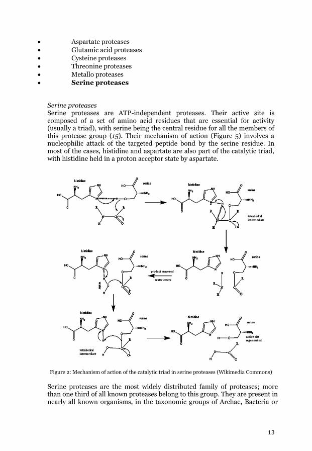
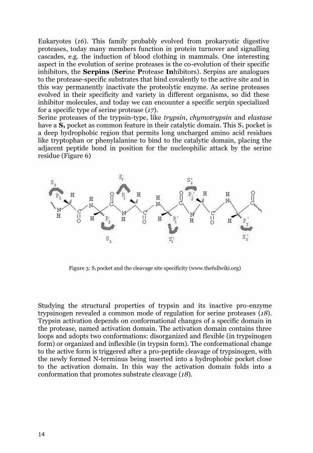
Eukaryotes (16). This family probably evolved from prokaryotic digestive proteases, today many members function in protein turnover and signalling cascades, e.g. the induction of blood clothing in mammals. One interesting aspect in the evolution of serine proteases is the co-evolution of their specific inhibitors, the Serpins (Serine Protease Inhibitors). Serpins are analogues to the protease-specific substrates that bind covalently to the active site and in this way permanently inactivate the proteolytic enzyme. As serine proteases evolved in their specificity and variety in different organisms, so did these inhibitor molecules, and today we can encounter a specific serpin specialized for a specific type of serine protease (17). Serine proteases of the trypsin-type, like trypsin, chymotrypsin and elastase have a S1 pocket as common feature in their catalytic domain. This S1 pocket is a deep hydrophobic region that permits long uncharged amino acid residues like tryptophan or phenylalanine to bind to the catalytic domain, placing the adjacent peptide bond in position for the nucleophilic attack by the serine residue (Figure 6)
Figure 3: S1 pocket and the cleavage site specificity (www.thefullwiki.org)
Studying the structural properties of trypsin and its inactive pro-enzyme trypsinogen revealed a common mode of regulation for serine proteases (18). Trypsin activation depends on conformational changes of a specific domain in the protease, named activation domain. The activation domain contains three loops and adopts two conformations: disorganized and flexible (in trypsinogen form) or organized and inflexible (in trypsin form). The conformational change to the active form is triggered after a pro-peptide cleavage of trypsinogen, with the newly formed N-terminus being inserted into a hydrophobic pocket close to the activation domain. In this way the activation domain folds into a conformation that promotes substrate cleavage (18).
15
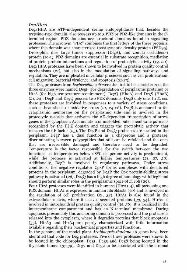
Deg/HtrA Deg/HtrA are ATP-independent serine endopeptidases that, besides the trypsine-type domain, also possess up to 3 PDZ or PDZ-like domains in the C-terminal region. PDZ domains are structural domains found in signalling proteases. The acronym “PDZ” combines the first letters of the three proteins, where this domain was characterized (post synaptic density protein (PSD95), Drosophila disc large tumor suppressor (DlgA), and zonula occludens-1 protein (zo-1). PDZ domains are essential in substrate recognition, mediation of protein-protein interactions and regulation of proteolytic activity (19, 20). Deg/HtrA proteases have been shown to be involved in protein quality control mechanisms (21), but also in the modulation of signalling pathways and regulation. They are implicated in cellular processes such as cell proliferation, cell migration, bacterial virulence, and apoptosis (21-23). The Deg proteases from Escherichia coli were the first to be characterized, the three enzymes were named DegP (for degradation of periplasmic proteins) or HtrA (for high temperature requirement), DegQ (HhoA) and DegS (HhoB) (21, 24). DegP and DegQ possess two PDZ domains, DegS only one. In E. coli these proteases are involved in responses to a variety of stress conditions, such as heat shock or oxidative stress (21, 24-26). DegS is anchored to the cytoplasmic membrane on the periplasmic side and is involved in the proteolytic cascade that activates the σE-dependent transcription of stress genes in the cytoplasm. Accumulation of misfolded outer membrane porins is recognized by the PDZ domain and triggers the proteolytic activity that releases the σE factor (25). The DegP and DegQ proteases are located in the periplasm. DegP has a dual function as a chaperone and a protease, discriminating between polypeptides that still can be refolded and the ones that are irreversible damaged and therefore need to be degraded. Temperature is the factor responsible for the switch between the two functions, at temperatures below 28ºC chaperone activity is predominant, while the protease is activated at higher temperatures (21, 27, 28). Additionally, DegP is involved in regulatory pathways. Under stress conditions, the negative regulator CpxP forms complexes with denatured proteins in the periplasm, degraded by DegP the Cpx protein-folding stress pathway is activated (26). DegQ has a high degree of homology with DegP and should perform similar roles in the periplasmic space of E. coli (29). Four HtrA proteases were identified in humans (HtrA1-4), all possessing one PDZ domain. HtrA1 is expressed in human fibroblasts (30) and is involved in the regulation of cell proliferation (31, 32). HtrA1 is also found in the extracellular matrix, where it cleaves secreted proteins (33, 34). HtrA2 is involved in mitochondrial protein quality control (35, 36). It is localized in the intermembrane compartment and has an N-terminal membrane. During apoptosis presumably this anchoring domain is processed and the protease is released into the cytoplasm, where it degrades proteins that block apoptosis (35). HtrA3 and HtrA4 are poorly characterized with little information available regarding their biochemical properties and functions. In the genome of the model plant Arabidopsis thaliana 16 genes have been identified that code for Deg proteases. Five of these proteases were shown to be located in the chloroplast: Deg1, Deg5 and Deg8 being located in the thylakoid lumen (37-39), Deg7 and Deg2 to be associated with the stromal
16
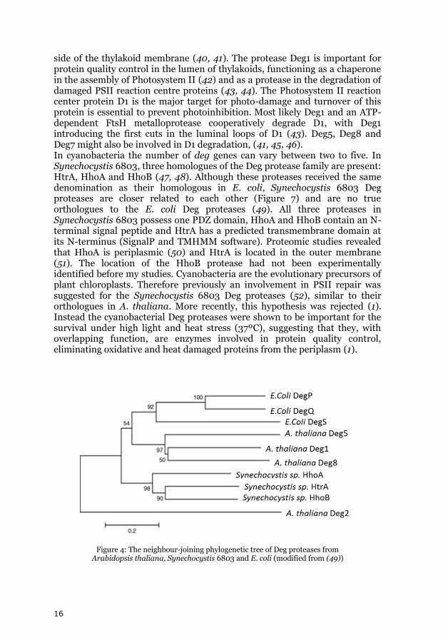
side of the thylakoid membrane (40, 41). The protease Deg1 is important for protein quality control in the lumen of thylakoids, functioning as a chaperone in the assembly of Photosystem II (42) and as a protease in the degradation of damaged PSII reaction centre proteins (43, 44). The Photosystem II reaction center protein D1 is the major target for photo-damage and turnover of this protein is essential to prevent photoinhibition. Most likely Deg1 and an ATP-dependent FtsH metalloprotease cooperatively degrade D1, with Deg1 introducing the first cuts in the luminal loops of D1 (43). Deg5, Deg8 and Deg7 might also be involved in D1 degradation, (41, 45, 46). In cyanobacteria the number of deg genes can vary between two to five. In Synechocystis 6803, three homologues of the Deg protease family are present: HtrA, HhoA and HhoB (47, 48). Although these proteases received the same denomination as their homologous in E. coli, Synechocystis 6803 Deg proteases are closer related to each other (Figure 7) and are no true orthologues to the E. coli Deg proteases (49). All three proteases in Synechocystis 6803 possess one PDZ domain, HhoA and HhoB contain an N-terminal signal peptide and HtrA has a predicted transmembrane domain at its N-terminus (SignalP and TMHMM software). Proteomic studies revealed that HhoA is periplasmic (50) and HtrA is located in the outer membrane (51). The location of the HhoB protease had not been experimentally identified before my studies. Cyanobacteria are the evolutionary precursors of plant chloroplasts. Therefore previously an involvement in PSII repair was suggested for the Synechocystis 6803 Deg proteases (52), similar to their orthologues in A. thaliana. More recently, this hypothesis was rejected (1). Instead the cyanobacterial Deg proteases were shown to be important for the survival under high light and heat stress (37ºC), suggesting that they, with overlapping function, are enzymes involved in protein quality control, eliminating oxidative and heat damaged proteins from the periplasm (1).
Figure 4: The neighbour-joining phylogenetic tree of Deg proteases from Arabidopsis thaliana, Synechocystis 6803 and E. coli (modified from (49))
17
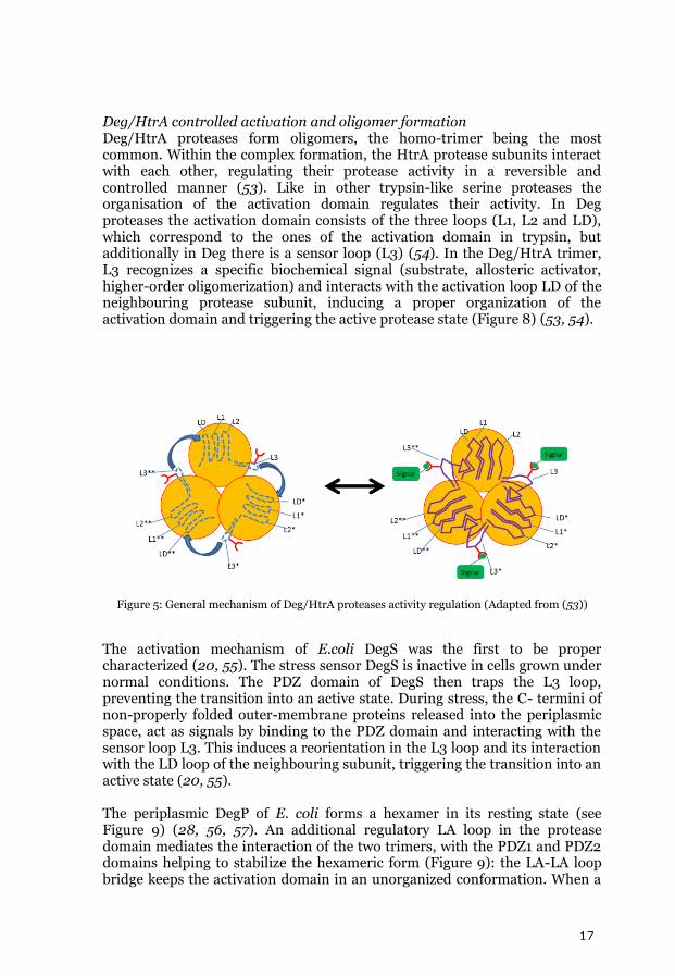
Deg/HtrA controlled activation and oligomer formation Deg/HtrA proteases form oligomers, the homo-trimer being the most common. Within the complex formation, the HtrA protease subunits interact with each other, regulating their protease activity in a reversible and controlled manner (53). Like in other trypsin-like serine proteases the organisation of the activation domain regulates their activity. In Deg proteases the activation domain consists of the three loops (L1, L2 and LD), which correspond to the ones of the activation domain in trypsin, but additionally in Deg there is a sensor loop (L3) (54). In the Deg/HtrA trimer, L3 recognizes a specific biochemical signal (substrate, allosteric activator, higher-order oligomerization) and interacts with the activation loop LD of the neighbouring protease subunit, inducing a proper organization of the activation domain and triggering the active protease state (Figure 8) (53, 54).
Figure 5: General mechanism of Deg/HtrA proteases activity regulation (Adapted from (53))
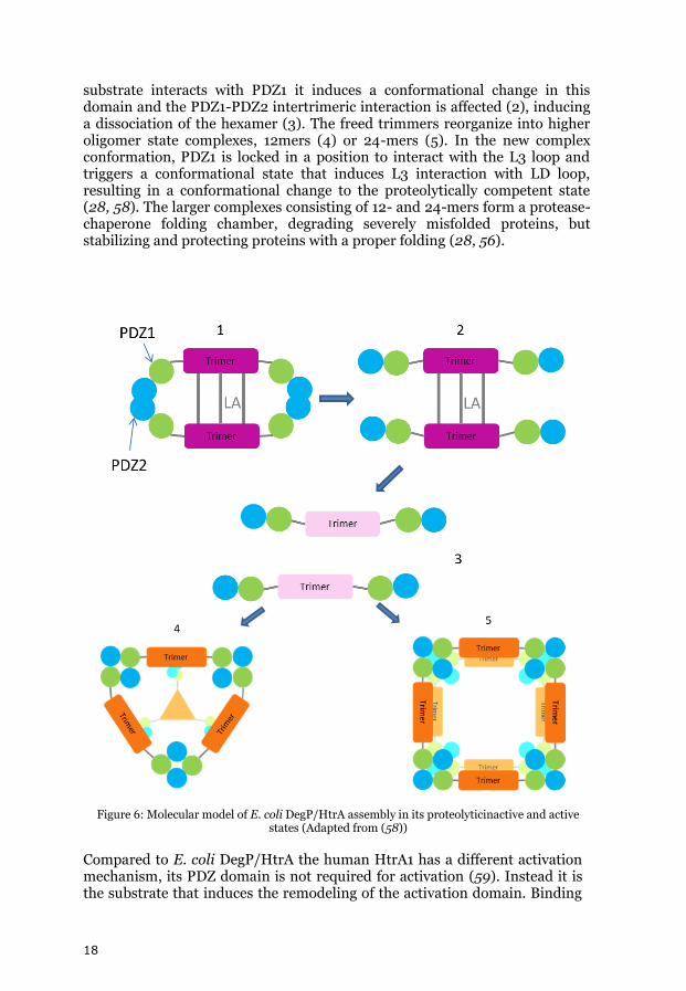
The activation mechanism of E.coli DegS was the first to be proper characterized (20, 55). The stress sensor DegS is inactive in cells grown under normal conditions. The PDZ domain of DegS then traps the L3 loop, preventing the transition into an active state. During stress, the C- termini of non-properly folded outer-membrane proteins released into the periplasmic space, act as signals by binding to the PDZ domain and interacting with the sensor loop L3. This induces a reorientation in the L3 loop and its interaction with the LD loop of the neighbouring subunit, triggering the transition into an active state (20, 55). The periplasmic DegP of E. coli forms a hexamer in its resting state (see Figure 9) (28, 56, 57). An additional regulatory LA loop in the protease domain mediates the interaction of the two trimers, with the PDZ1 and PDZ2 domains helping to stabilize the hexameric form (Figure 9): the LA-LA loop bridge keeps the activation domain in an unorganized conformation. When a
18
substrate interacts with PDZ1 it induces a conformational change in this domain and the PDZ1-PDZ2 intertrimeric interaction is affected (2), inducing a dissociation of the hexamer (3). The freed trimmers reorganize into higher oligomer state complexes, 12mers (4) or 24-mers (5). In the new complex conformation, PDZ1 is locked in a position to interact with the L3 loop and triggers a conformational state that induces L3 interaction with LD loop, resulting in a conformational change to the proteolytically competent state (28, 58). The larger complexes consisting of 12- and 24-mers form a protease-chaperone folding chamber, degrading severely misfolded proteins, but stabilizing and protecting proteins with a proper folding (28, 56).
Figure 6: Molecular model of E. coli DegP/HtrA assembly in its proteolyticinactive and active states (Adapted from (58))
Compared to E. coli DegP/HtrA the human HtrA1 has a different activation mechanism, its PDZ domain is not required for activation (59). Instead it is the substrate that induces the remodeling of the activation domain. Binding
19
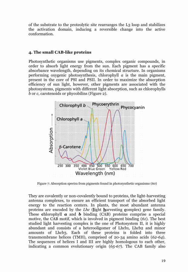
of the substrate to the proteolytic site rearranges the L3 loop and stabilizes the activation domain, inducing a reversible change into the active conformation. 4. The small CAB-like proteins Photosynthetic organisms use pigments, complex organic compounds, in order to absorb light energy from the sun. Each pigment has a specific absorbance wavelength, depending on its chemical structure. In organisms performing oxygenic photosynthesis, chlorophyll a is the main pigment, present in the core of PSI and PSII. In order to maximize the absorption efficiency of sun light, however, other pigments are associated with the photosystems, pigments with different light absorption, such as chlorophylls b or c, carotenoids or phycobilins (Figure 2).
Figure 7: Absorption spectra from pigments found in photosynthetic organisms (60)
They are covalently or non-covalently bound to proteins, the light-harvesting antenna complexes, to ensure an efficient transport of the absorbed light energy to the reaction centers. In plants, the most abundant antenna proteins are encoded by the Lhc (light harvesting complex) gene family. These chlorophyll a and b binding (CAB) proteins comprise a special motive, the CAB motif, which is involved in pigment binding (61). The best studied light harvesting complex is the one of Photosystem II, it is highly abundant and consists of a heterooligomer of Lhcb1, Lhcb2 and minor amounts of Lhcb3. Each of these proteins is folded into three transmembrane helices (TMH), comprised of 20-34 amino acids (62-64). The sequences of helices I and III are highly homologous to each other, indicating a common evolutionary origin (65-67). The CAB family also
20
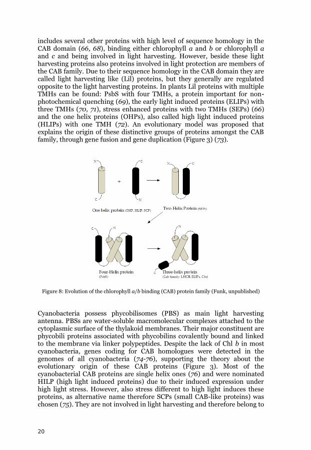
includes several other proteins with high level of sequence homology in the CAB domain (66, 68), binding either chlorophyll a and b or chlorophyll a and c and being involved in light harvesting. However, beside these light harvesting proteins also proteins involved in light protection are members of the CAB family. Due to their sequence homology in the CAB domain they are called light harvesting like (Lil) proteins, but they generally are regulated opposite to the light harvesting proteins. In plants Lil proteins with multiple TMHs can be found: PsbS with four TMHs, a protein important for non-photochemical quenching (69), the early light induced proteins (ELIPs) with three TMHs (70, 71), stress enhanced proteins with two TMHs (SEPs) (66) and the one helix proteins (OHPs), also called high light induced proteins (HLIPs) with one TMH (72). An evolutionary model was proposed that explains the origin of these distinctive groups of proteins amongst the CAB family, through gene fusion and gene duplication (Figure 3) (73).
Figure 8: Evolution of the chlorophyll a/b binding (CAB) protein family (Funk, unpublished)
Cyanobacteria possess phycobilisomes (PBS) as main light harvesting antenna. PBSs are water-soluble macromolecular complexes attached to the cytoplasmic surface of the thylakoid membranes. Their major constituent are phycobili proteins associated with phycobilins covalently bound and linked to the membrane via linker polypeptides. Despite the lack of Chl b in most cyanobacteria, genes coding for CAB homologues were detected in the genomes of all cyanobacteria (74-76), supporting the theory about the evolutionary origin of these CAB proteins (Figure 3). Most of the cyanobacterial CAB proteins are single helix ones (76) and were nominated HILP (high light induced proteins) due to their induced expression under high light stress. However, also stress different to high light induces these proteins, as alternative name therefore SCPs (small CAB-like proteins) was chosen (75). They are not involved in light harvesting and therefore belong to
21
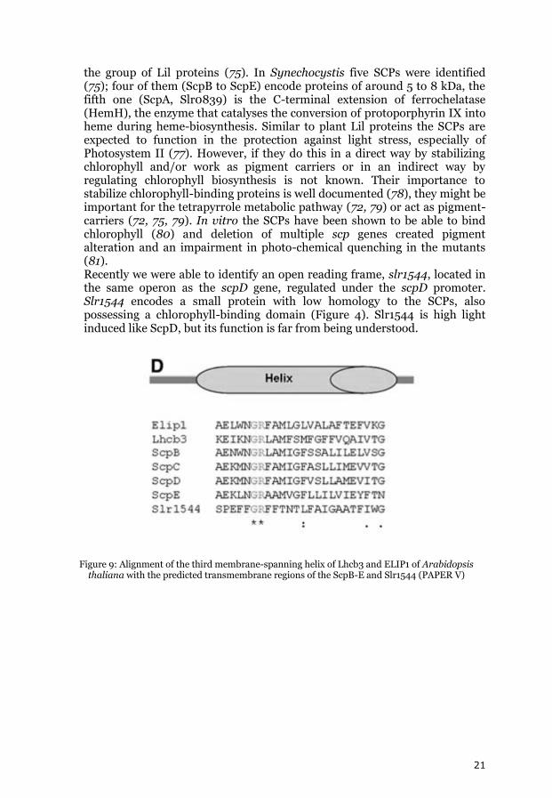
the group of Lil proteins (75). In Synechocystis five SCPs were identified (75); four of them (ScpB to ScpE) encode proteins of around 5 to 8 kDa, the fifth one (ScpA, Slr0839) is the C-terminal extension of ferrochelatase (HemH), the enzyme that catalyses the conversion of protoporphyrin IX into heme during heme-biosynthesis. Similar to plant Lil proteins the SCPs are expected to function in the protection against light stress, especially of Photosystem II (77). However, if they do this in a direct way by stabilizing chlorophyll and/or work as pigment carriers or in an indirect way by regulating chlorophyll biosynthesis is not known. Their importance to stabilize chlorophyll-binding proteins is well documented (78), they might be important for the tetrapyrrole metabolic pathway (72, 79) or act as pigment-carriers (72, 75, 79). In vitro the SCPs have been shown to be able to bind chlorophyll (80) and deletion of multiple scp genes created pigment alteration and an impairment in photo-chemical quenching in the mutants (81). Recently we were able to identify an open reading frame, slr1544, located in the same operon as the scpD gene, regulated under the scpD promoter. Slr1544 encodes a small protein with low homology to the SCPs, also possessing a chlorophyll-binding domain (Figure 4). Slr1544 is high light induced like ScpD, but its function is far from being understood.
Figure 9: Alignment of the third membrane-spanning helix of Lhcb3 and ELIP1 of Arabidopsis thaliana with the predicted transmembrane regions of the ScpB-E and Slr1544 (PAPER V)
22
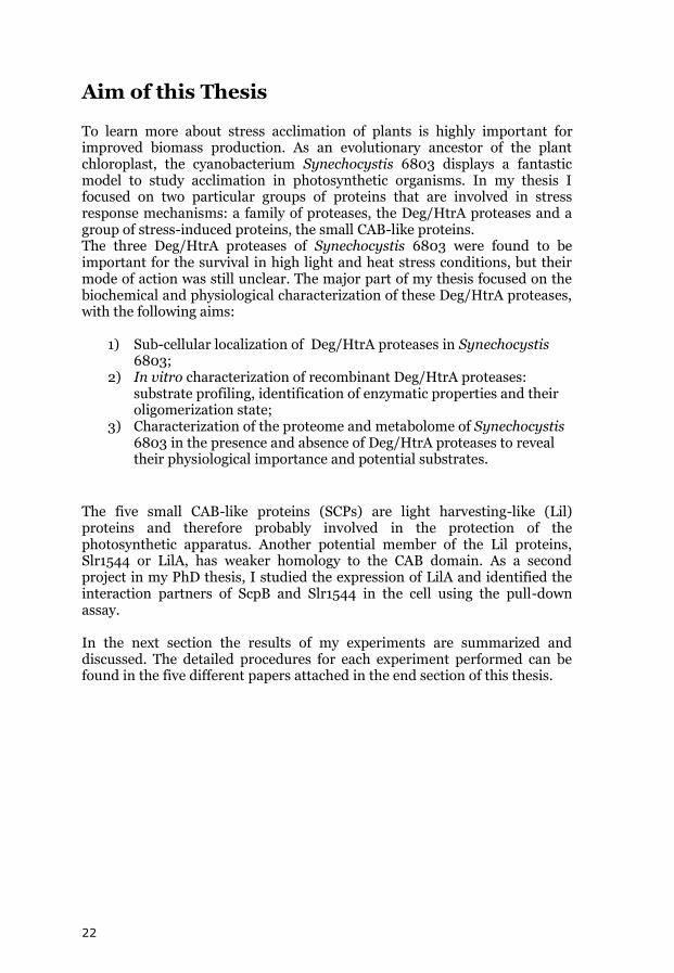
Aim of this Thesis To learn more about stress acclimation of plants is highly important for improved biomass production. As an evolutionary ancestor of the plant chloroplast, the cyanobacterium Synechocystis 6803 displays a fantastic model to study acclimation in photosynthetic organisms. In my thesis I focused on two particular groups of proteins that are involved in stress response mechanisms: a family of proteases, the Deg/HtrA proteases and a group of stress-induced proteins, the small CAB-like proteins. The three Deg/HtrA proteases of Synechocystis 6803 were found to be important for the survival in high light and heat stress conditions, but their mode of action was still unclear. The major part of my thesis focused on the biochemical and physiological characterization of these Deg/HtrA proteases, with the following aims:
1) Sub-cellular localization of Deg/HtrA proteases in Synechocystis 6803;
2) In vitro characterization of recombinant Deg/HtrA proteases: substrate profiling, identification of enzymatic properties and their oligomerization state;
3) Characterization of the proteome and metabolome of Synechocystis 6803 in the presence and absence of Deg/HtrA proteases to reveal their physiological importance and potential substrates.
The five small CAB-like proteins (SCPs) are light harvesting-like (Lil) proteins and therefore probably involved in the protection of the photosynthetic apparatus. Another potential member of the Lil proteins, Slr1544 or LilA, has weaker homology to the CAB domain. As a second project in my PhD thesis, I studied the expression of LilA and identified the interaction partners of ScpB and Slr1544 in the cell using the pull-down assay. In the next section the results of my experiments are summarized and discussed. The detailed procedures for each experiment performed can be found in the five different papers attached in the end section of this thesis.
23
Results and discussion
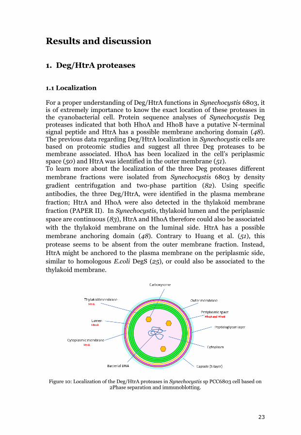
1. Deg/HtrA proteases 1.1 Localization For a proper understanding of Deg/HtrA functions in Synechocystis 6803, it is of extremely importance to know the exact location of these proteases in the cyanobacterial cell. Protein sequence analyses of Synechocystis Deg proteases indicated that both HhoA and HhoB have a putative N-terminal signal peptide and HtrA has a possible membrane anchoring domain (48). The previous data regarding Deg/HtrA localization in Synechocystis cells are based on proteomic studies and suggest all three Deg proteases to be membrane associated. HhoA has been localized in the cell’s periplasmic space (50) and HtrA was identified in the outer membrane (51). To learn more about the localization of the three Deg proteases different
membrane fractions were isolated from Synechocystis 6803 by density
gradient centrifugation and two-phase partition (82). Using specific
antibodies, the three Deg/HtrA, were identified in the plasma membrane
fraction; HtrA and HhoA were also detected in the thylakoid membrane
fraction (PAPER II). In Synechocystis, thylakoid lumen and the periplasmic
space are continuous (83), HtrA and HhoA therefore could also be associated
with the thylakoid membrane on the luminal side. HtrA has a possible
membrane anchoring domain (48). Contrary to Huang et al. (51), this
protease seems to be absent from the outer membrane fraction. Instead,
HtrA might be anchored to the plasma membrane on the periplasmic side,
similar to homologous E.coli DegS (25), or could also be associated to the
thylakoid membrane.
Figure 10: Localization of the Deg/HtrA proteases in Synechocystis sp PCC6803 cell based on 2Phase separation and immunoblotting.
24
1.2 In vitro characterization To study the biochemical properties of the three cyanobacterial Deg/HtrA proteases, HtrA, HhoA and HhoB were expressed as recombinant fusion proteins with a C-terminal His-tag using the E. coli expression system. In order to optimize the expression and purification yield of the proteases in vitro, the putative transmembrane-domain of HtrA, as well as the predicted signal peptides of HhoA and HhoB were truncated (PAPER I). Inactive versions of the recombinant fusion proteases (the serine residue in the catalytic site substituted by alanine) were also expressed. 1.2.1 Deg/HtrA proteases are potential protein quality control factors Model substrates such as bovine β-casein, BSA (Bovine Serum Albumine) and lysozyme were used to assay the activity and substrate specificity of the Deg/HtrA proteases (PAPER I). All three Deg proteases were able to degrade naturally unfolded β-casein, but not correctly folded lysozyme or globular BSA. Treatment with DTT to reduce of the disulfide bonds resulted in cleavage of partially unfolded lysozyme by HhoA and HtrA (to a lower extent), but not by HhoB (PAPER I). In vitro substrate profiling indicated that the Deg proteases of Synechocystis 6803 have a preference for (chemically) unfolded substrates, and therefore might act as protein quality control factors (1). Recombinant HhoA had the strongest proteolytic activity, recombinant HhoB was least active. Under the assumption that the activity of recombinant HhoB is not reduced only due to the construct chosen, it could be due to a more stringent substrate specificity (similar to its E. coli homonymous (84)), a requirement for specific allosteric activators for optimal activity (56, 85, 86); alternatively HhoB might function more as a chaperone rather than protease. 1.2.2 Degradation products and cleavage site specificity Proteases cleavage specificity in general is defined by amino acid residues that are positioned around the cleavage site, most importantly the non-prime site residues P3, P2 and P1 that precede the cleavage site, and the prime site residues P1’, P2’ and P3’following the cleavage site (87). Similar to other trypsin-type proteases, E. coli DegP has a strong preference for small hydrophobic residues (alanine, valine, threonine and isoleucine) in the P1 position, P2 and P3 non-prime sites are less specific (88). Interestingly, the binding site of PDZ1 domain and the catalytic domain in DegP exhibit extremely similar substrate specificities, both binding preferentially to small hydrophobic residues at the C-terminal (88). The PDZ1 domain would bind to the C-terminus of a substrate protein and direct the closest cleavage site (about 10 or 15 residues further) to the catalytic domain for peptide cutting. The introduced cut generates a new C-terminal that can bind to a nearby PDZ1 domain in the proteolytically active DegP oligomer, starting a new digestive cycle. This mechanism of “bind-cut-release-rebind” results in the processive digestion of a substrate protein into
25
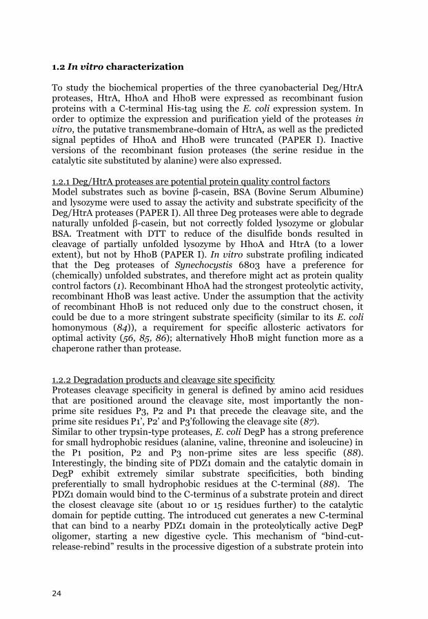
small peptides of 10 to 20 amino acid residues, without the accumulation of intermediates (28, 88). When degrading β-casein, all three Synechocystis 6803 Deg proteases generate distinct degradation products/intermediates (PAPER I). The PDZ domain of these proteases might bind preferentially to hydrophobic residues at the C-terminus, but the Deg/HtrA proteases of Synechocystis 6803 do not degrade proteins in a processive manner, releasing their substrates after cleavage, similar to E.coli DegS (25), Arabidopsis thaliana Deg2 (40) and Deg15 (89, 90). The processive degradation of E. coli DegP might be explained by the presence of a second PDZ domain in its structure, which could facilitate the “bind-cut-release-rebind” mechanism. 1.2.3 Enzymatic properties The enzymatic properties of Synechocystis 6803 HtrA, HhoA and HhoB were characterized using BODIPY TR-X-labeled casein as a fluorogenic substrate (EnzChek, Invitrogen detection technologies). The proteolytic degradation kinetics of fluorogenic substrate are correlated to fluorescence emission (Figure 11). Optimal pH and temperature for the proteolytic activity of the three Deg/HtrA proteases as well as the effect of divalent ions were tested (PAPER I).
Figure 11: Principle of EnzChek® Protease Assay Kit. Casein is labeled with a fluorophore that is intramolecular quenched. With the proteolytic degradation of casein, self-quenching of the fluorophores is abolished and a fluorescence signal increases proportionally to the protease activity (Invitrogen Life Science: Molecular Probes, The Handbook)
The protease activity of HtrA, HhoA and HhoB increases significantly in the presence of the divalent cations Mg2+ and Ca2+. The activity of HtrA and HhoA increased four to six times in the presence of CaCl2, maximal increase was reached at a concentration of 5 mM CaCl2. The activity of HhoB increased even more significantly (up to 14 fold), reaching a maximum at 10 mM CaCl2. A similar effect was observed after the addition of MgCl2 (PAPER I). The divalent cations could either interact with the substrate and make it more susceptible for degradation and/or they could bind to the Deg/HtrA protease affecting its structure and activity. One should note that
26
the proteolytic activity of recombinant HhoB was almost negligible in the absence of divalent cations. When measuring the proteolytic activity at different temperatures, the three Deg/HtrA proteases showed very low activity between 10⁰C and 25⁰C. HtrA displayed maximum activity between 30°C and 40°C, the activity of HhoA increased with rising temperature, reaching its maximum at around 55°C and decreased rapidly at higher temperatures (probably due to denaturation of the enzyme). HhoB behaved similar to HhoA, its activity reached a maximum at 45°C (PAPER I). Proteolytic activity was measured in a pH range from pH 4.5 to 8.0, in the presence or absence of CaCl2. Without addition of CaCl2, HtrA showed a maximal proteolytic activity between pH 5.5 and 6.0, whereas HhoA had its optimal activity at pH 6.5 (HhoB showed no detectable proteolytic activity) (PAPER I). Addition of 10 mM CaCl2 significantly changed the optimal pH for activity of HtrA and HhoA. HtrA displayed a broader pH optimum, with highest activity at pH 7.0. HhoA, showed a very unusual progressive increase in activity with higher pH. The proteolytic activity of HhoB increased in the presence of Ca2+ with an optimum in the range of pH 6.5 to 8.0 (PAPER I). The slightly acidic pH optimum of HtrA and HhoA and the observed effect of Ca2+ and Mg2+ on the activity of all three Deg/HtrA proteases may be particularly relevant for an effective response to high light and oxidative stress. Under strong light intensities the thylakoid lumen is acidified and the formation of ROS is increased (91). Oxidative damage on the photosynthetic complexes causes the release of Ca2+ from the thylakoid membranes to the lumen (92). High light stress conditions therefore would activate HtrA and HhoA present in the thylakoid lumen. The enzymatic kinetics of HtrA, HhoA and HhoB at elevated temperatures indicate if these proteases may contribute to the heat stress response. While the activity of HtrA decreased at temperatures higher than the optimal growth temperature of Synechocystis 6803, the activity of HhoA and HhoB increased to temperatures well above lethal conditions. HhoA and HhoB therefore could play a more important role in protein quality control at high temperatures than HtrA. 1.2.4 Oligomerisation states Based on structural studies on Deg/HtrA of E. coli and humans a homo-trimer was suggested to be the basic unit for Deg/HtrA proteases (20, 93, 94). Based on homology modeling studies also in Synechocystis 6803 the three Deg/HtrA proteases were predicted to form trimers (95). The oligomerisation state of the three proteases was assayed by size-exclusion chromatography using a calibrated pre-packed Superdex 200 column (GE Healthcare). As seen in Figure 12 the elution peaks of inactive versions of the recombinant Deg/HtrA proteases were directly proportional to the molecular weights of the protein complexes in the presence or absence of substrate.
27
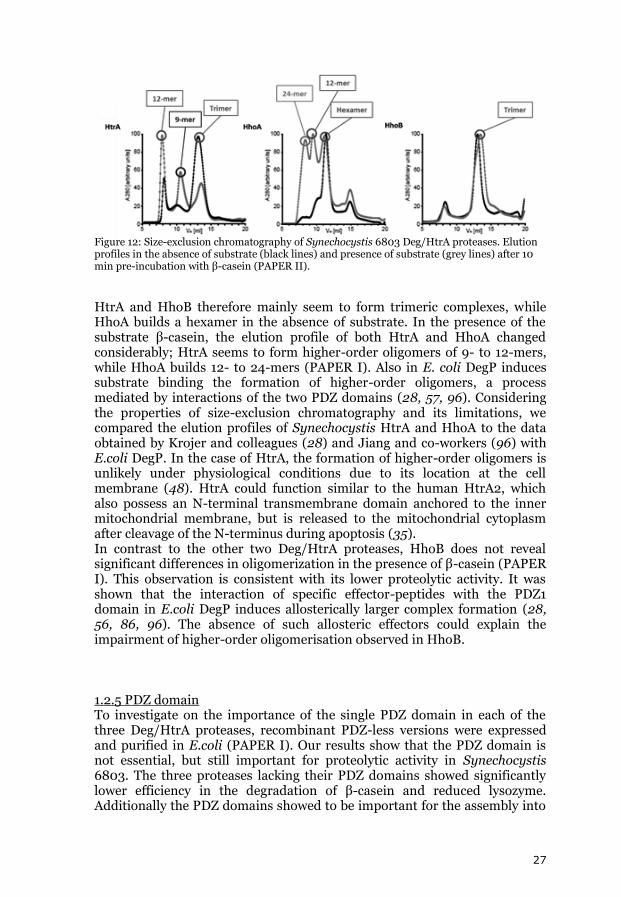
Figure 12: Size-exclusion chromatography of Synechocystis 6803 Deg/HtrA proteases. Elution profiles in the absence of substrate (black lines) and presence of substrate (grey lines) after 10 min pre-incubation with β-casein (PAPER II).
HtrA and HhoB therefore mainly seem to form trimeric complexes, while HhoA builds a hexamer in the absence of substrate. In the presence of the substrate β-casein, the elution profile of both HtrA and HhoA changed considerably; HtrA seems to form higher-order oligomers of 9- to 12-mers, while HhoA builds 12- to 24-mers (PAPER I). Also in E. coli DegP induces substrate binding the formation of higher-order oligomers, a process mediated by interactions of the two PDZ domains (28, 57, 96). Considering the properties of size-exclusion chromatography and its limitations, we compared the elution profiles of Synechocystis HtrA and HhoA to the data obtained by Krojer and colleagues (28) and Jiang and co-workers (96) with E.coli DegP. In the case of HtrA, the formation of higher-order oligomers is unlikely under physiological conditions due to its location at the cell membrane (48). HtrA could function similar to the human HtrA2, which also possess an N-terminal transmembrane domain anchored to the inner mitochondrial membrane, but is released to the mitochondrial cytoplasm after cleavage of the N-terminus during apoptosis (35). In contrast to the other two Deg/HtrA proteases, HhoB does not reveal significant differences in oligomerization in the presence of β-casein (PAPER I). This observation is consistent with its lower proteolytic activity. It was shown that the interaction of specific effector-peptides with the PDZ1 domain in E.coli DegP induces allosterically larger complex formation (28, 56, 86, 96). The absence of such allosteric effectors could explain the impairment of higher-order oligomerisation observed in HhoB. 1.2.5 PDZ domain To investigate on the importance of the single PDZ domain in each of the three Deg/HtrA proteases, recombinant PDZ-less versions were expressed and purified in E.coli (PAPER I). Our results show that the PDZ domain is not essential, but still important for proteolytic activity in Synechocystis 6803. The three proteases lacking their PDZ domains showed significantly lower efficiency in the degradation of β-casein and reduced lysozyme. Additionally the PDZ domains showed to be important for the assembly into
28
larger complexes in the presence of a substrate (PAPER I). Similarly in E. coli the PDZ domains of DegP are essential for the stabilization of higher order oligomers (57, 88). 1.3 Deg/HtrA proteases possibly acting as redox regulating proteases The oxygen-evolving Photosystem II is composed of more than thirty proteins located in the thylakoid membrane. The extrinsic PsbO protein plays an important role in the stabilization of the oxygen evolving complex (OEC) Mn4Ca-cluster (97, 98) and at least one copy of this protein seems to be present in all organisms performing oxygenic photosynthesis (99). PsbO contains two conserved cysteine residues that form an S-S bridge between its N-terminal loop and the β1 strand (100). The function of these two cysteins is controversial, but recently it was found that they were redox active, and possibly important for the regulation of PsbO (101, 102). While PsbO is very stable independent of temperature and pH variations, In Arabidopsis thaliana PsbO1 and PsbO2 were rapidly degraded after reduction by thioredoxin (101). This is not the first evidence shown of thioredoxin controlled proteolysis in plants and other organisms. It was previously shown that the degradation of seed storage proteins of rice (103) and Kunitz and Bowman-Birk soybean’s trypsin inhibitor proteins (104) was more significant when these proteins were reduced by thioredoxin. In animals, redox-controlled proteolysis was also observed for the bovine milk allergen β-lactoglobulin (105). Previously recombinant Deg1 of Arabidopsis thaliana was shown to degrade recombinant PsbO in vitro (44, 106). We were able to show that Deg/HtrA proteases are involved in redox-dependent PsbO degradation in situ, in plants and cyanobacteria. In Synechocystis 6803 PsbO has been localized in the thylakoid lumen but also associated to plasma membrane (107). HhoA localized in the thylakoid lumen and periplasmic space would be able to degrade reduced PsbO (PAPER II). Interestingly PsbO assembled to PSII is not susceptible to degradation (PAPER II), which is in agreement to the results by Li and colleagues (44) who found in extracts of Arabidopsis thaliana not mature PsbO degraded, but only fragments. 1.4 Functional studies of Synechocystis 6803 Deg/HtrA proteases The physiological functions of Deg/HtrA proteases in Synechocystis 6083 are far from being understood. It was believed that Deg/HtrA proteases could play a role similar to their orthologues in the Arabidopsis thaliana chloroplast lumen, and degrade the Photosystem II reaction centre protein D1 during photoinhibition (52). Later, Barker and colleagues (1) suggested that Deg/HtrA proteases are not involved in D1 turnover, but play an important role in the protein quality control in the periplasmic space, most importantly during high light and heat stress. The data obtained by these
29
authors also indicated that the three proteases have overlapping functions, as only a Deg/HtrA triple deletion mutant (Δdeg) showed growth impairment under high light and temperature stress, but not the single and double deletion mutants (1). To learn more about the function of the Synechocystis 6803 Deg/HtrA proteases, I used a proteomic and metabolomic approach and compared wild type cells with the Δdeg mutant grown at optimal conditions and exposed to different stresses. The gel-based Refraction-2D™ proteomics technique was used to identify proteins differently expressed in Synechocystis 6803 WT and the ∆deg mutant (PAPERS III and IV). In this technique, one standard and two different protein samples were labeled with fluorescence dyes (G-Dye 100, G-Dye 200 and G-Dye 300) and mixed before two-dimensional electrophoresis. The resulting 2D-gels were scanned with the excitation wavelengths of each dye in a sequence manner. The three different images for each gel were analyzed and the differential expression for each protein spot was calculated. To identify differences in the metabolite expression of WT and Δdeg, metabolomic samples were analyzed by gas chromatography-mass spectrometry (GC-MS) and detected metabolites constituted the variables for multivariate analysis. The data sets corresponding to samples of WT and Δdeg grown under different conditions, were explored by principle component analysis (PCA) and orthogonal projection to latent structures discriminant analysis (OPLS-DA) using the SIMCA-P+ 12.0 software (Umetrics, Umeå, Sweden) (PAPER III). From the OPLS-DA analysis, the variables (metabolites) significantly different between WT and Δdeg (under different conditions) were identified (PAPER III). 1.4.1 EPS production and S-layer formation is impaired in Δdeg Under normal growth conditions, glucose-6-phosphate (G6P) and fructose-6-phosphate (F6P) accumulate in the Δdeg mutant in comparison to WT, leading to a down-regulation of the enzymes GlpX and fructose-bisphosphate aldolase class II, which convert triose-phosphates into F6P during the regeneration phase of the Calvin cycle. The photosynthetic light reaction produces chemical energy and reductive power (ATP and NADPH), both are used for glucose biosynthesis in the Calvin cycle. With the Calvin cycle being less active, ATP could be redirected to other biosynthetic processes, the need for energy generation via the glucose catabolism therefore would be reduced. This in turn will lead to a down-regulation of glycolytic enzymes and ATP synthase, which we could observe in the Δdeg mutant (PAPER III). F6P is consumed in many biosynthetic processes in the cyanobacterial cell, e.g. it is used in the biosynthesis of different monosaccharides (e.g. mannose and glucose). Down-regulation of the enzyme GDP-D-mannose dehydratase was observed in the Δdeg mutant in all proteomic experiments (PAPERS III and IV). GDP-D-mannose dehydratase catalyzes the first and common reaction in the synthesis of the hexose deoxy sugars fucose, rhamnose and aldohexose talose (108-111), using GDP-D-mannose, the activated form of mannose, as substrate. Glucose and mannose as well as fucose and rhamnose are the major components of lipopolysaccharides (112) and
30
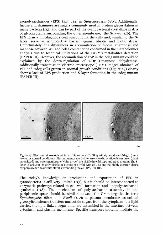
exopolysaccharides (EPS) (113, 114) in Synechocystis 6803. Additionally, fucose and rhamnose are sugars commonly used in protein glycosylation in many bacteria (115) and can be part of the cyanobacterial crystalline matrix of glycoproteins surrounding the outer membrane, the S-layer (116). The EPS form a mucilaginous coat surrounding the cells and, similar to the S-layer, serve as a protective barrier against abiotic and biotic stress. Unfortunately, the differences in accumulation of fucose, rhamnose and mannose between WT and Δdeg could not be confirmed in the metabolomics analysis due to technical limitations of the GC-MS metabolites detection (PAPER III). However, the accumulation of F6P in the Δdeg mutant could be explained by the down-regulation of GDP-D-mannose dehydratase. Additionally transmission electron microscope (TEM) images obtained of WT and Δdeg cells grown in normal growth conditions (Figure 13) clearly show a lack of EPS production and S-layer formation in the Δdeg mutant (PAPER III).
Figure 13: Electron microscopic picture of Synechocystis 6803 wild-type (a) and Δdeg (b) cells grown in normal conditions. Plasma membrane (white arrowhead), peptidoglycan layer (black arrowhead) and outer membrane (white arrow) are visible in wild-type and Δdeg mutant. The S-layer (black star) is only visible in picture of a wild-type cell, as are the highly electron-dense exopolysaccharides (white stars) surrounding the cell (PAPER III).
The today’s knowledge on production and exportation of EPS in cyanobacteria is still very limited (117), but it should be interconnected to enzymatic pathways related to cell wall formation and lipopolysaccharide synthesis (118). The mechanism of polysaccharide assembly in the periplasmic space should be similar between the Gram negative bacteria Synechocystis 6803 and E.coli (119): a plasma membrane associated glycosyltransferase transfers nucleotide sugars from the cytoplasm to a lipid carrier, the lipid-linked sugar units are assembled in the interface between cytoplasm and plasma membrane. Specific transport proteins mediate the
31
passage of these repeating units into the periplasmic space for final assembly into polysaccharides to be used in protein glycosylation in the periplasm (S-layer formation) and EPS production. Deg/HtrA proteases clearly are involved in this process, their specific mode of action still has to be determined. 1.4.2 Δdeg is more sensitive to high light and oxidative stress The Δdeg mutant is more sensitive to light and oxidative stress than WT, being unable to grow under high light conditions (1). Organisms suffer oxidative stress when there is an imbalance between the formation of reactive oxygen species (ROS) and the organism’s ability for a fast removal/ detoxification of the reactive intermediates or damage reparation. Oxidative stress can be a result of an increase in ROS formation (e.g. singlet oxygen, superoxide), decrease of antioxidant protection or less effective repair of ROS induced oxidative damage. The main ROS targets are membrane lipids, nuclear acids and proteins. The proteomic and metabolomic analyses of Synechocystis 6803 clearly indicate pronounced oxidative stress in the Δdeg mutant compared to WT, even in optimal growth conditions (PAPER III). The lipid-soluble α-tocopherol and the enzyme glutathione-S-transferase (GST) accumulate in Δdeg in optimal growth conditions (PAPER III). α-Tocopherol is an antioxidant that protects lipids from peroxidation (120). GST is an antioxidative enzyme that catalyzes reduced glutathione conjugation to electrophilic centers present in a large variety of substrates. It is responsible for the detoxification of degradation products of lipid hydroperoxide such as malondialdehyde and 4-hydroxynonental (121). The gene slr0315 codes for a protein that belongs to the short-chain dehydrogenases/reductases (SDR) family and is overexpressed in Δdeg. SDR proteins are hydrogen-peroxide induced in various organisms and are believed to be important for oxidative stress responses (122). Several proteins related to the protection of the photosynthetic apparatus were found up-regulated in the Δdeg mutant under normal growth conditions, e.g. the Photosystem I biogenesis protein BtpA, the Ycf53-like protein and the NDH-1 subunit I (PAPER III). The extrinsic thylakoid membrane protein BtpA is required for the enhanced stability of the Photosystem I (PSI) complex, protecting it from photoinhibitory damage (123). The Ycf53-like protein (sll0558) is an orthologous of Arabidopsis GUN4 and is able to bind porphyrin and to stimulate the Mg-chelatase (an important enzyme in chlorophyll biosynthesis). This protein also plays a role in photo-protection, preventing the porphyrin-mediated ROS formation in high light conditions (124). The NDH-1 subunit I belongs to the NAD(P)H:quinone oxireductase complex, which is involved in respiration, cyclic electron flow around PSI and CO2 fixation (125). Cyclic electron flow around PSI has a role in preventing photoinhibition of PSII (126). The GTP cyclohydrolase I (folE) was shown to be stronger-expressed in Δdeg in all proteomic experiments (PAPERS III and IV). This is the first enzyme in the folate biosynthesis pathways. Tetrahydrofolate (THF), the active form of folate, is a coenzyme involved in the one-carbon metabolism, transporting one-carbon units (methyl, methenyl, methylene or 10-formyl) in many
32
biosynthetic processes. THF is a key cofactor in glycine-serine inter-conversion (an important enzymatic step in plants photorespiration and cyanobacteria methane metabolism) and the biosynthesis of formyl-methyonine-tRNA, purines, pyrimidines and methionine. Methionine itself is the precursor of S-adenosyl methionine, a cofactor in chlorophyll (127, 128) and tocopherol (129) biosynthesis. THF synthesis is extremely important in photosynthetic organisms. In plants, maintenance of the THF one carbon pool is essential to cope with the high chlorophyll synthesis rate during growth (130). THF is very sensitive to chemical oxidation and is broken down by ROS action (131). The strong up-regulation of GTP cyclohydrolase I in Δdeg indicates a high synthesis of folate, which is required to compensate the breakdown of the THF coenzyme induced by ROS formation in Δdeg. In cyanobacteria and other photosynthetic organisms UV and excess light are the strongest factors for oxidative stress. In Synechocystis 6803 the S-layer and the EPS mucilaginous coat play an important role in protecting the cell from high light and ROS (132). The up-regulation of enzymes involved in the response to high light and oxidative stress in Δdeg, even when grown at optimal conditions, therefore very likely is a secondary effect caused by the impairment of EPS production and S-layer formation. 1.4.3 Twitching motility Twitching motility is a cell movement associated with phototaxis and is mediated by Type IV pili (T4P) (133). Δdeg has been found to exhibit pronounced phototaxis impairment (1). The function of Deg/HtrA proteases in the assembly of fucose-based polysacharides in the periplasmic space could explain this phenotype. Recently it was detected that T4P pilin fucose glycosylation is required for cell motility in Synechocystis 6803 (134). Burriesci and Bhaya (135) showed that phototactic movement is very limited also in isolated Synechocystis 6803 cells. The authors discuss that the cells need to modify the surface for twitching motility and they suggest that EPS secretion is necessary for significant phototaxis (135). 1.4.4 Peptidoglycan remodeling in Δdeg Peptidoglycan composes the cell wall in both Gram positive and Gram negative bacteria, guaranteeing its rigidity and stability. Peptidoglycan is partially degraded by peptidoglycan hydrolases during cell divison, peptidoglycan turnover and remodeling and cell wall expansion (136). With the interruption of peptidoglycan biosynthesis (e.g. during stationary phase) peptidoglycan hydrolases can promote autolysis (136). The activity of peptidoglycan hydrolases has to be strictly regulated, either through direct control of the enzyme activity or through substrate modifications. In the Gram positive Lactococcus lactis IL1403, deacetylation of peptidoglycan N-acetylglucosamine decreases cell autolysis, as deacetylated peptidoglycan is more resistant to AcmA autolysin action (137). A peptidoglycan deacetylase was also found to be important for the protection against lysozyme (an enzyme similar to peptidoglycan hydrolases) in the Gram negative pathogen
33
Helicobacter pylori (138). Sll1306 is a putative polysaccharide deacetylase that is outer membrane-bounded (periplasmic side) (139) and accumulates in Δdeg in normal growth and stress conditions (PAPERS III and IV), being a potential substrate for Deg/HtrA proteases. Its accumulation in Δdeg could impair the activity of peptidoglycan hydrolases and peptidoglycan turnover. Slr1534 was found to be down-regulated in Δdeg under normal growth conditions and mild heat stress (PAPER III). This putative carboxypeptidase belongs to the LD-carboxypeptidase (Muramoyl-tetrapeptide carboxypeptidase) family. LD-carboxypeptidase hydrolyses the amide bond between the di-basic amino acid and the C-terminal D-alanine in the peptidoglycan tetrapeptide. During cell wall degradation peptidoglycan tetrapeptides are produced and LD-carboxypeptidase should be important in peptidoglycan recycling. A defect in peptidoglycan recycling was shown to induce cell autolysis in E. coli (140). The down-regulation of Slr1534 could be caused by accumulation of Sll1306 in Δdeg. 1.4.5 Deg/HtrA and heat stress Proteomic data show that deletion of the Deg/HtrA proteases affects protein quality control under heat stress. In mild heat stress conditons, the GroEL protein 2 is stronger-expressed in Δdeg (PAPER III). GroEL protein 2 is a chaperonine with the function to promote proper protein folding in the cytoplasm and is up-regulated in heat stress (141, 142). Comparing the proteomes of WT and Δdeg isolated of cells exposed to heat shock or high light stress (PAPER IV), the amount of FutA1 and Ferredoxin-NADP reductase was found to be decreased in Δdeg. The iron binding FutA1 (Slr1295) protein is part of an ABC transporter involved in the uptake of Fe3+ in Synechocystis 6803 (143). FutA1 is mainly localized in the thylakoid lumen while its homologue FutA2 (Slr0513) is mainly localized in the periplasmic space (144, 145). FutA1 is important for the growth under iron limiting conditions, being responsible for an effective supply of iron to the Photosystem II D1/D2 reaction center heterodimer (145). Not only PSII is affected by iron limitation, but the entire photosynthetic apparatus as well, iron being an essential component of electron transport chains. Ferredoxin-NADP reductase catalyzes the terminal redox reaction in photosynthesis, creating reductive power in the form of NADPH (146, 147). Down-regulation of the ferredoxin-NADP reductase could indicate a lower efficiency of the photosynthetic apparatus due to lower iron availability. In the Δdeg mutant under heat shock, the accumulation of misfolded proteins in the thylakoid lumen could destabilize the iron transport in the thylakoid membrane. The accumulation of ROS in Δdeg under high light could have a similar effect. After the heat shock treatment, two proteins essential for the assembly of type IV pili were found to be in lower amounts in Δdeg, PilN (Slr1275) and PilO (Slr1276) (PAPER IV). Single deletion mutants of slr1275 and slr1276 genes are non-motile and non-competent, with the thick pili being completely absent (148).The proteomic results under heat shock indicate that, in the absence of Deg/HtrA proteases, the assembly of type IV pili could be more susceptible to damage.
34
2. Small CAB-like proteins
2.1 SCP associated with Photosystem II The Small CAB-like proteins (SCPs) are members of the light-harvesting like (Lil) proteins family found in cyanobacteria (61, 149). Unlike LHC proteins in plants, SCPs are not involved in the absorption of light, but instead, these proteins are induced in stress conditions. SCPs are involved in tetrapyrrole biosynthesis and chlorophyll stability (72, 79, 150), possibly functioning as chlorophyll pigment-carriers (70, 72, 75, 79) and involved in photoprotection. Five small CAB-like proteins were identified in Synechocystis 6803 (75). Previously, ScpC and ScpD were shown to be associated with PSII (77, 151), however, the localization of the other SCPs was not determined. We were able to tag ScpB for a pull-down assay and co-purify ScpE as well as PSII proteins together with ScpB (PAPER V). When expressed under the strong promoter of psbA2, ScpC and ScpD were rapidly degraded in cells grown at normal conditions, and only accumulated in cells grown under high light (77, 151). ScpB, however, when expressed under the strong psbA3 promoter, accumulated in the cells even under normal conditions (PAPER V). Regulated under their native promoter, ScpC and ScpD accumulated in cells only when grown under stress (75), and it was also previously shown that these proteins were degraded immediately when shifting cells from light stress to normal conditions (152). Altogether, it is a strong indication that both ScpC and ScpD are regulated post-translationally and their functions are only related to stress response. Also ScpE expression was found to be only induced under stress conditions (75, 152). Interestingly, the over-expression of ScpB induced the accumulation of both ScpB and ScpE in the cell even at normal growth conditions (PAPER V). ScpB could be a positive regulator of the ScpE promoter, a hypothesis consistent with the observations of He and colleagues (152), who showed that ScpE accumulation under high light was two hours delayed in comparison to ScpB. ScpC/D and ScpB/E might function as heterodimeric pairs (72, 79, 80). Single deletions of scp genes do not result in any phenotype (79), however, the deletion of both members of a pair does (72), suggesting that ScpC and ScpD might complement in function, similar to ScpB and ScpE. The concept of SCPs being able to form dimers is very captivating, however, as two membrane-spanning helices hypothetically are able of transient pigment-binding, similar to the helix 1 and 3 of the light-harvesting complex in plants. The open reading frame slr1544 forms an operon with scpD and encodes a small protein that possesses a predicted single transmembrane helix with the amino acids Gly-Arg of the CAB-motif (PAPER V). The Slr1544 protein might possibly share an evolutionary ancestor with the SCPs and has structural characteristics that place it in the family of light-harvesting-like proteins (Lils) (61). This protein, renamed LilA, was found to be associated with PSII and to accumulate in the Synechocystis cell under high light stress (PAPER V). Similar to ScpD, LilA should be particularly important for PSII turnover, under high light conditions.
35
Summary
In vitro characterization of recombinant Deg/HtrA proteases showed that the three cyanobacterial proteases differ in pH- and temperature optimum. All three proteases are activated by divalent cations. While their basic unit is the trimeric form, substrate binding induces higher oligomeric states in HtrA and HhoA. In Synechocystis 6803 the Deg/HtrA proteases are important protein quality control enzymes. All three proteases are localized in the periplasmic space associated with the plasma membrane, but two of them, HtrA and HhoA, were identified also in the thylakoid membrane fraction, most likely they are localized on the luminal side. HhoA was found to degrade PsbO, when it had disassembled from Photosystem II, interestingly this degradation was redox-dependent. Metabolomic and proteomic analyzes of a Synechocystis mutant depleted of all thre Deg/HtrA proteases (Δdeg) and wilde type showed that these proteases are not only involved in stress acclimation, but the Deg/HtrA proteases also regulate and influence peptidoglycan turnover and polysaccharides formation. The absence of S-layer formation and exopolysaccharide production in the Deg/HtrA triple deletion mutant can explain the observed phenotype: increased light sensitivity and reduced twitching motility (1). Deg/HtrA are further important for the stability of iron uptake in the thylakoid membrane under high light and heat shock conditions, as well as the assembly of thick pili under heat shock. The small CAB-like proteins are associated with Photosystem II, as is LilA (Slr1544). LilA and ScpD form an operon, regulated under the same promoter. While ScpC/D accumulation in the cell is only stable during stress, ScpB induces expression of ScpE and is detected under optimal growth conditions as well.
36
Outlook Deg/HtrA proteases Highly interesting was the finding that Deg/HtrA proteases are involved in peptidoglycan turnover and polysaccharide biosynthesis. Future research should focus on the identification of the exact point of action of Deg/HtrA proteases in these biosynthetic processes. Identifying the specific proteins interacting with the three proteases in the periplasmic space is essential. Affinity tag purification complemented with cross-linking experiments would be a good approach. Very little is known about exopolysaccharide production in cyanobacteria, especially in Synechocystis 6803. Microbial exopolysaccharides have many industrial applications, especially in the food and pharmaceutical industries. Investigating on the function of the Deg/HtrA proteases in polysaccharide biosynthesis and/or export to the medium would probably have high potential for biotechnological applications. SCPs The function of the SCPs in photoprotection still is not clear. Do they stabilize chlorophyll-binding proteins directly by binding chlorophyll and acting as pigment-carrier or do they regulate pigment-biosynthesis? Or are they involved indirectly by a so far unknown mechanism of non-photochemical quenching? Understanding the role of the Lil proteins will definitely help to understand the overall mechanism of photoprotection in cyanobacterium Synechocystis 6803.
37
Acknowledgments Acima de tudo e em primeirissímo lugar tenho que agradecer à minha
familia todo o apoio e forςa que me deu para esta grande aventura que foi a
minha vinda para a Suécia, em especial à minha mãe Matilde Henriques e
ao meu pai Luís Miranda, as duas pessoas a quem eu devo tudo. Um
especial agradecimento aos meus falecidos avós, José Henriques e
Lourenςo Miranda e a minha falecida tia Jóia, que sempre acreditaram
em mim e foram a minha fonte de inspiraςão. Com muito amor e carinho
agradeςo às minhas irmãs Anta e Margarida, às minhas avós Carolina e
Beatriz, à minha tia Augusta, aos meus primos Fanceni e Mohamed, às
minhas lindas sobrinhas Pilar, Ema e Luana, ao Jorge e à minha prima
Elsa.
Special special special thanks to my girlfriend Jeanette, without her I would
be TOTALLY lost! - In every way, believe me! Special thanks to my new
Swedish family and all the friends I made in Sweden, you really made me feel
at home. I am so grateful for you all!
I would like to thank my supervisor Professor Christiane Funk for giving
me the opportunity to work in this field full of potential that is the
cyanobacteria world. Thank you Christiane, for your patience and support!
I would also like to thank my co-supervisor Professor Wolfgang Schröder
for his insight in the field of proteomics.
Thanks to Professor Iwona Adamska and Professor Wim Vermaas for
welcoming me to their research groups in my first year as a PhD student. It
was a very enriching experience!
Warm thanks to all my colleagues in the Biochemistry corridor, from the
past and present (Paulo, Dmitry, Patrick, Adriana, Maribel, Miguel,
Galyna, José, Liudmila, Michael, Raik, Marcus, Jörgen, Irma,
Susan, Tania, Giada, Ana, Kumar, Yogesh and Anett). It was great to
know you all and I hope we can keep in touch.
Special thanks to Pitter Huesgen for his collaboration in the in vitro
studies of the Deg proteases, Harald Aigner for his initial guiding in the
proteomic field, Otilia Cheregi for her precious insights on Synechocystis
sp. PCC 6803 and Rui Pinto for his tutoring in multivariate data analysis.
SPECIAL thanks (notice the capital letters!) to Barbara. You’re a
wonderful friend, always willing to help me when I needed (which was quite
often, I have to say).
38
References 1. Barker, M., de Vries, R., Nield, J., Komenda, J., Nixon, P. J. (2006) The deg proteases
protect Synechocystis sp. PCC 6803 during heat and light stresses but are not essential for removal of damaged D1 protein during the photosystem two repair cycle, J Biol Chem 281, 30347-30355.
2. Bryant, D. A., and Frigaard, N. U. (2006) Prokaryotic photosynthesis and
phototrophy illuminated, Trends Microbiol 14, 488-496.
3. Frei, R., Gaucher, C., Poulton, S. W., and Canfield, D. E. (2009) Fluctuations in
Precambrian atmospheric oxygenation recorded by chromium isotopes, Nature 461, 250-253.
4. Kaneko, T., Sato, S., Kotani, H., Tanaka, A., Asamizu, E., Nakamura, Y., Miyajima, N.,
Hirosawa, M., Sugiura, M., Sasamoto, S., Kimura, T., Hosouchi, T., Matsuno, A., Muraki, A., Nakazaki, N., Naruo, K., Okumura, S., Shimpo, S., Takeuchi, C., Wada, T., Watanabe, A., Yamada, M., Yasuda, M., and Tabata, S. (1996) Sequence analysis of the genome of the unicellular cyanobacterium Synechocystis sp. strain PCC6803. II. Sequence determination of the entire genome and assignment of potential protein-coding regions (supplement), DNA Res 3, 185-209.
5. Haslbeck, M., Franzmann, T., Weinfurtner, D., and Buchner, J. (2005) Some like it
hot: the structure and function of small heat-shock proteins, Nat Struct Mol Biol 12, 842-846.
6. Lehel, C., Wada, H., Kovacs, E., Torok, Z., Gombos, Z., Horvath, I., Murata, N., and
Vigh, L. (1992) Heat shock protein synthesis of the cyanobacterium Synechocystis PCC 6803: purification of the GroEL-related chaperonin, Plant Mol Biol 18, 327-336.
7. Renger, G., and Kuhn, P. (2007) Reaction pattern and mechanism of light induced
oxidative water splitting in photosynthesis, Biochim Biophys Acta 1767, 458-471.
8. Calvin, M. (1950) The path of carbon in photosynthesis, Harvey Lect Series 46, 218-
251.
9. Finazzi, G., Barbagallo, R. P., Bergo, E., Barbato, R., and Forti, G. (2001)
Photoinhibition of Chlamydomonas reinhardtii in State 1 and State 2 - Damages to the photosynthetic apparatus under linear and cyclic electron flow, Journal of Biological Chemistry 276, 22251-22257.
10. Vass, I., Styring, S., Hundal, T., Koivuniemi, A., Aro, E., and Andersson, B. (1992)
Reversible and irreversible intermediates during photoinhibition of photosystem II: stable reduced QA species promote chlorophyll triplet formation, Proc Natl Acad Sci U S A 89, 1408-1412.
11. Edreva, A. (2005) Generation and scavenging of reactive oxygen species in
chloroplasts: a submolecular approach, Agriculture Ecosystems & Environment 106, 119-133.
12. Mishra, N. P., and Ghanotakis, D. F. (1994) Exposure of a Photosystem-Ii Complex to
Chemically Generated Singlet Oxygen Results in D1 Fragments Similar to the Ones Observed during Aerobic Photoinhibition, Biochimica Et Biophysica Acta-Bioenergetics 1187, 296-300.
39
13. Aro, E. M., Virgin, I., and Andersson, B. (1993) Photoinhibition of Photosystem II.
Inactivation, protein damage and turnover, Biochim Biophys Acta 1143, 113-134.
14. Behrenfeld, M. J., Prasil, O., Kolber, Z. S., Babin, M., and Falkowski, P. G. (1998)
Compensatory changes in Photosystem II electron turnover rates protect photosynthesis from photoinhibition, Photosynthesis Research 58, 259-268.
15. Neurath, H. (1984) Evolution of proteolytic enzymes, Science 224, 350-357.
16. Rawlings, N. D., and Barrett, A. J. (1994) Families of serine peptidases, Methods
Enzymol 244, 19-61.
17. Carrell, R. W., Pemberton, P. A., and Boswell, D. R. (1987) The serpins: evolution and
adaptation in a family of protease inhibitors, Cold Spring Harb Symp Quant Biol 52, 527-535.
18. Huber, R., and Bode, W. (1978) Structural Basis of Activation and Action of Trypsin,
Accounts of Chemical Research 11, 114-122.
19. Sheng, M., and Sala, C. (2001) PDZ domains and the organization of supramolecular
complexes, Annual Review of Neuroscience 24, 1-29.
20. Wilken, C., Kitzing, K., Kurzbauer, R., Ehrmann, M., and Clausen, T. (2004) Crystal
structure of the DegS stress sensor: How a PDZ domain recognizes misfolded protein and activates a protease, Cell 117, 483-494.
21. Clausen, T., Southan, C., and Ehrmann, M. (2002) The HtrA family of proteases:
implications for protein composition and cell fate, Mol Cell 10, 443-455.
22. Chien, J., Ota, T., Aletti, G., Shridhar, R., Boccellino, M., Quagliuolo, L., Baldi, A., and
Shridhar, V. (2009) Serine protease HtrA1 associates with microtubules and inhibits cell migration, Mol Cell Biol 29, 4177-4187.
23. Fahrenkrog, B., Sauder, U., and Aebi, U. (2004) The S. cerevisiae HtrA-like protein
Nma111p is a nuclear serine protease that mediates yeast apoptosis, J Cell Sci 117, 115-126.
24. Pallen, M. J., and Wren, B. W. (1997) The HtrA family of serine proteases, Mol
Microbiol 26, 209-221.
25. Walsh, N. P., Alba, B. M., Bose, B., Gross, C. A., and Sauer, R. T. (2003) OMP peptide
signals initiate the envelope-stress response by activating DegS protease via relief of inhibition mediated by its PDZ domain, Cell 113, 61-71.
26. Isaac, D. D., Pinkner, J. S., Hultgren, S. J., and Silhavy, T. J. (2005) The
extracytoplasmic adaptor protein CpxP is degraded with substrate by DegP, Proc Natl Acad Sci U S A 102, 17775-17779.
27. Spiess, C., Beil, A., and Ehrmann, M. (1999) A temperature-dependent switch from
chaperone to protease in a widely conserved heat shock protein, Cell 97, 339-347.
40
28. Krojer, T., Sawa, J., Schafer, E., Saibil, H. R., Ehrmann, M., and Clausen, T. (2008) Structural basis for the regulated protease and chaperone function of DegP, Nature 453, 885-890.
29. Kolmar, H., Waller, P. R., and Sauer, R. T. (1996) The DegP and DegQ periplasmic
endoproteases of Escherichia coli: specificity for cleavage sites and substrate conformation, J Bacteriol 178, 5925-5929.
30. Zumbrunn, J., and Trueb, B. (1996) Primary structure of a putative serine protease
specific for IGF-binding proteins, FEBS Lett 398, 187-192.
31. Hou, J., Clemmons, D. R., and Smeekens, S. (2005) Expression and characterization
of a serine protease that preferentially cleaves insulin-like growth factor binding protein-5, J Cell Biochem 94, 470-484.
32. Campioni, M., Severino, A., Manente, L., Tuduce, I. L., Toldo, S., Caraglia, M., Crispi,
S., Ehrmann, M., He, X., Maguire, J., De Falco, M., De Luca, A., Shridhar, V., and Baldi, A. (2010) The serine protease HtrA1 specifically interacts and degrades the tuberous sclerosis complex 2 protein, Mol Cancer Res 8, 1248-1260.
33. Milner, J. M., Patel, A., and Rowan, A. D. (2008) Emerging roles of serine proteinases
in tissue turnover in arthritis, Arthritis Rheum 58, 3644-3656.
34. Grau, S., Baldi, A., Bussani, R., Tian, X., Stefanescu, R., Przybylski, M., Richards, P.,
Jones, S. A., Shridhar, V., Clausen, T., and Ehrmann, M. (2005) Implications of the serine protease HtrA1 in amyloid precursor protein processing, Proc Natl Acad Sci U S A 102, 6021-6026.
35. Vande Walle, L., Lamkanfi, M., and Vandenabeele, P. (2008) The mitochondrial
serine protease HtrA2/Omi: an overview, Cell Death Differ 15, 453-460.
36. Winklhofer, K. F., and Haass, C. (2010) Mitochondrial dysfunction in Parkinson's
disease, Biochimica Et Biophysica Acta-Molecular Basis of Disease 1802, 29-44.
37. Itzhaki, H., Naveh, L., Lindahl, M., Cook, M., and Adam, Z. (1998) Identification and
characterization of DegP, a serine protease associated with the luminal side of the thylakoid membrane, J Biol Chem 273, 7094-7098.
38. Peltier, J. B., Emanuelsson, O., Kalume, D. E., Ytterberg, J., Friso, G., Rudella, A.,
Liberles, D. A., Soderberg, L., Roepstorff, P., von Heijne, G., and van Wijk, K. J. (2002) Central functions of the lumenal and peripheral thylakoid proteome of Arabidopsis determined by experimentation and genome-wide prediction, Plant Cell 14, 211-236.
39. Schubert, M., Petersson, U. A., Haas, B. J., Funk, C., Schroder, W. P., and Kieselbach,
T. (2002) Proteome map of the chloroplast lumen of Arabidopsis thaliana, Journal of Biological Chemistry 277, 8354-8365.
40. Haussuhl, K., Andersson, B., and Adamska, I. (2001) A chloroplast DegP2 protease
performs the primary cleavage of the photodamaged D1 protein in plant photosystem II, Embo Journal 20, 713-722.
41. Sun, X., Fu, T., Chen, N., Guo, J., Ma, J., Zou, M., Lu, C., and Zhang, L. (2010) The
stromal chloroplast Deg7 protease participates in the repair of photosystem II after photoinhibition in Arabidopsis, Plant Physiol 152, 1263-1273.
41
42. Sun, X., Ouyang, M., Guo, J., Ma, J., Lu, C., Adam, Z., and Zhang, L. (2010) The
thylakoid protease Deg1 is involved in photosystem-II assembly in Arabidopsis thaliana, Plant J 62, 240-249.
43. Kapri-Pardes, E., Naveh, L., and Adam, Z. (2007) The thylakoid lumen protease Deg1
is involved in the repair of photosystem II from photoinhibition in Arabidopsis, Plant Cell 19, 1039-1047.
44. Li, J., Sun, X. W., and Zhang, L. X. (2010) Deg1 is involved in the degradation of the
PsbO oxygen-evolving protein of photosystem II in Arabidopsis, Chinese Science Bulletin 55, 3145-3148.
45. Sun, X. W., Wang, L. Y., and Zhang, L. X. (2007) Involvement of DEG5 and DEG8
proteases in the turnover of the photosystem II reaction center D1 protein under heat stress in Arabidopsis thaliana, Chinese Science Bulletin 52, 1742-1745.
46. Sun, X., Peng, L., Guo, J., Chi, W., Ma, J., Lu, C., and Zhang, L. (2007) Formation of
DEG5 and DEG8 complexes and their involvement in the degradation of photodamaged photosystem II reaction center D1 protein in Arabidopsis, Plant Cell 19, 1347-1361.
47. Sokolenko, A., Pojidaeva, E., Zinchenko, V., Panichkin, V., Glaser, V. M., Herrmann,
R. G., and Shestakov, S. V. (2002) The gene complement for proteolysis in the cyanobacterium Synechocystis sp. PCC 6803 and Arabidopsis thaliana chloroplasts, Curr Genet 41, 291-310.
48. Kieselbach, T., and Funk, C. (2003) The family of Deg/HtrA proteases: from
Escherichia coli to Arabidopsis, Physiologia Plantarum 119, 337-346.
49. Huesgen PF, Schuhmann H, and Adamska I. (2005) The family of deg proteases in
cyanobacteria and chloroplasts of higher plants, Physiologia Plantarum 123, 413-420.
50. Fulda, S., Huang, F., Nilsson, F., Hagemann, M., and Norling, B. (2000) Proteomics
of Synechocystis sp. strain PCC 6803. Identification of periplasmic proteins in cells grown at low and high salt concentrations, Eur J Biochem 267, 5900-5907.
51. Huang, F., Hedman, E., Funk, C., Kieselbach, T., Schroder, W. P., and Norling, B.
(2004) Isolation of outer membrane of Synechocystis sp. PCC 6803 and its proteomic characterization, Mol Cell Proteomics 3, 586-595.
52. Silva, P., Choi, Y. J., Hassan, H. A., and Nixon, P. J. (2002) Involvement of the HtrA
family of proteases in the protection of the cyanobacterium Synechocystis PCC 6803 from light stress and in the repair of photosystem II, Philos Trans R Soc Lond B Biol Sci 357, 1461-1467; discussion 1467-1470.
53. Clausen, T., Kaiser, M., Huber, R., and Ehrmann, M. (2011) HTRA proteases:
regulated proteolysis in protein quality control, Nat Rev Mol Cell Biol 12, 152-162.
54. Kim, D. Y., and Kim, K. K. (2005) Structure and function of HtrA family proteins, the
key players in protein quality control, J Biochem Mol Biol 38, 266-274.
55. Hasselblatt, H., Kurzbauer, R., Wilken, C., Krojer, T., Sawa, J., Kurt, J., Kirk, R.,
Hasenbein, S., Ehrmann, M., and Clausen, T. (2007) Regulation of the sigmaE stress response by DegS: how the PDZ domain keeps the protease inactive in the resting
42
state and allows integration of different OMP-derived stress signals upon folding stress, Genes Dev 21, 2659-2670.
56. Merdanovic, M., Mamant, N., Meltzer, M., Poepsel, S., Auckenthaler, A., Melgaard, R.,
Hauske, P., Nagel-Steger, L., Clarke, A. R., Kaiser, M., Huber, R., and Ehrmann, M. (2010) Determinants of structural and functional plasticity of a widely conserved protease chaperone complex, Nat Struct Mol Biol 17, 837-843.
57. Krojer, T., Sawa, J., Huber, R., and Clausen, T. (2010) HtrA proteases have a
conserved activation mechanism that can be triggered by distinct molecular cues, Nat Struct Mol Biol 17, 844-852.
58. Ortega, J., Iwanczyk, J., and Jomaa, A. (2009) Escherichia coli DegP: a structure-
driven functional model, J Bacteriol 191, 4705-4713.
59. Truebestein, L., Tennstaedt, A., Monig, T., Krojer, T., Canellas, F., Kaiser, M.,
Clausen, T., and Ehrmann, M. (2011) Substrate-induced remodeling of the active site regulates human HTRA1 activity, Nat Struct Mol Biol.
60. Sadava, D. E. (2008) Life, the science of biology, 8th ed., Sinauer Associates ; W.H.
Freeman and Co., Sunderland, MA. 61. Jansson, S. (1999) A guide to the Lhc genes and their relatives in Arabidopsis, Trends
in Plant Science 4, 236-240.
62. Kuhlbrandt, W., Wang, D. N., and Fujiyoshi, Y. (1994) Atomic model of plant light-
harvesting complex by electron crystallography, Nature 367, 614-621.
63. Liu, Z., Yan, H., Wang, K., Kuang, T., Zhang, J., Gui, L., An, X., and Chang, W. (2004)
Crystal structure of spinach major light-harvesting complex at 2.72 A resolution, Nature 428, 287-292.
64. Standfuss, J., Terwisscha van Scheltinga, A. C., Lamborghini, M., and Kuhlbrandt, W.
(2005) Mechanisms of photoprotection and nonphotochemical quenching in pea light-harvesting complex at 2.5 A resolution, EMBO J 24, 919-928.
65. Green, B. R., and Kuhlbrandt, W. (1995) Sequence Conservation of Light-Harvesting
and Stress-Response Proteins in Relation to the 3-Dimensional Molecular-Structure of Lhcii, Photosynthesis Research 44, 139-148.
66. Heddad, M., and Adamska, I. (2002) The evolution of light stress proteins in
photosynthetic organisms, Comp Funct Genomics 3, 504-510.
67. Garczarek, L., Poupon, A., and Partensky, F. (2003) Origin and evolution of
transmembrane Chl-binding proteins: hydrophobic cluster analysis suggests a common one-helix ancestor for prokaryotic (Pcb) and eukaryotic (LHC) antenna protein superfamilies, Fems Microbiology Letters 222, 59-68.
68. Durnford, D. G., Deane, J. A., Tan, S., McFadden, G. I., Gantt, E., and Green, B. R.
(1999) A phylogenetic assessment of the eukaryotic light-harvesting antenna proteins, with implications for plastid evolution, J Mol Evol 48, 59-68.
69. Li, X. P., Bjorkman, O., Shih, C., Grossman, A. R., Rosenquist, M., Jansson, S., and
Niyogi, K. K. (2000) A pigment-binding protein essential for regulation of photosynthetic light harvesting, Nature 403, 391-395.
43
70. Adamska, I. (1997) ELIPs - Light-induced stress proteins, Physiologia Plantarum
100, 794-805.
71. Montane, M. H., and Kloppstech, K. (2000) The family of light-harvesting-related
proteins (LHCs, ELIPs, HLIPs): was the harvesting of light their primary function?, Gene 258, 1-8.
72. Xu, H., Vavilin, D., Funk, C., and Vermaas, W. (2004) Multiple deletions of small
Cab-like proteins in the cyanobacterium Synechocystis sp. PCC 6803: consequences for pigment biosynthesis and accumulation, Journal of Biological Chemistry 279, 27971-27979.
73. Green, B. R., Pichersky, E., and Kloppstech, K. (1991) Chlorophyll a/b-binding
proteins: an extended family, Trends Biochem Sci 16, 181-186.
74. Dolganov, N. A., Bhaya, D., and Grossman, A. R. (1995) Cyanobacterial protein with
similarity to the chlorophyll a/b binding proteins of higher plants: evolution and regulation, Proc Natl Acad Sci U S A 92, 636-640.
75. Funk, C., and Vermaas, W. (1999) A cyanobacterial gene family coding for single-helix
proteins resembling part of the light-harvesting proteins from higher plants, Biochemistry 38, 9397-9404.
76. Bhaya, D., Dufresne, A., Vaulot, D., and Grossman, A. (2002) Analysis of the hli gene
family in marine and freshwater cyanobacteria, Fems Microbiology Letters 215, 209-219.
77. Yao, D., Kieselbach, T., Komenda, J., Promnares, K., Prieto, M. A., Tichy, M.,
Vermaas, W., and Funk, C. (2007) Localization of the small CAB-like proteins in photosystem II, J Biol Chem 282, 267-276.
78. Vavilin, D., Yao, D., and Vermaas, W. (2007) Small Cab-like proteins retard
degradation of photosystem II-associated chlorophyll in Synechocystis sp. PCC 6803: kinetic analysis of pigment labeling with 15N and 13C, J Biol Chem 282, 37660-37668.
79. Xu, H., Vavilin, D., and Vermaas, W. (2002) The presence of chlorophyll b in
Synechocystis sp. PCC 6803 disturbs tetrapyrrole biosynthesis and enhances chlorophyll degradation, J Biol Chem 277, 42726-42732.
80. Storm, P., Hernandez-Prieto, M. A., Eggink, L. L., Hoober, J. K., and Funk, C. (2008)
The small CAB-like proteins of Synechocystis sp. PCC 6803 bind chlorophyll. In vitro pigment reconstitution studies on one-helix light-harvesting-like proteins, Photosynthesis Research 98, 479-488.
81. Havaux, M., Guedeney, G., He, Q., and Grossman, A. R. (2003) Elimination of high-
light-inducible polypeptides related to eukaryotic chlorophyll a/b-binding proteins results in aberrant photoacclimation in Synechocystis PCC6803, Biochim Biophys Acta 1557, 21-33.
82. Norling, B., Zak, E., Andersson, B., and Pakrasi, H. (1998) 2D-isolation of pure
plasma and thylakoid membranes from the cyanobacterium Synechocystis sp. PCC 6803, FEBS Lett 436, 189-192.
44
83. van de Meene, A. M., Hohmann-Marriott, M. F., Vermaas, W. F., and Roberson, R. W. (2006) The three-dimensional structure of the cyanobacterium Synechocystis sp. PCC 6803, Arch Microbiol 184, 259-270.
84. Alba, B. M., Zhong, H. J., Pelayo, J. C., and Gross, C. A. (2001) degS (hhoB) is an
essential Escherichia coli gene whose indispensable function is to provide sigma(E) activity, Molecular Microbiology 40, 1323-1333.
85. Meltzer, M., Hasenbein, S., Mamant, N., Merdanovic, M., Poepsel, S., Hauske, P.,
Kaiser, M., Huber, R., Krojer, T., Clausen, T., and Ehrmann, M. (2009) Structure, function and regulation of the conserved serine proteases DegP and DegS of Escherichia coli, Res Microbiol 160, 660-666.
86. Meltzer, M., Hasenbein, S., Hauske, P., Kucz, N., Merdanovic, M., Grau, S., Beil, A.,
Jones, D., Krojer, T., Clausen, T., Ehrmann, M., and Kaiser, M. (2008) Allosteric activation of HtrA protease DegP by stress signals during bacterial protein quality control, Angew Chem Int Ed Engl 47, 1332-1334.
87. Schechter, I., and Berger, A. (1967) On the size of the active site in proteases. I.
Papain, Biochem Biophys Res Commun 27, 157-162.
88. Krojer, T., Pangerl, K., Kurt, J., Sawa, J., Stingl, C., Mechtler, K., Huber, R., Ehrmann,
M., and Clausen, T. (2008) Interplay of PDZ and protease domain of DegP ensures efficient elimination of misfolded proteins, Proc Natl Acad Sci U S A 105, 7702-7707.
89. Helm, M., Luck, C., Prestele, J., Hierl, G., Huesgen, P. F., Frohlich, T., Arnold, G. J.,
Adamska, I., Gorg, A., Lottspeich, F., and Gietl, C. (2007) Dual specificities of the glyoxysomal/peroxisomal processing protease Deg15 in higher plants, Proc Natl Acad Sci U S A 104, 11501-11506.
90. Schuhmann, H., Huesgen, P. F., Gietl, C., and Adamska, I. (2008) The DEG15 serine
protease cleaves peroxisomal targeting signal 2-containing proteins in Arabidopsis, Plant Physiol 148, 1847-1856.
91. Barber, J., and Andersson, B. (1992) Too much of a good thing: light can be bad for
photosynthesis, Trends Biochem Sci 17, 61-66.
92. Adir, N., Zer, H., Shochat, S., and Ohad, I. (2003) Photoinhibition - a historical
perspective, Photosynth Res 76, 343-370.
93. Krojer, T., Garrido-Franco, M., Huber, R., Ehrmann, M., and Clausen, T. (2002)
Crystal structure of DegP (HtrA) reveals a new protease-chaperone machine, Nature 416, 455-459.
94. Li, W. Y., Srinivasula, S. M., Chai, J. J., Li, P. W., Wu, J. W., Zhang, Z. J., Alnemri, E.
S., and Shi, Y. G. (2002) Structural insights into the pro-apoptotic function of mitochondrial serine protease HtrA2/Omi, Nat Struct Biol 9, 436-441.
95. Jansen, T., Kidron, H., Taipaleenmaki, H., Salminen, T., and Maenpaa, P. (2005)
Transcriptional profiles and structural models of the Synechocystis sp PCC 6803 Deg proteases, Photosynth Res 84, 57-63.
96. Jiang, J., Zhang, X., Chen, Y., Wu, Y., Zhou, Z. H., Chang, Z., and Sui, S. F. (2008)
Activation of DegP chaperone-protease via formation of large cage-like oligomers upon binding to substrate proteins, Proc Natl Acad Sci U S A 105, 11939-11944.
45
97. Bockholt, R., Masepohl, B., and Pistorius, E. K. (1991) Insertional inactivation of the
psbO gene encoding the manganese stabilizing protein of photosystem II in the cyanobacterium Synechococcus PCC7942. Effect on photosynthetic water oxidation and L-amino acid oxidase activity, Febs Letters 294, 59-63.
98. Popelkova, H., and Yocum, C. F. (2011) PsbO, the manganese-stabilizing protein:
Analysis of the structure-function relations that provide insights into its role in photosystem II, J Photochem Photobiol B.
99. De Las Rivas, J., and Barber, J. (2004) Analysis of the Structure of the PsbO Protein
and its Implications, Photosynth Res 81, 329-343.
100. Nikitina, J., Shutova, T., Melnik, B., Chernyshov, S., Marchenkov, V., Semisotnov, G.,
Klimov, V., and Samuelsson, G. (2008) Importance of a single disulfide bond for the PsbO protein of photosystem II: protein structure stability and soluble overexpression in Escherichia coli, Photosynth Res 98, 391-403.
101. Hall, M., Mata-Cabana, A., Akerlund, H. E., Florencio, F. J., Schroder, W. P., Lindahl,
M., and Kieselbach, T. (2010) Thioredoxin targets of the plant chloroplast lumen and their implications for plastid function, Proteomics 10, 987-1001.
102. Marchand, C., Le Marechal, P., Meyer, Y., and Decottignies, P. (2006) Comparative
proteomic approaches for the isolation of proteins interacting with thioredoxin, Proteomics 6, 6528-6537.
103. Yano, H., and Kuroda, M. (2006) Disulfide proteome yields a detailed understanding
of redox regulations: a model study of thioredoxin-linked reactions in seed germination, Proteomics 6, 294-300.
104. Jiao, J. A., Yee, B. C., Kobrehel, K., and Buchanan, B. B. (1992) Effect of Thioredoxin-
Linked Reduction on the Activity and Stability of the Kunitz and Bowman-Birk Soybean Trypsin-Inhibitor Proteins, J Agr Food Chem 40, 2333-2336.
105. del Val, G., Yee, B. C., Lozano, R. M., Buchanan, B. B., Ermel, R. W., Lee, Y. M., and
Frick, O. L. (1999) Thioredoxin treatment increases digestibility and lowers allergenicity of milk, J Allergy Clin Immun 103, 690-697.
106. Chassin, Y., Kapri-Pardes, E., Sinvany, G., Arad, T., and Adam, Z. (2002) Expression
and characterization of the thylakoid lumen protease DegP1 from Arabidopsis, Plant Physiol 130, 857-864.
107. Zak, E., Norling, B., Maitra, R., Huang, F., Andersson, B., and Pakrasi, H. B. (2001)
The initial steps of biogenesis of cyanobacterial photosystems occur in plasma membranes, Proc Natl Acad Sci U S A 98, 13443-13448.
108. Elbein, A. D., Heath, E. C. (1965) The Biosynthesis of Cell Wall Lipopolysaccharide in
Escherichia Coli. Ii. Guanosine Diphosphate 4-Keto-6-Deoxy-D-Mannose, an Intermediate in the Biosynthesis of Guanosine Diphosphate Colitose, J Biol Chem 240, 1926-1931.
109. Sullivan, F. X., Kumar, R., Kriz, R., Stahl, M., Xu, G. Y., Rouse, J., Chang, X. J.,
Boodhoo, A., Potvin, B., Cumming, D. A. (1998) Molecular cloning of human GDP-mannose 4,6-dehydratase and reconstitution of GDP-fucose biosynthesis in vitro, J Biol Chem 273, 8193-8202.
46
110. Kneidinger, B., Graninger, M., Adam, G., Puchberger, M., Kosma, P., Zayni, S.,
Messner, P. (2001) Identification of two GDP-6-deoxy-D-lyxo-4-hexulose reductases synthesizing GDP-D-rhamnose in Aneurinibacillus thermoaerophilus L420-91T, J Biol Chem 276, 5577-5583.
111. Suzuki, N., Nakano, Y., Yoshida, Y., Nezu, T., Terada, Y., Yamashita, Y., Koga, T.
(2002) Guanosine diphosphate-4-keto-6-deoxy-d-mannose reductase in the pathway for the synthesis of GDP-6-deoxy-d-talose in Actinobacillus actinomycetemcomitans, Eur J Biochem 269, 5963-5971.
112. Schmidt, W., Drews, G., Weckesser, J., Mayer, H. (1980) Lipopolysaccharides in Four
Strains of the Unicellular Cyanobacterium Synechocystis, Archives of Microbiology 127, 217-222.
113. Panoff, J. M., Priem, B., Morvan, H., Joset, F. (1988) Sulfated Exopolysaccharides
Produced by 2 Unicellular Strains of Cyanobacteria, Synechocystis Pcc-6803 and Pcc-6714, Archives of Microbiology 150, 558-563.
114. Panoff, J. M., Joset, F. (1988) Polysaccharides Excreted by Synechocystis Pcc-6714
and Pcc-6803, Plant Physiology and Biochemistry 26, 207-207.
115. Moens, S., Vanderleyden, J. (1997) Glycoproteins in prokaryotes, Archives of
Microbiology 168, 169-175.
116. Smarda, J., Smajs, D., Komrska, J., Krzyzanek, V. (2002) S-layers on cell walls of
cyanobacteria, Micron 33, 257-277.
117. Yoshimura, H., Okamoto, S., Tsumuraya, Y., and Ohmori, M. (2007) Group 3 sigma
factor gene, sigJ, a key regulator of desiccation tolerance, regulates the synthesis of extracellular polysaccharide in cyanobacterium Anabaena sp. strain PCC 7120, DNA Res 14, 13-24.
118. Mozzi, F., Savoy de Giori, G., and Font de Valdez, G. (2003) UDP-galactose 4-
epimerase: a key enzyme in exopolysaccharide formation by Lactobacillus casei CRL 87 in controlled pH batch cultures, J Appl Microbiol 94, 175-183.
119. Pereira, S., Zille, A., Micheletti, E., Moradas-Ferreira, P., De Philippis, R., and
Tamagnini, P. (2009) Complexity of cyanobacterial exopolysaccharides: composition, structures, inducing factors and putative genes involved in their biosynthesis and assembly, FEMS Microbiol Rev 33, 917-941.
120. Maeda, H., Sakuragi, Y., Bryant, D. A., and Dellapenna, D. (2005) Tocopherols
protect Synechocystis sp. strain PCC 6803 from lipid peroxidation, Plant Physiol 138, 1422-1435.
121. Yang, Y., Sharma, R., Sharma, A., Awasthi, S., and Awasthi, Y. C. (2003) Lipid
peroxidation and cell cycle signaling: 4-hydroxynonenal, a key molecule in stress mediated signaling, Acta Biochim Pol 50, 319-336.
122. Vandenbroucke, K., Robbens, S., Vandepoele, K., Inze, D., Van de Peer, Y., and Van
Breusegem, F. (2008) Hydrogen peroxide-induced gene expression across kingdoms: a comparative analysis, Mol Biol Evol 25, 507-516.
47
123. Zak, E., and Pakrasi, H. B. (2000) The BtpA protein stabilizes the reaction center proteins of photosystem I in the cyanobacterium Synechocystis sp. PCC 6803 at low temperature, Plant Physiol 123, 215-222.
124. Verdecia, M. A., Larkin, R. M., Ferrer, J. L., Riek, R., Chory, J., and Noel, J. P. (2005)
Structure of the Mg-chelatase cofactor GUN4 reveals a novel hand-shaped fold for porphyrin binding, PLoS Biol 3, e151.
125. Battchikova, N., and Aro, E. M. (2007) Cyanobacterial NDH-1 complexes: multiplicity
in function and subunit composition, Physiol Plant 131, 22-32.
126. Takahashi, S., Milward, S. E., Fan, D. Y., Chow, W. S., and Badger, M. R. (2009) How
Does Cyclic Electron Flow Alleviate Photoinhibition in Arabidopsis?, Plant Physiol 149, 1560-1567.
127. Van Wilder, V., De Brouwer, V., Loizeau, K., Gambonnet, B., Albrieux, C., Van Der
Straeten, D., Lambert, W. E., Douce, R., Block, M. A., Rebeille, F., and Ravanel, S. (2009) C1 metabolism and chlorophyll synthesis: the Mg-protoporphyrin IX methyltransferase activity is dependent on the folate status, New Phytol 182, 137-145.
128. Shepherd, M., Reid, J. D., and Hunter, C. N. (2003) Purification and kinetic
characterization of the magnesium protoporphyrin IX methyltransferase from Synechocystis PCC6803, Biochem J 371, 351-360.
129. Cheng, Z., Sattler, S., Maeda, H., Sakuragi, Y., Bryant, D. A., and DellaPenna, D.
(2003) Highly divergent methyltransferases catalyze a conserved reaction in tocopherol and plastoquinone synthesis in cyanobacteria and photosynthetic eukaryotes, Plant Cell 15, 2343-2356.
130. Jabrin, S., Ravanel, S., Gambonnet, B., Douce, R., and Rebeille, F. (2003) One-carbon
metabolism in plants. Regulation of tetrahydrofolate synthesis during germination and seedling development, Plant Physiol 131, 1431-1439.
131. Scott, J., Rebeille, F., and Fletcher, J. (2000) Folic acid and folates: the feasibility for
nutritional enhancement in plant foods, Journal of the Science of Food and Agriculture 80, 795-824.
132. Foster, J. S., Havemann, S. A., Singh, A. K., and Sherman, L. A. (2009) Role of mrgA
in peroxide and light stress in the cyanobacterium Synechocystis sp. PCC 6803, FEMS Microbiol Lett 293, 298-304.
133. Bhaya, D., Watanabe, N., Ogawa, T., and Grossman, A. R. (1999) The role of an
alternative sigma factor in motility and pilus formation in the cyanobacterium Synechocystis sp. strain PCC6803, Proc Natl Acad Sci U S A 96, 3188-3193.
134. Kim, Y. H., Kim, J. Y., Kim, S. Y., Lee, J. H., Lee, J. S., Chung, Y. H., Yoo, J. S., and
Park, Y. M. (2009) Alteration in the glycan pattern of pilin in a nonmotile mutant of Synechocystis sp. PCC 6803, Proteomics 9, 1075-1086.
135. Burriesci, M., and Bhaya, D. (2008) Tracking phototactic responses and modeling
motility of Synechocystis sp. strain PCC6803, J Photochem Photobiol B 91, 77-86.
136. Smith, T. J., Blackman, S. A., and Foster, S. J. (2000) Autolysins of Bacillus subtilis:
multiple enzymes with multiple functions, Microbiology 146 ( Pt 2), 249-262.
48
137. Meyrand, M., Boughammoura, A., Courtin, P., Mezange, C., Guillot, A., and Chapot-Chartier, M. P. (2007) Peptidoglycan N-acetylglucosamine deacetylation decreases autolysis in Lactococcus lactis, Microbiology 153, 3275-3285.
138. Wang, G., Maier, S. E., Lo, L. F., Maier, G., Dosi, S., and Maier, R. J. (2010)
Peptidoglycan deacetylation in Helicobacter pylori contributes to bacterial survival by mitigating host immune responses, Infect Immun 78, 4660-4666.
139. Rajalahti, T., Huang, F., Klement, M. R., Pisareva, T., Edman, M., Sjostrom, M.,
Wieslander, A., and Norling, B. (2007) Proteins in different Synechocystis compartments have distinguishing N-terminal features: a combined proteomics and multivariate sequence analysis, J Proteome Res 6, 2420-2434.
140. Templin, M. F., Ursinus, A., and Holtje, J. V. (1999) A defect in cell wall recycling
triggers autolysis during the stationary growth phase of Escherichia coli, Embo Journal 18, 4108-4117.
141. Chitnis, P. R., and Nelson, N. (1991) Molecular cloning of the genes encoding two
chaperone proteins of the cyanobacterium Synechocystis sp. PCC 6803, J Biol Chem 266, 58-65.
142. Suzuki, I., Simon, W. J., and Slabas, A. R. (2006) The heat shock response of
Synechocystis sp. PCC 6803 analysed by transcriptomics and proteomics, J Exp Bot 57, 1573-1578.
143. Katoh, H., Hagino, N., and Ogawa, T. (2001) Iron-binding activity of FutA1 subunit of
an ABC-type iron transporter in the cyanobacterium Synechocystis sp. Strain PCC 6803, Plant Cell Physiol 42, 823-827.
144. Katoh, H., Hagino, N., Grossman, A. R., and Ogawa, T. (2001) Genes essential to iron
transport in the cyanobacterium Synechocystis sp. strain PCC 6803, J Bacteriol 183, 2779-2784.
145. Tolle, J., Michel, K. P., Kruip, J., Kahmann, U., Preisfeld, A., and Pistorius, E. K.
(2002) Localization and function of the IdiA homologue Slr1295 in the cyanobacterium Synechocystis sp. strain PCC 6803, Microbiology 148, 3293-3305.
146. Omura, T., Sanders, E., Estabroo.Rw, Cooper, D. Y., and Rosentha.O. (1966) Isolation
from Adrenal Cortex of a Nonheme Iron Protein and a Flavoprotein Funct+Onal as a Reduced Triphosphoryridine Nucleotide-Cytochrome P-450 Reductase, Arch Biochem Biophys 117, 660-&.
147. Shin, M., Tagawa, K., and Arnon, D. I. (1963) Crystallization of Ferredoxin-Tpn
Reductase and Its Role in the Photosynthetic Apparatus of Chloroplasts, Biochem Z 338, 84-96.
148. Yoshihara, S., Geng, X., Okamoto, S., Yura, K., Murata, T., Go, M., Ohmori, M., and
Ikeuchi, M. (2001) Mutational analysis of genes involved in pilus structure, motility and transformation competency in the unicellular motile cyanobacterium Synechocystis sp. PCC 6803, Plant Cell Physiol 42, 63-73.
149. Klimmek, F., Sjodin, A., Noutsos, C., Leister, D., and Jansson, S. (2006) Abundantly
and rarely expressed Lhc protein genes exhibit distinct regulation patterns in plants, Plant Physiol 140, 793-804.
49
150. Tzvetkova-Chevolleau, T., Franck, F., Alawady, A. E., Dall'Osto, L., Carriere, F., Bassi, R., Grimm, B., Nussaume, L., and Havaux, M. (2007) The light stress-induced protein ELIP2 is a regulator of chlorophyll synthesis in Arabidopsis thaliana, Plant J 50, 795-809.
151. Promnares, K., Komenda, J., Bumba, L., Nebesarova, J., Vacha, F., and Tichy, M.
(2006) Cyanobacterial small chlorophyll-binding protein ScpD (HliB) is located on the periphery of photosystem II in the vicinity of PsbH and CP47 subunits, J Biol Chem 281, 32705-32713.
152. He, Q., Dolganov, N., Bjorkman, O., and Grossman, A. R. (2001) The high light-
inducible polypeptides in Synechocystis PCC6803. Expression and function in high light, J Biol Chem 276, 306-314.