Embed Size (px)
Citation preview
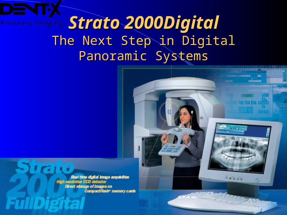
Strato 2000DigitalStrato 2000DigitalThe Next Step in Digital Panoramic The Next Step in Digital Panoramic
SystemsSystems
Advantages of Digital ImagingAdvantages of Digital Imagingo State-of-the art technologyo Eliminates films, chemicals, developer and related
costs of consumables and waste disposalo Dose reduction to the patient and extended X-ray tube
lifetimeo Real-time image acquisition: no waiting for film to
processo Image manipulation and storageo Reduction/elimination of retakes, thanks to the post-
processing compensation of exposure errors
Advantages of Digital ImagingAdvantages of Digital Imaging
o Integration between different image modalities (Intraoral sensor, camera, etc.)
o Integrated management of patient info
o Elimination of the film archive (one CD can store hundreds of images)
o Possibility to easily share images by e-mail, CD, or other media
Computer RequirementsComputer Requirements
o Intel 1.2 GHz chipset and processor
o 256 MB of RAM recommended
o 64 MB AGP graphics card
o USB 2.0 port
o CD-ROM drive and/or 3.5" (1.44 MB) diskette drive
o At least 8 GB hard disk
o Removable hard disk, Zip or Jaz backup system
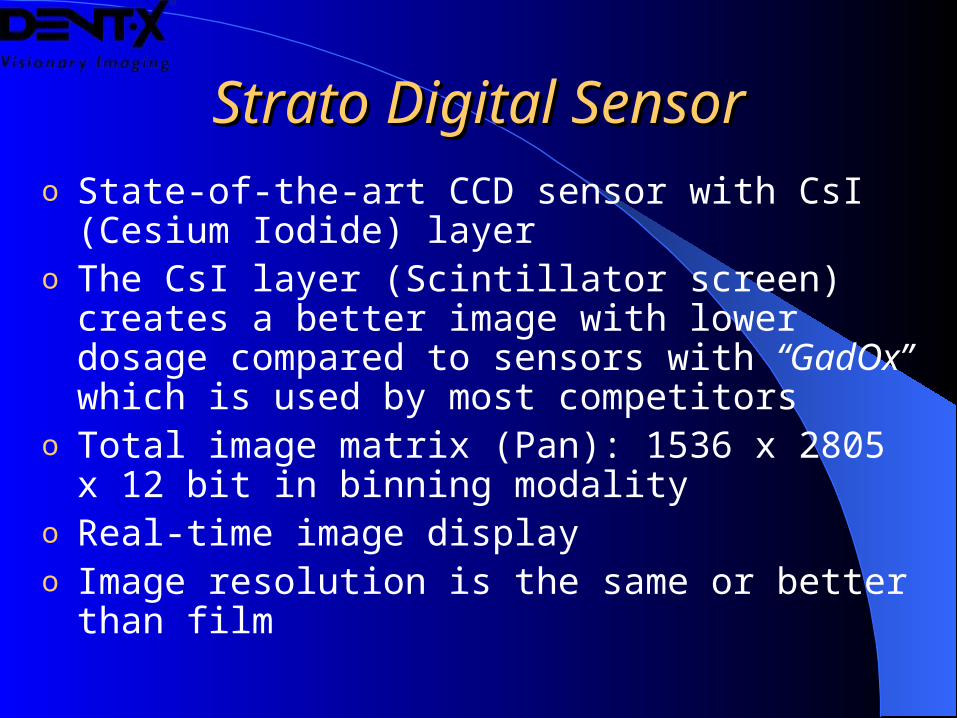
Strato Digital SensorStrato Digital Sensoro State-of-the-art CCD sensor with CsI (Cesium Iodide)
layero The CsI layer (Scintillator screen) creates a better image
with lower dosage compared to sensors with “GadOx” which is used by most competitors
o Total image matrix (Pan): 1536 x 2805 x 12 bit in binning modality
o Real-time image displayo Image resolution is the same or better than film
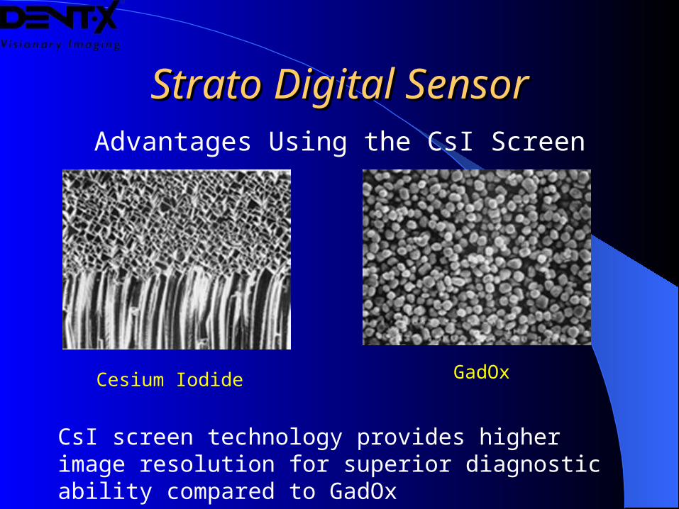
Strato Digital SensorStrato Digital SensorAdvantages Using the CsI Screen
Cesium Iodide GadOx
CsI screen technology provides higher image resolution for superior diagnostic ability compared to GadOx
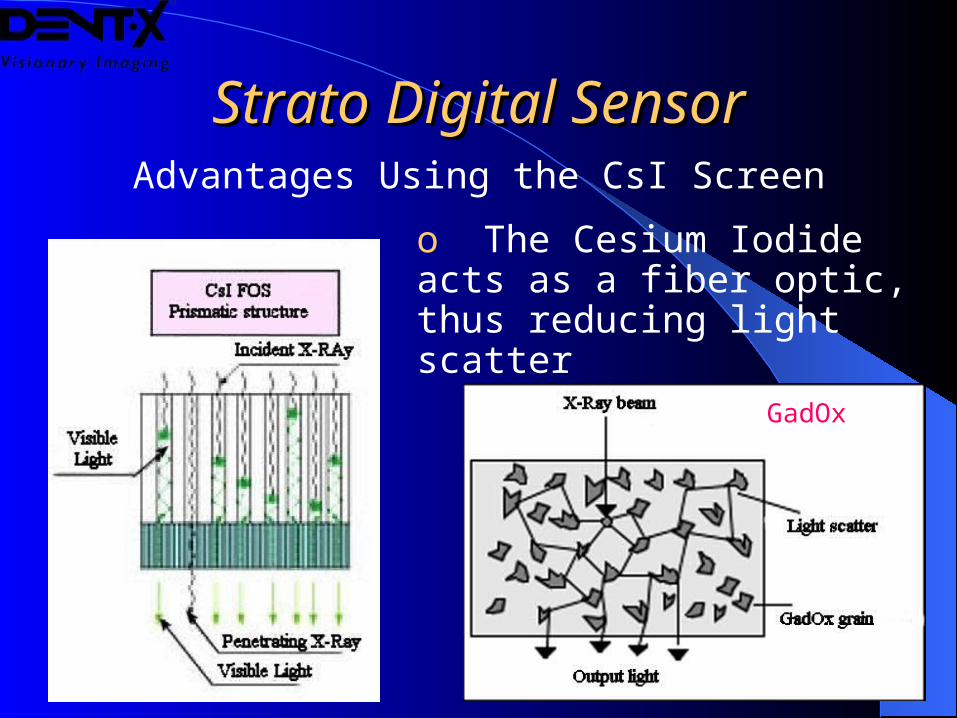
Strato Digital SensorStrato Digital SensorAdvantages Using the CsI Screen
o The Cesium Iodide acts as a fiber optic, thus reducing light scatter
GadOx
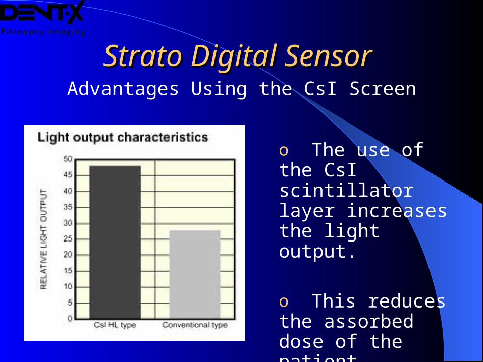
Strato Digital SensorStrato Digital SensorAdvantages Using the CsI Screen
o The use of the CsI scintillator layer increases the light output.
o This reduces the assorbed dose of the patient.
Strato Digital Image QualityStrato Digital Image Qualityo The advanced CCD sensor produces a superb
high-quality image in real-time
o The pixel size is 48µm and the theoretical resolution is 10.4 lp/mm
o The effective resolution is 5.2 lp/mm which is higher than conventional panoramic film
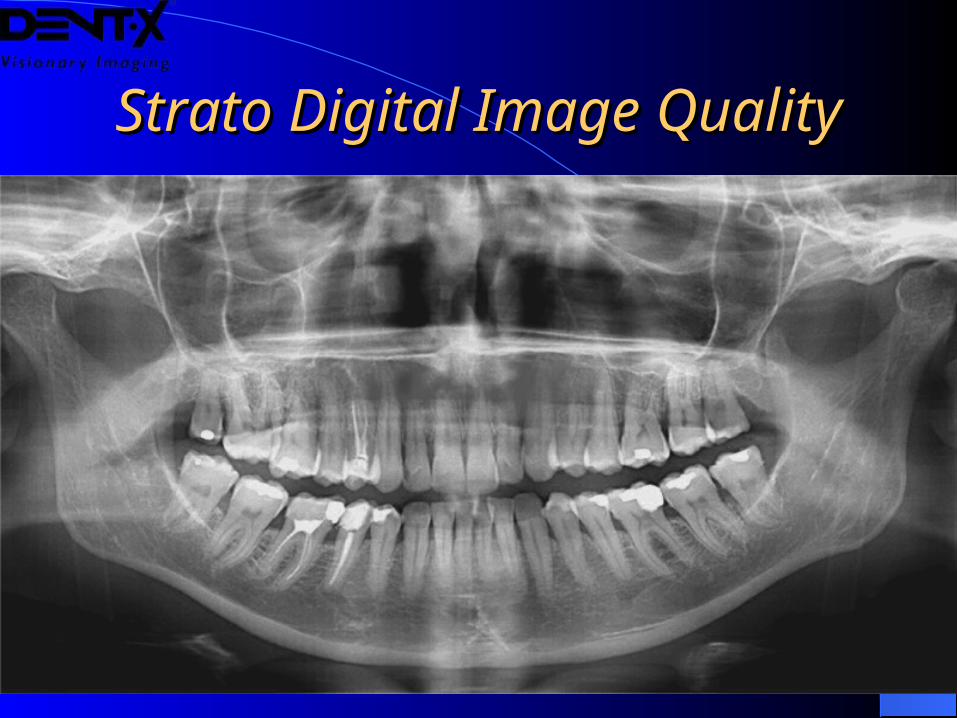
Strato Digital Image QualityStrato Digital Image Quality
Compact Flash CardsCompact Flash Cards
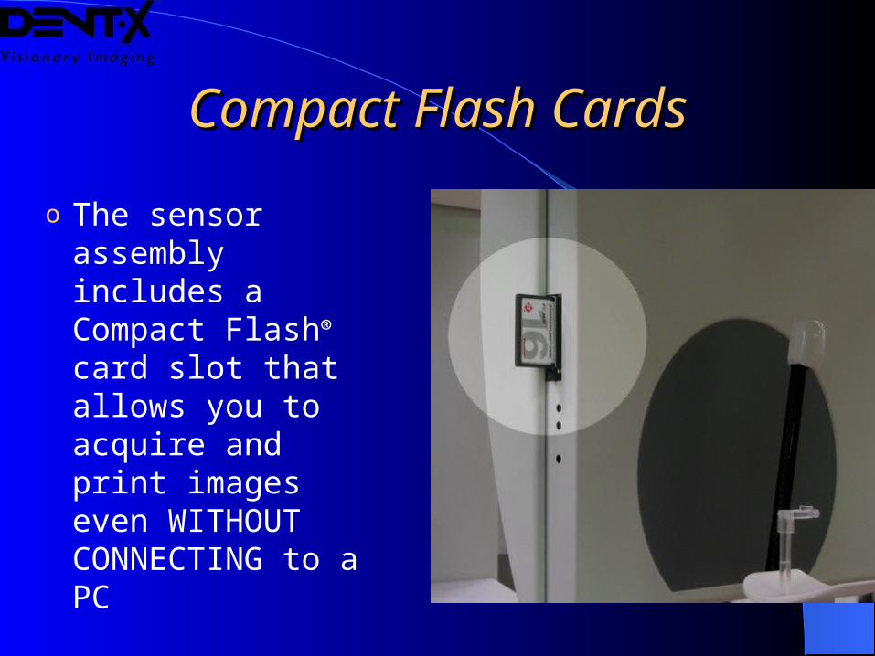
o The sensor assembly includes a Compact Flash® card slot that allows you to acquire and print images even WITHOUT CONNECTING to a PC
Compact Flash CardsCompact Flash Cards
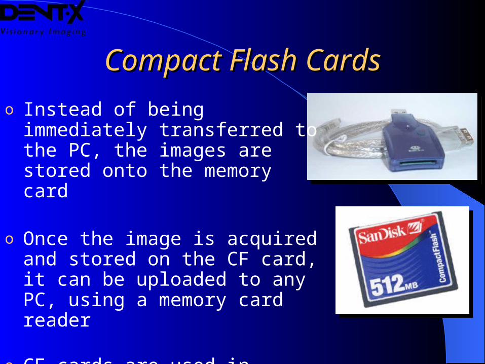
o Instead of being immediately transferred to the PC, the images are stored onto the memory card
o Once the image is acquired and stored on the CF card, it can be uploaded to any PC, using a memory card reader
o CF cards are used in consumer digital cameras and are widely available in the market in different sizes, up to 1 GB
USB 2.0 ConnectionUSB 2.0 Connection
o The new USB 2.0 interface connection allows high data transfer rate (up to 480Mb/s) between the sensor and the PC
o This translates into a real-time image display during the acquisition. The image is fully available at the very end of the tubehead rotation
USB 2.0 ConnectionUSB 2.0 Connection
o The sensor is connected to the PC via a USB cable
o There is no need to insert dedicated boards inside the PC or change any other hardware characteristics of the PC
o This means that the system can be connected to any PC, laptop or desktop, and it is not restricted to only one PC in the practice
o The Strato Digital and software is fully networkable
Software IntegrationSoftware Integration
o The exposure parameters and all of the pre-examination steps can be performed on the computer, including the setting of the patient data
o All of the information of the image, the patient and the exposure are stored in the computer for easy and complete retrieval
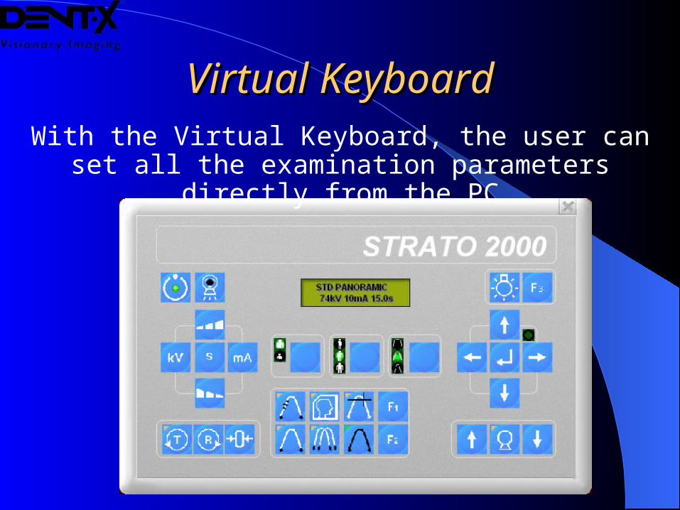
Virtual KeyboardVirtual KeyboardWith the Virtual Keyboard, the user can set all the
examination parameters directly from the PC
The SoftwareThe Software
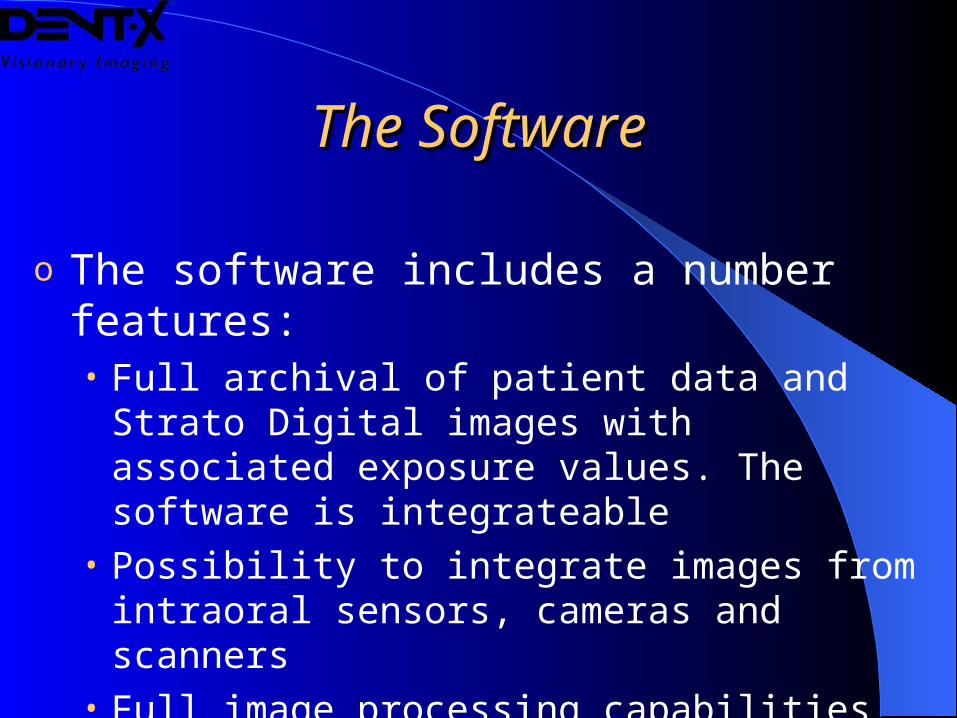
o The software includes a number features:• Full archival of patient data and Strato Digital images
with associated exposure values. The software is integrateable
• Possibility to integrate images from intraoral sensors, cameras and scanners
• Full image processing capabilities• Networking capabilities
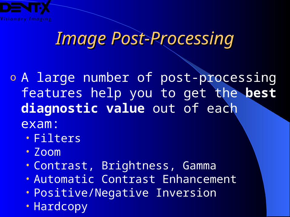
Image Post-ProcessingImage Post-Processing
o A large number of post-processing features help you to get the best diagnostic value out of each exam:• Filters• Zoom• Contrast, Brightness, Gamma• Automatic Contrast Enhancement• Positive/Negative Inversion• Hardcopy
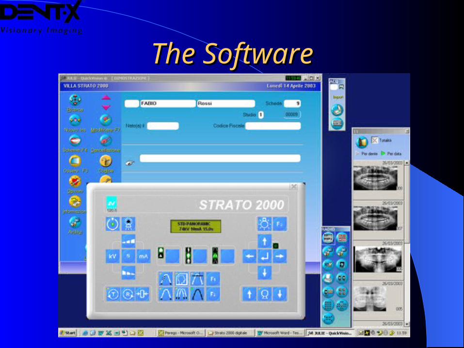
The SoftwareThe Software
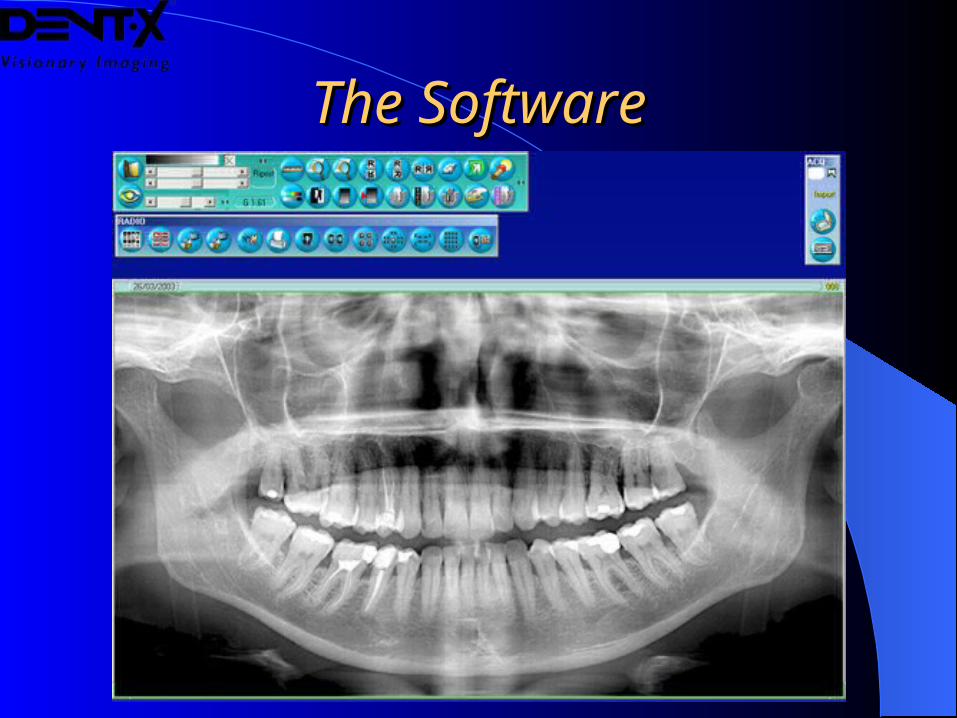
The SoftwareThe Software
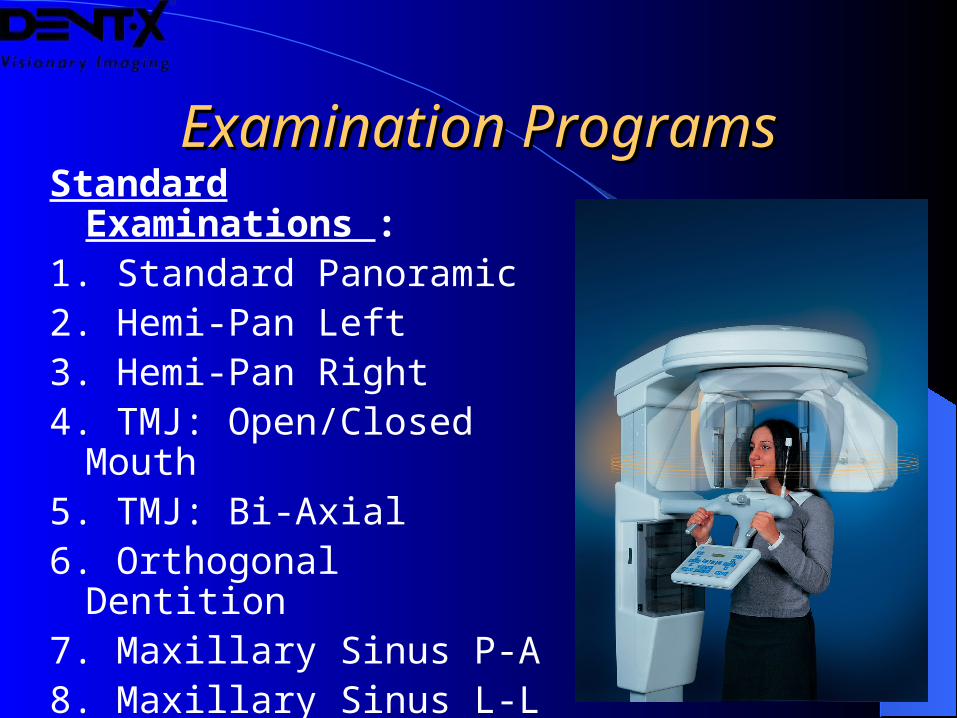
Examination ProgramsExamination ProgramsStandard Examinations :1. Standard Panoramic2. Hemi-Pan Left3. Hemi-Pan Right4. TMJ: Open/Closed Mouth5. TMJ: Bi-Axial6. Orthogonal Dentition7. Maxillary Sinus P-A8. Maxillary Sinus L-L
(All available in Child and Adult modes)
Examination ProgramsExamination Programs
Works In Progress
Will be added to the system by a firmware upgrade:
9. TMJ: P-A
10. Frontal Dentition
11. Reduced-Dosage Quick-Motion Panoramic
(all available in Child and Adult modes)
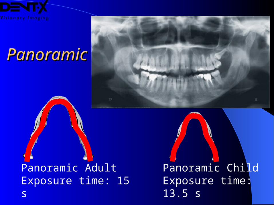
PanoramicPanoramic
Panoramic AdultExposure time: 15 s
Panoramic ChildExposure time: 13.5 s
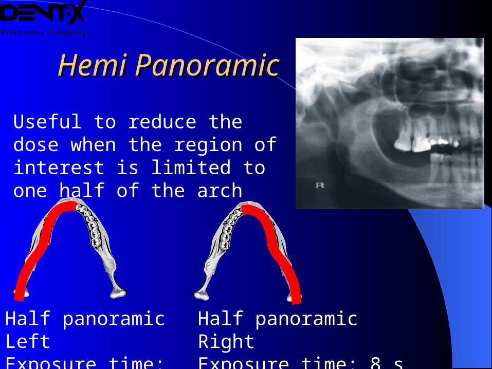
Hemi PanoramicHemi Panoramic
Half panoramic LeftExposure time: 8 s
Half panoramic RightExposure time: 8 s
Useful to reduce the dose when the region of interest is limited to one half of the arch
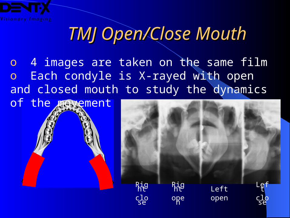
TMJ Open/Close Mouth TMJ Open/Close Mouth
o 4 images are taken on the same filmo Each condyle is X-rayed with open and closed mouth to study the dynamics of the movement
Right
close
Right
open
Leftopen
Leftclos
e
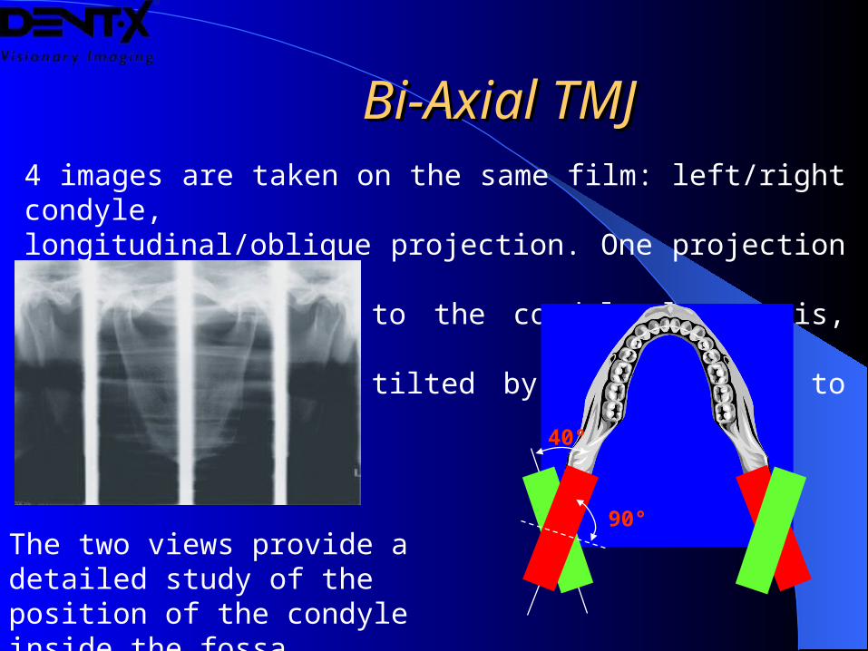
Bi-Axial TMJBi-Axial TMJ4 images are taken on the same film: left/right condyle,longitudinal/oblique projection. One projection is perpendicular
to the condyle long axis, the other is
tilted by 40° compared to the first.
40°
90°The two views provide a detailed study of the position of the condyle inside the fossa
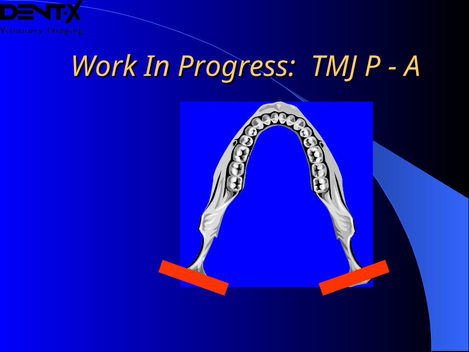
Work In Progress: TMJ P - AWork In Progress: TMJ P - A
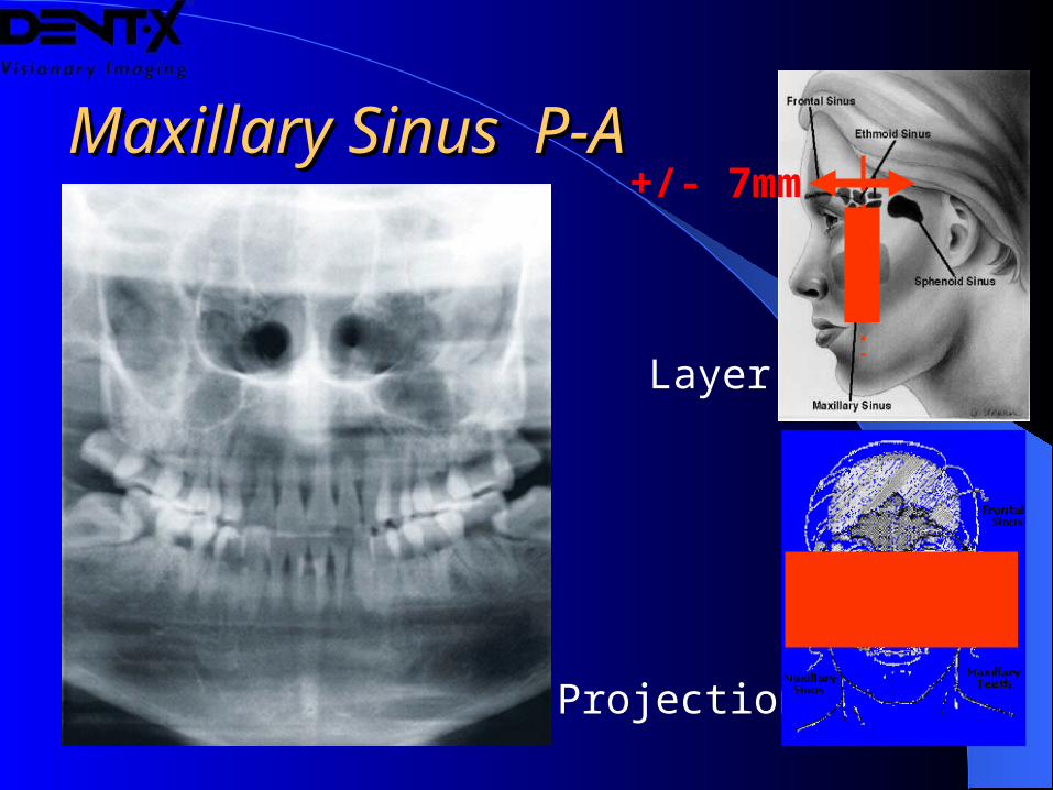
Maxillary Sinus P-AMaxillary Sinus P-A
Projection
Layer
+/- 7mm
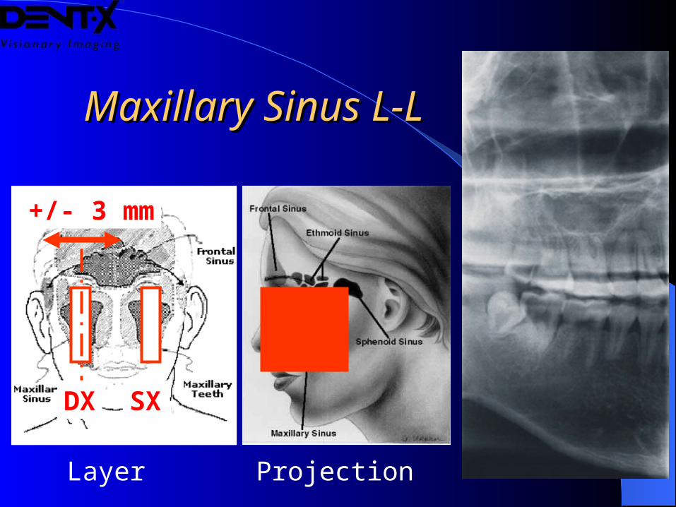
Maxillary Sinus L-LMaxillary Sinus L-L
ProjectionLayer
+/- 3 mm
SXDX
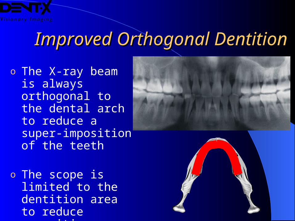
o The X-ray beam is always orthogonal to the dental arch to reduce a super-imposition of the teeth
o The scope is limited to the dentition area to reduce exposition area
Improved Improved Orthogonal Orthogonal DentitionDentition
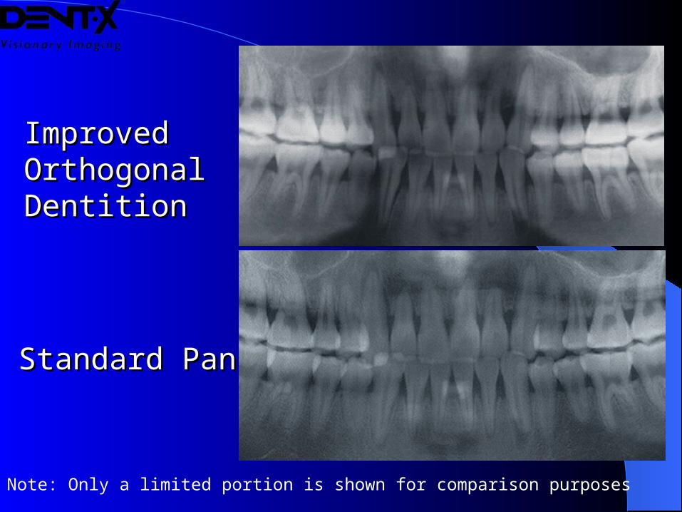
Improved Improved Orthogonal Orthogonal DentitionDentition
Standard PanStandard Pan
Note: Only a limited portion is shown for comparison purposes
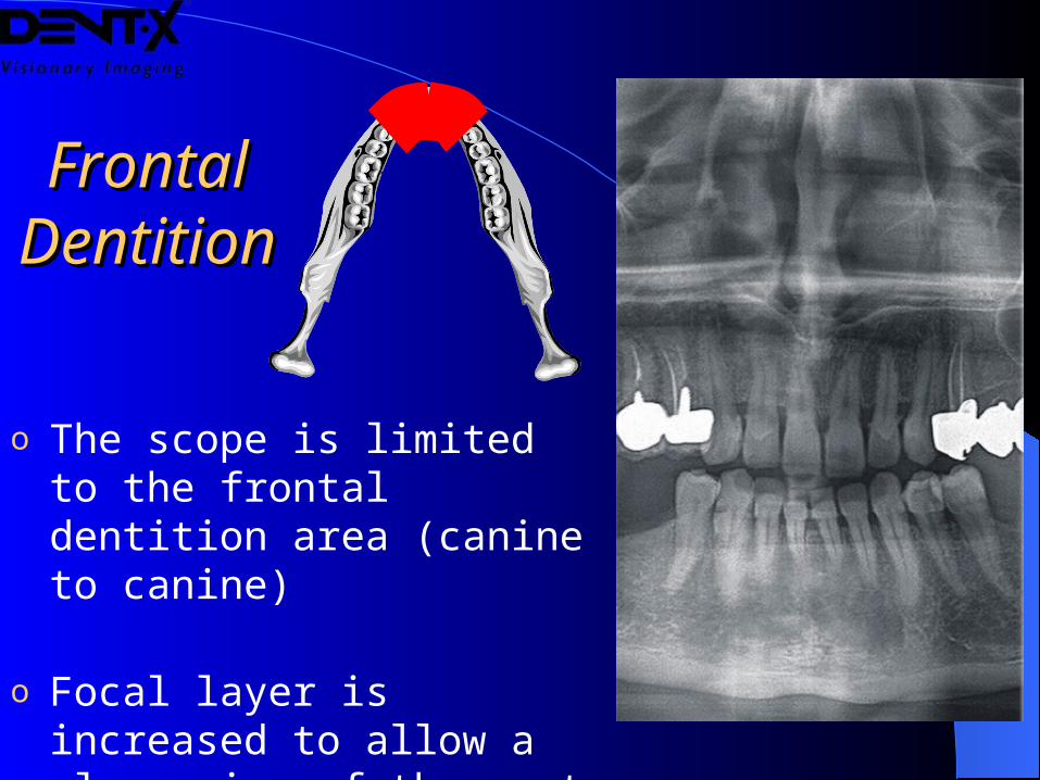
Frontal Frontal DentitionDentition
o The scope is limited to the frontal dentition area (canine to canine)
o Focal layer is increased to allow a clear view of the most critical details
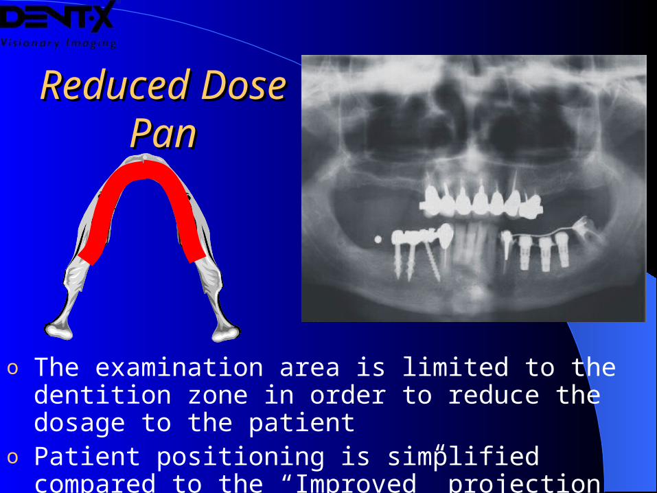
Reduced Dose Reduced Dose PanPan
o The examination area is limited to the dentition zone in order to reduce the dosage to the patient
o Patient positioning is simplified compared to the “Improved” projection
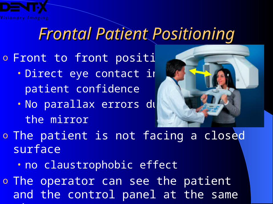
Frontal Patient PositioningFrontal Patient Positioningo Front to front positioning
• Direct eye contact increases
patient confidence• No parallax errors due to
the mirror
o The patient is not facing a closed surface• no claustrophobic effect
o The operator can see the patient and the control panel at the same time
Anatomical ProgramsAnatomical Programs
According to the anatomic program selected, the system automatically calculates:
o Shape of the focal layero Optimal kV valueo Optimal mA valueo Optimal exposure time
Constant MagnificationConstant Magnification
o All examination programs have constant magnification
o Exact measurements can be taken on the images
o In the panoramic exam the magnification is constant in the center of the focal layer
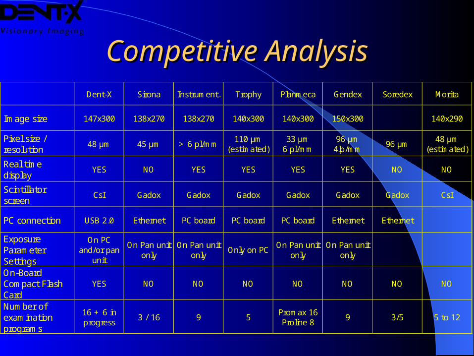
Competitive AnalysisCompetitive Analysis Dent-X Sirona Instrument. Trophy Planmeca Gendex Soredex Morita
Image size 147x300 138x270 138x270 140x300 140x300 150x300 140x290
Pixel size / resolution
48 µm 45 µm > 6 pl/mm 110 µm
(estimated) 33 µm
6 pl/mm 96 µm 4lp/mm
96 µm 48 µm
(estimated)
Real time display
YES NO YES YES YES YES NO NO
Scintillator screen
CsI Gadox Gadox Gadox Gadox Gadox Gadox CsI
PC connection USB 2.0 Ethernet PC board PC board PC board Ethernet Ethernet
Exposure Parameter Settings
On PC and/or pan
unit
On Pan unit only
On Pan unit only
Only on PC On Pan unit
only On Pan unit
only
On-Board Compact Flash Card
YES NO NO NO NO NO NO NO
Number of examination programs
16 + 6 in progress
3 / 16 9 5 Promax 16 Proline 8
9 3/5 5 to 12
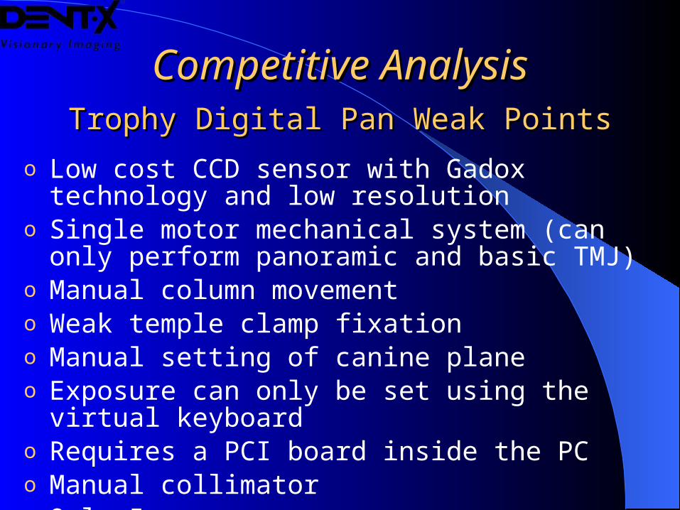
Competitive AnalysisCompetitive AnalysisTrophy Digital Pan Weak PointsTrophy Digital Pan Weak Points
o Low cost CCD sensor with Gadox technology and low resolution
o Single motor mechanical system (can only perform panoramic and basic TMJ)
o Manual column movement o Weak temple clamp fixationo Manual setting of canine planeo Exposure can only be set using the virtual keyboardo Requires a PCI board inside the PCo Manual collimatoro Only 5 programs

Contact Dent-XContact Dent-X
o For more information:For more information:• Visit www.Dent-X.com Visit www.Dent-X.com
• Call 800-225-1702Call 800-225-1702
• Email: [email protected] Email: [email protected]
• Contact your local dealerContact your local dealer
![MASTER REPORT REVIEW OF GENERAL PANORAMIC OPTICAL … · and security, panoramic endoscope, machine vision, panoramic projection system, and so on [1, 2]. Panoramic lens systems can](https://img.pdfslide.us/doc/110x75/5e184f54abc03831285efb0b/master-report-review-of-general-panoramic-optical-and-security-panoramic-endoscope.jpg)