Embed Size (px)
Citation preview

Dynamic Article LinksC<Soft Matter
Cite this: Soft Matter, 2011, 7, 10737
www.rsc.org/softmatter PAPER
Publ
ishe
d on
06
Oct
ober
201
1. D
ownl
oade
d by
Uni
vers
ity o
f C
hica
go o
n 28
/10/
2014
21:
24:1
0.
View Article Online / Journal Homepage / Table of Contents for this issue
Stimulus responsive self-assembly of Gemini Amphiphilic Pseudopeptides†
Jenifer Rubio,a Ignacio Alfonso,*b M. Isabel Burguetea and Santiago V. Luis*a
Received 27th July 2011, Accepted 30th August 2011
DOI: 10.1039/c1sm06435e
Amphiphilic amino acid derived compounds are very interesting for the design of building blocks able
to self-assemble into highly ordered nanostructures, in a hierarchical and controlled fashion. With this
aim, the modular synthesis and the full characterization of simple Gemini Amphiphilic Pseudopeptides
(GAPs) have been carried out. These compounds were designed to establish intermolecular interactions
in a hierarchical way to finally render supramolecular assemblies into well-ordered nanostructures,
such as fibers, tubes, tapes or spherical vesicles. Different structural variables have been implemented,
such as the amino acid side chain, the length of the central spacer and the nature of the hydrophobic
tails. Besides, the effect of the environment was systematically checked, by performing the studies in
solvents of different polarities (chloroform, methanol or aqueous methanol) and at different pHs
(neutral, basic and acidic). The non-covalent self-assembling abilities and the structural features of the
GAPs have been studied in the solid (SEM, TEM and FT-IR) and in the solution states (NMR, UV,
CD, FT-IR and fluorescence spectroscopy). Moreover, the connection between the solution and the
solid states has been established by monitoring the slow evaporation of the solvent by ATR FT-IR.
This study has allowed the establishment of a relationship between the chemical structures of the GAPs
and their abilities to form nanostructures. In some optimal cases (especially for the valine derivatives
with medium-length spacers and two decyloxybenzyl hydrophobic tails), they behaved as stimulus
responsive self-assembling nano-structures, which form amorphous materials from non-polar solvents,
nano-fibers from polar environments at neutral or basic pH and vesicles when become protonated at
acidic pH values. A reasonable structural model to explain the experimental observations can be
proposed through the combination of the results from the different techniques.
Introduction
The controlled self-assembly of small molecules into a well-
defined structure in the nanometric scale has become a very active
research field during the last decade.1–3 The formation of the
corresponding nanostructure is dictated by the combination of
intermolecular non-covalent interactions,4–9 which work cooper-
atively to define the shape and dimensions of the final nano-
object.10–12 This methodology has been called the bottom-up
approach to nanotechnology; it is a biomimetic strategy and
represents the natural approximation for chemists.4,6,13–20
Peptides and peptide-like molecules are among the most inter-
esting families of building blocks for this bottom-up
aDepartamento de Qu�ımica Inorg�anica y Org�anica, Universitat Jaume I,Avenida Sos Baynat, s/n, Castell�on, Spain. E-mail: [email protected]; Fax:+34 964728214; Tel: +34 964728239bDepartamento de Qu�ımica Biol�ogica y Modelizaci�on Molecular, Institutode Qu�ımica Avanzada de Catalu~na (IQAC-CSIC), Jordi Girona 18-26,Barcelona, Spain. E-mail: [email protected]; Fax: +34932045904; Tel: +34 934006100
† Electronic supplementary information (ESI) available: Full spectracharacterization of all the described compounds and additionalexperimental data. See DOI: 10.1039/c1sm06435e
This journal is ª The Royal Society of Chemistry 2011
construction.7,21–31 Amino acids are molecules with a high func-
tional density and a large potential for structural diversity, which
allows the implementation of many non-covalent interactions
with a minimum number of elements in the design.32–34 Accord-
ingly, the self-assembly of many peptide-like compounds into
nanostructures has been described in the literature, with prom-
ising applications in biomedicine and for the fabrication of new
materials.33,35–45 On the other hand, amphiphilic molecules are
also interesting species in the self-assembling field, since they are
able to display a dual behavior depending on the environment.46–50
Especially, they have been used for preparing nano-objects in
highly polar environments, with interesting potential to be used in
biological applications, as theymimic the structure of compounds
essential for the generation of important bioassemblies.51–53
The most interesting topic within this field should be the
possibility to make the self-assembling system able to respond to
a simple external stimulus, thus triggering the formation of
a given nanostructure, or for obtaining different nano-objects
from the same building blocks under controlled conditions.54–59
To this aim, different stimuli have been used like polarity of the
medium, pH, enzymatic reactions, dilution or the presence of
chemicals and reducing agents.60–69
Soft Matter, 2011, 7, 10737–10748 | 10737

Publ
ishe
d on
06
Oct
ober
201
1. D
ownl
oade
d by
Uni
vers
ity o
f C
hica
go o
n 28
/10/
2014
21:
24:1
0.
View Article Online
Here we report on the design, synthesis and characterization of
different Gemini Amphiphilic Pseudopeptides (thereafter named
GAPs)70 able to self-assemble into nanostructures displaying
different shapes like fibers, tubes, tapes or vesicles. More inter-
estingly, the self-assembling process can be controlled through
the polarity of the environment and, for some specific examples,
can be switched between fibers and vesicles by simple variation of
the pH.
Scheme 1 Synthesis of the GAPs by a reductive amination reaction.
Results and discussion
Design and synthesis of the GAPs building blocks
The GAPs considered in this work were designed to display
different intermolecular polar and non-polar interactions. Their
main structural characteristics are depicted in Fig. 1.
They possess two amino acid moieties connected through their
acid ends by a flexible spacer, and two hydrophobic tails at both
amino termini, rendering an overall C2 symmetry. This design
will allow the modular implementation of several functional
groups with the potential of establishing different interactions
(H-bond, dipole–dipole, p-stacking, hydrophobic or van der
Waals contacts, etc.). Moreover, the GAPs have also two
secondary amino nitrogens, which display the corresponding
acid–base properties, and the possibility of the systems to
respond to pH changes.
The synthesis of the proposed GAPs has been carried out
following a simple procedure as depicted in Scheme 1. First of all,
the corresponding C2 symmetrical bis(amidoamines) were
prepared using previously reported procedures.71 After that, the
N,N0-dialkylated compounds were prepared by a reductive
amination reaction with different aldehydes.72–74 The selection of
both the solvent and the reducing agent was not trivial for this
step, and, after an optimization study, we found that the
combination of chloroform and the borane–pyridine complex led
to the best yields and selectivity towards the dialkylated deriva-
tive (Table 1).70,75,76 The results mainly depend on the structure of
the starting aldehyde, leading to higher yields in the case of
aromatic aldehydes. Although the yields with the aliphatic
aldehydes were moderate to low, the complete selectivity towards
the dialkylated compound made this procedure of synthetic
interest. Besides, considering the low solubility of the final
compounds in these cases, we believe that the lower yields
observed can be associated to the problems found during their
isolation. In some cases, the corresponding monoalkylated
derivative was obtained as a by-product (yields in brackets in
Table 1), being readily separated from the GAPs by flash chro-
matography. The reaction can be performed with pseudopep-
tides having aliphatic (Me, iPr, iBu) or aromatic (Bn) side chains,
and with different spacers between amino acid moieties. Some
Fig. 1 General structure of the GAPs.
10738 | Soft Matter, 2011, 7, 10737–10748
compounds bearing shorter tails (entries 14 and 15) were also
prepared as references for the self-assembling studies (see below).
For a clearer discussion, we will use a systematic code for the
naming of the compounds (Table 1) as follows: ANXxx_a(tail)
where N means the total number of carbon atoms of the central
spacer (and thus, N ¼ n + 2), Xxx is the three letter code for the
corresponding amino acid, and the tail is named as a8 for octyl,
a1obenz for methoxybenzyl, a4obenz for butyloxybenzyl and
a10obenz for decyloxybenzyl. Thus, for instance, A4Val_a10o-
benz would be the GAP derived from valine with a butylenic
spacer and two decyloxybenzyl tails.
Ultrastructure in the solid state
In order to study the ability of the GAPs to self-assemble into
supramolecular nano-structures in the solid state we performed
Scanning Electron Microscopy (SEM) experiments for samples
(ca. 2 mg mL�1) slowly evaporated onto aluminium surfaces.
Also, for selected examples, Transmission Electron Microscopy
(TEM) images were taken, rendering similar morphologies to
those observed by SEM, although with slightly smaller sizes,
probably due to the differences in sample preparation for both
techniques. The samples were grown from solvents of different
polarities such as CHCl3, MeOH or aqueous (33% v/v) MeOH in
order to study the effect of the environment in the aggregation
behavior. Besides, the protonation of the secondary amino
groups of the GAPs was also studied by preparing the samples in
aqueous methanol containing a slight excess of acid. The
observations are summarized in Table 2 and selected images are
shown in Fig. 2–4, the rest being provided as ESI (Fig. S1–S4†).
Different nano-structures were observed ranging from fibrils,
fibers, tapes, tubes or even spheres, depending on the structure of
the GAPs and on the solvent used for growing the samples.77–79
Several general trends can be concluded from this study. First
of all, the GAPs show low tendency to self-organize in chloro-
form, since only a few of them form nanostructures when
evaporated from this medium. Mainly the compounds bearing
aliphatic tails and ethylene (A2Val_a8, entry 9) or propylene
(A3Val_a8, entry 10) spacers self-aggregate in chloroform
yielding hollow tubes or tapes (Fig. 2), respectively. The saddle
structural changes rendering important supramolecular differ-
ences suggested that the morphology of the nanostructures is
dictated by the conformation of the central spacer, which leads to
completely different disposition and shape of the overall GAP.
However, upon increasing the polarity of the solvent, we
observed the formation of nanostructures for many of the GAPs,
most of them appearing as fibers with different sizes and widths
This journal is ª The Royal Society of Chemistry 2011

Table 1 Synthesis of the GAPs
Spacer (n) Aaa (R) Tail (CH2R0) Compound Yielda
1 (CH2)2 (0) Val (iPr) CH2C6H4OC10H21 A2Val_a10obenz 63 [5]b
2 (CH2)3 (1) Val (iPr) CH2C6H4OC10H21 A3Val_a10obenz 68 [15]b
3 (CH2)4 (2) Val (iPr) CH2C6H4OC10H21 A4Val_a10obenz 66 [19]b
4 (CH2)5 (3) Val (iPr) CH2C6H4OC10H21 A5Val_a10obenz 38 [19]b
5 (CH2)6 (4) Val (iPr) CH2C6H4OC10H21 A6Val_a10obenz 67 [9]b
6 (CH2)2 (0) Phe (Bn) CH2C6H4OC10H21 A2Phe_a10obenz 66 [21]b
7 (CH2)3 (1) Phe (Bn) CH2C6H4OC10H21 A3Phe_a10obenz 61 [19]b
8 (CH2)4 (2) Phe (Bn) CH2C6H4OC10H21 A4Phe_a10obenz 74 [23]b
9 (CH2)2 (0) Val (iPr) C8H17 A2Val_a8 2610 (CH2)3 (1) Val (iPr) C8H17 A3Val_a8 2111 (CH2)4 (2) Val (iPr) C8H17 A4Val_a8 3212 (CH2)4 (2) Ala (Me) CH2C6H4OC10H21 A4Ala_a10obenz 48 [26]b
13 (CH2)4 (2) Leu (iBu) CH2C6H4OC10H21 A4Leu_a10obenz 63 [12]b
14 (CH2)4 (2) Val (iPr) CH2C6H4OCH3 A4Val_a1obenz 7715 (CH2)4 (2) Val (iPr) CH2C6H4OC4H9 A4Val_a4obenz 70 [12]b
a Isolated yields (%) for the one-pot two-steps reductive amination reaction. b Numbers in brackets correspond to the monoalkylated by-product.
Publ
ishe
d on
06
Oct
ober
201
1. D
ownl
oade
d by
Uni
vers
ity o
f C
hica
go o
n 28
/10/
2014
21:
24:1
0.
View Article Online
(Fig. 3). Among all the measured compounds in MeOH, only
three of them were unable to form nanostructures (entries 1, 2
and 6). It is worth mentioning that those observations corre-
spond to GAPs having the smaller spacers: A2 (entries 1 and 6)
or A3 (entry 2). Very interestingly, the compounds with benzy-
loxyalkyl tails having short alkyl groups (entries 14 and 15 for
Me and Bu alkyl groups, respectively) also showed the formation
of fibers when evaporated from MeOH. This suggests that the
central pseudopeptidic moiety must be playing a major role in the
aggregation process, maybe through the establishment of inter-
molecular H-bonding interactions. This proposal is also in line
with the formation of fibers due to the high directionality of H-
bonds, which would induce the growing of the nanostructures in
a preferred direction. The further increase of the polarity by
using 33% (v/v) of water in the MeOH did not significantly
change the aggregation abilities (Fig. 3d). In this mixture of
solvents, we also tested the effect of the pH, by performing the
Table 2 Self-assembling properties of the GAPs
Compound CHCl3a MeOHa MeOH (aq)a MeOH/H+a CMCc
1 A2Val_a10obenz Fibrils No No No 852 A3Val_a10obenz No No No Vesicles 163 A4Val_a10obenz No Fibers Fibers Vesicles 134 A5Val_a10obenz No Fibers Fibers Vesicles 205 A6Val_a10obenz No Fibers Fibers No 636 A2Phe_a10obenz No No No No >1007 A3Phe_a10obenz No Fibers Fibers No >1008 A4Phe_a10obenz No Fibers Fibers No 489 A2Val_a8 Tubes Fibers Fibers Fibers n.md
10 A3Val_a8 Tapes Tapes Tapes Non-hom.b n.md
11 A4Val_a8 No Fibers Fibers Fibers n.md
12 A4Ala_a10obenz Fibrils Fibers Fibers Vesicles 1613 A4Leu_a10obenz No Fibrils Fibers No 6214 A4Val_a1obenz No Fibers Fibers No >10015 A4Val_a4obenz No Fibers Fibers No >100
a Nanostructures observed by electron microscopy in samples grownfrom different solvents. b Non-hom.: SEM images showed non-homogeneous formation of both fibrils and spheres, accompanied withamorphous material. c CMC (mM): critical micellar concentration inwater at pH 2.7 (AcOH/AcONa buffer) determined by fluorescencespectroscopy using pyrene as a probe. d n.m.: not measured.
This journal is ª The Royal Society of Chemistry 2011
experiments in neutral, basic and acidic media. No important
changes were observed from neutral to basic media. However,
the situation in the presence of acid is completely different. The
fiber-like nanostructures were destroyed in most of the studied
examples upon protonation, except for the derivatives with
completely aliphatic tails (entries 9–11 in Table 2). Very
surprisingly, for some specific cases having decyloxybenzyl tails
and Val or Ala amino acids, the formation of spheres of micro-
metric size was observed (entries 2, 3, 4 and 12 and Fig. 4).
Besides, the formation of microspheres is observed for
compounds having an intermediate number of atoms in the
Fig. 2 SEM micrographs of (a) A4Val_a1obenz, (b) A4Phe_a10obenz,
(c and d) A2Val_a8 and (e and f) A3Val_a8 grown fromCHCl3 solutions.
The corresponding scale bars are shown for every picture.
Soft Matter, 2011, 7, 10737–10748 | 10739

Fig. 3 Selected SEM micrographs for (a) A4Val_a1obenz, (b)
A2Val_a8, (c) A4Leu_a10obenz and (d) A6Val_a10obenz grown from
MeOH (a–c) or 2 : 1 MeOH : H2O (d) solutions. The corresponding scale
bars are shown for every picture.
Publ
ishe
d on
06
Oct
ober
201
1. D
ownl
oade
d by
Uni
vers
ity o
f C
hica
go o
n 28
/10/
2014
21:
24:1
0.
View Article Online
central spacer (from three to five, n ¼ 1–3). Either shorter or
longer spacers produced amorphous materials in acidic medium.
Therefore, there is a strong correlation between the chemical
structure of the GAP and its ability to self-assemble into spheres.
Thus, for instance, the compound having the butylenic spacer
(A4Val_a10obenz, with a length in the middle of those forming
spheres) can be considered as a stimulus dependent self-assem-
bling GAP, since it forms amorphous material in CHCl3, fibers in
Fig. 4 Effect of the environment on the self-assembling of
A4Val_a10obenz. SEMmicrographs of A4Val_a10obenz grown from (a)
CHCl3, (b) MeOH, (c) 2 : 1 MeOH : H2O and (d) 2 : 1 MeOH : H2O +
HCl. TEM images of A4Val_a10obenz grown from (e) MeOH and (f)
2 : 1 MeOH : H2O + HCl.
10740 | Soft Matter, 2011, 7, 10737–10748
MeOH or aqueous MeOH at neutral and basic pH, while
providing spheres in acidic medium (Fig. 4). These morphologies
have been studied by both SEM and TEM techniques, which
suggested that the spheres are hollow and, therefore, can be
classified as vesicles attending to their dimensions and the
molecular size of the building blocks (Fig. 4d and f). Within the
same central pseudopeptidic frame, the nature of the tails is
highly important for the formation of these vesicles.
The decyloxybenzyl groups seem to be an essential feature in
this regard since the compounds having shorter alkyloxybenzyl
(A4Val_a1obenz and A4Val_a4obenz) or fully aliphatic residues
(A2Val_a8, A3Val_a8 and A4Val_a8) at the amino nitrogens,
either failed to form any nanostructure (entries 14 and 15 in
Table 2), or led to a fibrilar morphology (entries 9 and 11) or to
a non-homogeneous mixture (entry 10). Overall, we have found
that with a delicate tune of the molecular architecture and the
environment conditions, a control over the self-assembling in the
solid state can be carried out. Thus, transition from amorphous
materials to fibers or vesicles can be observed in several cases,
especially in Val derivatives with decyloxybenzyl tails and
a spacer of a medium length (n ¼ 1–3).
Solution structure of the GAPs
With the aim of getting deeper information on the conforma-
tional and structural behavior of the GAPs in a pre-aggregated
state, before the formation of the self-assembled nanostructures,
we have carried out a systematic study of the compounds in
solution, by using a broad variety of spectroscopic techniques.
Thus, we have performed NMR, UV, CD and fluorescence
studies of selected examples under different conditions. Finally,
we have also studied the transition from the diluted solution to
the solid state by monitoring the evaporation of the solvent by
ATR FT-IR spectroscopy.
NMR spectroscopy. The 1H NMR spectra of some selected
GAPs were studied in different solvents (CDCl3 and CD3OD), in
the absence and in the presence of acid (DCl in D2O), at different
temperatures and at two overall concentrations (1 mM and 20
mM). Considering the results obtained in the solid state, we
selected the compounds displaying a more distinct self-assem-
bling behavior in different media.
Therefore, the Val derivatives with the decyloxybenzyl tails
and bearing spacers of different lengths were selected for this
systematic NMR study (Fig. S5–S15 and Tables S1–S3†). First of
all, a very small effect was observed when changing the overall
concentration of the GAPs from 1 mM to 20 mM, which sug-
gested that no significant aggregation was present over that
concentration range. Thus, the NMR study should mainly
correspond to fully solvated pre-aggregated species. Anyway,
knowledge of the conformational behavior and the inter/intra-
molecular interactions is of interest to rationalize the self-
assembling observations in the solid state. Clear differences were
observed on the NMR spectra when changing the polarity of the
medium. Thus, for instance, we studied the 1H NMR spectra in
different conditions for A4Val_a10obenz (Fig. 5 and ESI†). The
chemical shift (>7.00 ppm) and temperature dependence (DdNH/
DT ¼ �3.5 ppb K�1) of the amide NH protons signal in CDCl3suggested the presence of partially H-bonded amide groups. The
This journal is ª The Royal Society of Chemistry 2011

Fig. 5 1H NMR (500 MHz, 303 K) spectra of A4Val_a10obenz in
CDCl3 (down), CD3OD (middle) and CD3OD + DCl (up). Assignations
of the signals are shown following the arbitrary lettering of the chemical
substructure.
Publ
ishe
d on
06
Oct
ober
201
1. D
ownl
oade
d by
Uni
vers
ity o
f C
hica
go o
n 28
/10/
2014
21:
24:1
0.
View Article Online
high chemical shift values of protons at a and b positions are also
in line with this proposal. Besides, the methylene vicinal to the
amide group (A in Fig. 5) showed chemical non-equivalence [Dd
(HA/A0) ¼ 0.052 ppm at 294 K], which was decreased upon
heating [Dd(HA/A0)¼ 0.038 ppm at 323 K]. All these observations
suggested that the pseudopeptidic moiety is intramolecularly H-
bonded in a relatively rigid conformation. However, when
acquiring the 1H NMR spectrum in methanol, the signals cor-
responding to the protons from the pseudopeptidic moiety (a, b,
g and A) moved up-field, while those from the hydrophobic tails
did not significantly change. More importantly, the hydrogen
atoms of the methylenes in the A position lost their chemical
non-equivalence, appearing as a broad triplet. These observa-
tions were consistent with the breaking of the intramolecularly
H-bonded pseudopeptidic structure, leading to an unfolded and
more flexible conformation in solution. Protonation of
A4Val_a10obenz in CD3OD also affected the NMR spectrum.
Thus, the presence of a positive charge at the amino nitrogen
produced large downfield shifts at the positions 1, a, b and g.
Besides, the deshielding of protons at 2, 3 and 4 suggested that
the anisole substructure was highly polarized upon nitrogen
protonation. On the other hand, the signals from the hydro-
phobic tail suffered a slight up-field shift (0.01–0.02 ppm).
Surprisingly, the anisochrony of the protons at position A [Dd
(HA/A0) ¼ 0.043 ppm] again suggested a restricted mobility of the
butylenic spacer for the protonated form of A4Val_a10obenz,
and a different chemical environment for both faces of the
spacer.
We observed similar trends for the other derivatives (see ESI†
for details), suggesting that the central pseudopeptidic spacer
behaved in a similar way. Thus, we can conclude that the
different behavior in the transition from the fibers to the vesicles
is an effect of the structural relationship between this moiety and
the hydrophobic tails (see below).
This journal is ª The Royal Society of Chemistry 2011
Molecular electronic spectroscopy (UV-CD). Those selected
GAPs were also studied by electronic spectroscopy (UV) and
circular dichroism (CD) in solution. The large absorbance of
chloroform at 220–230 nm precluded the accurate study of the
systems in this solvent. Thus, we focused on the effect of the
protonation in MeOH solution, bearing in mind that the changes
in the self-assembling behavior were also accompanied by
a change in the flexibility of the GAPs in solution, as suggested
by the 1H NMR spectra, in the presence and in the absence of
acid. Moreover, the UV-CD spectra were acquired at different
concentrations (0.012–0.583 mM) showing a perfect linear
dependence of the absorbance versus the concentration, which
also suggested the absence of aggregated species at this concen-
tration range. Different structural parameters in the GAPs were
varied, like the length of the central spacer, the amino acid side
chain and the hydrophobic tails.
The length of the central spacer had an important impact on
the UV-CD spectra. The UV spectra in MeOH at neutral pH
showed two characteristic bands: one very intense at 226–230 nm
(p–p* transition) and another less intense at 273–276 nm (n–p*
transition). The corresponding UV-CD spectra for compounds
ANVal_a10obenz (with N ¼ 2–6) are superposed in Fig. 6A. The
molar extinction coefficient is dependent on the length of the
spacer, being highest with the butylenic one (n ¼ 2,
A4Val_a10obenz). Regarding the CD spectra in MeOH, all the
compounds showed a weak negative Cotton effect at 230 nm.
Interestingly, if we represent the D3 of the CD spectra versus the
spacer (Fig. 6B), once again the most intense CD signal was
obtained for the medium-sized spacers. This observation corre-
lates with the fact that these were the GAPs showing a higher
propensity to form fibers in SEM.
The addition of acid produced important changes in the UV-
CD spectra, which were significantly different for each
compound (Fig. 6C). Thus, the variation of the UV absorbance
strongly depended on the length of the spacer (Fig. 6D), showing
a strong hypochromic effect of both maxima for n ¼ 2
(A4Val_a10obenz). Concomitantly, an almost complete disap-
pearance of the CD signal was observed upon protonation. The
reduction of the CD signal due to the transition from fibers to
vesicles has been previously reported61,80,81 but, in our case, the
CD measurements were performed in a pre-aggregated state.
Thus, we ascribed the almost silent CD spectra to the presence
of a mixture of conformations in solution with a complementary
geometrical disposition of the chromophores, finally rendering
a null effect on the CD spectra (see below). Another interesting
observation, after the protonation, is the appearance of a band at
lower energy at 330 nm, which can be assigned to a charge
transfer (CT) band (Fig. 6E). This CT can be produced by the
interaction between the anisole chromophore on one side of the
GAP and the ammonium group on the other side, in a folded
conformation. According to this hypothesis, this CT band is
maximum for the shortest spacer (close proximity between
groups), but does not uniformly decay with the separation
between the interacting groups. Once again, we observed
a discontinuity for the butylenic spacer showing a slightly more
effective CT than the propylenic one (Fig. 6F) and suggesting
a more efficient folding of the GAP with this spacer.
Other structural parameters were also studied by UV-CD
spectroscopy, by keeping constant the butylenic spacer (N ¼ 4).
Soft Matter, 2011, 7, 10737–10748 | 10741

Fig. 6 (A) UV-CD spectra of ANVal_a10obenz (N¼ 2–6) inMeOH. (B)
Plot of the D3 of the CD spectra of ANVal_a10obenz (N¼ 2–6) inMeOH
versus n (n¼N�2). (C) UV-CD spectra of ANVal_a10obenz (N¼ 2–6) in
MeOH in the presence of acid. (D) Plot of the variation of UV extinction
coefficients at the corresponding maxima due to protonation versus n. (E)
Zoomed UV spectra of ANVal_a10obenz (N¼ 2–6) in MeOH in the long
wavelength region. (F) Plot of the variation of UV extinction coefficient
for the CT band.
Publ
ishe
d on
06
Oct
ober
201
1. D
ownl
oade
d by
Uni
vers
ity o
f C
hica
go o
n 28
/10/
2014
21:
24:1
0.
View Article Online
For instance, in order to understand the role of the aliphatic tail
attached to the benzyloxy moiety, we measured the UV-CD
spectra of the GAP bearing a short methyl group (A4Val_a1o-
benz). Interestingly, for this compound, there was practically no
change of the UV and CD spectra upon protonation (Fig. 7A).
Thus, the conformational changes associated to protonation
(observable by UV and CD) require the presence of the long
hydrophobic chain in the tails.
Fig. 7 (A) UV-CD spectra of A4Val_a10obenz and A4Val_a1obenz in
MeOH in the absence and in the presence of acid. (B) Plot of the variation
of UV extinction coefficients at the corresponding maxima due to
protonation for several amino acid side chains.
10742 | Soft Matter, 2011, 7, 10737–10748
Considering the observations in the solid state, this fact must
also be related to the ability of the systems to produce the
transition from fibers to vesicles upon protonation. On the other
hand, the effect of the amino acid side chain was also evaluated
by UV (Fig. 7B). The most dramatic decrease of the UV absor-
bance upon protonation occurred with Val derivatives, when
compared to Ala or Leu. This is in agreement with the ability for
the formation of vesicles (previous section and below) and must
be related to the presence of a substituent in the b-carbon of the
amino acid moiety.61–63
Overall, taking into account all the UV-CD measurements,
some general conclusion can be drawn. First of all, the polarity of
the media and the protonation in the GAPs produced important
changes in the UV and CD spectra. Considering the protonation,
those changes are more dramatic for the compounds bearing
a butylenic spacer. These changes seem to be related to a folding
of the chromophores and require the presence of the long
hydrophobic chain on the tails. Among all the amino acid side
chains tested, the Val derivative showed the most marked
differences in the protonation process, which is in agreement
with the ability shown to form vesicles.
Fluorescence spectroscopy. Taking into account the formation
of vesicles by the GAPs in acidic polar environments, we decided
to study their aggregation in water at acidic pH in a more
quantitative way. To this aim, we have measured the critical
micellar concentration (CMC) by fluorescence spectroscopy
using pyrene as the fluorescent probe.82,83 This method takes
advantage of the sensitivity of the pyrene emission spectrum to
the polarity of the medium. All the measurements were per-
formed in aqueous acetic/acetate buffer at pH 2.7. The CMCwas
obtained by plotting the ratio between the first and the third
band of the emission spectrum of pyrene (I1/I3) versus the overall
concentration of the GAP (in log scale). The CMC was
Fig. 8 (A) Plots of I1/I3 fluorescence intensity of pyrene versus log C of
the GAPs for ANVal_a10obenz (N¼ 2–6) to study the effect of the spacer
on the CMC. (B) Plot of the CMC values as a function of the spacer
length (n). (C) Effect of the hydrophobic tail on the I1/I3 plot. (D) Effect
of the amino acid side chain on the I1/I3 plot.
This journal is ª The Royal Society of Chemistry 2011

Fig. 9 Selected regions of several ATR FT-IR spectra of A4Val_a10o-
benz: (A) amide A in CHCl3, (B) amide A in MeOH, (C) amide I in
CHCl3, (D) amide I in MeOH, (E) C–H stretching in MeOH, and (F) C–
H stretching in MeOH with acid.
Publ
ishe
d on
06
Oct
ober
201
1. D
ownl
oade
d by
Uni
vers
ity o
f C
hica
go o
n 28
/10/
2014
21:
24:1
0.
View Article Online
calculated by extrapolation of the slopes for the two straight lines
observed (Fig. 8).
The values for the CMC are given in Table 2 showing that the
lower is the CMC, the more efficient is the assembly in water. The
results obtained are in good agreement with those observed by
UV-CD experiments, regarding the tendency to self-assemble
into vesicles. For instance, the spacers with an intermediate
length are more efficient as observed in Fig. 8A, the butylenic one
being the best in this regard (Fig. 8B, entries 1–5 in Table 2). The
effect of the hydrophobic tails was also studied (Fig. 8C) by
Table 3 ATR FT-IR data (cm�1) of A4Val_a10obenz in different media
Solv. Samp.a Amide Ab
CHCl3 1 3364 (b)2 3347 (b)3 3303 (m)Dry 3301 (m-s)
MeOH 1 3345 (b)2 3300 (b-m)3 3300 (b-m)Dry 3300 (s)
MeOH/H+ 1 Overlapped2 Overlapped3 OverlappedDry Overlapped
a Samples ‘1–3’ corresponded to acquisition at increasing concentrations obsample. b The letters in parenthesis describe the shape of the band (b: broad,
This journal is ª The Royal Society of Chemistry 2011
measuring the corresponding methyl (A4Val_a1obenz, black
symbols) and butyl (A4Val_a4obenz, red symbols) GAPs which
did not produce changes in the fluorescence of pyrene up to 1
mM concentrations. We also studied the corresponding mono-
alkylated compound (A4Val_a10obenz_mono, Fig. 8C, blue
symbols) obtained as a by-product in the synthesis of
A4Val_a10obenz. The results supported that two hydrophonic
tails are needed for the vesicle formation. Finally, the effect of the
side chain was also studied, rendering the Val derivative as the
most efficient pseudopeptide for the assembly in acidic water
(Table 2 and Fig. 8D). These data are in agreement with the
results obtained in solid state by microscopy and in solution by
other spectroscopic techniques, and rendered the compound
A4Val_a10obenz as the most interesting GAP regarding effi-
ciency and diversity in the formation of nanostructures upon the
action of external stimuli, such as solvent polarity or pH.
Attenuated total reflectance Fourier transform infrared (ATR
FT-IR) spectroscopy. The connection between the solution
behavior (pre-assembled state) and the solid state nanostructures
can be carried out by ATR FT-IR spectroscopy, which is very
suitable for the study of the self-assembling process itself.84 First
of all, the vibrational modes of some characteristic functional
groups are very sensitive to the environment and to the inter/
intramolecular interactions. Besides, FT-IR can be performed
with practically all the states of matter and thus, results in
solution and in solid state can be directly compared.
Accordingly, we decided to monitor the self-assembling
process from the pre-aggregated state in solution to the final solid
state nanostructures. For this study, we selected the compound
A4Val_a10obenz, which displayed the most interesting behavior
regarding its stimulus dependent self-assembly. For this, 20 mM
solutions of this GAP (the most concentrated solutions studied
by NMR, showing no aggregation) were placed onto the ATR
sampler holder, and the corresponding FT-IR spectra were
acquired at different time intervals during the slow evaporation
of the solvent (concentration of the sample) until obtaining the
dry nanostructure (Fig. 9). With this procedure, the variation of
some representative IR bands can be monitored while the self-
assembly of the GAP is taking place onto the ATR sampler
holder. This study was performed in chloroform, MeOH and
C–H stretchb Amide Ib Amide IIb
2945, 2866 (b) 1686 (b) 1527 (b)2928, 2859 (b) 1657 (b) 1515 (b)2922, 2856 (m) 1637 (m) 1548, 1515(m)2922, 2856 (m) 1634 (m-s) 1546, 1509(m)2943, 2830 (b) 1645 (b) 1549, 1515 (b)2920, 2851 (m) 1636 (m) 1549, 1515(m)2920, 2853 (s) 1636 (m) 1549, 1515(m)2929, 2858 (s) 1634 (s) 1549, 1515(m)2947, 2830 (b) 1669 (b) 1451 (b)2947, 2833 (m) 1661 (b) 1451 (b)2947, 2833 (s) 1661 (b) 1451 (b)2947, 2833 (s) 1661 (b) 1451 (b)
tained by the evaporation of the solvent while ‘dry’ states for the solidm: medium, s: sharp).
Soft Matter, 2011, 7, 10737–10748 | 10743
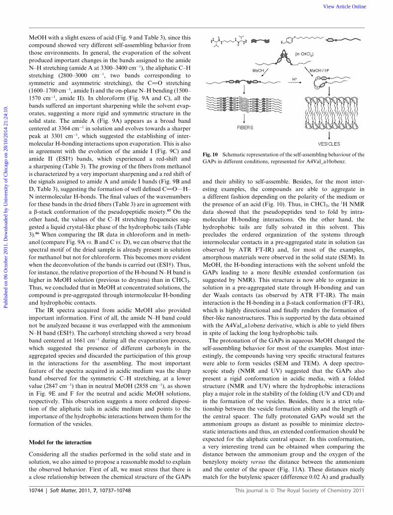
Fig. 10 Schematic representation of the self-assembling behaviour of the
GAPs in different conditions, represented for A4Val_a10obenz.
Publ
ishe
d on
06
Oct
ober
201
1. D
ownl
oade
d by
Uni
vers
ity o
f C
hica
go o
n 28
/10/
2014
21:
24:1
0.
View Article Online
MeOH with a slight excess of acid (Fig. 9 and Table 3), since this
compound showed very different self-assembling behavior from
those environments. In general, the evaporation of the solvent
produced important changes in the bands assigned to the amide
N–H stretching (amide A at 3300–3400 cm�1), the aliphatic C–H
stretching (2800–3000 cm�1, two bands corresponding to
symmetric and asymmetric stretching), the C]O stretching
(1600–1700 cm�1, amide I) and the on-plane N–H bending (1500–
1570 cm�1, amide II). In chloroform (Fig. 9A and C), all the
bands suffered an important sharpening while the solvent evap-
orates, suggesting a more rigid and symmetric structure in the
solid state. The amide A (Fig. 9A) appears as a broad band
centered at 3364 cm�1 in solution and evolves towards a sharper
peak at 3301 cm�1, which suggested the establishing of inter-
molecular H-bonding interactions upon evaporation. This is also
in agreement with the evolution of the amide I (Fig. 9C) and
amide II (ESI†) bands, which experienced a red-shift and
a sharpening (Table 3). The growing of the fibers from methanol
is characterized by a very important sharpening and a red shift of
the signals assigned to amide A and amide I bands (Fig. 9B and
D, Table 3), suggesting the formation of well defined C]O/H–
N intermolecular H-bonds. The final values of the wavenumbers
for these bands in the dried fibers (Table 3) are in agreement with
a b-stack conformation of the pseudopeptidic moiety.85 On the
other hand, the values of the C–H stretching frequencies sug-
gested a liquid crystal-like phase of the hydrophobic tails (Table
3).86 When comparing the IR data in chloroform and in meth-
anol (compare Fig. 9A vs. B and C vs.D), we can observe that the
spectral motif of the dried sample is already present in solution
for methanol but not for chloroform. This becomes more evident
when the deconvolution of the bands is carried out (ESI†). Thus,
for instance, the relative proportion of the H-bound N–H band is
higher in MeOH solution (previous to dryness) than in CHCl3.
Thus, we concluded that in MeOH at concentrated solutions, the
compound is pre-aggregated through intermolecular H-bonding
and hydrophobic contacts.
The IR spectra acquired from acidic MeOH also provided
important information. First of all, the amide N–H band could
not be analyzed because it was overlapped with the ammonium
N–H band (ESI†). The carbonyl stretching showed a very broad
band centered at 1661 cm�1 during all the evaporation process,
which suggested the presence of different carbonyls in the
aggregated species and discarded the participation of this group
in the interactions for the assembling. The most important
feature of the spectra acquired in acidic medium was the sharp
band observed for the symmetric C–H stretching, at a lower
value (2847 cm�1) than in neutral MeOH (2858 cm�1), as shown
in Fig. 9E and F for the neutral and acidic MeOH solutions,
respectively. This observation suggests a more ordered disposi-
tion of the aliphatic tails in acidic medium and points to the
importance of the hydrophobic interactions between them for the
formation of the vesicles.
Model for the interaction
Considering all the studies performed in the solid state and in
solution, we also aimed to propose a reasonable model to explain
the observed behavior. First of all, we must stress that there is
a close relationship between the chemical structure of the GAPs
10744 | Soft Matter, 2011, 7, 10737–10748
and their ability to self-assemble. Besides, for the most inter-
esting examples, the compounds are able to aggregate in
a different fashion depending on the polarity of the medium or
the presence of an acid (Fig. 10). Thus, in CHCl3, the1H NMR
data showed that the pseudopeptides tend to fold by intra-
molecular H-bonding interactions. On the other hand, the
hydrophobic tails are fully solvated in this solvent. This
precludes the ordered organization of the systems through
intermolecular contacts in a pre-aggregated state in solution (as
observed by ATR FT-IR) and, for most of the examples,
amorphous materials were observed in the solid state (SEM). In
MeOH, the H-bonding interactions with the solvent unfold the
GAPs leading to a more flexible extended conformation (as
suggested by NMR). This structure is now able to organize in
solution in a pre-aggregated state through H-bonding and van
der Waals contacts (as observed by ATR FT-IR). The main
interaction is the H-bonding in a b-stack conformation (FT-IR),
which is highly directional and finally renders the formation of
fiber-like nanostructures. This is supported by the data obtained
with the A4Val_a1obenz derivative, which is able to yield fibers
in spite of lacking the long hydrophobic tails.
The protonation of the GAPs in aqueous MeOH changed the
self-assembling behavior for most of the examples. Most inter-
estingly, the compounds having very specific structural features
were able to form vesicles (SEM and TEM). A deep spectro-
scopic study (NMR and UV) suggested that the GAPs also
present a rigid conformation in acidic media, with a folded
structure (NMR and UV) where the hydrophobic interactions
play a major role in the stability of the folding (UV and CD) and
in the formation of the vesicles. Besides, there is a strict rela-
tionship between the vesicle formation ability and the length of
the central spacer. The fully protonated GAPs would set the
ammonium groups as distant as possible to minimize electro-
static interactions and thus, an extended conformation should be
expected for the aliphatic central spacer. In this conformation,
a very interesting trend can be obtained when comparing the
distance between the ammonium group and the oxygen of the
benzyloxy moiety versus the distance between the ammonium
and the center of the spacer (Fig. 11A). These distances nicely
match for the butylenic spacer (difference 0.02 �A) and gradually
This journal is ª The Royal Society of Chemistry 2011

Fig. 11 (A) Structural relationships (distances in �A) and molecular
models for ANVal_a10obenz (N ¼ 2–6). (B) Schematic representation of
the proposal for the formation of bilayers.
Fig. 13 (A) Proposed equilibrium between the two conformers of
protonated A4Val_a10obenz, with the chromophores highlighted. (B)
Superimposed structures in several views. For clarity, the hydrogen
atoms have been omitted and the carbon atoms have been represented in
a different color for each minimum.
Publ
ishe
d on
06
Oct
ober
201
1. D
ownl
oade
d by
Uni
vers
ity o
f C
hica
go o
n 28
/10/
2014
21:
24:1
0.
View Article Online
become unfit as far as the spacer is either longer or shorter.
Molecular mechanics calculations showed the possibility of
stable energy minima with a conformation setting the aromatic
rings folded over the pseudopeptidic moiety.
Besides, for the best matched distances (A3, A4 and A5), this
conformation favors the van der Waals contacts between the
long hydrophobic tails, being the best structural complemen-
tarity for A4. This observation is in agreement with the ability to
form vesicles, which was optimal for the A4Val_a10obenz
compound. Actually, the cationic gemini surfactant-like dispo-
sition in a T-shaped molecular structure would explain the
assembly in a curved bilayer finally rendering the formation of
vesicles, as experimentally observed (Fig. 11B). The van der
Waals contacts between the tails are much less efficient for the
Fig. 12 Correlation between structural and spectroscopic parameters.
This journal is ª The Royal Society of Chemistry 2011
shortest (A2) and the longest (A6) spacer, which nicely explain
our experimental observations. Actually, if we plot the discrep-
ancy in the previously commented distances and some experi-
mental data (UV changes due to protonation or CMC measured
by fluorescence) versus the length of the spacer, a very good
correlation between them is observed (Fig. 12).
This model is also in good agreement with other spectroscopic
observations in solution state. For instance, the large aniso-
chrony in the 1H NMR signals of the methylenes of the spacer
directly attached to the N (protons A in Fig. 5) can be explained
by the large anisotropy effect of the anisole ring placed at one
face of the spacer (like in our model). Additionally, this geometry
would correlate with the red-shift of the symmetric C–H
stretching bands in the IR spectra, since the efficient contacts
between the tails would rigidify their conformation. This dispo-
sition would also favor an electronic communication between the
anisole ring and the pseudopeptidic moiety (Fig. S28† for
the frontier orbitals on a simplified model) which could explain
the changes observed in the UV spectra upon protonation.
Regarding the silent CD signal, for protonated A4Val_a10obenz,
we found two energetically very close conformations with
a complementary disposition of the chromophores, but retaining
the same molecular shape and interactions (Fig. 13). Considering
the dipole moment changes associated to the transitions, these
structures would produce mirror CD signals and thus, we
concluded that the co-existence of both geometries might
produce low or null CD spectra.
Conclusions
The syntheses and full characterization (including ultrastructure
characterization) of new Gemini Amphiphilic Pseudopeptides
(GAPs) have been carried out. The formation of a large variety
Soft Matter, 2011, 7, 10737–10748 | 10745

Publ
ishe
d on
06
Oct
ober
201
1. D
ownl
oade
d by
Uni
vers
ity o
f C
hica
go o
n 28
/10/
2014
21:
24:1
0.
View Article Online
of nanostructures was observed in the solid state, rendering
fibers, tubes, tapes or spheres. The structural parameters and
environmental effects have been systematically varied in order to
get reasonable structure/nanostructure relationships, based on
the intermolecular non-covalent interactions between the corre-
sponding building blocks. For some specific structural features,
a stimulus dependent self-assembling behavior was observed,
which produced amorphous materials in chloroform, fibers in
polar solvents and vesicles upon protonation. This behavior was
especially efficient for the GAPs derived from Valine, with
a spacer of medium length (from 3 to 5 methylenes) and bearing
two decyloxybenzyl hydrophobic tails. Solution state studies
using different spectroscopic techniques (NMR, UV, CD, fluo-
rescence and ATR FT-IR) were employed to correlate the pre-
aggregated species with the formation of the final nano-
structures. The absence of nanostructures in non-polar medium
can be related to the ability of the GAPs to establish intra-
molecular H-bond interactions in a partially folded conforma-
tion, and to the efficient solvation of the hydrophobic tails. In
polar environments, the H-bonds with the solvent would unfold
the GAPs leading to an extended conformation which self-
assemble upon concentration through intermolecular H-bonding
and van der Waals contacts. The main role of the H-bonds was
expressed through the directionality of the aggregation into
fibers. The protonation of these GAPs produced a conforma-
tional change to a new folded structure. For some specific GAPs,
those species are able to self-assemble into spherical vesicles
mainly due to the intermolecular hydrophobic contacts between
tails, in a cationic surfactant-like mode. Thus, we have been able
to rationalize our results in the formation of nanostructures on
the basis of structural parameters. We envision that the under-
standing of the aggregation process within these simple amphi-
philic amino acid derivatives will allow us to further design
pseudopeptidic molecules able to form a desired nanostructure
under given conditions and, more interestingly, to undergo
controlled transitions between different shapes and sizes of the
nano-objects.
Experimental section
General
Reagents and solvents were purchased from commercial
suppliers (Adrich, Fluka or Merck) and were used without
further purification. The C2 symmetrical bis(amidoamines) were
prepared as previously described.71
Electron microscopy
Scanning Electron Microscopy was performed either in a LEO
440I or in a JEOL 7001F microscope with a digital camera.
Samples were obtained by slow evaporation of a solution of the
compounds (�1 to 2 mg ml�1) directly onto the sample holder,
and were conventionally coated previous to the measurement.
Transmission Electron Microscopy was carried out in a JEOL
2100 microscope at 120 KV. The micrographs were obtained
from �1 mg ml�1 solutions onto a holey carbon copper grid. The
samples were sonicated for 10 minutes previous to the
measurement, one drop added onto the grid and collected
directly without staining.
10746 | Soft Matter, 2011, 7, 10737–10748
NMR spectroscopy
The NMR experiments were carried out on a Varian INOVA 500
spectrometer (500 MHz for 1H and 125 MHz for 13C) or on
a Varian UNITY 300 (300 MHz for 1H and 75 MHz for 13C).
Chemical shifts are reported in ppm using TMS as a reference.
Infrared spectroscopy
FT-IR spectra were acquired in a JASCO 6200 equipment having
a MIRacle Single Reflection ATR Diamond/ZnSe accessory. A
20 mM sample of the corresponding pseudopeptide was prepared
and seeded onto the ATR sample holder. The FT-IR spectra
were sequentially collected until complete solvent evaporation.
The raw IR data were processed with the JASCO spectral
manager software and the deconvolution of the bands was per-
formed with Origin software, using Gaussian-shaped ideal peaks.
UV and CD spectroscopy
Spectra were recorded with a JASCO J-810 spectropolarimeter at
RT. The normalized CD spectra were obtained by transforming
the molar circular dichroic absorption data (D3, cm2 mmol�1)
using the formula: D3 ¼ q/(32980Cl), in which q is the measured
ellipticity (in mdeg), C is the concentration (in M) and l is the
path length (in cm). Molar extinction coefficients of the UV
spectra were obtained by linear regression between the measured
absorbance and the sample concentration.
Steady-state fluorescence spectroscopy
Steady-state fluorescence spectra were recorded in a Spex Fluo-
rog 3–11 equipped with a 450 W xenon lamp. Fluorescence
spectra were recorded in the front face mode. All the samples
were measured in aerated conditions, unless otherwise stated.
Molecular modeling
All the theoretical calculations were performed with Spartan 06
software, using the MMFF level of theory for the geometry
optimizations.
General procedure for the reductive amination reaction
Synthesis of A2Val_a10obenz. The corresponding pseudo-
peptidic bis(amidoamine) precursor (216.0 mg, 0.836 mmol) was
dissolved in 5 mL of CHCl3 and the solution was placed inside
a flask under nitrogen atmosphere. Then, 4-decylox-
ybenzaldehyde (476.1 mL, 452.3 mg, 1.672 mmol) was dissolved
in 5 mL of CHCl3, this solution was added over the solution of
the diamine and afterwards, 7 mL of CHCl3 were added until
a final volume of 17 mL (0.05 M final concentration each). The
mixture was stirred overnight, then a large excess of Py$BH3
(889.0 mL, 817.9 mg, 8.36 mmol) was carefully added at 35 �C,and the mixture was allowed to react for 24 h before being
hydrolyzed (conc. HCl, to acidity) and evaporated to dryness.
The residue obtained was dissolved in water, basified with 1N
NaOH, and extracted with CHCl3. The combined organic layers
were dried (MgSO4) and evaporated in vacuum. The product was
purified by flash chromatography on silica gel using CH2Cl2 as
eluent, increasing slowly the polarity with MeOH and several
This journal is ª The Royal Society of Chemistry 2011

Publ
ishe
d on
06
Oct
ober
201
1. D
ownl
oade
d by
Uni
vers
ity o
f C
hica
go o
n 28
/10/
2014
21:
24:1
0.
View Article Online
drops of aqueous ammonia. Yield ¼ 63%; mp 95–104 �C; [a]25D ¼�27.0 (c¼ 0.01, CHCl3); IR (ATR) 3303, 2921, 2851, 1641, 1555,
1513 cm�1; 1H-NMR (500 MHz, CDCl3) d 0.79 (m, 18H), 0.86
(m, 24H), 1.23 (m, 4H), 1.38 (s, 2H), 1.48 (m, 4H), 1.7 (m, 2H),
2.01 (m, 2H), 2.87 (m, 4H), 3.35 (dd, 2H, J ¼ 5.2, 8.1 Hz), 3.45
(dd, 2H, J ¼ 5.4, 8.2 Hz), 3.61 (t, 4H, J ¼ 6.6 Hz), 6.77 (d, 4H, J
¼ 6.77 Hz), 7.10 (d, 4H, J ¼ 7.10 Hz), 7.53 (s, 2H); 13C-NMR
(125 MHz, CDCl3) d 14.3, 18.0, 19.8, 22.9, 26.3, 29.5, 29.8, 29.9,
31.5, 32.1, 39.6, 53.1, 68.0, 68.3, 114.8, 129.5, 131.7, 158.7, 174.7;
HRMS (ESI-TOF)+ measured for C46H78N4O4 (M + H)+:
751.6101; found 751.6102. Anal. Calcd for C46H78N4O4$H2O: C,
71.93; H, 11.17; N, 6.70; found: C, 71.83; H, 10.98; N, 6.90%. A
small amount of the monoalkylated by-product was also iso-
lated: yield ¼ 5%; mp 78–82 �C; [a]25D ¼ �2.5 (c ¼ 0.01, CHCl3);
IR (ATR) 3299, 2958, 2852, 1631, 1553, 1513, 1467, 1244 cm�1;1H-NMR (500 MHz, CDCl3) d 0.81 (d, 6H, J ¼ 6.7 Hz), 0.87 (m,
3H), 0.95 (d, 6H, J ¼ 6.0 Hz), 1.30 (m, 12H), 1.43 (m, 2H), 1.65
(m, 2H), 1.76 (td, 1H, J ¼ 6.6, 13.0 Hz), 2.08 (m, 1H), 2.25 (m,
1H), 2.96 (s, 1H), 3.19 (s, 1H), 3.40 (t, 4H, J ¼ 12.5 Hz), 3.55 (d,
1H, J ¼ 12.8 Hz), 3.69 (d, 1H, J ¼ 11.4 Hz), 3.93 (t, 2H, J ¼ 6.5
Hz), 6.84 (m, 2H), 7.19 (m, 2H), 7.61 (s, 2H), 7.69 (s, 2H); 13C-
NMR (75 MHz, CDCl3) d 14.3, 16.4, 18.1, 19.7, 22.9, 26.3, 29.5,
29.6, 29.8, 31.0, 31.4, 32.1, 39.3, 39.8, 52.9, 60.4, 67.9, 68.3, 114.8,
129.6, 131.4, 158.8, 174.7, 175.1; HRMS (ESI-TOF)+ calcd for
C29H52N4O3 (M + H)+: 505.4118; found 505.4114. Anal. Calcd
for C29H52N4O3: C, 69.01; H, 10.38; N, 11.10; found: C, 68.85;
H, 10.70; N, 11.43%.
Acknowledgements
This work was supported by the Spanish Ministry of Science and
Innovation (CTQ2009-14366-C02) and UJI-Bancaixa (P1-1B-
2009-59). J.R. thanks MICINN for personal financial support
(FPU fellowship). The support of the SCIC of the UJI for the
different instrumental techniques is acknowledged.
Notes and references
1 Y. Kim, M. F. Mayer and S. C. Zimmerman, Angew. Chem., Int. Ed.,2003, 42, 1121–1126.
2 Y. Ma, S. V. Kolotuchin and S. C. Zimmerman, J. Am. Chem. Soc.,2002, 124, 13757–13769.
3 T. Park and S. C. Zimmerman, J. Am. Chem. Soc., 2006, 128, 11582–11590.
4 G. M. Whitesides and B. Grzybowski, Science, 2002, 295, 2418–2421.5 G. M. Whitesides, J. P. Mathias and C. T. Seto, Science, 1991, 254,1312–1319.
6 J. M. Lehn, Science, 2002, 295, 2400–2403.7 S. Cavalli, F. Albericio and A. Kros, Chem. Soc. Rev., 2010, 39, 241–263.
8 D. N. Reinhoudt and M. Crego-Calama, Science, 2002, 295, 2403–2407.
9 M. Surin, P. G. A. Janssen, R. Lazzaroni, P. Lecl�ere, E.W.Meijer andA. P. H. J. Schenning, Adv. Mater., 2009, 21, 1126–1130.
10 J. M. Lehn, Angew. Chem., Int. Ed. Engl., 1990, 29, 1304–1319.11 J. M. Lehn, Supramolecular Chemistry, Concepts and Perspectives,
Wiley-VCH, Weinheim, 1995.12 E. Gorrea, P. Nolis, E. Torres, E. Da Silva, D. B. Amabilino,
V. Branchadell and R. M. Ortu~no, Chem.–Eur. J., 2011, 17, 4588–4597.
13 V. Berl, I. Huc, R. G. Khoury, M. J. Krische and J. M. Lehn, Nature,2000, 407, 720–723.
14 R. Feynman, Eng. Sci., 1960, 23, 22–36.15 O. Ramstr€om, T. Bunyapaiboonsri, S. Lohmann and J. M. Lehn,
Biochim. Biophys. Acta, Gen. Subj., 2002, 1572, 178–186.
This journal is ª The Royal Society of Chemistry 2011
16 P. Pramod, K. G. Thomas and M. V. George, Chem.–Asian J., 2009,4, 806–823.
17 T. Nakanishi, K. Ariga, T. Michinobu, K. Yoshida, H. Takahashi,T.Teranishi,H.M€ohwald andD.G.Kurth,Small, 2007,3, 2019–2023.
18 A. Petitjean, L. A. Cuccia, J.-M. Lehn, H. Nierengarten andM. Schmutz, Angew. Chem., Int. Ed., 2002, 41, 1195–1198.
19 C. A. E. Hauser and S. Zhang, Chem. Soc. Rev., 2010, 39, 2780–2790.20 A. Aggeli, I. A. Nyrkova, M. Bell, R. Harding, L. Carrick,
T. C. B. McLeish, A. N. Semenov and N. Boden, Proc. Natl. Acad.Sci. U. S. A., 2001, 98, 11857–11862.
21 J. D. Hartgerink, E. Beniash and S. I. Stupp, Science, 2001, 294, 1684–1688.
22 H. Cui, M. J. Webber and S. I. Stupp, Biopolymers, 2010, 94, 1–18.23 S. I. Stupp, Nano Lett., 2010, 10, 4783–4786.24 I. Cherny and E. Gazit, Angew. Chem., Int. Ed., 2008, 47, 4062–4069.25 R. V. Ulijn and A. M. Smith, Chem. Soc. Rev., 2008, 37, 664–675.26 E. Gazit, Chem. Soc. Rev., 2007, 36, 1263–1269.27 X. Zhao and S. Zhang, Chem. Soc. Rev., 2006, 35, 1105–1110.28 S. Zhang, Nat. Biotechnol., 2003, 21, 1171–1178.29 L. P. Hern�andez-Egu�ıa, R. J. Brea, L. Castedo, P. Ballester and
J. R. Granja, Chem.–Eur. J., 2011, 17, 1220–1229.30 M. J. Krysmann, V. Castelletto, J. E. McKendrick, L. A. Clifton,
I. W. Hamley, P. J. F. Harris and S. M. King, Langmuir, 2008, 24,8158–8162.
31 N. Amdursky, M. Molotskii, E. Gazit and G. Rosenman, J. Am.Chem. Soc., 2010, 132, 15632–15636.
32 J. Berg, J. Tymoczko, L. Stryer and N. D. Clarke, Biochemistry, W.H.Freeman and Co., 2002.
33 I. Imaz, M. Rubio-Mart�ınez, W. J. Saletra, D. B. Amabilino andD. Maspoch, J. Am. Chem. Soc., 2009, 131, 18222–18223.
34 R. J. Brea, C. Reiriz and J. R. Granja, Chem. Soc. Rev., 2010, 39,1448–1456.
35 T. Muraoka, C. Y. Koh, H. Cui and S. I. Stupp, Angew. Chem., Int.Ed., 2009, 48, 5946–5949.
36 J. N. Shera and X. S. Sun, Biomacromolecules, 2009, 10, 2446–2450.37 S. Ghosh and S. Verma, Tetrahedron, 2008, 64, 6202–6208.38 K. Lu, L. Guo, A. K. Mehta, W. S. Childers, S. N. Dublin,
S. Skanthakumar, V. P. Conticello, P. Thiyagarajan,R. P. Apkarian and D. G. Lynn, Chem. Commun., 2007, 2729–2731.
39 L. S. Birchall, S. Roy, V. Jayawarna, M. Hughes, E. Irvine,G. T. Okorogheye, N. Saudi, E. de Santis, T. Tuttle, A. A. Edwardsand R. V. Ulijn, Chem. Sci., 2011, 2, 1349–1355.
40 A. Mata, L. Hsu, R. Capito, C. Aparicio, K. Henrikson andS. I. Stupp, Soft Matter, 2009, 5, 1228–1236.
41 E. Torres, J. Puigmart�ı-Luis, A. P�erez Del Pino, R. M. Ortu~no andD. B. Amabilino, Org. Biomol. Chem., 2010, 8, 1661–1665.
42 D.B.AmabilinoandJ.Puigmart�ı-Luis,SoftMatter, 2010,6, 1605–1612.43 R. Garc�ıa-Fandi~no, J. R. Granja, M. D’Abramo and M. Orozco, J.
Am. Chem. Soc., 2009, 131, 15678–15686.44 C. s. Reiriz, R. J. Brea, R. o. Arranz, J. L. Carrascosa, A. Garibotti,
B. Manning, J. M. Valpuesta, R. n. Eritja, L. Castedo andJ. R. Granja, J. Am. Chem. Soc., 2009, 131, 11335–11337.
45 G. P. Spada, S. Lena, S. Masiero, S. Pieraccini, M. Surin andP. Samorı̀, Adv. Mater., 2008, 20, 2433–2438.
46 W. Cai, G. T.Wang, Y. X. Xu, X. K. Jiang and Z. T. Li, J. Am. Chem.Soc., 2008, 130, 6936–6937.
47 T. Kawasaki, M. Tokuhiro, N. Kimizuka and T. Kunitake, J. Am.Chem. Soc., 2001, 123, 6792–6800.
48 I. W. Hamley, Soft Matter, 2011, 7, 4122–4138.49 V. Castelletto and I. W. Hamley, Biophys. Chem., 2009, 141, 169–174.50 A. Kelarakis, C. Chaibundit, M. J. Krysmann, V. Havredaki,
K. Viras and I. W. Hamley, J. Colloid Interface Sci., 2009, 330, 67–72.51 H. Cui, E. T. Pashuck, Y. S. Velichko, S. J. Weigand,
A. G. Cheetham, C. J. Newcomb and S. I. Stupp, Science, 2010,327, 555–559.
52 R. J. Williams, A. M. Smith, R. Collins, N. Hodson, A. K. Das andR. V. Ulijn, Nat. Nanotechnol., 2009, 4, 19–24.
53 S.Ghosh, S.K. SinghandS.Verma,Chem.Commun., 2007, 2296–2298.54 B. S. Kim, D. J. Hong, J. Bae and M. Lee, J. Am. Chem. Soc., 2005,
127, 16333–16337.55 N. D�ıaz, F.-X. Simon, M. Schmutz, M. Rawiso, G. Decher, J. Jestin
and P. J. M�esini, Angew. Chem., Int. Ed., 2005, 44, 3260–3264.56 H. J. Kim, T. Kim and M. Lee, Acc. Chem. Res., 2011, 44, 72–82.57 T. B. Schuster, D. De Bruyn Ouboter, E. Bordignon, G. Jeschke and
W. Meier, Soft Matter, 2010, 6, 5596–5604.
Soft Matter, 2011, 7, 10737–10748 | 10747

Publ
ishe
d on
06
Oct
ober
201
1. D
ownl
oade
d by
Uni
vers
ity o
f C
hica
go o
n 28
/10/
2014
21:
24:1
0.
View Article Online
58 P. Kumaraswamy, R. Lakshmanan, S. Sethuraman andU. M. Krishnan, Soft Matter, 2011, 7, 2744–2754.
59 Y. Lin, Y. Qiao, P. Tang, Z. Li and J. Huang, Soft Matter, 2011, 7,2762–2769.
60 X. Hou, W. Guo and L. Jiang, Chem. Soc. Rev., 2011, 40, 2385–2401.61 S. R. Diegelmann, J. M. Gorham and J. D. Tovar, J. Am. Chem. Soc.,
2008, 130, 13840–13841.62 P. P. Bose, A. K. Das, R. P. Hegde, N. Shamala and B. A. Banerjee,
Chem. Mater., 2007, 19, 6150–6157.63 Y. Song, S. R. Challa, C. J. Medforth, Y. Qiu, R. K. Watt, D. Pena,
J. E. Miller, F. v. Swol and J. A. Shelnutt, Chem. Commun., 2004,1044–1045.
64 H. Matsui, J. Phys. Chem. B, 2000, 104, 3385–3386.65 H. Matsui and C. Holtman, Nano Lett., 2002, 2, 887–889.66 J. Naskar and A. Banerjee, Chem.–Asian J., 2009, 4, 1817–1823.67 X. Yan, Q. He, K. Wang, L. Duan, Y. Cui and J. Li, Angew. Chem.,
Int. Ed., 2007, 46, 2431–2434.68 A. Ajayaghosh, R. Varghese, S. Mahesh and V. K. Praveen, Angew.
Chem., Int. Ed., 2006, 45, 7729–7732.69 S. J. Choi, W. J. Jeong, T. H. Kim and Y. B. Lim, Soft Matter, 2011,
7, 1675–1677.70 J. Rubio, I. Alfonso, M. Bru, M. I. Burguete and S. V. Luis,
Tetrahedron Lett., 2010, 51, 5861–5867.71 J. Becerril, M. Bolte, M. I. Burguete, F. Galindo, E. Garc�ıa-Espa~na,
S. V. Luis and J. F. Miravet, J. Am. Chem. Soc., 2003, 125, 6677–6686.
72 R. N. Salvatore, C. H. Yoon and K. W. Jung, Tetrahedron, 2001, 57,7785–7811.
10748 | Soft Matter, 2011, 7, 10737–10748
73 I. Alfonso, M. Bolte, M. Bru, M. Isabel Burguete and S. V. Luis,Chem.–Eur. J., 2008, 14, 8879–8891.
74 I. Alfonso, M. Bolte, M. Bru, M. I. Burguete, S. V. Luis and J. Rubio,J. Am. Chem. Soc., 2008, 130, 6137–6144.
75 F. Campbell, J. Plante, C. Carruthers, M. J. Hardie, T. J. Prior andA. J. Wilson, Chem. Commun., 2007, 2240–2242.
76 F. Campbell, C. A. Kilner and A. J. Wilson, Tetrahedron Lett., 2010,51, 1361–1363.
77 I. Alfonso, M. Bolte, M. Bru, M. I. Burguete, S. V. Luis andC. Vicent, Org. Biomol. Chem., 2010, 8, 1329–1339.
78 I. Alfonso, M. I. Burguete, F. Galindo, S. V. Luis and L. Vigara, J.Org. Chem., 2007, 72, 7947–7956.
79 I. Alfonso, M. I. Burguete and S. V. Luis, J. Org. Chem., 2006, 71,2242–2250.
80 X. Yan, Q. He, K. Wang, L. Duan, Y. Cui and J. Li, Angew. Chem.,Int. Ed., 2007, 46, 2431–2434.
81 A. Ajayaghosh, R. Varghese, S. Mahesh and V. K. Praveen, Angew.Chem., Int. Ed., 2006, 45, 7729–7732.
82 K. S. Sharma, C. Rodgers, R.M. Palepu andA. K. Rakshit, J. ColloidInterface Sci., 2003, 268, 482–488.
83 T. Yoshimura, T. Ichinokawa, M. Kaji and K. Esumi, Colloids Surf.,A, 2006, 273, 208–212.
84 I. Alfonso, M. Bru, M. Isabel Burguete, E. Garc�ıa-Verdugo andS. V. Luis, Chem.–Eur. J., 2010, 16, 1246–1255.
85 H. S. Kim, J. D. Hartgerink and M. R. Ghadiri, J. Am. Chem. Soc.,1998, 120, 4417–4424.
86 M. C. Hull, L. R. Cambrea and J. S. Hovis, Anal. Chem., 2005, 77,6096–6099.
This journal is ª The Royal Society of Chemistry 2011