Embed Size (px)
Citation preview
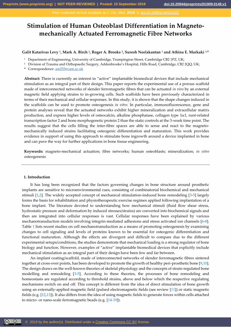
Stimulation of Human Osteoblast Differentiation in Magneto-mechanically Actuated Ferromagnetic Fibre Networks
Galit Katarivas Levy 1, Mark A. Birch 2, Roger A. Brooks 2, Suresh Neelakantan 1 and Athina E. Markaki 1,*
1 Department of Engineering, University of Cambridge, Trumpington Street, Cambridge CB2 1PZ, UK; 2 Division of Trauma and Orthopaedic Surgery, Addenbrooke’s Hospital, Hills Road, Cambridge, CB2 2QQ, UK;
* Correspondence: [email protected]
Abstract: There is currently an interest in “active” implantable biomedical devices that include mechanical
stimulation as an integral part of their design. This paper reports the experimental use of a porous scaffold
made of interconnected networks of slender ferromagnetic fibres that can be actuated in vivo by an external
magnetic field applying strains to in-growing cells. Such scaffolds have been previously characterized in
terms of their mechanical and cellular responses. In this study, it is shown that the shape changes induced in
the scaffolds can be used to promote osteogenesis in vitro. In particular, immunofluorescence, gene and
protein analyses reveal that the actuated networks exhibit higher mineralization and extracellular matrix
production, and express higher levels of osteocalcin, alkaline phosphatase, collagen type 11, runt-related
transcription factor 2 and bone morphogenetic protein 2 than the static controls at the 3-week time point. The
results suggest that the cells filling the inter-fibre spaces are able to sense and react to the magneto-
mechanically induced strains facilitating osteogenic differentiation and maturation. This work provides
evidence in support of using this approach to stimulate bone ingrowth around a device implanted in bone
and can pave the way for further applications in bone tissue engineering.
Keywords: magneto-mechanical actuation; fibre networks; human osteoblasts; mineralization; in vitro
osteogenesis
1. Introduction
It has long been recognized that the factors governing changes in bone structure around prosthetic
implants are sensitive to microenvironmental cues, consisting of combinatorial biochemical and mechanical
stimuli [1,2]. The widely accepted concept of mechanical stimulation-induced bone remodeling [3-5] largely
forms the basis for rehabilitation and physiotherapeutic exercise regimes applied following implantation of a
bone implant. The literature devoted to understanding how mechanical stimuli (fluid flow shear stress,
hydrostatic pressure and deformation by induced stresses/strains) are converted into biochemical signals and
then are integrated into cellular responses is vast. Cellular responses have been explained by various
mechanotransduction models involving integrin-mediated adhesions and stress activated ion channels [6-8].
Table 1 lists recent studies on cell mechanotransduction as a means of promoting osteogenesis by examining
changes to cell signaling and levels of proteins known to be essential for osteogenic differentiation and
functional maturation. Although the effects are divergent and difficult to compare due to the different
experimental setups/conditions, the studies demonstrate that mechanical loading is a strong regulator of bone
biology and function. However, examples of “active” implantable biomedical devices that explicitly include
mechanical stimulation as an integral part of their design have been few and far between.
An implant coating/scaffold, made of interconnected networks of slender ferromagnetic fibres sintered
together at cross-over points, has been developed to promote the growth of healthy peri-prosthetic bone [9,10].
The design draws on the well-known theories of skeletal physiology and the concepts of strain-regulated bone
modelling and remodeling [3-5]. According to these theories, the processes of bone remodeling and
homeostasis are regulated according to threshold strains, above and below which the respective regulating
mechanisms switch on and off. This concept is different from the idea of direct stimulation of bone growth
using an externally-applied magnetic field (pulsed electromagnetic fields (see review [11]) or static magnetic
fields (e.g. [12,13]). It also differs from the idea of using magnetic fields to generate forces within cells attached
to micro- or nano-scale ferromagnetic beads (e.g. [14-19]).
Preprints (www.preprints.org) | NOT PEER-REVIEWED | Posted: 15 September 2019 doi:10.20944/preprints201909.0149.v1
© 2019 by the author(s). Distributed under a Creative Commons CC BY license.
Peer-reviewed version available at J. Clin. Med. 2019, 8; doi:10.3390/jcm8101522
2 of 17
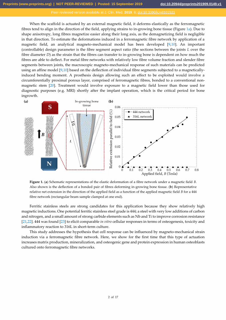
When the scaffold is actuated by an external magnetic field, it deforms elastically as the ferromagnetic
fibres tend to align in the direction of the field, applying strains to in-growing bone tissue (Figure 1a). Due to
shape anisotropy, long fibres magnetize easier along their long axis, as the demagnetizing field is negligible
in that direction. To estimate the deformations induced in a ferromagnetic fibre network by application of a
magnetic field, an analytical magneto-mechanical model has been developed [9,10]. An important
(controllable) design parameter is the fibre segment aspect ratio (the sections between the joints L over the
fibre diameter D) as the strain that the fibres can transfer to in-growing bone is dependent on how much the
fibres are able to deflect. For metal fibre networks with relatively low fibre volume fraction and slender fibre
segments between joints, the macroscopic magneto-mechanical response of such materials can be predicted
using an affine model [9,10] based on the deflection of individual fibre segments subjected to a magnetically-
induced bending moment. A prosthesis design allowing such an effect to be exploited would involve a
circumferentially proximal porous layer, comprised of ferromagnetic fibres, bonded to a conventional non-
magnetic stem [20]. Treatment would involve exposure to a magnetic field lower than those used for
diagnostic purposes (e.g. MRI) shortly after the implant operation, which is the critical period for bone
ingrowth.
Figure 1. (a) Schematic representations of the elastic deformation of a fibre network under a magnetic field B.
Also shown is the deflection of a bonded pair of fibres deforming in-growing bone tissue. (b) Representative
relative net extension in the direction of the applied field as a function of the applied magnetic field B for a 444
fibre network (rectangular beam sample clamped at one end).
Ferritic stainless steels are strong candidates for this application because they show relatively high
magnetic inductions. One potential ferritic stainless steel grade is 444; a steel with very low additions of carbon
and nitrogen, and a small amount of strong carbide elements such as Nb and Ti to improve corrosion resistance
[21,22]. 444 was found [23] to elicit comparable in vitro cellular responses in terms of osteogenesis, toxicity and
inflammatory reaction to 316L in short-term culture.
This study addresses the hypothesis that cell response can be influenced by magneto-mechanical strain
induction via a ferromagnetic fibre network. Here, we show for the first time that this type of actuation
increases matrix production, mineralization, and osteogenic gene and protein expression in human osteoblasts
cultured onto ferromagnetic fibre networks.
Preprints (www.preprints.org) | NOT PEER-REVIEWED | Posted: 15 September 2019 doi:10.20944/preprints201909.0149.v1
Peer-reviewed version available at J. Clin. Med. 2019, 8; doi:10.3390/jcm8101522
3 of 17
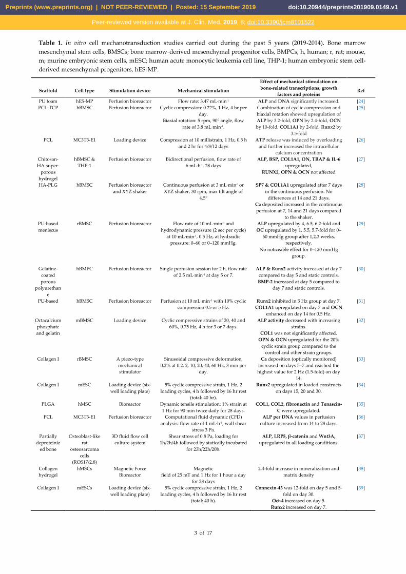
Table 1. In vitro cell mechanotransduction studies carried out during the past 5 years (2019-2014). Bone marrow
mesenchymal stem cells, BMSCs; bone marrow-derived mesenchymal progenitor cells, BMPCs, h, human; r, rat; mouse,
m; murine embryonic stem cells, mESC; human acute monocytic leukemia cell line, THP-1; human embryonic stem cell-
derived mesenchymal progenitors, hES-MP.
Scaffold Cell type Stimulation device Mechanical stimulation
Effect of mechanical stimulation on
bone-related transcriptions, growth
factors and proteins Ref
PU foam hES-MP Perfusion bioreactor Flow rate: 3.47 mL·min-1 ALP and DNA significantly increased. [24]
PCL‐TCP hBMSC Perfusion bioreactor Cyclic compression: 0.22%, 1 Hz, 4 hr per
day.
Biaxial rotation: 5 rpm, 90° angle, flow
rate of 3.8 mL·min-1.
Combination of cyclic compression and
biaxial rotation showed upregulation of
ALP by 3.2‐fold, OPN by 2.4‐fold, OCN
by 10‐fold, COL1A1 by 2‐fold, Runx2 by
3.5-fold
[25]
PCL MC3T3-E1 Loading device Compression at 10 millistrain, 1 Hz, 0.5 h
and 2 hr for 4/8/12 days
ATP release was induced by overloading
and further increased the intracellular
calcium concentration
[26]
Chitosan-
HA super-
porous
hydrogel
hBMSC &
THP-1
Perfusion bioreactor Bidirectional perfusion, flow rate of
6 mL·h-1, 28 days
ALP, BSP, COL1A1, ON, TRAP & IL-6
upregulated,
RUNX2, OPN & OCN not affected
[27]
HA-PLG hBMSC Perfusion bioreactor
and XYZ shaker
Continuous perfusion at 3 mL·min-1 or
XYZ shaker, 30 rpm, max tilt angle of
4.5°
SP7 & COL1A1 upregulated after 7 days
in the continuous perfusion. No
differences at 14 and 21 days.
Ca deposited increased in the continuous
perfusion at 7, 14 and 21 days compared
to the shaker.
[28]
PU-based
meniscus
rBMSC Perfusion bioreactor Flow rate of 10 mL·min-1 and
hydrodynamic pressure (2 sec per cycle)
at 10 mL·min-1, 0.5 Hz, at hydraulic
pressure: 0–60 or 0–120 mmHg.
ALP upregulated by 4, 6.5, 6.2-fold and
OC upregulated by 1, 5.5, 5.7-fold for 0–
60 mmHg group after 1,2,3 weeks,
respectively.
No noticeable effect for 0–120 mmHg
group.
[29]
Gelatine-
coated
porous
polyurethan
e
hBMPC Perfusion bioreactor Single perfusion session for 2 h, flow rate
of 2.5 mL·min-1 at day 5 or 7.
ALP & Runx2 activity increased at day 7
compared to day 5 and static controls.
BMP-2 increased at day 5 compared to
day 7 and static controls.
[30]
PU-based hBMSC Perfusion bioreactor Perfusion at 10 mL·min-1 with 10% cyclic
compression 0.5 or 5 Hz.
Runx2 inhibited in 5 Hz group at day 7.
COL1A1 upregulated on day 7 and OCN
enhanced on day 14 for 0.5 Hz.
[31]
Octacalcium
phosphate
and gelatin
mBMSC Loading device Cyclic compressive strains of 20, 40 and
60%, 0.75 Hz, 4 h for 3 or 7 days.
ALP activity decreased with increasing
strains.
COL1 was not significantly affected.
OPN & OCN upregulated for the 20%
cyclic strain group compared to the
control and other strain groups.
[32]
Collagen I rBMSC A piezo-type
mechanical
stimulator
Sinusoidal compressive deformation,
0.2% at 0.2, 2, 10, 20, 40, 60 Hz, 3 min per
day.
Ca deposition (optically monitored)
increased on days 5–7 and reached the
highest value for 2 Hz (1.5-fold) on day
14.
[33]
Collagen I mESC Loading device (six-
well loading plate)
5% cyclic compressive strain, 1 Hz, 2
loading cycles, 4 h followed by 16 hr rest
(total: 40 hr).
Runx2 upregulated in loaded constructs
on days 15, 20 and 30.
[34]
PLGA hMSC Bioreactor Dynamic tensile stimulation: 1% strain at
1 Hz for 90 min twice daily for 28 days.
COL1, COL2, fibronectin and Tenascin-
C were upregulated.
[35]
PCL MC3T3-E1 Perfusion bioreactor Computational fluid dynamic (CFD)
analysis: flow rate of 1 mL·h-1, wall shear
stress 3 Pa.
ALP per DNA values in perfusion
culture increased from 14 to 28 days.
[36]
Partially
deproteiniz
ed bone
Osteoblast-like
rat
osteosarcoma
cells
(ROS17/2.8)
3D fluid flow cell
culture system
Shear stress of 0.8 Pa, loading for
1h/2h/4h followed by statically incubated
for 23h/22h/20h.
ALP, LRP5, β-catenin and Wnt3A,
upregulated in all loading conditions.
[37]
Collagen
hydrogel
hMSCs Magnetic Force
Bioreactor
Magnetic
field of 25 mT and 1 Hz for 1 hour a day
for 28 days
2.4-fold increase in mineralization and
matrix density
[38]
Collagen I mESCs Loading device (six-
well loading plate)
5% cyclic compressive strain, 1 Hz, 2
loading cycles, 4 h followed by 16 hr rest
(total: 40 h).
Connexin-43 was 12-fold on day 5 and 5-
fold on day 30.
Oct-4 increased on day 5.
Runx2 increased on day 7.
[39]
Preprints (www.preprints.org) | NOT PEER-REVIEWED | Posted: 15 September 2019 doi:10.20944/preprints201909.0149.v1
Peer-reviewed version available at J. Clin. Med. 2019, 8; doi:10.3390/jcm8101522
4 of 17
2. Experimental
2.1. Substrates - Fibre Networks
This study utilized two types of solid-state sintered stainless-steel fibre networks made of 444 ferritic
stainless steel (Nikko Techno Ltd, Japan) and 316L austenitic stainless steel (Bekaert SA, Belgium), containing
~15 vol% of fibres (Table 2). 444 networks were produced by shaving 60 µm fibres off a 100 µm thick coiled
metal foil, which led to a rectangular cross-sectional shape. The 316L networks were made of bundle-drawn
fibres in a hexagonal cross-sectional shape. The diagonal length of the fibre cross-sectional area was about 40
µm. Details on the manufacturing of these networks are described elsewhere [40,41]. Information on the 444
and 316L networks are presented in Table 2 [41,42]. Although the fibre volume fractions and fibre orientation
distributions are very similar between the two networks, as illustrated in Table 2, there are differences in the
fibre cross-sectional shape and size. Because the 444 networks are comprised of coarser fibres (60 × 100 µm2),
they have less fibres per unit area, and consequently larger inter-fibre spaces compared to 316L networks, of
the same fibre volume fraction, made of finer fibres (diagonal length 40 µm, side length 20 µm). Due to these
differences, 316L networks were included in the study as a control, rather than an experimental group.
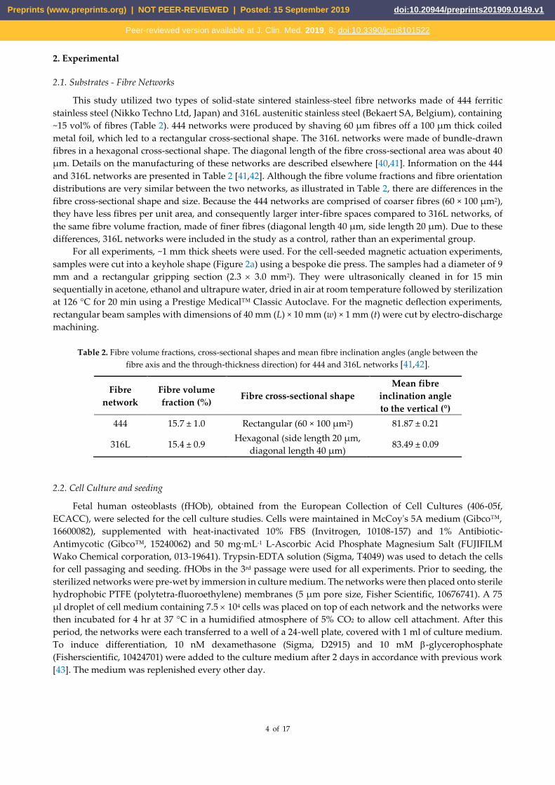
For all experiments, ~1 mm thick sheets were used. For the cell-seeded magnetic actuation experiments,
samples were cut into a keyhole shape (Figure 2a) using a bespoke die press. The samples had a diameter of 9
mm and a rectangular gripping section (2.3 3.0 mm2). They were ultrasonically cleaned in for 15 min
sequentially in acetone, ethanol and ultrapure water, dried in air at room temperature followed by sterilization
at 126 °C for 20 min using a Prestige Medical™ Classic Autoclave. For the magnetic deflection experiments,
rectangular beam samples with dimensions of 40 mm (L) × 10 mm (w) × 1 mm (t) were cut by electro-discharge
machining.
Table 2. Fibre volume fractions, cross-sectional shapes and mean fibre inclination angles (angle between the
fibre axis and the through-thickness direction) for 444 and 316L networks [41,42].
Fibre
network
Fibre volume
fraction (%) Fibre cross-sectional shape
Mean fibre
inclination angle
to the vertical (°)
444 15.7 ± 1.0 Rectangular (60 × 100 µm2) 81.87 ± 0.21
316L 15.4 ± 0.9 Hexagonal (side length 20 µm,
diagonal length 40 µm) 83.49 ± 0.09
2.2. Cell Culture and seeding
Fetal human osteoblasts (fHOb), obtained from the European Collection of Cell Cultures (406-05f,
ECACC), were selected for the cell culture studies. Cells were maintained in McCoy's 5A medium (Gibco™,
16600082), supplemented with heat-inactivated 10% FBS (Invitrogen, 10108-157) and 1% Antibiotic-
Antimycotic (Gibco™, 15240062) and 50 mg·mL-1 L-Ascorbic Acid Phosphate Magnesium Salt (FUJIFILM
Wako Chemical corporation, 013-19641). Trypsin-EDTA solution (Sigma, T4049) was used to detach the cells
for cell passaging and seeding. fHObs in the 3rd passage were used for all experiments. Prior to seeding, the
sterilized networks were pre-wet by immersion in culture medium. The networks were then placed onto sterile
hydrophobic PTFE (polytetra-fluoroethylene) membranes (5 μm pore size, Fisher Scientific, 10676741). A 75
μl droplet of cell medium containing 7.5 104 cells was placed on top of each network and the networks were
then incubated for 4 hr at 37 °C in a humidified atmosphere of 5% CO2 to allow cell attachment. After this
period, the networks were each transferred to a well of a 24-well plate, covered with 1 ml of culture medium.
To induce differentiation, 10 nM dexamethasone (Sigma, D2915) and 10 mM -glycerophosphate
(Fisherscientific, 10424701) were added to the culture medium after 2 days in accordance with previous work
[43]. The medium was replenished every other day.
Preprints (www.preprints.org) | NOT PEER-REVIEWED | Posted: 15 September 2019 doi:10.20944/preprints201909.0149.v1
Peer-reviewed version available at J. Clin. Med. 2019, 8; doi:10.3390/jcm8101522
5 of 17
2.3. Cell adhesion and cytoskeleton organization
The samples were fixed with 4% (w/v) paraformaldehyde (Sigma–Aldrich, 252549) for 15 min at room
temperature, permeabilized with 0.1% Triton X-100 (Sigma, T878) and 0.1% TWEEN-20 (Sigma, P6585) for 15
min and incubated with 1% (w/v) bovine serum albumin (BSA) in PBS at 37 °C for 10 min to block non-
specific binding. The actin cytoskeleton was visualized using FITC-labelled phalloidin (1:100) (Sigma, P5282)
followed by cellular nuclei staining with Vectashield antifade mountant containing 4,6-diamidino-2-
phenylindole (DAPI), (Sigma, F6057). The fluorescence images were obtained using a Zeiss Axio-Obsorber.Z1
fluorescence microscope.
2.4. Cell Mineralization
2.4.1. OsteoImageTM Mineralization Assay
Mineralization was assessed using the OsteoImageTM Mineralization Assay (Lonza, PA-1503) according
to the manufacturer's instructions. This assay is based on the specific binding of a fluorescent staining reagent
to bone-like minerals deposited by cells. Briefly, cells were fixed with 4% formaldehyde in PBS for 30 min at
room temperature and stained with the staining reagent (1:100 in staining reagent dilution buffer) for 30 min,
protected from light. Samples were washed with the washing buffer and mounted with DAPI. The
hydroxyapatite of the bone nodules was measured qualitatively using a Zeiss Axio-Obsorber.Z1 fluorescence
microscope and quantitatively by ImageJ software (7 images for sample, 3 samples per group).
2.4.2. Alizarin Red Staining
Alizarin Red Staining (Sigma, A5533) was used to visualize the calcium deposition indicating
mineralization of cells after 16 and 21 days for all magnetic and static groups. Briefly, the samples were washed
with phosphate-buffered saline (PBS) and fixed in 4% (v/v) formaldehyde at room temperature for 30 min.
After washing with excess distilled water (dH2O), Alizarin Red solution (2% w/v in dH2O adjusted to pH 4.2
using 0.5% ammonium hydroxide) was used to cover the samples for 30 min. After aspiration of the
unincorporated dye, the samples were washed thoroughly with dH2O. The samples were visualized using a
Zeiss Axio-Obsorber.Z1 fluorescence microscope (fluorescence emission 580 nm). To assess relative levels of
matrix mineralization the Alizarin Red stain was extracted from the samples (3 samples per group) by adding
800 μl of 10% acetic acid followed by 30 minutes incubation at room temperature as described previously [44].
The absorbance at 405 nm of the solubilised Alizarin Red dye from the samples was measured using a
FLUOstar® Omega plate reader (BMG Labtech, UK). An Alizarin Red staining standard curve was established
with a known concentration of the dye.
2.5. Gene expression analysis
At day 21 of culture, the total RNA was extracted from the cell-seeded scaffolds using RNeasy Protect
Mini Kit (Qiagen, 74124) according to the manufacturer's instructions. Complementary deoxyribonucleic acid
(cDNA) was synthesized by reverse transcriptase–polymerase chain reaction (RT-PCR) using QuantiTect®
Reverse Transcription Kits (Qiagen, 205311) according to the manufacturer's instructions. RT-PCR was
conducted using the QuantiFast SYBR Green PCR Kit (Qiagen, 204056) with the following primers:
Glyceraldehyde-3-phosphate dehydrogenase (GAPDH), bone morphogenetic protein 2 (BMP-2), collagen type
11 (COL1A1), alkaline phosphatase (ALP), osteocalcin (OCN), runt-related transcription factor 2 (Runx2) and
vascular endothelial growth factor (VEGF), which amplify transcripts characteristic of osteoblasts. The primers
were obtained from Qiagen and reconstituted according to the manufacturer’s instructions. The cycle
conditions were performed with a 5 min at 95 °C activation step followed by 40 cycles with a 10 sec at 95 °C
denaturation and 30 min at 60 °C extension step. GAPDH expression served as an internal control. Relative
expression was calculated using the 2-ΔΔCT method according to Livak and Schmittgen [45]. Results were
presented as fold change expression normalized to their non-actuated counterparts to determine the effect of
the magneto-mechanical actuation.
Preprints (www.preprints.org) | NOT PEER-REVIEWED | Posted: 15 September 2019 doi:10.20944/preprints201909.0149.v1
Peer-reviewed version available at J. Clin. Med. 2019, 8; doi:10.3390/jcm8101522

6 of 17
2.6. Protein release
Human BMP-2 (Sigma, RAB0028-1KT) and Human Osteocalcin (Sigma, RAB1073-1KT) were used
according to the manufacturer’s instructions for quantitative measurement of the chosen proteins in the cell
culture supernatants. Briefly, cell culture media were removed after 21 days, pipetted in polypropylene
eppendorf tubes and stored at -80 °C until analysis in an enzyme-linked immunosorbent assay (ELISA) assay.
The antibodies, employed in this assay, were specific for Human BMP-2 and OCN and were coated on the 96-
well plate provided. Standards and samples from the fibre networks were pipetted into the wells and BMP-2/
OCN present in a sample was bound to the wells by the immobilized antibody. The wells were washed and
biotinylated anti-Human BMP-2 or OCN antibody were added. After washing away unbound biotinylated
antibody, HRP-conjugated streptavidin was pipetted to the wells. The wells were washed again, a TMB
substrate solution was added to the wells and color developed in proportion to the amount of BMP-2 and
OCN bound. The Stop Solution changed the color from blue to yellow, and the intensity of the color was
measured as absorbance using the FLUOstar® Omega plate reader (BMG Labtech, UK) at 450 nm. With the
help of a standard curve, the protein content was determined and set in relation to the respective controls.
2.7. Magneto-Mechanical Actuation
2.7.1. Magneto-Mechanical Actuation of Cell-seeded Fibre Networks
The cell actuation experiments were carried out using a 1.6 Tesla DC coil electromagnet, with a
homogeneous operating region of 75 75 75 mm3, designed by Hirst Magnetic Instruments Ltd (Cornwall,
UK). The field coils were powered by an 11 KW 30 V/ 500A power supply with an internal main breaker of
three modules of 40 A each. A customized OKO-lab incubating chamber was supplied by Indigo Scientific Ltd
(Herts, UK). The system was an electrically-heated CO2 incubator equipped with temperature, gas and
humidity controllers. CO2 levels inside the chamber were maintained at 5% by purging with a 5% CO2/air
medical gas mixture.
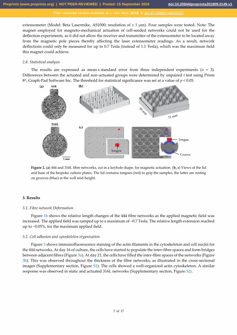
Cell culture plates: Bespoke polystyrene cell culture plates were produced by micro-machining
(Protolabs, Telford, UK). A schematic depiction of the plates is shown in Figure 2b-c. Figure 2b shows the lid
and base of the bespoke culture plates. The lid contained tongues (marked in red, Figure 2c) which were used
to grip the rectangular section of the keyhole samples while the circular area of the keyhole samples (Figure
2a) was free to deform during the actuation process. The samples were resting on grooves (Figure 2c – blue)
at mid-height of the well providing intra-well space for medium flow and ease in sample handling. An offset
spacer was introduced in the plate corner, to allow a 0.5 mm gap for air circulation when the base of the plate
was covered with the lid. Air circulation allowed equilibration of the well medium with the gas phase
(humidified atmosphere of 5% CO2/95% air). The temperature was maintained at 37° C during the actuation
process. This was verified by measuring in situ the media temperature of the wells containing the actuated
fibre networks and also the air temperature above the polystyrene cell culture plates.
Actuation protocol: All actuation experiments involved static culture for 7 days (to allow cells to colonize
the inter-fibre spaces), followed by daily actuation for 5 hr (equivalent to 3,600 cycles per day) for 14 days. The
magnetic field varied sinusoidally from 0.3 to 1.1 Tesla at a frequency of 0.2 Hz. In all studies, the response of
444 magneto-mechanically actuated networks (444_M) was normalized against the control samples (444 non-
actuated networks, 444_S). To investigate any direct effects of the magnetic field on the cells, 316L (non-
magnetic) networks underwent the same actuation protocols (316L_M) and were compared to non-actuated
316L samples (316L_S).
2.7.2. Magneto-mechanical Network Deflection
Rectangular beam samples were magneto-mechanically actuated using a 0.7 Tesla coil electromagnet. The
setup involved two water-cooled coils, which were separately powered using a 60V-20A DC power supply.
The samples were placed between the pole pieces of the magnet, with their long axis parallel to the applied
magnetic field, and held in a mechanical clamping arrangement, secured at one end (Supplementary section,
Figure S4). The field strength was ramped up stepwise by increasing the voltage of the power supply and the
applied magnetic field strength was measured using a Hirst transverse hall probe. The corresponding
deflections along the length (i.e. parallel to the applied field) were captured using a laser scanning
Preprints (www.preprints.org) | NOT PEER-REVIEWED | Posted: 15 September 2019 doi:10.20944/preprints201909.0149.v1
Peer-reviewed version available at J. Clin. Med. 2019, 8; doi:10.3390/jcm8101522
7 of 17
extensometer (Model: Beta Lasermike, AS1000; resolution of ± 1 μm). Four samples were tested. Note: The
magnet employed for magneto-mechanical actuation of cell-seeded networks could not be used for the
deflection experiments, as it did not allow the receiver and transmitter of the extensometer to be located away
from the magnetic pole pieces thereby affecting the laser extensometer readings. As a result, network
deflections could only be measured for up to 0.7 Tesla (instead of 1.1 Tesla), which was the maximum field
this magnet could achieve.
2.8. Statistical analysis
The results are expressed as mean ± standard error from three independent experiments (n = 3).
Differences between the actuated and non-actuated groups were determined by unpaired t test using Prism
8®, Graph-Pad Software Inc. The threshold for statistical significance was set at a value of p < 0.05.
Figure 2. (a) 444 and 316L fibre networks, cut in a keyhole shape, for magnetic actuation. (b, c) Views of the lid
and base of the bespoke culture plates. The lid contains tongues (red) to grip the samples, the latter are resting
on grooves (blue) at the well mid-height.
3. Results
3.1. Fibre network Deformation
Figure 1b shows the relative length changes of the 444 fibre networks as the applied magnetic field was
increased. The applied field was ramped up to a maximum of ~0.7 Tesla. The relative length extension reached
up to ~0.05%, for the maximum applied field.
3.2. Cell adhesion and cytoskeleton organization
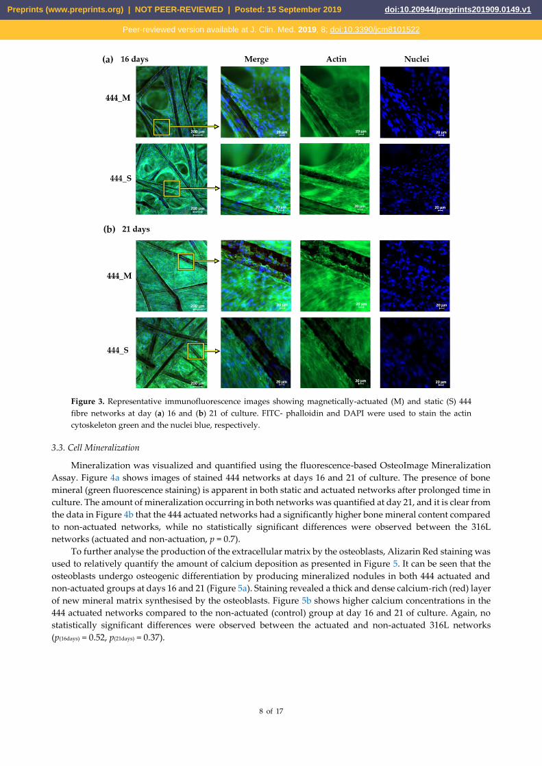
Figure 3 shows immunofluorescence staining of the actin filaments in the cytoskeleton and cell nuclei for
the 444 networks. At day 16 of culture, the cells have started to populate the inter-fibre spaces and form bridges
between adjacent fibres (Figure 3a). At day 21, the cells have filled the inter-fibre spaces of the networks (Figure
3b). This was observed throughout the thickness of the fibre networks, as illustrated in the cross-sectional
images (Supplementary section, Figure S1). The cells showed a well-organized actin cytoskeleton. A similar
response was observed in static and actuated 316L networks (Supplementary section, Figure S2).
Preprints (www.preprints.org) | NOT PEER-REVIEWED | Posted: 15 September 2019 doi:10.20944/preprints201909.0149.v1
Peer-reviewed version available at J. Clin. Med. 2019, 8; doi:10.3390/jcm8101522
8 of 17
Figure 3. Representative immunofluorescence images showing magnetically-actuated (M) and static (S) 444
fibre networks at day (a) 16 and (b) 21 of culture. FITC- phalloidin and DAPI were used to stain the actin
cytoskeleton green and the nuclei blue, respectively.
3.3. Cell Mineralization
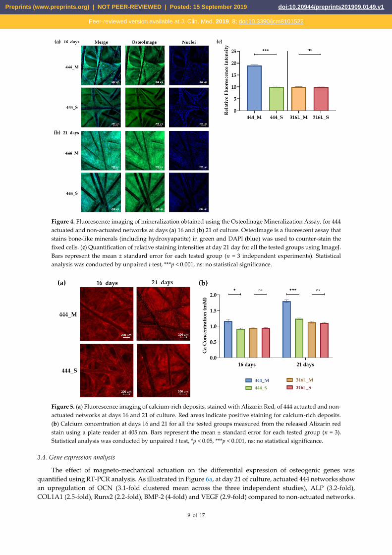
Mineralization was visualized and quantified using the fluorescence-based OsteoImage Mineralization
Assay. Figure 4a shows images of stained 444 networks at days 16 and 21 of culture. The presence of bone
mineral (green fluorescence staining) is apparent in both static and actuated networks after prolonged time in
culture. The amount of mineralization occurring in both networks was quantified at day 21, and it is clear from
the data in Figure 4b that the 444 actuated networks had a significantly higher bone mineral content compared
to non-actuated networks, while no statistically significant differences were observed between the 316L
networks (actuated and non-actuation, p = 0.7).
To further analyse the production of the extracellular matrix by the osteoblasts, Alizarin Red staining was
used to relatively quantify the amount of calcium deposition as presented in Figure 5. It can be seen that the
osteoblasts undergo osteogenic differentiation by producing mineralized nodules in both 444 actuated and
non-actuated groups at days 16 and 21 (Figure 5a). Staining revealed a thick and dense calcium-rich (red) layer
of new mineral matrix synthesised by the osteoblasts. Figure 5b shows higher calcium concentrations in the
444 actuated networks compared to the non-actuated (control) group at day 16 and 21 of culture. Again, no
statistically significant differences were observed between the actuated and non-actuated 316L networks
(p(16days) = 0.52, p(21days) = 0.37).
Preprints (www.preprints.org) | NOT PEER-REVIEWED | Posted: 15 September 2019 doi:10.20944/preprints201909.0149.v1
Peer-reviewed version available at J. Clin. Med. 2019, 8; doi:10.3390/jcm8101522
9 of 17
Figure 4. Fluorescence imaging of mineralization obtained using the OsteoImage Mineralization Assay, for 444
actuated and non-actuated networks at days (a) 16 and (b) 21 of culture. OsteoImage is a fluorescent assay that
stains bone-like minerals (including hydroxyapatite) in green and DAPI (blue) was used to counter-stain the
fixed cells. (c) Quantification of relative staining intensities at day 21 day for all the tested groups using ImageJ.
Bars represent the mean ± standard error for each tested group (n = 3 independent experiments). Statistical
analysis was conducted by unpaired t test, ***p < 0.001, ns: no statistical significance.
Figure 5. (a) Fluorescence imaging of calcium-rich deposits, stained with Alizarin Red, of 444 actuated and non-
actuated networks at days 16 and 21 of culture. Red areas indicate positive staining for calcium-rich deposits.
(b) Calcium concentration at days 16 and 21 for all the tested groups measured from the released Alizarin red
stain using a plate reader at 405 nm. Bars represent the mean ± standard error for each tested group (n = 3).
Statistical analysis was conducted by unpaired t test, *p < 0.05, ***p < 0.001, ns: no statistical significance.
3.4. Gene expression analysis
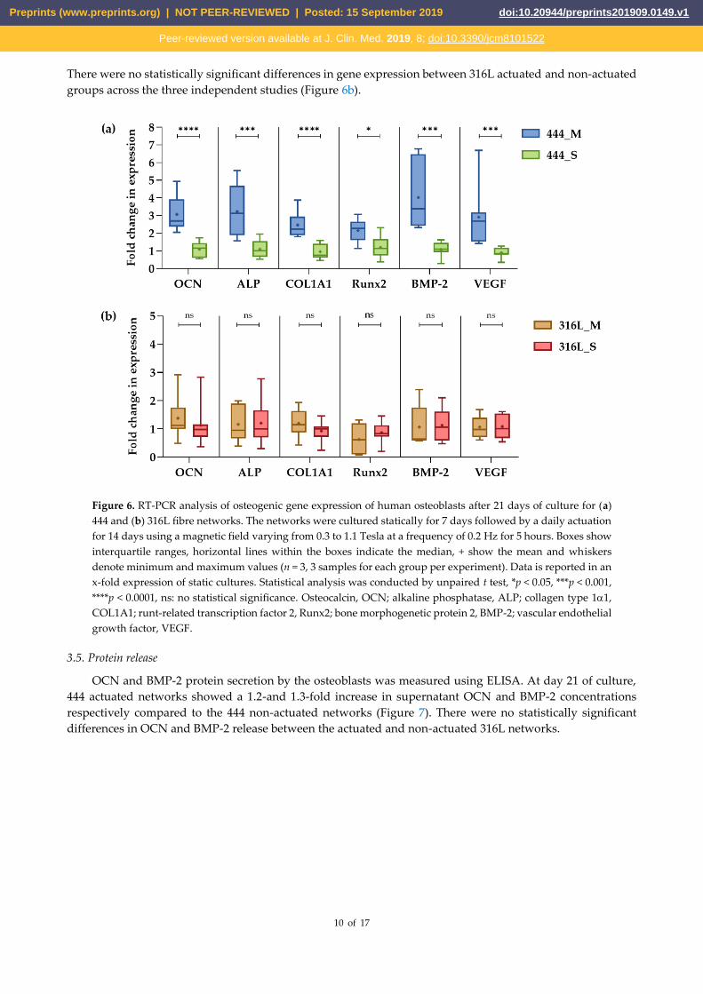
The effect of magneto-mechanical actuation on the differential expression of osteogenic genes was
quantified using RT-PCR analysis. As illustrated in Figure 6a, at day 21 of culture, actuated 444 networks show
an upregulation of OCN (3.1-fold clustered mean across the three independent studies), ALP (3.2-fold),
COL1A1 (2.5-fold), Runx2 (2.2-fold), BMP-2 (4-fold) and VEGF (2.9-fold) compared to non-actuated networks.
Preprints (www.preprints.org) | NOT PEER-REVIEWED | Posted: 15 September 2019 doi:10.20944/preprints201909.0149.v1
Peer-reviewed version available at J. Clin. Med. 2019, 8; doi:10.3390/jcm8101522
10 of 17
There were no statistically significant differences in gene expression between 316L actuated and non-actuated
groups across the three independent studies (Figure 6b).
Figure 6. RT-PCR analysis of osteogenic gene expression of human osteoblasts after 21 days of culture for (a)
444 and (b) 316L fibre networks. The networks were cultured statically for 7 days followed by a daily actuation
for 14 days using a magnetic field varying from 0.3 to 1.1 Tesla at a frequency of 0.2 Hz for 5 hours. Boxes show
interquartile ranges, horizontal lines within the boxes indicate the median, + show the mean and whiskers
denote minimum and maximum values (n = 3, 3 samples for each group per experiment). Data is reported in an
x-fold expression of static cultures. Statistical analysis was conducted by unpaired t test, *p < 0.05, ***p < 0.001,
****p < 0.0001, ns: no statistical significance. Osteocalcin, OCN; alkaline phosphatase, ALP; collagen type 11,
COL1A1; runt-related transcription factor 2, Runx2; bone morphogenetic protein 2, BMP-2; vascular endothelial
growth factor, VEGF.
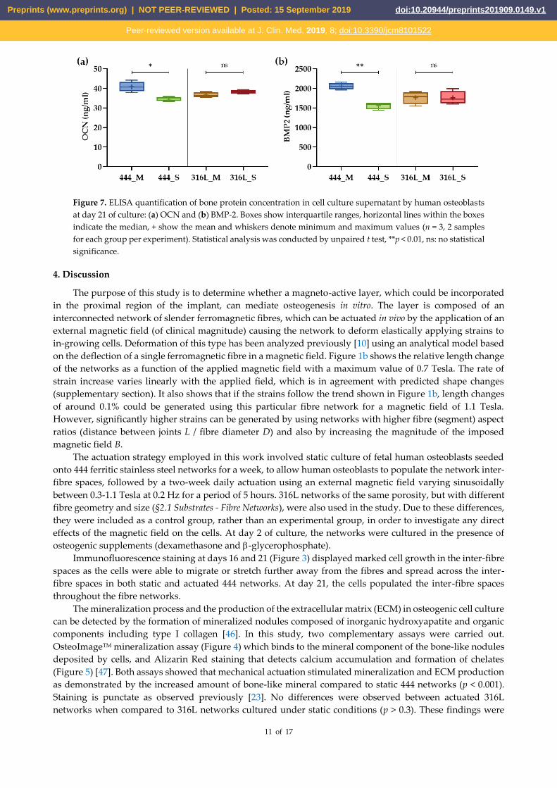
3.5. Protein release
OCN and BMP-2 protein secretion by the osteoblasts was measured using ELISA. At day 21 of culture,
444 actuated networks showed a 1.2-and 1.3-fold increase in supernatant OCN and BMP-2 concentrations
respectively compared to the 444 non-actuated networks (Figure 7). There were no statistically significant
differences in OCN and BMP-2 release between the actuated and non-actuated 316L networks.
Preprints (www.preprints.org) | NOT PEER-REVIEWED | Posted: 15 September 2019 doi:10.20944/preprints201909.0149.v1
Peer-reviewed version available at J. Clin. Med. 2019, 8; doi:10.3390/jcm8101522
11 of 17
Figure 7. ELISA quantification of bone protein concentration in cell culture supernatant by human osteoblasts
at day 21 of culture: (a) OCN and (b) BMP-2. Boxes show interquartile ranges, horizontal lines within the boxes
indicate the median, + show the mean and whiskers denote minimum and maximum values (n = 3, 2 samples
for each group per experiment). Statistical analysis was conducted by unpaired t test, **p < 0.01, ns: no statistical
significance.
4. Discussion
The purpose of this study is to determine whether a magneto-active layer, which could be incorporated
in the proximal region of the implant, can mediate osteogenesis in vitro. The layer is composed of an
interconnected network of slender ferromagnetic fibres, which can be actuated in vivo by the application of an
external magnetic field (of clinical magnitude) causing the network to deform elastically applying strains to
in-growing cells. Deformation of this type has been analyzed previously [10] using an analytical model based
on the deflection of a single ferromagnetic fibre in a magnetic field. Figure 1b shows the relative length change
of the networks as a function of the applied magnetic field with a maximum value of 0.7 Tesla. The rate of
strain increase varies linearly with the applied field, which is in agreement with predicted shape changes
(supplementary section). It also shows that if the strains follow the trend shown in Figure 1b, length changes
of around 0.1% could be generated using this particular fibre network for a magnetic field of 1.1 Tesla.
However, significantly higher strains can be generated by using networks with higher fibre (segment) aspect
ratios (distance between joints L / fibre diameter D) and also by increasing the magnitude of the imposed
magnetic field B.
The actuation strategy employed in this work involved static culture of fetal human osteoblasts seeded
onto 444 ferritic stainless steel networks for a week, to allow human osteoblasts to populate the network inter-
fibre spaces, followed by a two-week daily actuation using an external magnetic field varying sinusoidally
between 0.3-1.1 Tesla at 0.2 Hz for a period of 5 hours. 316L networks of the same porosity, but with different
fibre geometry and size (§2.1 Substrates - Fibre Networks), were also used in the study. Due to these differences,
they were included as a control group, rather than an experimental group, in order to investigate any direct
effects of the magnetic field on the cells. At day 2 of culture, the networks were cultured in the presence of
osteogenic supplements (dexamethasone and -glycerophosphate).
Immunofluorescence staining at days 16 and 21 (Figure 3) displayed marked cell growth in the inter-fibre
spaces as the cells were able to migrate or stretch further away from the fibres and spread across the inter-
fibre spaces in both static and actuated 444 networks. At day 21, the cells populated the inter-fibre spaces
throughout the fibre networks.
The mineralization process and the production of the extracellular matrix (ECM) in osteogenic cell culture
can be detected by the formation of mineralized nodules composed of inorganic hydroxyapatite and organic
components including type I collagen [46]. In this study, two complementary assays were carried out.
OsteoImageTM mineralization assay (Figure 4) which binds to the mineral component of the bone-like nodules
deposited by cells, and Alizarin Red staining that detects calcium accumulation and formation of chelates
(Figure 5) [47]. Both assays showed that mechanical actuation stimulated mineralization and ECM production
as demonstrated by the increased amount of bone-like mineral compared to static 444 networks (p < 0.001).
Staining is punctate as observed previously [23]. No differences were observed between actuated 316L
networks when compared to 316L networks cultured under static conditions (p > 0.3). These findings were
Preprints (www.preprints.org) | NOT PEER-REVIEWED | Posted: 15 September 2019 doi:10.20944/preprints201909.0149.v1
Peer-reviewed version available at J. Clin. Med. 2019, 8; doi:10.3390/jcm8101522
12 of 17
supported by upregulation of the COL1A1 by 2.5-fold (p < 0.0001), shown in Figure 6a, for the 444 actuated
group compared to the 444 static group. COL1A1 is the main component in osseous extracellular matrix [48].
It can mediate cell adhesion, contribute to the mature osteoblast phenotype and provide a template for
mineralization [49].
To further analyze the effect of magneto-mechanical actuation on the osteoblast osteogenic capacity, bone
formation markers such as ALP and OCN as well as Runx2, BMP-2 and VEGF, which have known functions
in bone biology, were evaluated (Figures 6 and 7). OCN is the second most abundant protein in bone after
collagen that can be found in a fully-mineralized matrix (late markers of osteoblast differentiation) and
promotes deposition of mineral substance [50]. ALP is an ectoenzyme, highly expressed in active osteoblasts
[46] and plays a role in bone mineralization by controlling the concentrations of mineralization inhibitors and
phosphate ions. In this study, both ALP (3.2-fold (p < 0.001)) and OCN (gene-level: 3-fold (p < 0.0001), protein-
level: 1.2-fold (p < 0.05)) were upregulated for the 444 actuated group compared to the static group across three
independent studies, while no significant differences were observed in the 316L groups. These results are in
line with previous studies [51-53], that reported upregulation of the above markers at both gene and protein
levels upon application of mechanical stimulation and have previously been associated with calcium binding
and bone matrix mineralization process.
Runx2 is an essential transcription factor for osteoblastic differentiation and bone formation, and directly
regulates the expression of OCN [54]. Also, Runx2 mutations in humans, that reduce the level of this gene’s
functional activity, are responsible for cleidocranial dysplasia [55]. In this study, mechanical actuation
increased the Runx2 expression by 2.2-fold (p < 0.05). This is in line with previous reports [34,56,57], that
demonstrated upregulation of Runx2 as a result of mechanical stimulation such as cyclic compression (5 & 10
% strain) and tension (1%). BMP-2 is accumulated in ECM and is shown to stimulate osteoblastic
differentiation in vitro [58]. It exhibits this osteogenic action by regulating transcription of osteogenic genes
such as ALP, COL1A1 and OCN. Moreover, BMP-2 is known to control the expression and functions of Runx2
[54]. For example, osteoblasts cultured onto polycaprolactone (PCL) fibre networks showed an upregulation
of BMP-2 (17-fold), Runx2 (1.6-fold), ALP (2.5-fold), COL1A1 (2.2-fold) and OCN (3.4-fold) compared to static
networks when the scaffolds, after 4 weeks of culture, were subjected to 10% cyclic compressive strain at 0.5 Hz
for 4 hours [57].
VEGF is the main angiogenic growth factor involved in bone healing and has an important role in bone
repair by stimulating osteoblasts [46,59]. The synergy between BMP-2 and VEGF has been reported [60], in
which there is an intimate relation to bone development and healing that is advantageous for bone
regeneration procedures. In the present study, BMP-2 was upregulated by 4-fold while magnetic actuation
augmented the expression of VEGF by 2.9-fold (p < 0.001). Also, a 1.3-fold increase in supernatant BMP-2
concentration was measured compared to the static networks (p < 0.01).
Magneto-mechanical actuation of 316L (non-magnetic) networks showed a similar deposition of bone-
like minerals, gene expression, and protein release to the supernatant compared to those cultured without
actuation suggesting that the magnetic field has no direct effect on the cellular responses. The effects observed
in cells within the 444 (magnetic) networks can be attributed to mechanical deformations induced in in-
growing cells via magneto-mechanical actuation of the fibre networks. During the first weeks of culture, cells
form bridges between connecting fibres and will then begin to form cell-matrix accumulations in the corners
of the inter-fibre spaces where two fibres meet by curvature driven tissue growth as previously described for
MC3T3-E1 mouse osteoblasts [61]. The cells are linked to each other or to the extracellular matrix and thereby
linked either directly or indirectly to the fibres. In response to the application of a magnetic field, the networks
deform elastically, transmitting stresses and strains to in-growing cells via small local fibre deflections.
Deformation of the fibre networks will also cause fluid flow within the networks producing shear stresses
(tangential frictional forces produced when fluid flows over the cell surface). To induce the changes in cell
behavior and matrix deposition seen, these forces must be converted into biochemical responses by acting
through mechanosensitive molecules within focal adhesion complexes, ion channels and the cytoskeleton [62].
In summary, the results of this study show that mechanical actuation increases the expression of genes
important for osteoblast differentiation (Runx2, BMP-2), matrix deposition and mineralization (COL1A1, ALP,
OCN) and vascularization (VEGF). There was a concomitant increase in the amount of BMP-2 and osteocalcin
produced by the cells and enhanced deposition of mineralized matrix. These results demonstrate that the
strains produced during magneto-mechanical actuation of ferromagnetic fibre networks can increase the in
Preprints (www.preprints.org) | NOT PEER-REVIEWED | Posted: 15 September 2019 doi:10.20944/preprints201909.0149.v1
Peer-reviewed version available at J. Clin. Med. 2019, 8; doi:10.3390/jcm8101522
13 of 17
vitro osteogenic differentiation of human osteoblasts growing within the networks. Future work will focus on
the effect of this actuation on self-assembly of endothelial cells and supporting cell populations (e.g.
osteoblasts) into vessel-like structures, which is important for bone formation.
5. Conclusions
In this study, magneto-mechanical actuation was investigated as a means of promoting osteogenesis in
vitro in highly porous ferromagnetic fibre networks by examining mineralization, ECM production and the
resulting expression of genes, proteins and transcription factors known to be essential for osteogenic
differentiation and functional maturation. We showed that the actuation can enhance mineralization, ECM
production and upregulate osteogenesis by inducing OCN, ALP, COL1A1, Runx2 and BMP-2 expression, and
result in the synthesis of proteins involved in osteoblast differentiation and mineralized matrix deposition.
Furthermore, actuation increased gene expression of the pro-angiogenic molecule VEGF. In contrast, non-
magnetic scaffolds showed no significant effects in response to magneto-mechanical actuation over respective
static controls suggesting that there are no direct effects of the magnetic field on cellular responses. The results
corroborate that controlled shape changes achieved via application of an external magnetic field to a
ferromagnetic fibrous scaffold can be used to induce changes in cellular behavior. This concept can be
exploited in developing biomedical devices with the potential for controlled actuation in vivo.
Funding: This research was supported by the EPSRC (EP/R511675/1), the Blavatnik Family Foundation and the European
Research Council (Grant No. 240446). Financial support for GKL has been provided via the Blavatnik Family Foundation
and Reuben Foundation. Financial support for RAB has been provided via the National Institute for Health Research
(NIHR).
Acknowledgments: The authors would like to acknowledge Dr V.N. Malheiro for the design of the bespoke tissue culture
plates for the magnetic actuation experiments and Dr A. Symeonidou for useful discussions.
Conflicts of Interest: The authors declare no conflict of interest.
References
1. Clarke, B. Normal bone anatomy and physiology. Clinical journal of the American Society of Nephrology 2008, 3, S131-
S139.
2. Dimitriou, R.; Jones, E.; McGonagle, D.; Giannoudis, P.V. Bone regeneration: current concepts and future
directions. BMC medicine 2011, 9, 66.
3. Lanyon, L.E.; Goodship, A.E.; Pye, C.J.; MacFie, J.H. Mechanically adaptive bone remodelling. J Biomech 1982, 15,
141-154.
4. Rubin, C.T.; Lanyon, L.E. Regulation of bone mass by mechanical strain magnitude. Calcif Tissue Int 1985, 37, 411-
417.
5. Frost, H.M. Bone “mass” and the “mechanostat”: a proposal. The anatomical record 1987, 219, 1-9.
6. Ross, T.D.; Coon, B.G.; Yun, S.; Baeyens, N.; Tanaka, K.; Ouyang, M.; Schwartz, M.A. Integrins in
mechanotransduction. Curr Opin Cell Biol 2013, 25, 613-618, doi:10.1016/j.ceb.2013.05.006.
7. Orr, A.W.; Helmke, B.P.; Blackman, B.R.; Schwartz, M.A. Mechanisms of mechanotransduction. Dev Cell 2006, 10,
11-20, doi:10.1016/j.devcel.2005.12.006.
8. Iqbal, J.; Zaidi, M. Molecular regulation of mechanotransduction. Biochem Biophys Res Commun 2005, 328, 751-755,
doi:10.1016/j.bbrc.2004.12.087.
9. Markaki, A.E.; Clyne, T.W. Magneto-mechanical actuation of bonded ferromagnetic fibre arrays. Acta materialia
2005, 53, 877-889.
Preprints (www.preprints.org) | NOT PEER-REVIEWED | Posted: 15 September 2019 doi:10.20944/preprints201909.0149.v1
Peer-reviewed version available at J. Clin. Med. 2019, 8; doi:10.3390/jcm8101522
14 of 17
10. Markaki, A.E.; Clyne, T.W. Magneto-mechanical stimulation of bone growth in a bonded array of ferromagnetic
fibres. Biomaterials 2004, 25, 4805-4815.
11. Daish, C.; Blanchard, R.; Fox, K.; Pivonka, P.; Pirogova, E. The Application of Pulsed Electromagnetic Fields
(PEMFs) for Bone Fracture Repair: Past and Perspective Findings. Ann Biomed Eng 2018, 46, 525-542,
doi:10.1007/s10439-018-1982-1.
12. Kotani, H.; Kawaguchi, H.; Shimoaka, T.; Iwasaka, M.; Ueno, S.; Ozawa, H.; Nakamura, K.; Hoshi, K. Strong static
magnetic field stimulates bone formation to a definite orientation in vitro and in vivo. J Bone Miner Res 2002, 17,
1814-1821, doi:10.1359/jbmr.2002.17.10.1814.
13. Yan, Q.C.; Tomita, N.; Ikada, Y. Effects of static magnetic field on bone formation of rat femurs. Med Eng Phys
1998, 20, 397-402.
14. Pankhurst, Q.; Thanh, N.; Jones, S.; Dobson, J. Progress in applications of magnetic nanoparticles in biomedicine.
Journal of Physics D: Applied Physics 2009, 42, 224001.
15. Dobson, J. Remote control of cellular behaviour with magnetic nanoparticles. Nature nanotechnology 2008, 3, 139.
16. Yuge, L.; Okubo, A.; Miyashita, T.; Kumagai, T.; Nikawa, T.; Takeda, S.; Kanno, M.; Urabe, Y.; Sugiyama, M.;
Kataoka, K. Physical stress by magnetic force accelerates differentiation of human osteoblasts. Biochem Biophys
Res Commun 2003, 311, 32-38.
17. Yuan, Z.; Memarzadeh, K.; Stephen, A.S.; Allaker, R.P.; Brown, R.A.; Huang, J. Development of a 3D Collagen
Model for the In Vitro Evaluation of Magnetic-assisted Osteogenesis. Scientific reports 2018, 8, 16270.
18. Henstock, J.R.; Rotherham, M.; El Haj, A.J. Magnetic ion channel activation of TREK1 in human mesenchymal
stem cells using nanoparticles promotes osteogenesis in surrounding cells. Journal of tissue engineering 2018, 9,
2041731418808695.
19. Rotherham, M.; Nahar, T.; Goodman, T.; Telling, N.; Gates, M.; El Haj, A. Magnetic Mechanoactivation of Wnt
Signaling Augments Dopaminergic Differentiation of Neuronal Cells. Advanced Biosystems 2019, 1900091.
20. Markaki, A.; Justin, A. A magneto-active scaffold for stimulation of bone growth. Materials Science and Technology
2014, 30, 1590-1598.
21. Dowling, N.; Kim, Y.-H.; Ahn, S.-K.; Lee, Y.-D. Effect of alloying elements and residuals on corrosion resistance
of type 444 stainless steel. Corrosion 1999, 55, 187-199.
22. Asami, K.; Hashimoto, K. Importance of initial surface film in the degradation of stainless steels by atmospheric
exposure. Corrosion Science 2003, 45, 2263-2283.
23. Malheiro, V.N.; Spear, R.L.; Brooks, R.A.; Markaki, A.E. Osteoblast and monocyte responses to 444 ferritic
stainless steel intended for a magneto-mechanically actuated fibrous scaffold. Biomaterials 2011, 32, 6883-6892.
24. Bhaskar, B.; Owen, R.; Bahmaee, H.; Rao, P.S.; Reilly, G.C. Design and assessment of a dynamic perfusion
bioreactor for large bone tissue engineering scaffolds. Applied biochemistry and biotechnology 2018, 185, 555-563.
25. Ravichandran, A.; Wen, F.; Lim, J.; Chong, M.S.K.; Chan, J.K.; Teoh, S.H. Biomimetic fetal rotation bioreactor for
engineering bone tissues—Effect of cyclic strains on upregulation of osteogenic gene expression. Journal of tissue
engineering and regenerative medicine 2018.
26. Liu, L.; Li, H.; Cui, Y.; Li, R.; Meng, F.; Ye, Z.; Zhang, X. Calcium Channel Opening Rather than the Release of
ATP Causes the Apoptosis of Osteoblasts Induced by Overloaded Mechanical Stimulation. Cellular Physiology and
Biochemistry 2017, 42, 441-454.
27. Beşkardeş, I.G.; Hayden, R.S.; Glettig, D.L.; Kaplan, D.L.; Gümüşderelioğlu, M. Bone tissue engineering with
scaffold-supported perfusion co-cultures of human stem cell-derived osteoblasts and cell line-derived osteoclasts.
Process Biochemistry 2017.
28. Mitra, D.; Whitehead, J.; Yasui, O.W.; Leach, J.K. Bioreactor culture duration of engineered constructs influences
bone formation by mesenchymal stem cells. Biomaterials 2017, 146, 29-39.
Preprints (www.preprints.org) | NOT PEER-REVIEWED | Posted: 15 September 2019 doi:10.20944/preprints201909.0149.v1
Peer-reviewed version available at J. Clin. Med. 2019, 8; doi:10.3390/jcm8101522
15 of 17
29. Tang, X.; Teng, S.; Liu, C.; Jagodzinski, M. Influence of hydrodynamic pressure on the proliferation and
osteogenic differentiation of bone mesenchymal stromal cells seeded on polyurethane scaffolds. Journal of
Biomedical Materials Research Part A 2017, 105, 3445-3455.
30. Filipowska, J.; Reilly, G.C.; Osyczka, A.M. A single short session of media perfusion induces osteogenesis in
hBMSCs cultured in porous scaffolds, dependent on cell differentiation stage. Biotechnology and bioengineering
2016, 113, 1814-1824.
31. Teng, S.; Liu, C.; Guenther, D.; Omar, M.; Neunaber, C.; Krettek, C.; Jagodzinski, M. Influence of biomechanical
and biochemical stimulation on the proliferation and differentiation of bone marrow stromal cells seeded on
polyurethane scaffolds. Experimental and therapeutic medicine 2016, 11, 2086-2094.
32. Yamada, M.; Anada, T.; Masuda, T.; Takano-Yamamoto, T.; Suzuki, O. Effect of mechanical stress on
differentiation of mouse mesenchymal stem cells seeded into an octacalcium phosphate–gelatin scaffold. Sensors
and Actuators B: Chemical 2015, 220, 125-130.
33. Tanaka, S.M.; Tachibana, K. Frequency-dependence of mechanically stimulated osteoblastic calcification in
tissue-engineered bone in vitro. Annals of biomedical engineering 2015, 43, 2083-2089.
34. Damaraju, S.; Matyas, J.R.; Rancourt, D.E.; Duncan, N.A. The role of gap junctions and mechanical loading on
mineral formation in a collagen-I scaffold seeded with osteoprogenitor cells. Tissue Engineering Part A 2015, 21,
1720-1732.
35. Subramony, S.D.; Su, A.; Yeager, K.; Lu, H.H. Combined effects of chemical priming and mechanical stimulation
on mesenchymal stem cell differentiation on nanofiber scaffolds. Journal of biomechanics 2014, 47, 2189-2196.
36. Bartnikowski, M.; Klein, T.J.; Melchels, F.P.; Woodruff, M.A. Effects of scaffold architecture on mechanical
characteristics and osteoblast response to static and perfusion bioreactor cultures. Biotechnology and bioengineering
2014, 111, 1440-1451.
37. Jia, Y.-Y.; Li, F.; Geng, N.; Gong, P.; Huang, S.-J.; Meng, L.-X.; Lan, J.; Ban, Y. Fluid flow modulates the expression
of genes involved in the Wnt signaling pathway in osteoblasts in 3D culture conditions. International journal of
molecular medicine 2014, 33, 1282-1288.
38. Henstock, J.R.; Rotherham, M.; Rashidi, H.; Shakesheff, K.M.; El Haj, A.J. Remotely activated
mechanotransduction via magnetic nanoparticles promotes mineralization synergistically with bone
morphogenetic protein 2: applications for injectable cell therapy. Stem cells translational medicine 2014, 3, 1363-
1374.
39. Damaraju, S.; Matyas, J.R.; Rancourt, D.E.; Duncan, N.A. The effect of mechanical stimulation on mineralization
in differentiating osteoblasts in collagen-I scaffolds. Tissue Engineering Part A 2014, 20, 3142-3153.
40. Malheiro, V.N.; Skepper, J.N.; Brooks, R.A.; Markaki, A.E. In vitro osteoblast response to ferritic stainless steel
fiber networks for magneto‐active layers on implants. Journal of Biomedical Materials Research Part A 2013, 101,
1588-1598.
41. Spear, R.L.; Brooks, R.A.; Markaki, A.E. Short-term in vitro responses of human peripheral blood monocytes to
ferritic stainless steel fiber networks. J Biomed Mater Res A 2013, 101, 1456-1463, doi:10.1002/jbm.a.34451.
42. Neelakantan, S.; Bosbach, W.; Woodhouse, J.; Markaki, A.E. Characterization and deformation response of
orthotropic fibre networks with auxetic out-of-plane behaviour. Acta Materialia 2014, 66, 326-339.
43. Langenbach, F.; Handschel, J. Effects of dexamethasone, ascorbic acid and β-glycerophosphate on the osteogenic
differentiation of stem cells in vitro. Stem cell research & therapy 2013, 4, 117.
44. Gregory, C.A.; Gunn, W.G.; Peister, A.; Prockop, D.J. An Alizarin red-based assay of mineralization by adherent
cells in culture: comparison with cetylpyridinium chloride extraction. Anal Biochem 2004, 329, 77-84,
doi:10.1016/j.ab.2004.02.002.
Preprints (www.preprints.org) | NOT PEER-REVIEWED | Posted: 15 September 2019 doi:10.20944/preprints201909.0149.v1
Peer-reviewed version available at J. Clin. Med. 2019, 8; doi:10.3390/jcm8101522
16 of 17
45. Livak, K.J.; Schmittgen, T.D. Analysis of relative gene expression data using real-time quantitative PCR and the
2− ΔΔCT method. Methods 2001, 25, 402-408.
46. Blair, H.C.; Larrouture, Q.C.; Li, Y.; Lin, H.; Beer-Stoltz, D.; Liu, L.; Tuan, R.S.; Robinson, L.J.; Schlesinger, P.H.;
Nelson, D.J. Osteoblast differentiation and bone matrix formation in vivo and in vitro. Tissue Engineering Part B:
Reviews 2017, 23, 268-280.
47. Pettersson, L.F.; Kingham, P.J.; Wiberg, M.; Kelk, P. In vitro osteogenic differentiation of human mesenchymal
stem cells from jawbone compared with dental tissue. Tissue engineering and regenerative medicine 2017, 14, 763-
774.
48. Hiemer, B.; Ziebart, J.; Jonitz-Heincke, A.; Grunert, P.C.; Su, Y.; Hansmann, D.; Bader, R. Magnetically induced
electrostimulation of human osteoblasts results in enhanced cell viability and osteogenic differentiation.
International journal of molecular medicine 2016, 38, 57-64.
49. Chen, W.; Liu, X.; Chen, Q.; Bao, C.; Zhao, L.; Zhu, Z.; Xu, H.H. Angiogenic and osteogenic regeneration in rats
via calcium phosphate scaffold and endothelial cell co‐culture with human bone marrow mesenchymal stem cells
(MSCs), human umbilical cord MSCs, human induced pluripotent stem cell‐derived MSCs and human embryonic
stem cell‐derived MSCs. Journal of tissue engineering and regenerative medicine 2018, 12, 191-203.
50. Rutkovskiy, A.; Stensløkken, K.-O.; Vaage, I.J. Osteoblast differentiation at a glance. Medical science monitor basic
research 2016, 22, 95.
51. Dumas, V.; Perrier, A.; Malaval, L.; Laroche, N.; Guignandon, A.; Vico, L.; Rattner, A. The effect of dual frequency
cyclic compression on matrix deposition by osteoblast-like cells grown in 3D scaffolds and on modulation of
VEGF variant expression. Biomaterials 2009, 30, 3279-3288.
52. Sittichockechaiwut, A.; Scutt, A.M.; Ryan, A.J.; Bonewald, L.F.; Reilly, G.C. Use of rapidly mineralising osteoblasts
and short periods of mechanical loading to accelerate matrix maturation in 3D scaffolds. Bone 2009, 44, 822-829.
53. Zong ming, W.; Jian yu, L.; Rui xin, L.; Hao, L.; Yong, G.; Lu, L.; Xin chang, Z.; Xi zheng, Z. Bone formation in
rabbit cancellous bone explant culture model is enhanced by mechanical load. Biomedical engineering online 2013,
12, 35.
54. Matsubara, T.; Kida, K.; Yamaguchi, A.; Hata, K.; Ichida, F.; Meguro, H.; Aburatani, H.; Nishimura, R.; Yoneda,
T. BMP2 regulates Osterix through Msx2 and Runx2 during osteoblast differentiation. Journal of biological chemistry
2008, 283, 29119-29125.
55. Lou, Y.; Javed, A.; Hussain, S.; Colby, J.; Frederick, D.; Pratap, J.; Xie, R.; Gaur, T.; Van Wijnen, A.J.; Jones, S.N. A
Runx2 threshold for the cleidocranial dysplasia phenotype. Human molecular genetics 2008, 18, 556-568.
56. Ignatius, A.; Blessing, H.; Liedert, A.; Schmidt, C.; Neidlinger-Wilke, C.; Kaspar, D.; Friemert, B.; Claes, L. Tissue
engineering of bone: effects of mechanical strain on osteoblastic cells in type I collagen matrices. Biomaterials 2005,
26, 311-318.
57. Rath, B.; Nam, J.; Knobloch, T.J.; Lannutti, J.J.; Agarwal, S. Compressive forces induce osteogenic gene expression
in calvarial osteoblasts. Journal of biomechanics 2008, 41, 1095-1103.
58. Takeuchi, Y.; Suzawa, M.; Kikuchi, T.; Matsumoto, T. Interaction of matrix collagen with osteoblastic cells
enhances stimulatory effects of bone morphogenetic protein (BMP)-2 on the differentiation of osteoblasts. Bone
1995, 6, 568.
59. Li, Q.; Wang, Z. Influence of mesenchymal stem cells with endothelial progenitor cells in co-culture on
osteogenesis and angiogenesis: an in vitro study. Archives of medical research 2013, 44, 504-513.
60. Samee, M.; Kasugai, S.; Kondo, H.; Ohya, K.; Shimokawa, H.; Kuroda, S. Bone morphogenetic protein-2 (BMP-2)
and vascular endothelial growth factor (VEGF) transfection to human periosteal cells enhances osteoblast
differentiation and bone formation. Journal of pharmacological sciences 2008, 0809050139-0809050139.
Preprints (www.preprints.org) | NOT PEER-REVIEWED | Posted: 15 September 2019 doi:10.20944/preprints201909.0149.v1
Peer-reviewed version available at J. Clin. Med. 2019, 8; doi:10.3390/jcm8101522

17 of 17
61. Rumpler, M.; Woesz, A.; Dunlop, J.W.; van Dongen, J.T.; Fratzl, P. The effect of geometry on three-dimensional
tissue growth. Journal of the Royal Society Interface 2008, 5, 1173-1180.
62. Mammoto, T.; Ingber, D.E. Mechanical control of tissue and organ development. Development 2010, 137, 1407-
1420.
Preprints (www.preprints.org) | NOT PEER-REVIEWED | Posted: 15 September 2019 doi:10.20944/preprints201909.0149.v1
Peer-reviewed version available at J. Clin. Med. 2019, 8; doi:10.3390/jcm8101522