Embed Size (px)
Citation preview

1
Stimulating Cardiac Muscle by Light: Cardiac Optogenetics by Cell Delivery
Running title: Jia et al.; Cell-based cardiac optogenetics
Zhiheng Jia1, MS, Virginijus Valiunas2, PhD, Zongju Lu2, PhD, Harold Bien1, MD/PhD, Huilin
Liu2, MS, Hong-Zhang Wang2, PhD, Barbara Rosati2, 3, PhD, Peter R. Brink2, 3, PhD, Ira S.
Cohen2, 3, MD/PhD, and Emilia Entcheva1, 2, 3, PhD
1Department of Biomedical Engineering; 2Department of Physiology & Biophysics; 3Institute for
Molecular Cardiology; Stony Brook University, Stony Brook, NY
Correspondence:
Emilia Entcheva, PhD
HSC T18-30, Department of Biomedical Engineering,
Stony Brook University
Stony Brook, NY, 11794-8181
Phone: (631) 444 2368.
Fax: (631) 444 6646.
E-mail: [email protected]
Journal Subject Codes: [150] Imaging, [152] Ion channels/membrane transport; [120] Pacemaker
logyyyyyyy &&&&&&& BBBBBBBioioioioioioiophphphphphphphyysyyyg g; p y gy p y
e Y
g g; p y gy p y
ecular Cardiology; Stony Brook University, Stony Brook, NY
by guest on June 14, 2018http://circep.ahajournals.org/
Dow
nloaded from
by guest on June 14, 2018http://circep.ahajournals.org/
Dow
nloaded from
by guest on June 14, 2018http://circep.ahajournals.org/
Dow
nloaded from
by guest on June 14, 2018http://circep.ahajournals.org/
Dow
nloaded from
by guest on June 14, 2018http://circep.ahajournals.org/
Dow
nloaded from
by guest on June 14, 2018http://circep.ahajournals.org/
Dow
nloaded from
by guest on June 14, 2018http://circep.ahajournals.org/
Dow
nloaded from
by guest on June 14, 2018http://circep.ahajournals.org/
Dow
nloaded from
by guest on June 14, 2018http://circep.ahajournals.org/
Dow
nloaded from
by guest on June 14, 2018http://circep.ahajournals.org/
Dow
nloaded from
by guest on June 14, 2018http://circep.ahajournals.org/
Dow
nloaded from
by guest on June 14, 2018http://circep.ahajournals.org/
Dow
nloaded from
by guest on June 14, 2018http://circep.ahajournals.org/
Dow
nloaded from
by guest on June 14, 2018http://circep.ahajournals.org/
Dow
nloaded from
by guest on June 14, 2018http://circep.ahajournals.org/
Dow
nloaded from
by guest on June 14, 2018http://circep.ahajournals.org/
Dow
nloaded from
by guest on June 14, 2018http://circep.ahajournals.org/
Dow
nloaded from
by guest on June 14, 2018http://circep.ahajournals.org/
Dow
nloaded from
by guest on June 14, 2018http://circep.ahajournals.org/
Dow
nloaded from
by guest on June 14, 2018http://circep.ahajournals.org/
Dow
nloaded from
2
Abstract:
Background - After the recent cloning of light-sensitive ion channels and their expression in
mammalian cells, a new field, optogenetics, emerged in neuroscience, allowing for precise
perturbations of neural circuits by light. However, functionality of optogenetic tools has not been
fully explored outside neuroscience; and a non-viral, non-embryogenesis based strategy for
optogenetics has not been shown before.
Methods and Results - We demonstrate the utility of optogenetics to cardiac muscle by a tandem
cell unit (TCU) strategy, where non-excitable cells carry exogenous light-sensitive ion channels,
and when electrically coupled to cardiomyocytes, produce optically-excitable heart tissue. A
stable channelrhodopsin2 (ChR2) expressing cell line was developed, characterized and used as a
cell delivery system. The TCU strategy was validated in vitro in cell pairs with adult canine
myocytes (for a wide range of coupling strengths) and in cardiac syncytium with neonatal rat
cardiomyocytes. For the first time, we combined optical excitation and optical imaging to
capture light-triggered muscle contractions and high-resolution propagation maps of light-
triggered electrical waves, found to be quantitatively indistinguishable from electrically-triggered
waves.
Conclusions- Our results demonstrate feasibility to control excitation and contraction in cardiac
muscle by light using the TCU approach. Optical pacing in this case uses less energy, offers
superior spatiotemporal control, remote access and can serve not only as an elegant tool in
arrhythmia research, but may form the basis for a new generation of light-driven cardiac
pacemakers and muscle actuators. The TCU strategy is extendable to (non-viral) stem cell
therapy and is directly relevant to in vivo applications.
Key words: optogenetics, channelrhodopsin2, light-sensitive ion channels, cardiac, optical mapping
ally excitable hehehehhehh
oped,d,ddddd cccc ccchahahahahahaharararararararactctctctctctcterererererereriziziziziziziz
Th TCU t t lid t d i itd in cell pairs with a
e h
r m
e p
waves found to be quantitatively indistinguishable from elec
The TCU strategy was validated in vitrod in cell pairs with a
e range of coupling strengths) and in cardiac syncytium with
r the first time, we combined optical excitation and optical imd
ed muscle contractions and high-resolution propagation map
waves found to be quantitatively indistinguishable from elec by guest on June 14, 2018http://circep.ahajournals.org/
Dow
nloaded from
3
Introduction
The simplest known opto-electrical transducers in nature are a class of light-sensitive
transmembrane proteins, best represented by bacteriorhodopsin, converting photons into
transmembrane voltage via proton pumping. Since their discovery1, the prokaryote rhodopsins
have been viewed as potential bioelectronics components2 due to offered ultra-fine
spatiotemporal control by light. The latter is of equal interest in excitability control of eukaryotic
cells. The cloning of Channelrhodopsin2 (ChR2) by Nagel, Hegemann and colleagues3 expanded
the field beyond microorganisms. These ion channels provide excitatory current with relatively
fast kinetics and can effectively trigger electrical impulses (action potentials) in excitable cells.
Since 20054, 5, numerous neuroscience applications in vitro and in vivo delineated a new research
area, termed “optogenetics”4, 6-12– the precise interrogation, stimulation and control by light of
excitable tissue, genetically altered to become light-sensitive.
Use of optogenetics in other excitable tissues, e.g. cardiac, skeletal, smooth muscle, has
been virtually non-existent until very recently13-15. At the end of 2010, Bruegmann et al.13
combined viral expression of a ChR2 variant with a CAG promoter into mouse embryonic stem
cells (ESCs) with targeted differentiation and purification of ESC-derived cardiomyocytes for in
vitro demonstration of optical pacing. They also generated transgenic mice with cardiac ChR2
expression, in which normal rhythm was perturbed by light pulses and focal arrhythmias were
induced by long pulses. A transgenic zebra fish was used by Arrenberg et al.14 to spatially map
the exact cardiac pacemaking region by structured illumination. At the same time, our group
succeeded in developing the first non-viral strategy to optogenetics that does not rely on
embryogenesis and is applicable at the syncytial level – work presented in abstract form in
201015, and reported here.
nnnnnnn p p pppppotototototototenenenenenenentititititititialalalalalalals)s)s)s)s)s)s) i i i ii i innnn nn n eeeeeee
erous neuroscience applications in vitro and in vivo delineate
e
e
ti i th it bl ti di k l t l
erous neuroscience applications in vitro and in vivo delineate
enetics”4, 6-12– the precise interrogation, stimulation and con
etically altered to become light-sensitive.
ti i th it bl ti di k l t l by guest on June 14, 2018http://circep.ahajournals.org/
Dow
nloaded from
4
Unlike the brain, cardiac tissue is composed of densely packed, highly coupled
cardiomyocytes, integrating electrical and mechanical function. The heart’s electromechanical
function requires synchrony of excitation waves for efficient global contraction, achieved by
cell-cell coupling via gap junction channels formed by Connexin43 (Cx43) in the ventricular
portion of the heart. Here, we exploit the heart’s high coupling aspect, to develop and validate a
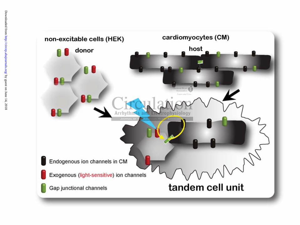
non-viral cell delivery system for expression of light-sensitive ion channels. Fig. 1 illustrates the
concept of a “tandem cell unit” (TCU), formed by a host cardiomyocyte and a non-excitable
donor cell, carrying exogenous ion channels, e.g. ChR2. Biophysically, for this unit to be
functional (to fire an action potential upon light excitation), low-resistance coupling is needed for
closing the local electric circuits. Our group has previously validated this concept for generation
of a two-cell pacemaking unit using cardiomyocytes and stem cells expressing a pacemaking ion
channel16.
The TCU strategy, if proven successful, has potential safety advantages over viral
delivery methods used in all prior optogenetics studies, and may be applicable for study and
treatment of cardiac rhythm disorders. In this work, we demonstrate the utility of optogenetics in
the development of a more robust and energy-efficient solution for cardiac stimulation/actuation
by: 1) employing a non-viral, cell delivery strategy to create optically-excitable cardiac muscle,
extendable to in vivo applications; 2) applying biophysical methods to validate the TCU strategy
in cell pairs and in cardiac syncytium in vitro; 3) demonstrating an all-optical sensing and
actuation in a cardiac syncytium by combining optical stimulation with high-resolution optical
mapping for quantification of wave properties under optical vs. electrical stimulation.
resistance coupupupupupupupl
ateddddddd t t t t t t hihihihihihihis s ss s s cococococococoncncncncncncncepe
aki unit usin cardio oc es and stem cells ex essi a
ategy, if proven successful, has potential safety advantages o
aking unit using cardiomyocytes and stem cells expressing a
ategy, if proven successful, has potential safety advantages o
by guest on June 14, 2018http://circep.ahajournals.org/
Dow
nloaded from
5
Methods
Detailed description of the methods is available as Online Supplement.
As a proof-of-principle cell delivery system, we developed a stable HEK cell line
expressing ChR2 and capable of Cx43-mediated coupling16 to cardiomyocytes to generate
optically-excitable cardiac tissue. The ChR2 plasmid, developed by the Deisseroth’s lab, was
obtained from Addgene, Cambridge, MA (pcDNA3.1/hChR2(H134R)-EYFP), grown in
replication-deficient bacteria, purified and sequenced to confirm the published map. Transfection
of the HEK293 cells (ATCC, Manassas, VA) was done using Lipofectamine™ 2000 (Invitrogen,
Carlsbad, CA), followed by 500 g/ml Geneticin (GIBCO Invitrogen) selection to achieve >98%
expression.
We used whole-cell patch clamp to confirm ChR2-mediated inward current inducible by
pulses of blue light (470/40nm) in the developed cell line. The TCU concept was validated using
a dual-patch technique16 in cell pairs of adult canine cardiomyocytes (CM), isolated as described
before16 and HEK-ChR2 cells, as well as in co-culture of neonatal rat CM17-19 with ChR2-
expressing HEK cells at initial plating ratio 100:1 or 45:1. Without explicit suppression of
proliferation, within the 2-3 days to experiments, the effective ratio was several folds lower.
These in vitro syncytia were used for tissue-level optical mapping of excitation and contraction.
In some experiments, carbenoxolone (CBX), a gap-junction uncoupler, was applied as described
to probe the role of cellular coupling in the TCU approach.
Electrical stimulation was provided through Platinum electrodes, driven by a computer-
controlled stimulator. For most experiments, optical stimulation was through the dish bottom, by
focused light from a blue LED (470nm, 1.35 cd, 20mA) or a fiber-optics coupled high-power
blue LED (470nm, 1.6A), connected to the TTL output of a second computer-driven stimulator.
ogogogogogogogenenenenenenen) ) ) ) ) ) ) seseseseseseselelelelelelelectctctctctctctioioioioioioionn nnnnn tttttt
o e
( s
16 i ll i f d l i di (C ) i l
ole-cell patch clamp to confirm ChR2-mediated inward curre
(470/40nm) in the developed cell line. The TCU concept was
16 i ll i f d l i di (C ) i l by guest on June 14, 2018http://circep.ahajournals.org/
Dow
nloaded from
6
Optical tracking of excitation waves was done using Rhod-4, a calcium-sensitive probe,
with excitation at 525/40nm and emission at 610/75nm to avoid interference of the stimulation
light with the dye’s excitation spectrum. A non-conventional distributed tangential illumination
was used here to accommodate optical stimulation and for superior contrast. Emitted light was
collected through high-NA optics and a fast intensified CMOS camera. Records of contractility
were done with Hamamatsu ImagEM EMCCD camera on a confocal Olympus
FluoViewTM FV1000.
Data from multiple samples are presented, as indicated, using standard deviation, standard
error of the mean or 95% confidence intervals. Statistical testing for functional data on
propagation was done in Matlab using a standard two-way ANOVA, followed by a Tukey-
Kramer correction for multiple comparisons; p-values are listed for all comparisons. Normality
of the distributions was confirmed by Kolmogorov-Smirnov test in Matlab, except for data on
coupling in cell pairs, where nonparamateric population statistics was applied (median and
interquartile range, IQR). Curve fitting was done also in Matlab using the robust nonlinear least-
squares fit method, with pre-specified equations for the desired curve types, e.g. a sigmoidal
curve or exponential decay.
Results and Discussion
Development and characterization of a cell delivery system for non-viral optogenetics
To validate the TCU strategy for cardiac optogenetics, as a proof of principle, we
developed a stable HEK cell line expressing a variant of ChR2. Fig. 2a-e illustrates the
properties of such donor cell line. Preserved expression and functionality were established for the
HEK-ChR2 cell line after multiple freeze-thaw cycles and multiple passages (passages 2 to 30
fofofofofofofor r r r r r fufufufufufufuncncncncncncnctititititititionononononononalalalalalalal d d d d d d d
VA,,,,,, fofofofofofofollllllllllllllowowowowowowowededededededed bbbbby
or multiple comparisons; p-values are listed for all compariso
was confirmed b Kolm orov-Smirnov test in Matlab, exce
s m
IQR) Curve fitting was done also in Matlab using the robust
or multiple comparisons; p-values are listed for all compariso
was confirmed by Kolmogorov-Smirnov test in Matlab, exce
s, where nonparamateric population statistics was applied (m
IQR) Curve fitting was done also in Matlab using the robust by guest on June 14, 2018http://circep.ahajournals.org/
Dow
nloaded from
7
were used for functional experiments), Fig. 2a. Successful expression is possible in other non-
excitable cell types, including mesenchymal stem cells, Supplementary Fig. 1, that may yield
more clinically relevant cell delivery systems.
Confirmation of ChR2 functionality was done by whole-cell voltage clamp.
Quantification of the steady-state light-sensitive ion current in single HEK-ChR2 cells (Fig. 2b-
c) revealed that the channel is closed and non-contributing during dark periods regardless of
transmembrane voltage, and has a mildly inwardly rectifying current-voltage (I-V) relationship
when blue light is applied. Overall, significantly higher steady-state current densities were seen
in our donor cells at all voltages in the I-V relationship, even at the low irradiance used here
(0.24 mW/mm2), compared to previously reported light-induced ChR2 current in HEK cells20, C.
elegans muscle cells10 or in cardiomyocytes13. For example, for comparable irradiance levels, at
holding potential of -40mV, about 20 times higher steady-state current densities were measured
in our donor cells compared to ventricular myocytes from a transgenic mouse expressing
ChR213. These data confirm high expression levels and/or functionality of ChR2 in the
developed cell delivery system, important for optimizing light stimulation parameters.
Recent comprehensive characterization of ChR2 current kinetics indicates fast activation
(<5ms), deactivation (<10ms) and inactivation (<50ms)20, thus making it suitable as excitatory
(action potential – generating) current for cardiomyocytes during external optical pacing at
relevant frequencies (5-12 Hz for rodents, 1-3 Hz for humans). Indeed, our kinetics
characterization (Fig. 2d-e) estimates the activation and deactivation time constants for ChR2-
mediated current in the ms range. Therefore suitable rates for cardiac pacing are attainable even
without genetic modifications, as previously done for neural applications, where faster
hehehehehhh l l llowowowoowoo i i i iiiirrrrrrrrrrr adadadaddddiaiaiaiaiaiaiancncncncncncnc
ChR2R2R2R2R2R2R2 cccccccurururururururrererererererentntntntntntnt iiinp p y p g
d
ompared to ventricular myocytes from a transgenic mouse x
p p y p g
10 or in cardiomyocytes13. For example, for comparable irrad
-40mV, about 20 times higher steady-state current densities
ompared to ventricular myocytes from a transgenic mouse ex
by guest on June 14, 2018http://circep.ahajournals.org/
Dow
nloaded from
8
optogenetic tools in conjunction with much shorter action potentials allowed for pacing rates up
to 200Hz9.
In contrast to the robust expression of ChR2 in HEK cells, much lower yield was seen
when directly transfecting cardiomyocytes with ChR2 using nucleofector electroporation. This
prevented direct synthesis of a large-scale ventricular syncytium from ChR2-expressing
myocytes. Nevertheless, individual ChR2-expressing neonatal rat ventricular myocytes were
excitable and contracting when optically stimulated and quiescent otherwise (Supplementary
Movie 1).
Validation of the TCU strategy for cardiac excitation
The TCU approach for cardiac optogenetics, i.e. inscribing light-sensitivity into
cardiomyocytes and cardiac tissue without their direct genetic modifications, was validated in
cell pairs of CM and HEK-ChR2 cells, as well as in a synthesized large-scale cardiac syncytium.
ChR2-expressing donor cells were most often found to aggregate in small clusters among the
neonatal CMs rather than disperse as single cells, Fig. 3a. The donor cells expressed a significant
amount of Cx43, as confirmed by a Western blot (Fig. 3b). Interestingly, substantially more
Cx43 protein was seen in the HEK-ChR2 cells compared to the parental cell line without ChR2 –
an observation that warrants further investigation.
Functional response of TCUs to optical stimulation was first confirmed in cell pairs of
adult canine CM and HEK-ChR2 cells using dual-clamp to estimate coupling (Fig 3c-d) and to
record light-triggered action potentials in the cardiomyocytes, Fig. 3e-f. A histogram of
measured coupling in spontaneously formed cell pairs (n=31) of canine CM and HEK-ChR2
C
p i
a
CU strategy for cardiac excitation
proach for cardiac optogenetics, i.e. inscribing light-sensitivi
cardiac tissue without their direct genetic modifications, wa
by guest on June 14, 2018http://circep.ahajournals.org/
Dow
nloaded from
9
over 48 hour period is shown, Fig 3c. A robust response was seen in a wide range of coupling
values spanning an order of magnitude, starting as low as 1.5 nS. Interestingly, a similar low
critical coupling value (1.5 – 2 nS), below which TCU functionality failed, was found previously
in the generation of a two-cell pacemaking unit by a donor cell carrying HCN2 (a gene encoding
for the pacemaking current If) and a cardiomyocyte16. Extreme uncoupling abolished the light-
sensitivity of the myocytes in the TCUs – values below 1.5nS and pharmacological uncoupling
with cabenoxolone provided further proof for gap junctions’ critical role in the TCU
functionality for neonatal rat and for adult canine myocytes, Fig 3f and Supplementary Movie
2.
In a functional TCU pair, the cardiomyocytes generated normal action potentials upon
stimulation by blue light (470nm, 0.13 mW/mm2, 10ms pulses), Fig 3e, indistinguishable from
electrically-triggered ones. The donor cell’s membrane potential followed passively by a low-
pass filtered version of an action potential, Fig. 3e. In a spatially-extended (several centimetres)
two-dimensional cardiac syncytium of randomly mixed neonatal rat CMs and HEK-ChR2 (45:1
initial plating ratio), robust synchronous contractions were registered upon stimulation by blue
light 2-3 days after plating (Supplementary Movie 3).
Wave properties of cardiac syncytium in response to optical vs. electrical stimulation
Synthesized optically-excitable cardiac tissue was then subjected to further functional
testing. Synchronized wave propagation is essential for the heart’s normal functionality and
efficient mechanical contraction; lethal arrhythmias occur when the generation or propagation of
these excitation waves is altered (failure to initiate, abnormal propagation velocity and/or path).
Accordingly, we have developed an ultra-high resolution optical mapping system18, 19 to dissect
cardiac wave propagation during external pacing or arrhythmic activity over a centimeter-scale
normamamamamamamalllllll acacacacacacactititititititiononononononon pppppppop y y g p
l g
d v
r
p y y g p
light (470nm, 0.13 mW/mm2, 10ms pulses), Fig 3e, indisting
d ones. The donor cell’s membrane potential followed passiv
of an action potential, f Fig. 3e. In a spatially-extended (sever
by guest on June 14, 2018http://circep.ahajournals.org/
Dow
nloaded from
10
(>2cm) with subcellular resolution (22 m/pix) at 200 fps using fast voltage and calcium-
sensitive dyes18. This optical mapping system was made compatible with simultaneous optical
excitation, Fig. 4a, so that excitation light for the fluorescence measurements did not induce
ChR2 excitation and ChR2 excitation did not interfere with the measurements. While mapping
was done here with Rhod-4, a calcium-sensitive fluorescent dye, suitable voltage-sensitive
probes with similar spectral properties can also be used, e.g. di-4 or di-8-ANEPPS18. In normal
pacing conditions, cardiac calcium transients are an excellent surrogate for action potentials, and
calcium dyes outperform voltage-sensitive dyes in signal-to-noise ratio.
Optical mapping of propagating waves triggered by localized electrical and optical
stimulation in the same sample, revealed similar conduction velocities and calcium transient
morphologies, thus confirming equivalent triggering abilities for both modes of stimulation, Fig.
4b-e and Supplementary Movie 4. Pure cardiomyocyte cultures and co-cultures of
cardiomyocytes and HEK cells without ChR2 served as controls. At the mixing ratios used here,
the presence of HEK cells, with or without ChR2, did not alter the recorded calcium transients
(Fig. 4c-d), p=0.36 with ChR2, p=0.44 without ChR2. However, the mixing ratio was a
significant factor (p<0.0001) in modulating CV, as revealed by a two-way ANOVA, i.e about
30% drop in CV was seen at initial plating ratios of 45:1 (CM:HEK) while a ratio of 100:1 lead
to a smaller (non-significant, p=0.06) reduction, Fig. 4e. The presence of ChR2 did not
contribute as a significant factor in CV modulation (p=0.16), even though a slight trend to a
decrease was seen. Further titration (higher mixing ratios) and/or localized spatial distribution
are likely to minimize these effects. Electrical and optical pacing in light-sensitive samples
(CM:HEK+ChR2) resulted in identical wave propagation properties. The controls (CM only and
izizizizizzizededededededed e e ee eeeleleleleleleectctctctctctctririririririricacacacacacacal ll l l l l aaaananana
citieeeeeesssssss anananananannddddddd cacacacacacacalclclclclclclciiup ,
c s
n s
r
p ,
confirming equivalent triggering abilities for both modes of s
ntary Movie 4. Pure cardiomyocyte cultures and co-cultures
HEK cells without ChR2 served as controls. At the mixing rd
by guest on June 14, 2018http://circep.ahajournals.org/
Dow
nloaded from
11
CM+HEK without ChR2) were quiescent and never produced excitation in response to light
triggers.
The response of the syncytium to optical excitation was captured by constructing a
strength-duration curve, describing minimum irradiance over a range of pulse duration values for
a point excitation of the 2D syncytium (2mm fiber-optic coupled controllable LED), Fig 4f. The
fitted curve revealed a particularly low minimal irradiance levels (average rheobase21 for
excitation of about 0.05mW/mm2) – at least an order of magnitude lower than previously shown
values for optical stimulation of ventricular or atrial tissue13. Within the tested diameter for light
delivery, macroscopic excitability remained uniform across the tissue. However, we anticipate
that donor cell clustering (Fig. 3a) needs to be addressed in order to achieve maximal spatial
resolution of excitation, down to the single cell level.
Considering the electromechanical nature of cardiomyocytes, we also show direct light-
triggered muscle contraction, confirming intact excitation-contraction coupling in single
myocytes or hybrid cardiac tissue (Fig. 4g and Supplementary Movies 1-3). This demonstration
of mechanical response triggered by light-sensitive ion channels suggests possible development
of light-driven actuators with efficient energy transfer and illustrates the feasibility for direct
optogenetic control in other muscles.
Energy needs in cardiac optogenetics
Previous studies in neuroscience have reported optical energies used to excite single
neurons or brain tissue4, 6, 22 in a wide range of high values (approximately 8 to 75 mW/mm2).
The well-coupled spatially-extended cardiac tissue was expected to present higher load for
iisssssssssss ueueueeueee. . . . HoHoHoHoHoHoHoweweweweweeevevevevevevever,r,r,r,r,r,r,
r to aaaaaaachchchchchchchieieieieieieieveveveveveveve mmmmmmmaxaaaag ( g )
i
o
ntraction, confirming intact excitation contraction coupling i
g ( g )
ion, down to the single cell level.
the electromechanical nature of cardiomyocytes, we also sho
ntraction, confirming intact excitation-contraction coupling i by guest on June 14, 2018http://circep.ahajournals.org/
Dow
nloaded from
12
optical stimulation, thus possibly requiring even higher irradiance values. Yet, surprisingly, in
Bruegmann et al.’s study13, significantly lower light levels (0.5 to 7 mW/mm2) were sufficient to
optically stimulate cardiac tissue in vitro or in vivo for a wide range of pulse durations.
In our TCU approach, during optical pacing in the cell pairs or in the two-dimensional
cardiac syncytium, we measured irradiance at 470nm as low as 0.006 mW/mm2. The strength-
duration curve (Fig 4f) further corroborated low irradiance needed across pulse durations.
Interestingly, this is much lower than reported for neuroscience applications and about 1-2 orders
of magnitude lower than previously reported values for cardiac excitation in cells and tissue13 for
comparable pulse durations. The significantly lower optical energy needed in our study can be
explained, at least partially, by the superior light sensitivity of the donor cells as seen in the
higher ChR2 current densities, Fig 2c. Further differences when compared to optical excitation
of ventricular and atrial tissue may stem from dimensionality (2D here vs. 3D13).
It is important to note, that in contrast to a previous study13, no (pro-arrhythmic) re-
excitations were observed during longer stimulation pulses (up to 1s) at our low illumination
intensities, close to the rheobase of the strength-duration curve. This low light intensity is an
important factor in minimizing heat-related effects, phototoxicity and in considering future
implantable devices.
An interesting question concerns comparison of energy needed for electrical vs. optical
pacing. In our system, energy needed for supra-threshold stimulation of two-dimensional cardiac
syncytium can be estimated as follows: For typical electrical pacing (5V, 0.2A, 0.01s pulses), we
obtain 10mJ. For optical stimulation with an LED (5V, 0.02A, 0.01 – 0.05s pulses), we obtain 1
to 5mJ. Considering the possibility for optimization of light focusing, as well as the active
development of more efficient light-emitting diodes (more lumens per watt), it is likely that
gygygygygygygy n nn neeeeeeeeee dededededededed d dd ininininininin o o o o o o ouuururuuu
e dooooooonononononononorrrrrrr cececececececellllllllllllllsssssss aaaasasaay y p g y
t t
r
nt to note, that in contrast to a previous study , no (pro arrhy
y y p g y
t densities, Fig 2c. Further differences when compared to opt
rial tissue may stem from dimensionality (2D here vs. 3D13)
nt to note, that in contrast to a previous study13, no (pro-arrhy by guest on June 14, 2018http://circep.ahajournals.org/
Dow
nloaded from
13
optical stimulation may be more energy efficient than its electrical counterpart. As pacemaking
(unlike cardioversion) is a topologically simple problem, i.e. a spatially-localized (light-
sensitive) cell ensemble can be used, the TCU strategy may prove particularly valuable in
pursuing potential clinical applications, given the above energy considerations. However, the
challenges of achieving efficient light delivery to a densely packed tissue, such as the
myocardium, should not be underestimated, and ultimately in vivo testing is needed.
Potential benefits of the TCU strategy and in vivo considerations
When comparing the TCU strategy to the direct expression of ChR2 in native myocytes
(mostly by viral methods), several important differences are worth mentioning, see also
Supplementary Fig 2: (1) the donor (D) cells are non-excitable and typically do not have major
repolarizing currents, i.e. the ChR2 inward current is their main current, unlike native CMs; (2)
the D-cells have higher membrane impedance at rest due to smaller/negligible inward rectifier,
IK1, and typically have a more depolarized resting potential; (3) compared to CM-CM coupling,
the D-CM coupling is typically somewhat reduced. We ask the question: What factors in the
TCU approach affect the ease of excitation and how is the TCU response to light different than
the response of a homogeneous myocardial tissue with direct expression of ChR2?
For a spatially-extended tissue, when the cell pair (TCU) is connected to some “load” of
excitable cells (CMs), the system can be abstracted to a Source-Neighbor-Load (S-N-L) triad for
easier analysis. A simplified equivalent circuit of such a triad is presented in Supplementary Fig
2d and analysis is provided in detail in the Online Supplement.
on oooooooff f f f ff ChChChChChChChR2R2R2R2R2R2R2 i i i iiiin n n nn n n nnananannana
h s
g o
s, i.e. the ChR2 inward current is their main current, unlike n
h b i d t t d t ll / li ibl i
hods), several important differences are worth mentioning, s
g 2: (1) the donor (D) cells are non-excitable and typically doa
s, i.e. the ChR2 inward current is their main current, unlike n
h b i d t t d t ll / li ibl i by guest on June 14, 2018http://circep.ahajournals.org/
Dow
nloaded from
14
The main results of this analysis (Supplementary Fig 2d) are that the ease of optical
excitation will improve with: (1) higher membrane impedance for the source/donor cell (as seen
for a D vs. CM source cell), since that makes it closer to an ideal current source, and a higher
excitation current is available for the neighboring CMs for the same light-induced current in the
source; (2) lower membrane impedance of the immediate CM neighbors, determined by their K+
conductance; (3) better S-N coupling but reduced coupling in the load and reduced overall
impedance of the load. It is important to note, that for spatially dispersed donor cells, as here, the
S-N coupling in the S-N-L triad plays a dual role, i.e. it affects both the S-N charge transfer and
the equivalent “load” presented by the tissue. Since the two have opposite effects on the ease to
excite, there may be an optimal D-CM coupling for excitation via the TCU strategy.
Having a dedicated donor cell may also provide additional benefits by allowing donor
cell optimization independent of the properties of the target tissue, i.e. maximizing the light
sensitivity by achieving high current densities in the donor with proper cellular environment for
the function of the ChR2 channels, for example.
The presented cell delivery method can be viewed as an easy and accessible research tool
for basic studies in vitro. Its feasibility for potential in vivo cardiac pacing is supported by prior
relevant studies, including one using MSCs with a depolarizing current, If, in a canine heart23,
where the cells’ survival and functionality was demonstrated for at least several weeks. In that
study, about 0.7mil cells were delivered, 40% of them carrying the HCN2 gene, encoding for an
If-like current, expressed at comparable densities to our ChR2 current24. The cell number needed
for localized excitation can be further inferred from considerations of tissue properties, i.e.
estimates of “liminal length” (the minimum size of tissue capable of exciting the rest)25, 26, which
in some cases can be derived from the strength-duration curve. Recent estimates by computer
opposite effeectctccccc s
a theheeeeee T T TTTTTCUCUCUCUCUCUCU s s s s s sstrtrtrtrtrtrtraaaaatata e
d l
d n
v e
dicated donor cell may also provide additional benefits by all
dependent of the properties of the target tissue, i.e. maximizin
ving high current densities in the donor with proper cellular en
by guest on June 14, 2018http://circep.ahajournals.org/
Dow
nloaded from
15
simulations of a related problem – localized generation of early or delayed afterdepolarizations
(EADs/DADs)27 - yielded about 0.7 mil cells in a cluster required for maximum load (worst case
scenario), i.e. 3D highly coupled ventricular myocardium. With decreased coupling and other
load reductions, this cell number also decreased. Since we did not perform experiments with a
confined cell region in 2D, it is hard to directly relate to these estimates. Nevertheless, these
studies support the general feasibility of in vivo cell delivery for optical pacing by the TCU
approach, provided that an appropriate light delivery solution is found. Optimizing the light
sensitivity of the donor cells partially alleviates these challenges. Compared to pacing,
cardioversion and defibrillation are topologically more complicated problems due to their
inherent spatial component and requirement for spatially distributed light-sensitivity and light
delivery. Stability and long-term functionality of ChR2 in vivo have not been studied rigorously,
but preliminary data from ChR2 use in primate brains over several months are promising in
terms of persistence and lack of toxicity28.
Conclusions
In summary, our study highlights the utility of optogenetics for cardiac applications by
using a strategy inspired by the specific properties of cardiac tissue, i.e. high cell-cell coupling.
The optogenetic approach offers high spatiotemporal resolution for precise interrogation and
control of excitation, seemingly without interfering with essential cardiac tissue properties.
Therefore, it presents a new versatile actuation tool in cardiac research for dissection of
arrhythmias. Furthermore, cardiac optogenetics based on the TCU strategy, presented here, may
evolve in a more translational direction and lead to a new generation of optical pacemakers and
ted problems ddddddduuuu
uted d lilililililil ghghghghghghghtttttt-sssssssenenenenenenensisisisisisisititi
n u
r
nd long-term functionality of ChR2 in vivo have not been stu
from ChR2 use in primate brains over several months are pr
and lack of toxicity28.
by guest on June 14, 2018http://circep.ahajournals.org/
Dow
nloaded from
16
potentially cardioverter/defibrillators. The feasibility of this is supported by several critical
features of the method presented here: 1) desirable pacing rates achievable with the current
kinetics of ChR2; 2) finer control of excitation and repolarization in shaping cardiac action
potentials, and potentially in terminating arrhythmias is possible by a combination of light-
sensitive ion channels providing outward current7, 8 and ChR2; 3) the cell delivery platform
demonstrated here may offer a safer alternative to viral delivery for in vivo applications; 4)
optical fibers are inherently more biocompatible than metal electrode leads for in vivo pacing; 5)
preliminary energy estimates point to potential fold improvements in energy consumption with
optical vs. electrical pacing – important for extending battery life in implantable devices.
Acknowledgements The authors thank Jacqueline Guenther for cardiac cell culture preparation, Joan Zuckerman for canine cell isolation, Yuanjian Guo for canine mesenchymal stem cell isolation, Laima Valiuniene for heterologous cell culture with canine myocytes, Chris Gordon for Western blots and Qinghong Yan, for help with expansion and purification of the ChR2 plasmid.
Funding Sources: We acknowledge support by NIH-NIGMS funded Systems Biology Center in New York State (GM071558, EE), NLHBI grant HL094410 (ISC), NIGMS grant RO1GM088181 (VV) and a grant from the Institute for Molecular Cardiology at Stony Brook University (EE).
Conflict of Interest Disclosures: None
References
1. Oesterhelt D, Stoeckenius W. Rhodopsin-like protein from the purple membrane of halobacterium halobium. Nat New Biol. 1971;233:149-152.
2. Vsevolodov NN. Biomolecular electronics. An introduction via photosensitive proteins.Boston: Birkhauser; 1998.
3. Nagel G, Szellas T, Huhn W, Kateriya S, Adeishvili N, Berthold P, Ollig D, Hegemann P, Bamberg E. Channelrhodopsin-2, a directly light-gated cation-selective membrane channel. Proc Natl Acad Sci U S A. 2003;100:13940-13945.
fe in implantablblblblblblblee
s la
l
s The authors thank Jacqueline Guenther for cardiac cell culcanine cell isolation, Yuanjian Guo for canine mesenchyma
liuniene for heterologous cell culture with canine myocytes, d Qinghong Yan, for help with expansion and purification oh
by guest on June 14, 2018http://circep.ahajournals.org/
Dow
nloaded from

17
4. Boyden ES, Zhang F, Bamberg E, Nagel G, Deisseroth K. Millisecond-timescale, genetically targeted optical control of neural activity. Nat Neurosci. 2005;8:1263-1268.
5. Li X, Gutierrez DV, Hanson MG, Han J, Mark MD, Chiel H, Hegemann P, Landmesser LT, Herlitze S. Fast noninvasive activation and inhibition of neural and network activity by vertebrate rhodopsin and green algae channelrhodopsin. Proc Natl Acad Sci U S A.2005;102:17816-17821.
6. Wang H, Peca J, Matsuzaki M, Matsuzaki K, Noguchi J, Qiu L, Wang D, Zhang F, Boyden E, Deisseroth K, Kasai H, Hall WC, Feng G, Augustine GJ. High-speed mapping of synaptic connectivity using photostimulation in channelrhodopsin-2 transgenic mice. Proc Natl Acad Sci U S A. 2007;104:8143-8148.
7. Chow BY, Han X, Dobry AS, Qian X, Chuong AS, Li M, Henninger MA, Belfort GM, Lin Y, Monahan PE, Boyden ES. High-performance genetically targetable optical neural silencing by light-driven proton pumps. Nature. 2010;463:98-102.
8. Han X, Boyden ES. Multiple-color optical activation, silencing, and desynchronization of neural activity, with single-spike temporal resolution. PLoS One. 2007;2:e299.
9. Gunaydin LA, Yizhar O, Berndt A, Sohal VS, Deisseroth K, Hegemann P. Ultrafast optogenetic control. Nat Neurosci. 2010;13:387-392.
10. Nagel G, Brauner M, Liewald JF, Adeishvili N, Bamberg E, Gottschalk A. Light activation of channelrhodopsin-2 in excitable cells of caenorhabditis elegans triggers rapid behavioral responses. Curr Biol. 2005;15:2279-2284.
11. Airan RD, Thompson KR, Fenno LE, Bernstein H, Deisseroth K. Temporally precise in vivo control of intracellular signalling. Nature. 2009;458:1025-1029.
12. Huber D, Petreanu L, Ghitani N, Ranade S, Hromadka T, Mainen Z, Svoboda K. Sparse optical microstimulation in barrel cortex drives learned behaviour in freely moving mice. Nature. 2008;451:61-64.
13. Bruegmann T, Malan D, Hesse M, Beiert T, Fuegemann CJ, Fleischmann BK, Sasse P. Optogenetic control of heart muscle in vitro and in vivo. Nat Methods. 2010;7:897-900.
14. Arrenberg AB, Stainier DY, Baier H, Huisken J. Optogenetic control of cardiac function. Science. 2010;330:971-974.
15. Jia Z, Lu Z, Bien H, Liu H, Rosati B, Cohen IS, Entcheva E. Optically activated light-sensitive channels can pace cardac tissue and generate propagating cardiac impulses. Circulation Research. 2010;107:Late Breaking Basic Science Abstracts, AHA.
16. Valiunas V, Kanaporis G, Valiuniene L, Gordon C, Wang HZ, Li L, Robinson RB, Rosen MR, Cohen IS, Brink PR. Coupling an hcn2-expressing cell to a myocyte creates a two-cell pacing unit. J Physiol. 2009;587:5211-5226.
17. Chung CY, Bien H, Entcheva E. The role of cardiac tissue alignment in modulating electrical function. J Cardiovasc Electrophysiol. 2007;18:1323-1329.
18. Entcheva E, Bien H. Macroscopic optical mapping of excitation in cardiac cell networks with ultra-high spatiotemporal resolution. Prog.Biophys.Mol.Biol. 2006;92:232-257.
;;
hhhhh KKKKKKK, , HeHeHeHeHeHeHegegegegegegegemamamamamamamannnnnnnnnnnnnn P P PPPPP
a Ag
o
hompson KR, Fenno LE, Bernstein H, Deisseroth K. Tempo
auner M, Liewald JF, Adeishvili N, Bamberg E, Gottschalk Achannelrhodopsin-2 in excitable cells of caenorhabditis eleg
oral responses. Curr Biol. 2005;15:2279-2284.
hompson KR, Fenno LE, Bernstein H, Deisseroth K. Tempoof intracellular signalling. Nature. 2009;458:1025-1029.
by guest on June 14, 2018http://circep.ahajournals.org/
Dow
nloaded from
18
19. Bien H, Yin L, Entcheva E. Calcium instabilities in mammalian cardiomyocyte networks. Biophys.J. 2006;90:2628-2640.
20. Chater TE, Henley JM, Brown JT, Randall AD. Voltage- and temperature-dependent gating of heterologously expressed channelrhodopsin-2. J Neurosci Methods.2010;193:7-13.
21. Malmivuo J, Plonsey R. Bioelectromagnetism. New York: Oxford University Press, Ch. 3 (http://www.bem.fi/book/index.htm); 1995.
22. Cardin JA, Carlen M, Meletis K, Knoblich U, Zhang F, Deisseroth K, Tsai LH, Moore CI. Targeted optogenetic stimulation and recording of neurons in vivo using cell-type-specific expression of channelrhodopsin-2. Nat Protoc. 2010;5:247-254.
23. Plotnikov AN, Shlapakova I, Szabolcs MJ, Danilo P, Jr., Lorell BH, Potapova IA, Lu Z, Rosen AB, Mathias RT, Brink PR, Robinson RB, Cohen IS, Rosen MR. Xenografted adult human mesenchymal stem cells provide a platform for sustained biological pacemaker function in canine heart. Circulation. 2007;116:706-713.
24. Potapova I, Plotnikov A, Lu Z, Danilo P, Jr., Valiunas V, Qu J, Doronin S, Zuckerman J, Shlapakova IN, Gao J, Pan Z, Herron AJ, Robinson RB, Brink PR, Rosen MR, Cohen IS. Human mesenchymal stem cells as a gene delivery system to create cardiac pacemakers. Circ Res. 2004;94:952-959.
25. Rushton WAH. Intitiation of the propagated disturbance. Proc R Soc Lond B.1937;124:210-243.
26. Fozzard HA, Schoenberg M. Strength-duration curves in cardiac purkinje fibres: Effects of liminal length and charge distribution. J Physiol. 1972;226:593-618.
27. Xie Y, Sato D, Garfinkel A, Qu Z, Weiss JN. So little source, so much sink: Requirements for afterdepolarizations to propagate in tissue. Biophys J. 2010;99:1408-1415.
28. Han X, Qian X, Bernstein JG, Zhou HH, Franzesi GT, Stern P, Bronson RT, Graybiel AM, Desimone R, Boyden ES. Millisecond-timescale optical control of neural dynamics in the nonhuman primate brain. Neuron. 2009;62:191-198.
Figure Legends:
Figure 1. The functional “tandem cell unit” (TCU) concept of donor-host cells. Non-excitable
cells (e.g. HEK cells here) are transfected to express a light-sensitive ion channel (ChR2). When
coupled via gap junctions to excitable cardiomyocytes (CM) they form an optically controllable
QQQQQQu J,, Doronnininininininin BrBrBBBBB ininininininink k k k k k k PRPRPRPRPRPRPR,, , , , , , RoRoRoRoRoRoRosesesesesesesennnnnnnm too cc c ccccrerererererereatatatatatatate e e e e e e cacacacacacacardrdrdrdrdrr i
04;94:952-959.
A n0
en
04;94:952-959.
AH. Intitiation of the propagated disturbance. Proc R Soc Lon0-243.
Schoenberg M. Strength-duration curves in cardiac purkinjength and charge distribution. J Physiol. 1972;226:593-618.
by guest on June 14, 2018http://circep.ahajournals.org/
Dow
nloaded from
19
functional TCU, i.e. the CM will generate an action potential upon light-triggered opening of the
depolarizing ChR2 in the HEK cell.
Figure 2. Development and functional characterization of a cell delivery system for ChR2. a:
Stable HEK-ChR2 cell line – shown is EYFP-reported ChR2 expression in the 10th passage after
transfection and purification; scale bar is 50 m. b: Voltage-clamp test protocol and example
traces for quantification of the steady-state ChR2 current in single HEK-ChR2 cells with 500ms
voltage pulses in the range (-80 to +50mV) with and without excitation light for ChR2 on
(470nm, 0.24 mW/mm2). c: Example curves for the light-sensitive component after subtraction
of current in dark, and the resultant average current-voltage (I-V) relationship for n=12 cells, cell
capacitance 43.3 7.5pF, data are presented as mean SD. d: Magnitude of the light-triggered
current does not depend on the duration of rest ( t rest) or activation ( t act), thus indicating
relatively fast deactivation in the examined range. Holding potential is -80mV. e: Kinetics of
activation (on) and deactivation (off), quantified by a sl parameter in the sigmoid curve fits to
the light-controlled current transitions (see inset); bar graphs represent mean SEM.
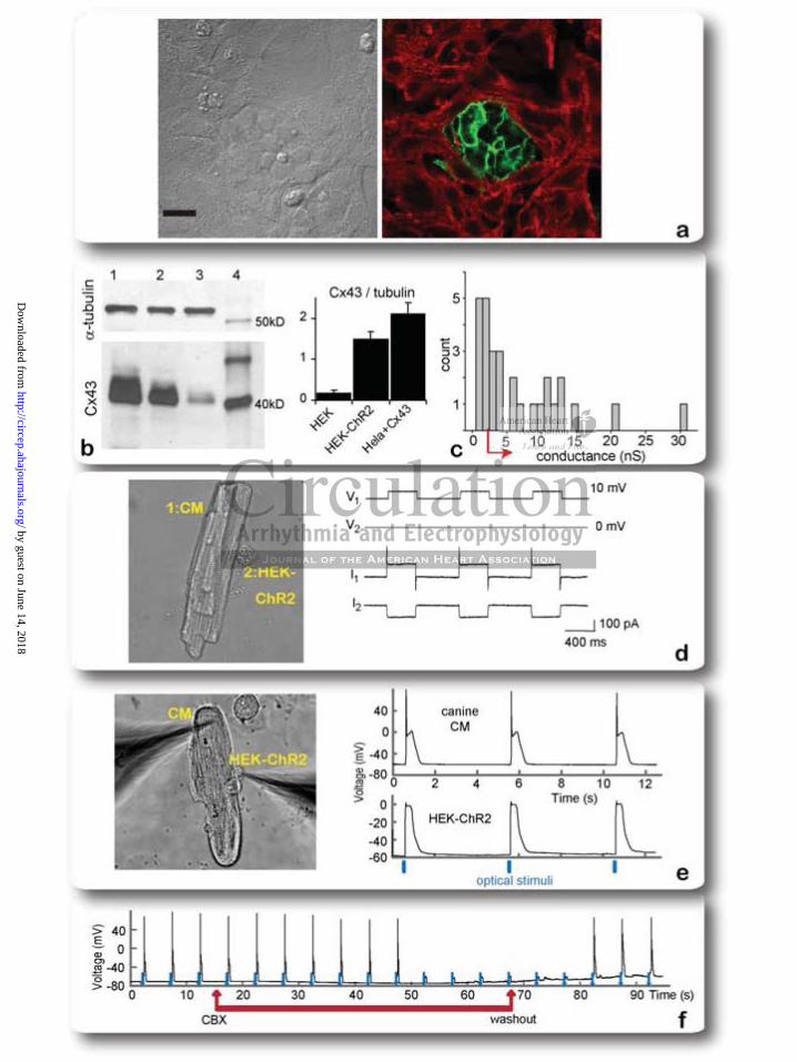
Figure 3. Implementation and validation of the TCU concept for neonatal rat CM and adult
canine CM coupled to HEK+ChR2 cells. a: Phase and fluorescence images of neonatal rat CM
and HEK-ChR2 co-culture. Immunostaining in red for -actinin (CMs), green is EYFP-ChR2-
expressing HEK cells, typically forming small clusters as shown. Scale bar is 20 m. b: Western
blot for Cx43 and -tubulin (at 55kD) in the cell delivery system (HEK-ChR2), column 2;
column 1 shows a positive control of stably transfected HeLa-Cx43 cells; column 3 shows
parental HEK cells without ChR2; column 4 shows the ladder – MagicMarkTM bands in kDa;
Normalized (Cx43/tubulin) expression is provided for four gels (mean 95%CI). c: Histogram of
))))))) rrrrrelelelelelelelatatatatatatatioioioioioioionsnsnsnsnsnsnshihhihihihihippp p p pp fofofofofofofo
5pF, data are presented as mean : Magnitude of the lig
u
v e
5pF, data are presented as mean SD. d: Magnitude of the lig
end on the duration of rest ( t rest) or activation ( t act), thu
vation in the examined range. Holding potential is -80mV. e
by guest on June 14, 2018http://circep.ahajournals.org/
Dow
nloaded from
20
measured coupling conductances in TCUs of canine CMs and HEK-ChR2 cells, n=31, median
value of 4nS and IQR (2 – 11nS); red arrow indicates coupling levels allowing optical
excitability of the TCUs. d: Dual whole-cell voltage clamp of a TCU - adult canine ventricular
CM (1) and HEK-ChR2 cell (2). Voltage steps (V1=10mV, 0.4s), applied to the canine CM (cell
1), induced junctional currents (I2) in this cell pair (estimated g.j. conductance of 11nS). e:
Action potentials in a cell pair (canine CM and HEK-ChR2 cell, phase image on the left) in
response to optical pacing (0.13 mW/mm2, 10ms pulses). Due to coupling, the HEK cell exhibits
a low-pass filtered version of the CM-generated action potentials. f: Action potentials in a cell
pair (canine CM and HEK-ChR2 cell) in response to continuous optical pacing before, during
and after washout of uncoupler carbenoxolone (CBX).
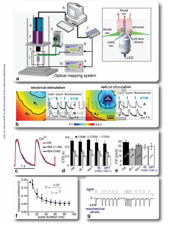
Figure 4. Optical control of cardiac tissue function over space-time: light-triggered excitation
waves and light-triggered contractions. a: Experimental setup for ultra-high resolution high-
speed optical imaging and optical control of cardiac excitation: 1) experimental chamber with
tangential light illumination for calcium imaging, focused LED illumination on a moveable stage
for ChR2 excitation (see inset on the right); 2) high-NA optics for high-resolution macroscopic
imaging; 3) Gen III MCP intensifier; 4) pco 1200hs CMOS camera; 5) light source, excitation
filter and optical light guides for tangential excitation; 6) computer system and software for data
acquisition and control of electrical and optical stimulation; 7) interface for stimulation control;
8) controllable stimulator for electrical pacing (analog output); 9) controllable stimulator for
optical pacing (TTL output). 10) LED for ChR2 excitation, driven by the TTL stimulator output.
b: Activation maps in a cardiac monolayer by electrical and optical pacing at 0.5Hz. Color
represents time of activation; isochrones are shown in black at 0.15s. Calcium transient traces in
optical pacingngggggg b b
o e
g
ontrol of cardiac tissue function over space-time: light-trigge
gered contractions. a: Experimental setup for ultra-high reso
by guest on June 14, 2018http://circep.ahajournals.org/
Dow
nloaded from
21
response to electrical or optical stimulation are shown from 2 locations (A and B), normalized
fluorescence. Blue marks indicate time of stimulation (electrical pulses were 10ms, optical –
20ms each). c: Normalized Ca2+ transients from CM monolayer (red), CM:HEK (black) and
CM:HEK+ChR2 co-culture 100:1 (blue) at 1Hz pacing. d: Quantification of calcium transient
duration (CTD) – CTD25, CTD50 and CTD80 for pure CM monolayer, 45:1 and 100:1
CM:HEK, as well as 100:1 CM:(HEK+ChR2) co-culture under electrical and optical pacing at
1Hz. e: Comparison of conduction velocity (CV) among the same 5 groups as in (d); for d and e
optical pacing was at irradiance of 0.01- 0.04mW/mm2, 50ms pulses; data are shown as
mean±95%CI; listed number of samples applies to both; f: Strength-duration curve (along with
the equation for the fitted curve) obtained for optical pacing in co-cultures (100:1 CM:HEK
ratio) at 30°C, n=8, mean±SEM. g: Example contractility recording from optically-driven
CM+HEK+ChR2 – displacement normalized to cell length. Scale bar is 1s.
gth duration cucuuucuuurr
o-cululullllltutututututuurerererererer s s s s s s s (1(1(1(1(1(1(100000000000000:
m l
d
mean±SEM. g: Example contractility recording from optical
displacement normalized to cell length. Scale bar is 1s.d
by guest on June 14, 2018http://circep.ahajournals.org/
Dow
nloaded from
by guest on June 14, 2018http://circep.ahajournals.org/
Dow
nloaded from

by guest on June 14, 2018http://circep.ahajournals.org/
Dow
nloaded from
by guest on June 14, 2018http://circep.ahajournals.org/
Dow
nloaded from
by guest on June 14, 2018http://circep.ahajournals.org/
Dow
nloaded from
Rosati, Peter R. Brink, Ira S. Cohen and Emilia EntchevaZhiheng Jia, Virginijus Valiunas, Zongju Lu, Harold Bien, Huilin Liu, Hong-Zhang Wang, Barbara
Stimulating Cardiac Muscle by Light: Cardiac Optogenetics by Cell Delivery
Print ISSN: 1941-3149. Online ISSN: 1941-3084 Copyright © 2011 American Heart Association, Inc. All rights reserved.
Dallas, TX 75231is published by the American Heart Association, 7272 Greenville Avenue,Circulation: Arrhythmia and Electrophysiology
published online August 9, 2011;Circ Arrhythm Electrophysiol.
http://circep.ahajournals.org/content/early/2011/08/09/CIRCEP.111.964247World Wide Web at:
The online version of this article, along with updated information and services, is located on the
http://circep.ahajournals.org/content/suppl/2011/08/09/CIRCEP.111.964247.DC1Data Supplement (unedited) at:
http://circep.ahajournals.org//subscriptions/
is online at: Circulation: Arrhythmia and Electrophysiology Information about subscribing to Subscriptions:
http://www.lww.com/reprints Information about reprints can be found online at: Reprints:
document. Permissions and Rights Question and Answerinformation about this process is available in the
requested is located, click Request Permissions in the middle column of the Web page under Services. FurtherCenter, not the Editorial Office. Once the online version of the published article for which permission is being
can be obtained via RightsLink, a service of the Copyright ClearanceCirculation: Arrhythmia and Electrophysiology Requests for permissions to reproduce figures, tables, or portions of articles originally published inPermissions:
by guest on June 14, 2018http://circep.ahajournals.org/
Dow
nloaded from

1
SUPPLEMENTAL MATERIAL
I. Detailed Methods
II. Supplementary Figures (2)
III. Supplementary Movies (4)
IV. Equivalent Circuit Analysis of TCUs
I. DETAILED METHODS
Development of a ChR2-expressing stable cell line
A bacterial stock containing the pcDNA3.1/hChR2(H134R)-EYFP plasmid was obtained from
Addgene and amplified in selective Luria-Bertani (LB) medium. Plasmid DNA was extracted
using Qiagen HiSpeed Plasmid kit (Qiagen, Valencia, CA), ethanol-precipitated and
resuspended in endotoxin-free water for use in cell trasnfections. After verification of identity by
restriction digestion and sequencing, it was stored at -20C at the obtained concentration
(typically 2-4 g/ml), later diluted to 1 g/ml for transfection.
HEK293 cells (ATCC, Manassas, VA) were transfected with the plasmid using Lipofectamine™
2000 (Invitrogen) as directed: 4 g of DNA and 10g of Lipofectamine in 250 l medium for a
35mm dish with cells. Gene expression was examined by EYFP signal the next day. 48 hours
after transfection, cells were switched to selection medium, containing 500 g/ml Geneticin
(GIBCO Invitrogen). The selected cells with a high fluorescence signal were maintained in
Geneticin (500 g/ml) containing culture medium at 37 in a humidified atmosphere incubator
with 5% CO2 and 95% air. Expanded HEK cell cultures showing near 100% expression were
frozen at -80C for later use. Immediately prior to use, the HEK-ChR2 cells were grown in DMEM

2
(Dulbecco’s Modified Eagle’s Medium, GIBCO Invitrogen) supplemented with 10% FBS (fetal
bovine serum, Sigma-Aldrich, St Louis, MO) and 1% penicillin-streptomycin (Sigma) at 37, 5%
CO2. Expression and functional properties were confirmed in passages 2 to 20 and used in co-
culture experiments with cardiomyocytes.
Confirmation and analysis of light-triggered ChR2-current in the cell delivery system
ChR2 cell membrane expression was confirmed in virtually 100% of the transfected HEK cells
(Fig. 2b) using EYFP fluorescence as a marker. For functional measurements of the ChR2-
current, the HEK-ChR2 cells were harvested by trypsinization, replated at low density on
polylysine-coated coverslips and stored in DMEM medium at 37 in a humidified atmosphere
incubator with 5% CO2. The membrane current was recorded in single cell by whole-cell patch
clamp with an Axopatch 1D amplifier (Axon instruments Inc, Foster City, CA). Borosilicate glass
pipettes (World Precision Instruments Inc., Sarasota, FL) were pulled on a Flaming-Brown–type
pipette puller (Sutter Instrument Co, Novato, CA) and heat-polished before use. Pipette
resistances measured in Tyrode's solution were 3-4 MΩ when filled with pipette solution. The
pipette solution contained (mmol/L) potassium aspartate 80, KCl 50, MgCl2 1, MgATP 3, EGTA
10 and HEPES 10 (pH 7.4 with KOH). The external solution contained (mmol/L) KCl 5.4, NaCl
140, MgCl2 1,CaCl2 1.8, HEPES 10 and Glucose 10 (pH 7.4 with NaOH). Membrane currents
were recorded, digitized (DIGIDATA 1320A, Axon Instruments) and stored for offline analysis.
There was a liquid junction potential of ~10 mV between the bath solutions and the electrode
solution. The current was recorded as depolarizing 500ms pulses from -80 mV to +50mV with
and without illumination (Fig. 2). The light-triggered ChR2- current was determined by
subtracting the “off” light trace from the recorded response of light “on”. Illumination pulses were
generated using the microscope-attached fluorescence light unit, filtered at 470nm. The light-
triggered inward ChR2-current was reproducible upon repeated on/off light pulses.
3
The kinetics of light-triggered ChR2-current was examined as the cells were clamped at -80mV
after obtaining whole-cell configuration, and light pulses of variable duration and spacing were
applied sequentially. For the analysis of the current kinetics – activation and deactivation time-
constants - nonlinear sigmoidal curve fit was applied to the rising and the falling portion upon
light on/off pulse (Fig. 2d-e). The slope parameter (sl) was quantified.
Optically-excitable cardiac syncytium: primary cardiomyocyte cell culture and co-culture
with HEK-ChR2 cells
Neonatal Sprague-Dawley rats were sacrificed and cardiomyocytes were isolated by an
approved Stony Brook University IACUC protocol as previously described1-5. Briefly, the
ventricular portion of the hearts was excised and washed free of blood. The tissue was cut into
small pieces and enzymatically digested overnight with trypsin at 4 (1mg/ml, USB, Cleveland,
OH), and then with collagenase at 37 (1mg/ml, Worthington, Lakewood, NJ) the next morning.
Cardiac fibroblasts were removed by a two-stage pre-plating process. In some transfection
experiments, these cardiac fibroblasts were used in conjunction with electroporation.
Cardiomyocytes were then plated onto fibronectin-coated glass coverslips at high density: 4×10⁵
cells/cm2 for the control myocyte group and 3.5×10⁵ cells/cm2 for the co-culture groups, mixed
with approximately 7,700 or 3,500 HEK cells (for 45:1 and 100:1 initial plating ratios) onto glass
bottom dishes in M199 medium (GIBCO Invitrogen) supplemented with 10% fetal bovine serum
(GIBCO Invitrogen) for the first 2 days and then reduced to 2%. Cultures were maintained in an
incubator at 37 with 5% CO2 for 4 to 5 days before functional measurements.
4
Direct expression of ChR2-EYFP in cardiomyocytes, cardiac fibroblasts and
mesenchymal stem cells
Freshly isolated neonatal rat cardiomyocytes and cardiac fibroblasts, as well as mesenchymal
stem cells, were transfected by electroporation using a Nucleofector device (Amaxa Lonza,
Gaithersburg, MD) as follows: 4 g of plasmid DNA was mixed with 100 l of nucleofector
solution for transfecting 4x10⁶ cells. Human mesenchymal stem cells (hMSC) were purchased
from Clonetics/BioWhittaker, Walkersville, MD, USA, and cultured in mesenchymal stem cell
growth medium - Poietics-MSCGM (BioWhittaker). Canine mesenchymal stem cells (cMSC)
were isolated from the bone marrow of adult dogs and cultured in Poietics-MSCGM. Flow
cytometry revealed 93.9% CD44+ and 6.1% cells were CD34+. Cells with spindle-like
morphology were selected after flow cytometry characterization and replated for use.
Transfected cells were incubated in normal culture conditions. Similar conditions were used to
transfect cardiac fibroblasts or stem cells via electroporation. Expression of fluorescence was
detected 24 to 48 hours after transfection using confocal fluorescence imaging.
Images were processed as follows: background fluorescence was first subtracted for images of
control (non-transfected) and transfected cells of the same type. Then the remaining integral
fluorescence over identical areas was used to form a ratio (transfected/average control) in order
to quantify and compare different cell types, see Suppl. Fig 1.
Demonstration of TCU functionality in cell pairs of adult canine ventricular myocytes and
HEK-ChR2
Adult mongrel dogs were euthanized as per IACUC protocol at Stony Brook University by
intravenous injection of sodium pentobarbitone (80mg/kg body weight) and the heart was
removed. Canine ventricular cells were isolated using a modified Langendorff procedure6
5
perfusing a wedge of the left ventricle through a coronary artery with 0.5 mg ml−1 collagenase
(Worthington) and 0.08 mg ml−1 protease (Sigma) for 10 min before tissue digestion. Prior to
plating, isolated cardiomyocytes were stored in Kraft-Brühe (KB) solution (in mM: KCl, 83;
K2HPO4, 30; MgSO4, 5; Na-Pyruvic Acid, 5; -OH-Butyric Acid, 5; Creatine, 5; Taurine, 20;
Glucose, 10; EGTA, 0.5; KOH, 2; and Na2-ATP, 5; pH was adjusted to 7.2 with KOH) at room
temperature. The canine ventricular myocytes were plated onto laminin-coated glass coverslips
(10 g/ml, Invitrogen) and incubated in a 37 to ensure attachment. HEK-ChR2 cells were
added within 24h at low density to stimulate formation of individual cell pairs and the co-culture
was maintained in Medium 199 (Gibco) supplemented with 15% FBS, 2 mm l-glutamine, 100 U
ml−1 penicillin, 100 μg ml−1 streptomycin and 50 μg ml−1 gentamicin.
Dual patch clamp experiments were performed within 48 hours after plating. Briefly,
experiments were carried out on heterologous (HEK-ChR2 - canine myocyte) cell pairs within 48
hours after plating, as described previously7. A dual whole-cell voltage-clamp method was used
to control and record the membrane potential of both cells and to measure associated
membrane and junctional currents7-9. Each cell of a pair was voltage clamped at the same
potential by two separate patch clamp amplifiers (Axopatch 200, Axon Instruments). To record
junctional conductance, brief voltage steps (±10 mV, 400 ms) were applied to one cell of a pair,
whereas the other cell was held at constant voltage and the junctional currents were recorded
from the unstepped cell. Membrane and action potentials were recorded in current-clamp mode.
For electrical recordings, glass coverslips with adherent cells were transferred to an
experimental chamber mounted on the stage of an inverted microscope (Olympus-IX71)
equipped with a fluorescence imaging system. The chamber was perfused at room temperature
(~22 C) with bath solution containing (in mM): NaCl, 140; Mg Cl2, 1; KCl, 5; CaCl2, 2; HEPES,
5 (pH 7.4); glucose, 10. Perfusion with 200M of carbenoxolone (Sigma) was used to block cell-
cell communication. The patch pipettes were filled with solution containing (in mM): K+
6
aspartate-, 120; NaCl, 10; MgATP, 3; HEPES, 5 (pH 7.2); EGTA, 10 (pCa ~8). Patch pipettes
were pulled from glass capillaries (code GC150F-10; Harvard Apparatus) with a horizontal puller
(DMZ-Universal, Zeitz-Instrumente). When filled, the resistance of the pipettes measured 1-4M
.
Immunostaining of co-cultures
For immunocytochemistry, the co-cultures were fixed and permeabilized with 3.7%
formaldehyde and 0.02% Triton-X 100 before being stained with a monoclonal mouse antibody
against sarcomeric -actinin (Sigma). Samples were visualized using goat anti-mouse antibody
conjugated with fluorophore Alexa 546 (Invitrogen) and imaged on the Olympus FluoView
confocal system.
Western blots of Cx43 and -tubulin
Cells from three groups (HEK293, HEK+ChR2 and stably transfected HeLa+Cx43 cells)
were collected, lysed and centrifuged to obtain a pellet. The pellets were re-suspended in cold
RIPA buffer (R0278, Sigma), protease Inhibitor cocktail (P2714, Sigma), sodium orthovanadate
(S-6508, Sigma) and PMSF (P-7626, Sigma); after centrifugation, the supernatants were
transferred to pre chilled microtubes. Protein concentration for each sample was determined by
the Bradford assay. Total protein of 30 micrograms from each lysate was mixed with equal
volume of laemmli sample buffer (161-0737, Bio-Rad, Hercules, CA) containing β-
mercaptoethanol and boiled for 5 minutes at 95˚C. After centrifugation, samples were loaded on
a SDS-polacrylamide gel. MagicMark XP Protein Standard (LC5602, Invitrogen) was loaded
along with the samples. After separation by electrophoresis at 115 V for 90 minutes in tris-
glycine/SDS buffer, proteins were transferred to immobilon-P membrane (Millipore, Billerica,
MA) by electrophoresis at 100 V for 60 minutes in tris-glycine/methanol buffer. Nonspecific
7
antibody binding was blocked for 1 hour by 5% blotting grade blocker non-fat dry milk (Bio-Rad)
dissolved in 1x TBST. The following antibodies were used: primary anti-Cx43 antibody raised in
rabbit (C 6219, Sigma), secondary goat-anti-rabbit antibody (sc-2004, Santa Cruz); a primary
antibody for -tubulin at 55kD (sc-8035, Santa Cruz), and a secondary goat-anti-mouse
antibody for tubulin from Pierce, Rockford, IL. The secondary antibodies were detected using
SuperSignal West Femto Maximum Sensitivity Substrate (34095, Pierce) and images obtained
by exposing the membrane to HyBlot CL autoradiography film. Quantification of the Cx43 bands
relative to the -tubulin bands was done using a buil-in routine in ImageG.
Ultra-high resolution optical mapping of cardiac excitation waves triggered by light in co-
cultures
Two-dimensional optical mapping over a large field of view (about 2.2cm) was done with a
custom-developed macroscopic system allowing for ultra-high spatiotemporal resolution4, 5. The
system (Fig. 4a) includes a CMOS camera (pco, Germany) recording images at 200 frames per
second (fps) over 1,280×1,024 pixels), a Gen III fast-response intensifier (Video Scope
International, Dulles, VA), collecting optics (Navitar Platinum lens, 50mm, f/1.0) and filters,
excitation light source (Oriel with fiber optics lights guides) and an adjustable imaging stage.
Subcellular spatial resolution was achieved – about 22 m per pixel. All measurements were
done in normal Tyrode’s solution at room temperature. Quest Rhod-4 (AAT Bioquest,
Sunnyvale, CA) was used to label the cells for tracking Ca2+ waves. This optical dye was
chosen for wavelength compatibility with ChR2 and EYFP excitations/emissions. Excitation light
(525nm) for Ca2+ recording was delivered through non-conventional distributed tangential
illumination (90 angle with respect to the optical axis) to accommodate optical stimulation but
also to achieve superior contrast by complete uncoupling of the Rhod-4 excitation from the light-
gathering optics, Fig. 4a. Excitation light for Rhod-4 was provided by a QTH lamp with a
8
branching liquid light guide, attached to a custom designed experimental chamber with reflective
inner walls and open bottom surface, accommodating a 35mm dish with the sample. Emitted
Rhod-4 fluorescence was collected at 585nm through an emission filter in front of the intensified
camera on top of the sample.
All movies of propagation were acquired using CamWare (pco, Germany) data acquisition
software. Raw data were binned (2×2) and analyzed in custom-developed Matlab software to
extract quantitative information about calcium transient morphology, conduction velocity etc.
Activation maps (based on time of maxiumum rise in Ca2+) and phase movies (using the Hilbert
transform) were generated after filtering spatially (Bartlett filter, 5-pixel kernel) and temporally
(Savitsky-Golay, order 2, width 7)3, 5.
Electrical and optical pacing
For records of electrically-triggered activity, cells were paced by Pt electrodes, connected to a
computer-driven Myopacer stimulator (IonOptix, Milton, MA). Excitation pulses for light-triggered
activity were delivered through the bottom of the dish from an optically focused light from an
LED (470nm, 1.35 cd, 20mA, 5mm, 30deg angle) from Optek Technology (Carrollton, TX) or
from a fiber-optics coupled high-power blue LED (470nm, 1.6A), Thor Labs (Newton, NJ),
connected to the TTL output of a second computer-driven Myopacer stimulator. Irradiance (in
mW/mm2) was measured at the cell monolayer site using a Newport digital optical power meter
Model 815 (Newport, Irvine, CA), with a sensor area of 0.4cmx0.4cm, with wavelength set at
470nm. Values are reported for each experiment.
Recording light-triggered contractions
9
Microscopic imaging for confirmation of gene expression or for documenting contractions by
optical excitation was done with the Olympus FluoViewTM FV1000 confocal system at room
temperature. Hamamatsu ImagEM EMCCD camera (Hamamatsu, Bridgewater, NJ), attached to
the Olympus FluoViewTM FV1000 microscope, was used to record contractility movies at 20 fps
with a 60X oil lens (NA=1.42), using SlideBook 5 software (Intelligent Imaging Innovations,
Denver, CO). In addition, contractility response was also documented by automatic optical
tracking of cell length1 at 250Hz using an IonOptix videosystem (IonOptix) attached to a Nikon
TE2000 inverted microscope.
Carbenoxolone treatment to test effects of cell coupling on TCU
Carbenoxolone, CBX, (Sigma), a gap junctional uncoupler8, was used at a concentration 200M
in the dual-patch experiments with canine CM and HEK-ChR2 or in the cardiac syncytium of
neonatal rat CM and HEK-ChR2. In the latter case, CBX was applied for 20min (without
perfusion) in the co-cultures of HEK-ChR2 cells and cardiomyocytes. Contractility movies were
recorded in response to optical pacing before and during administration of carbenoxolone, and
upon washout to assess the role of gap junctional coupling in the functionality of the TCU.
10
II. Supplementary Figures
11
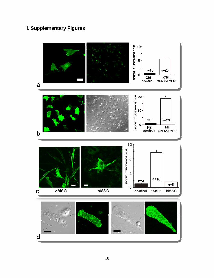
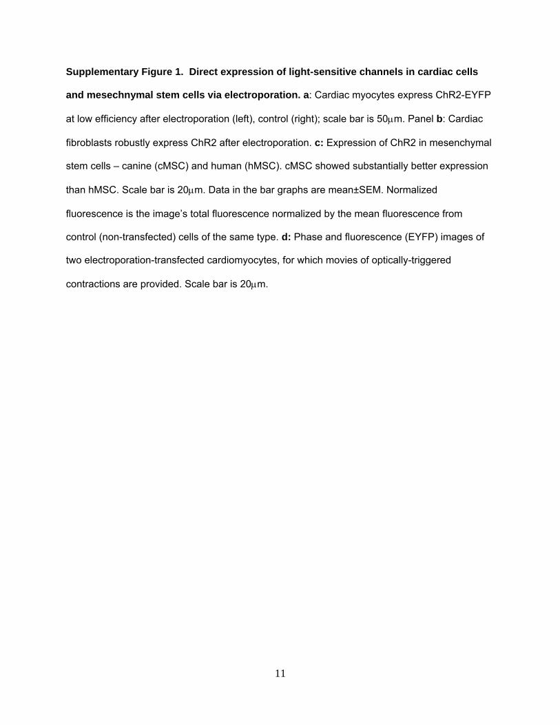
Supplementary Figure 1. Direct expression of light-sensitive channels in cardiac cells
and mesechnymal stem cells via electroporation. a: Cardiac myocytes express ChR2-EYFP
at low efficiency after electroporation (left), control (right); scale bar is 50m. Panel b: Cardiac
fibroblasts robustly express ChR2 after electroporation. c: Expression of ChR2 in mesenchymal
stem cells – canine (cMSC) and human (hMSC). cMSC showed substantially better expression
than hMSC. Scale bar is 20m. Data in the bar graphs are mean±SEM. Normalized
fluorescence is the image’s total fluorescence normalized by the mean fluorescence from
control (non-transfected) cells of the same type. d: Phase and fluorescence (EYFP) images of
two electroporation-transfected cardiomyocytes, for which movies of optically-triggered
contractions are provided. Scale bar is 20m.

12
Supplementary Figure 2. Equivalent circuit for TCU-mediated excitation of cardiac
tissue. a: CM-CM cell pair where both cell carry the excitatory current, and equivalent circuit. b:
TCU of a donor (D) cell and a CM, with differences listed. c: abstraction using a Source-
Neighbor-Load (S-N-L) triad for a spatially-extended system. d: simplified equivalent circuit of
the S-N-L; red arrows indicate the direction of contribution of the different circuit elements
towards “ease of excitation”, as analyzed below.

13
III. Supplementary Movies
Supplementary Movie 1: CM-only ChR2 contractions (compressed)
Shown are two examples of contractions in optically-paced neonatal rat cardiomyocytes, directly
transfected with ChR2-EYFP by electroporation using nucleofector technology. ChR2
expression was firstly confirmed using Olympus FluoViewTM FV1000 confocal microscope, as
shown in the still fluorescence image before each contractility movie: green shows the reporter
gene EYFP. Contractility was recorded at 20 fps using an EMCCD camera with 60X oil lens
(NA=1.42). Optical stimulation was achieved by switching on/off blue light (470nm), easily seen
by the brightness in the field of view and the lightening up of the cardiomyocytes during pacing
pulses. The black scale bar in the contractility movie is 10µm.
Supplementary Movie 2: CM-HEK pair coupling test (carbenoxolone)
The role of gap junctional coupling in forming a “tandem cell unit” that responds to light was
tested here. This is a sparse co-culture of HEK-ChR2-EYFP and neonatal rat cardiomyocytes
(CM). The movie shows optical excitation (and resulting CM contraction) before and during
treatment with an uncoupling agent – carbenoxolone (200M, 20min) and after washout of the
drug. Contractility was recorded at 20 fps using an EMCCD camera with 60X oil lens (NA=1.42).
Optical stimulation was achieved by switching on/off blue light (470nm), easily seen by the
brightness in the field of view and the lightening up of the HEK cells during pacing pulses. The
black scale bar in the contractility movie is 10µm.
Supplementary Movie 3: CM+HEK-contractions-optical stimulation-confocal
The movie shows two examples of contractions in optically-paced cardiac tissue (room
temperature), in which neonatal rat cardiomyocytes were co-cultured with ChR2-expressing
HEK cells at initial ratio of 45:1. Movie was recorded at 20 fps using an EMCCD camera with
60X oil lens (NA=1.42), attached to an Olympus FluoViewTM FV1000 microscope. Optical
stimulation was achieved by switching on/off blue light (470nm), easily seen by the brightness in
14
the field of view and the lighting up of the HEK cells during pacing pulses (due to some
wavelength overlap with excitation of the reporter gene EYFP). Scale bar in the movie is 10µm.
Supplementary Movie 4: Optical mapping of propagation in CM+HEK+ChR2-3.4 speed
Electrical pacing and all-optical interrogation of cardiac tissue are shown (room temperature,
45:1 initial mixing ratio). Ca2+ wave propagation in cardiac tissue, in which neonatal rat
cardiomyocytes were co-cultured with ChR2-expressing HEK cells, was done with Rhod-4. In
the top panels, blue line shows the timing of electrical stimulation (in this case – a line Pt
electrode on the left) or optical stimulation from a blue LED (470nm),: 0 is “off”, 1 is “on”. The
black trace is the corresponding Ca2+ transient from a pixel in the image (indicated by ‘*’ in the
bottom panel). The bottom panels show color-coded (Hilbert) phase movies of Ca2+ wave
propagation acquired by our custom-developed ultra-high resolution macroscopic system and
processed in Matlab (color indicates phase during a cycle, black line denotes the wave-front;
details are given in the Methods section). Movie is played at ¾ real speed. The scale bar in the
movie is 2mm.

15
IV. Equivalent Circuit Analysis of TCU
When comparing the TCU strategy to direct expression of ChR2 in the native myocytes
(mostly by viral methods), several important differences are worth mentioning, Supplementary
Fig 2: (1) the donor (D) cells are non-excitable and typically do not have major repolarizing
currents, i.e. the ChR2 inward current is their main current, unlike native CMs; (2) the D-cells
have higher membrane impedance at rest due to smaller/negligible inward rectifier, IK1, and
typically have a more depolarized resting potential; (3) compared to CM-CM coupling, the D-CM
coupling is typically somewhat reduced. We ask the question: What factors in the TCU
approach affect the ease of excitation and how is the TCU response different than the response
of a homogeneous myocardial tissue with direct expression of ChR2?
For a spatially-extended tissue, when the cell pair (TCU) is connected to some “load” of
excitable cells (CMs), the system can be abstracted to a Source-Neighbor-Load (S-N-L) triad for
easier analysis. A simplified equivalent circuit of such a triad is presented in Supplementary
Fig 2d, derived under the following assumptions: use of equivalent impedances (Z); assuming
negligibly small extracellular impedance Ze; for the immediate neighbor cells to be charged (N),
we consider the membrane Zm and intracellular (coupling) impedance Zi, but for the rest of the
tissue – consider a lumped/load impedance, ZL. Red arrows indicated the direction of
contribution of the different circuit elements towards “ease of excitation”, as analyzed below.
For the simplified circuit in Supplementary Fig 2d, we can derive the following
expressions, representing the underlying current-divider circuits and the total current drawn by
the circuit, IT, as well as the current to excite the immediate neighbors, i.e. the current of
interest, Im:
(1)
(2)
DTi
mT I
ZZ
DZI
)(
TLm
Lm I
ZZ
ZI
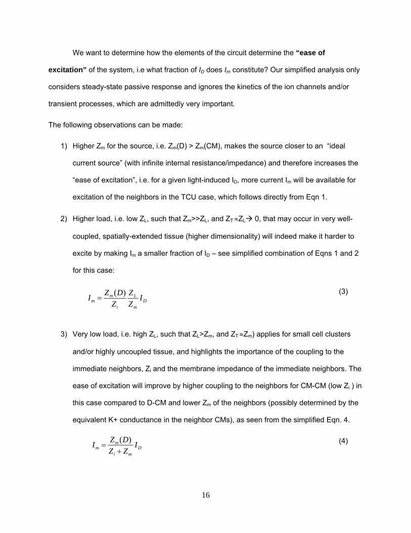
16
We want to determine how the elements of the circuit determine the “ease of
excitation” of the system, i.e what fraction of ID does Im constitute? Our simplified analysis only
considers steady-state passive response and ignores the kinetics of the ion channels and/or
transient processes, which are admittedly very important.
The following observations can be made:
1) Higher Zm for the source, i.e. Zm(D) > Zm(CM), makes the source closer to an “ideal
current source” (with infinite internal resistance/impedance) and therefore increases the
“ease of excitation”, i.e. for a given light-induced ID, more current Im will be available for
excitation of the neighbors in the TCU case, which follows directly from Eqn 1.
2) Higher load, i.e. low ZL, such that Zm>>ZL, and ZT ZL 0, that may occur in very well-
coupled, spatially-extended tissue (higher dimensionality) will indeed make it harder to
excite by making Im a smaller fraction of ID – see simplified combination of Eqns 1 and 2
for this case:
(3)
3) Very low load, i.e. high ZL, such that ZL>Zm, and ZT Zm) applies for small cell clusters
and/or highly uncoupled tissue, and highlights the importance of the coupling to the
immediate neighbors, Zi and the membrane impedance of the immediate neighbors. The
ease of excitation will improve by higher coupling to the neighbors for CM-CM (low Zi ) in
this case compared to D-CM and lower Zm of the neighbors (possibly determined by the
equivalent K+ conductance in the neighbor CMs), as seen from the simplified Eqn. 4.
(4)
Dm
L
i
mm I
Z
Z
Z
DZI
)(
Dmi
mm I
ZZ
DZI
)(

17
4) Overall, better coupling for S-N, but reduced coupling (and equivalent conductivity) for
N-L works towards ease of excitation. It is important to note, however, that for spatially
distributed D-cells, the S-N coupling in the S-N-L triad plays a dual role, i.e. it affects
both the S-N charge transfer and the equivalent “load” presented by the tissue. All
parameters indicated in d act in parallel and actual simulations, quantification is needed
to fully uncover the extent of their individual contribution.
18
REFERENCES
1. Entcheva E, Bien H. Tension development and nuclear eccentricity in topographically controlled cardiac syncytium. J Biomed Microdev. 2003;5:163-168
2. Yin L, Bien H, Entcheva E. Scaffold topography alters intracellular calcium dynamics in cultured cardiomyocyte networks. Am J Physiol Heart Circ Physiol. 2004;287:H1276-H1285
3. Chung CY, Bien H, Entcheva E. The role of cardiac tissue alignment in modulating electrical function. J Cardiovasc Electrophysiol. 2007;18:1323-1329
4. Entcheva E, Bien H. Macroscopic optical mapping of excitation in cardiac cell networks with ultra-high spatiotemporal resolution. Prog.Biophys.Mol.Biol. 2006;92:232-257
5. Bien H, Yin L, Entcheva E. Calcium instabilities in mammalian cardiomyocyte networks. Biophys.J. 2006;90:2628-2640
6. Yu H, Gao J, Wang H, Wymore R, Steinberg S, McKinnon D, Rosen MR, Cohen IS. Effects of the renin-angiotensin system on the current i(to) in epicardial and endocardial ventricular myocytes from the canine heart. Circ Res. 2000;86:1062-1068
7. Valiunas V, Kanaporis G, Valiuniene L, Gordon C, Wang HZ, Li L, Robinson RB, Rosen MR, Cohen IS, Brink PR. Coupling an hcn2-expressing cell to a myocyte creates a two-cell pacing unit. J Physiol. 2009;587:5211-5226
8. Valiunas V, Kanaporis G, Valiuniene L, Gordon C, Wang H, Li L, Robinson RB, Rosen MR, Cohen IS, Brink PR. Coupling an hcn2 expressing cell to a myocyte creates a two cell pacing unit. J Physiol. 2009
9. Valiunas V, Beyer EC, Brink PR. Cardiac gap junction channels show quantitative differences in selectivity. Circ.Res. 2002;91:104-111