Embed Size (px)
Citation preview
CaMKII Autonomy Is Substrate-dependent and FurtherStimulated by Ca2�/Calmodulin*□S
Received for publication, September 23, 2009, and in revised form, March 29, 2010 Published, JBC Papers in Press, March 30, 2010, DOI 10.1074/jbc.M109.069351
Steven J. Coultrap, Isabelle Buard, Jaqueline R. Kulbe, Mark L. Dell’Acqua, and K. Ulrich Bayer1
From the Department of Pharmacology, University of Colorado Denver School of Medicine, Aurora, Colorado 80045
A hallmark feature of Ca2�/calmodulin (CaM)-dependentprotein kinase II (CaMKII) regulation is the generation of Ca2�-independent autonomous activity by Thr-286 autophosphory-lation. CaMKII autonomyhas been regarded a form ofmolecularmemory and is indeed important in neuronal plasticity andlearning/memory. Thr-286-phosphorylated CaMKII is thoughtto be essentially fully active (�70–100%), implicating that it isno longer regulated and that its dramatically increased Ca2�/CaM affinity is of minor functional importance. However, thisstudy shows that autonomy greater than 15–25%was the excep-tion, not the rule, and required a special mechanism (T-sitebinding; by the T-substrates AC2 or NR2B). Autonomous activ-ity toward regular R-substrates (including tyrosine hydroxylaseand GluR1) was significantly further stimulated by Ca2�/CaM,both in vitro and within cells. AlteredKm andVmaxmade auton-omy also substrate- (and ATP) concentration-dependent, butonly over anarrow range,with remarkable stability at physiolog-ical concentrations. Such regulation still allowsmolecularmem-ory of previous Ca2� signals, but prevents complete uncouplingfrom subsequent cellular stimulation.
Ca2�/calmodulin (CaM)2-dependent protein kinase II (CaMKII)can phosphorylate a large variety of substrate proteins and is akey player in many Ca2�-regulated cellular events (for reviewsee Refs. 1–4). However, CaMKII is best know for its regulationof long term potentiation of synaptic strength (LTP) (5, 6),likely by increasing both number (7) and single channel con-ductance (8, 9) of synaptic AMPA-type glutamate receptors,and possibly by stimulating BDNF production (10, 11) (forreview see Refs. 1–4). CaMKII autophosphorylation at Thr-286generates Ca2�-independent autonomous activity (12–14), aprocess regarded as molecular memory (for review see Ref. 2)and indeed important in learning and memory (15).Phosphorylation in the activation loop is a necessary step to
generate full activity of many kinases, including PKA, PKC, andseveral CaMKs (for review see Refs. 16, 17). By contrast,CaMKII is thought to be fully activated by Ca2�/CaM alone,without requirement for phosphorylation. Its Thr-286 is not
located in the activation loop, but in the N-terminal half of theautoinhibitory �-helix, which binds to the T-site (Thr-286-in-teraction site; Ref. 18) in the basal state of CaMKII (19) (Fig.1A). The C-terminal portion of the autoinhibitory �-helixextends to block the substrate binding site (S-site)(19) (Fig. 1A).Ca2�/CaM binding to the autoinhibitory �-helix relieves theS-site block, andmakes Thr-286 accessible for phosphorylationby a neighboring kinase subunit within the 12meric CaMKIIholoenzyme (20–22). Phospho-T286 then prevents completere-binding of the autoinhibitory �-helix.The dual role of Ca2�/CaM in Thr-286 autophosphorylation
(for kinase activation and substrate presentation) allows com-putation of temporal patterns in Ca2� signaling, and indeed,CaMKII autonomy is dependent on the frequency of stimulation(23,24).Suchdecodingof temporalpatternsclearly requires short-term molecular memory of the stimulation history. Ever since itsdiscovery, CaMKII autonomy has been speculated to also providelong-term memory storage (Refs. 12, 13; for review see Ref. 2).Indeed, autonomy-deficient T286A mutant mice are impaired inboth LTP and in learning/memory (15).It is generally believed that Thr-286-phosphorylatedCaMKII is
almost fully active (�70–100%) (for review see Refs. 1–4),implicating that it can no longer be regulated and that its dra-matically increased affinity for CaM (25) is of minor functionalimportance in regulating kinase activity. However, while thereare indeed many reports of high autonomy (�70%)(13, 23, 26),there are also reports of much lower autonomy (�20%)(12, 27)as well as some reports of medium levels of autonomy (�40–60%)(14, 28), in studies that included the same substrates. Highautonomy levels have prevailed in the general perception asbetter estimates, likely because it was easier to explain findingsof low autonomy by submaximal experimental conditions thanexplaining high autonomy by supermaximal conditions.Surprisingly, the results of this study show that CaMKII
autonomy is substrate-dependent: Autonomous activity to-ward regular substrates was found to be low (15–25%) andsignificantly further stimulated by Ca2�/CaM. Higherautonomy (�65%) seen for the same kinase preparation inparallel assays was found to be an exception that required aspecialmechanism (T-site binding of the substrate). The resultsdirectly show that generation of autonomous CaMKII activitydoes not lead to a complete uncoupling from subsequent neu-ronal Ca2� signaling.
EXPERIMENTAL PROCEDURES
Proteins and Peptides—CaMKII and CaM were purified asdescribed (18). MAP2 was purchased (Sigma). Peptides wereobtained from GenScript or CHI Scientific. mGFP-CaMKII
* This work was supported, in whole or in part, by National Institutes of HealthGrants P30NS048154 (UCD Center Grant), R01NS040701 (to M. L. D.), andR01NS052644 (to K. U. B.).
□S The on-line version of this article (available at http://www.jbc.org) containssupplemental Figs. S1–S7 and Tables S1 and S2.
1 To whom correspondence should be addressed. E-mail: [email protected].
2 The abbreviations used are: CaM, Ca2�/calmodulin; PIPES, 1,4-piperazinedi-ethanesulfonic acid; GFP, green fluorescent protein; HA, hemagglutinin;TH, tyrosine hydroxylase; GST, glutathione S-transferase.
THE JOURNAL OF BIOLOGICAL CHEMISTRY VOL. 285, NO. 23, pp. 17930 –17937, June 4, 2010© 2010 by The American Society for Biochemistry and Molecular Biology, Inc. Printed in the U.S.A.
17930 JOURNAL OF BIOLOGICAL CHEMISTRY VOLUME 285 • NUMBER 23 • JUNE 4, 2010
by guest on May 25, 2018
http://ww
w.jbc.org/
Dow
nloaded from
wild type and mutants were expressed in HEK cells; their con-centration in the extracts was determined by fluorescence andnormalized for total protein (29). GST-NR2B-C (containing thecytoplasmic C terminus of rat NR2B from amino acids 1,120–1,482) was expressed in bacteria as described (18, 30), and puri-fied by glutathione affinity chromatography.CaMKII Peptide Substrate Phosphorylation—Phosphate
incorporation into peptide substrates was assessed as described(18, 29). Assays were done at 30 °C for 1 min and contained 2.5nM CaMKII kinase subunits, 50 mM PIPES, pH 7.0–7.2, 0.1%bovine serum albumin, 10 mM MgCl, 100 �M [�-32P]ATP (�1mCi/�mol), and 75�M substrate peptide, or as indicated. Stim-ulated activity was generally measured for naïve CaMKII (butalso for prephosphorylated kinase where indicated) in presenceof CaCl2 (1 mM) and CaM (2 �M; 1 �M in Fig. 1C); autonomousactivity assays contained EGTA (0.5mM) instead. CaMKII (50–250 nM) Thr-286 prephosphorylation was done in stimulationbuffer, but without substrate and 32P, for�5min on ice. Beforeactivity assays, CaM dissociation was induced by EGTA (or inFigs. 1C and 3A by EGTA/EDTA) for at least 3 min. CaMKIIactivity was calculated as pmol of phosphorylated substrate perpmol of CaMKII subunits per min (resulting in the unit prod-uct/kinase/min); CaMKII autonomy reflects the ratio of auton-omous activity to maximal stimulated activity.Western Blot Analysis and Protein Phosphorylation—SDS-
PAGE, transfer onto polyvinylidene difluoride membrane, andimmunodetection were done as described, utilizing antibodiesagainst CaMKII� (CB�2), all CaMKII isoforms (BD Pharmin-gen) phospho-T286 or -T305 CaMKII, GluR1 phospho-S831(PhosphoSolutions), total GluR1 (Calbiochem), or NR2B phos-pho-S1303 (Millipore) (29). Chemoluminescence was imagedby exposure to Hyperfilm (Amersham Biosciences), or, forquantification, by a CCD camera (alpha Innotech).
Proteins were phosphorylatedin the same buffer as peptides,but substrate concentration was200 nM MAP2 (or 1 �M GST-NR2B-C). After PAGE and transferto polyvinylidene difluoride mem-brane, phosphorylation of MAP2was quantified using a Storm Sys-tem and ImageQuant software(Molecular Dynamics).Phosphorylation within PC12
Cells—PC12 cells were culturedin high serum medium (Dulbecco’smodified Eagle’s medium (DMEM),10% horse serum, 5% fetal calf se-rum)onpoly-D-lysine/laminin coatedplates, differentiated inNGF (10 ng/ml) containing low-serum medium(DMEM, 1% horse serum), andtransfected on day 5 using Lipo-fectamine 2000 (Invitrogen). On thenext day, cells were stimulated for 5min by KCl depolarization (in 25mM HEPES pH 7.4, 90 mM KCl, 85mMNaCl, 2mMCaCl2, 2mMMgCl2,
and 30 mM glucose) or treated for 5 min with control solution(as above, but containing 2.5mMKCl and 119mMNaCl instead)immediately before fixation for immunocytochemistry, done asdescribed (30, 31) using rabbit anti-phospho-S19 TH (p1580–19; PhosphoSolutions) or mouse anti-total TH (mAb318;Chemicon; both at 1:500 dilution).Phosphorylation within HEK Cells—HEK cells were cultured
and transfected by the calcium phosphatemethod, as done pre-viously (31). 24 h after transfection, cells were stimulated for 3min by 10�M ionomycin, and immediately harvested in SDS gelloading buffer. Phospho-S831 and total GluR1 in the extractswere determined by Western blot analysis as described above.
RESULTS
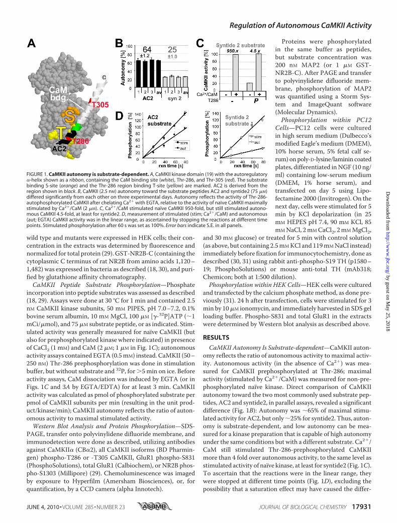
CaMKII Autonomy Is Substrate-dependent—CaMKII auton-omy reflects the ratio of autonomous activity to maximal activ-ity. Autonomous activity (in the absence of Ca2�) was mea-sured for CaMKII prephosphorylated at Thr-286; maximalactivity (stimulated by Ca2�/CaM) was measured for non-pre-phosphorylated naïve kinase. Direct comparison of CaMKIIautonomy toward the twomost commonly used substrate pep-tides, AC2 and syntide2, in parallel assays, revealed a significantdifference (Fig. 1B): Autonomy was �65% of maximal stimu-lated activity forAC2, but only�25% for syntide2. Thus, auton-omy is substrate-dependent, and low autonomy can be mea-sured for a kinase preparation that is capable of high autonomyunder the same conditions but with a different substrate. Ca2�/CaM still stimulated Thr-286-prephosphorylated CaMKIImore than 4 fold over autonomous activity, to the same level asstimulated activity of naïve kinase, at least for syntide2 (Fig. 1C).To ascertain that the reactions were in the linear range, theywere stopped at different time points (Fig. 1D), excluding thepossibility that a saturation effect may have caused the differ-
FIGURE 1. CaMKII autonomy is substrate-dependent. A, CaMKII kinase domain (19) with the autoregulatory�-helix shown as a ribbon, containing the CaM binding site (white), Thr-286, and Thr-305 (red). The substratebinding S-site (orange) and the Thr-286 region binding T-site (yellow) are marked. AC2 is derived from theregion shown in black. B, CaMKII (2.5 nM) autonomy toward the substrate peptides AC2 and syntide2 (75 �M)differed significantly from each other on three experimental days. Autonomy reflects the activity of Thr-286-autophosphorylated CaMKII after chelating Ca2� with EGTA, relative to the activity of naïve CaMKII maximallystimulated by Ca2�/CaM (2 �M). C, Ca2�/CaM stimulated naïve CaMKII 950-fold, but still stimulated autono-mous CaMKII 4.5-fold, at least for syntide2. D, measurement of stimulated (stim; Ca2�/CaM) and autonomous(aut; EGTA) CaMKII activity was in the linear range, as ascertained by stopping the reactions at different timepoints. Stimulated phosphorylation after 60 s was set as 100%. Error bars indicate S.E. in all panels.
Regulation of Autonomous CaMKII Activity
JUNE 4, 2010 • VOLUME 285 • NUMBER 23 JOURNAL OF BIOLOGICAL CHEMISTRY 17931
by guest on May 25, 2018
http://ww
w.jbc.org/
Dow
nloaded from

ence (see supplemental Fig. S1 for autonomy and phosphory-lation rates at each time point).Low Autonomy Is the Default—Three possible mechanisms
for substrate dependence of autonomy were ruled out experi-mentally (Fig. 2 and supplemental results). Briefly, substrate-dependent autonomy was not due to differences in stimulatedphosphorylation rates, Thr-286 phosphorylation, or inhibitoryThr-305 phosphorylation (which prevents Ca2�/CaM-binding;Refs. 32–34) (Fig. 2). Amajor remaining difference between thetwo substrates tested is their interactionwithCaMKII. Syntide2is a regular substrate (R-substrate) that interacts with the sub-strate binding site (S-site) only. By contrast, AC2 is derivedfrom the CaMKII autoregulatory region around Thr-286, andcan additionally bind at the T-site (T-substrate)(29)(see Fig.1A). Thus, we hypothesized that T-site interaction of AC2mayenhance the phospho-T286 induced partial displacement of
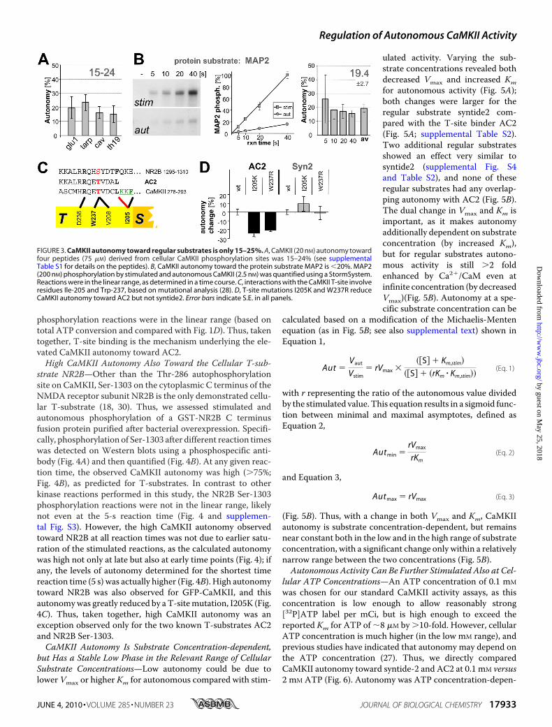
the autoinhibitory �-helix from theT-site, thereby enhancing autono-mous activity. If this is the case, AC2should be an exception, and auton-omy toward all regular substrates(S- but not T-site binding) shouldbe low, as seen for syntide2. Thus,a set of four additional peptides(derived from several importantCaMKII substrates sites) was exam-ined (Fig. 3A): glu1 (GluR1 Ser-831),tarp (Tarp �-4 Ser-259), cav (CaV1.2Ser-439), and TH19 (tyrosine hy-droxylase Ser-19) (see supplementalTable S1 for details on the peptidesand function of the phosphorylationsites). In these assays, CaMKII con-centrationwas increased, as the fourpeptides were poor substrates com-pared with AC2 and syntide2 (seesupplemental Fig. S2). For all thesepeptides, autonomy of 15–25% wasobserved (Fig. 3A), as was predictedfor regular substrates. Peptides areideal for determining the defaultactivity, as this minimizes thepotential for additional levels of reg-ulation that may confound theresults. To test if there may be aprinciple difference in phosphoryla-tion of protein substrates, we choseMAP2, an excellent CaMKII sub-strate with multiple phosphoryla-tion sites (35). Autonomy towardMAP2 was �20% (Fig. 3B), asexpected for a regular substrate. Apossible saturation effect was againexcluded by stopping the reactionsat different time points (Fig. 3B).Thus, for all regular substratestested, autonomous CaMKII activ-ity can be further stimulated 4–6
fold by Ca2� signals. Not surprisingly, the absolute phosphory-lation rates differed widely between the substrates (see Fig. 2,and supplemental Figs. S1 and S2). However, autonomy(reflecting the ratio of autonomous to stimulated phosphory-lation rates) was very similar for all regular substrates.T-site Binding Can Cause Higher Autonomy—Is AC2 really
the exception because of its T-site binding? To test this moredirectly, GFP-CaMKII wild type was compared with two T-sitemutants, I205K and W237, predicted to impair T-site inter-action of AC2 (Fig. 3C). Indeed, autonomy toward AC2 wassignificantly reduced by eithermutation (Fig. 3D). By contrast,themutations did not decrease autonomy toward syntide2 (Fig.3D) or the other regular substrate peptides (supplementalFig. S2); if any, autonomy appeared elevated in some cases(supplemental Fig. S2), consistent with fewer interactions of amutated T-site with the autoinhibitory�-helix (28). All peptide
FIGURE 2. Substrate dependence of CaMKII autonomy is not caused by differential effects on stimulatedactivity. A, between AC2 and syntide2, the stimulated rate of phosphorylation differed more than the auton-omous rate. B, stimulated rate of phosphorylation was not a predictor of autonomy, as two other substratespeptides (derived from AKAP79) with similar or lower stimulated rates than AC2 (left panel; compare A) showedthe same degree of autonomy as syntide2 (right panel). C, prephosphorylation of Thr-286 did not significantlyaffect stimulated activity toward AC2 or syntide2. Prephosphorylation of Thr-286 but not Thr-305 under theexperimental conditions was verified by Western blot. D, comparison of CaMKII wild type and T286A showedthat Thr-286 phosphorylation has no differential effect on AC2 and syntide2. Phosphorylation rates of syntide2were normalized to 100% to correct for differences between the two independent enzyme preparations.E, phosphorylation of CaMKII at Thr-305 and Thr-286 during mock-stimulated kinase activity assays (without32P), with and without added substrates, was assessed by Western analysis. CaMKII and phospho-T286 CaMKIIbands (arrow) versus shifted bands after autophosphorylation at additional sited (arrowhead) are indicated.F, quantification of analysis as in E showed that substrate further decreased the low level of inhibitory auto-phosphorylation at Thr-305, but AC2 even more so than syntide. Level of phosphorylated Thr-286 was notaffected by substrate. Error bars indicate S.E. in all panels.
Regulation of Autonomous CaMKII Activity
17932 JOURNAL OF BIOLOGICAL CHEMISTRY VOLUME 285 • NUMBER 23 • JUNE 4, 2010
by guest on May 25, 2018
http://ww
w.jbc.org/
Dow
nloaded from
phosphorylation reactions were in the linear range (based ontotal ATP conversion and compared with Fig. 1D). Thus, takentogether, T-site binding is the mechanism underlying the ele-vated CaMKII autonomy toward AC2.High CaMKII Autonomy Also Toward the Cellular T-sub-
strate NR2B—Other than the Thr-286 autophosphorylationsite on CaMKII, Ser-1303 on the cytoplasmic C terminus of theNMDA receptor subunit NR2B is the only demonstrated cellu-lar T-substrate (18, 30). Thus, we assessed stimulated andautonomous phosphorylation of a GST-NR2B C terminusfusion protein purified after bacterial overexpression. Specifi-cally, phosphorylation of Ser-1303 after different reaction timeswas detected on Western blots using a phosphospecific anti-body (Fig. 4A) and then quantified (Fig. 4B). At any given reac-tion time, the observed CaMKII autonomy was high (�75%;Fig. 4B), as predicted for T-substrates. In contrast to otherkinase reactions performed in this study, the NR2B Ser-1303phosphorylation reactions were not in the linear range, likelynot even at the 5-s reaction time (Fig. 4 and supplemen-tal Fig. S3). However, the high CaMKII autonomy observedtoward NR2B at all reaction times was not due to earlier satu-ration of the stimulated reactions, as the calculated autonomywas high not only at late but also at early time points (Fig. 4); ifany, the levels of autonomy determined for the shortest timereaction time (5 s) was actually higher (Fig. 4B). High autonomytoward NR2B was also observed for GFP-CaMKII, and thisautonomywas greatly reduced by aT-sitemutation, I205K (Fig.4C). Thus, taken together, high CaMKII autonomy was anexception observed only for the two known T-substrates AC2and NR2B Ser-1303.CaMKII Autonomy Is Substrate Concentration-dependent,
but Has a Stable Low Phase in the Relevant Range of CellularSubstrate Concentrations—Low autonomy could be due tolower Vmax or higher Km for autonomous compared with stim-
ulated activity. Varying the sub-strate concentrations revealed bothdecreased Vmax and increased Kmfor autonomous activity (Fig. 5A);both changes were larger for theregular substrate syntide2 com-pared with the T-site binder AC2(Fig. 5A; supplemental Table S2).Two additional regular substratesshowed an effect very similar tosyntide2 (supplemental Fig. S4and Table S2), and none of theseregular substrates had any overlap-ping autonomy with AC2 (Fig. 5B).The dual change in Vmax and Km isimportant, as it makes autonomyadditionally dependent on substrateconcentration (by increased Km),but for regular substrates autono-mous activity is still �2 foldenhanced by Ca2�/CaM even atinfinite concentration (by decreasedVmax)(Fig. 5B). Autonomy at a spe-cific substrate concentration can be
calculated based on a modification of the Michaelis-Mentenequation (as in Fig. 5B; see also supplemental text) shown inEquation 1,
Aut �Vaut
Vstim� rVmax �
��S� � Km,stim�
��S� � �rKm � Km,stim��(Eq. 1)
with r representing the ratio of the autonomous value dividedby the stimulated value. This equation results in a sigmoid func-tion between minimal and maximal asymptotes, defined asEquation 2,
Autmin �rVmax
rKm(Eq. 2)
and Equation 3,
Autmax � rVmax (Eq. 3)
(Fig. 5B). Thus, with a change in both Vmax and Km, CaMKIIautonomy is substrate concentration-dependent, but remainsnear constant both in the low and in the high range of substrateconcentration, with a significant change only within a relativelynarrow range between the two concentrations (Fig. 5B).Autonomous Activity Can Be Further Stimulated Also at Cel-
lular ATP Concentrations—An ATP concentration of 0.1 mM
was chosen for our standard CaMKII activity assays, as thisconcentration is low enough to allow reasonably strong[32P]ATP label per mCi, but is high enough to exceed thereported Km for ATP of �8 �M by �10-fold. However, cellularATP concentration is much higher (in the low mM range), andprevious studies have indicated that autonomy may depend onthe ATP concentration (27). Thus, we directly comparedCaMKII autonomy toward syntide-2 and AC2 at 0.1 mM versus2 mM ATP (Fig. 6). Autonomy was ATP concentration-depen-
FIGURE 3. CaMKII autonomy toward regular substrates is only 15–25%. A, CaMKII (20 nM) autonomy towardfour peptides (75 �M) derived from cellular CaMKII phosphorylation sites was 15–24% (see supplementalTable S1 for details on the peptides). B, CaMKII autonomy toward the protein substrate MAP2 is �20%. MAP2(200 nM) phosphorylation by stimulated and autonomous CaMKII (2.5 nM) was quantified using a StormSystem.Reactions were in the linear range, as determined in a time course. C, interactions with the CaMKII T-site involveresidues Ile-205 and Trp-237, based on mutational analysis (28). D, T-site mutations I205K and W237R reduceCaMKII autonomy toward AC2 but not syntide2. Error bars indicate S.E. in all panels.
Regulation of Autonomous CaMKII Activity
JUNE 4, 2010 • VOLUME 285 • NUMBER 23 JOURNAL OF BIOLOGICAL CHEMISTRY 17933
by guest on May 25, 2018
http://ww
w.jbc.org/
Dow
nloaded from
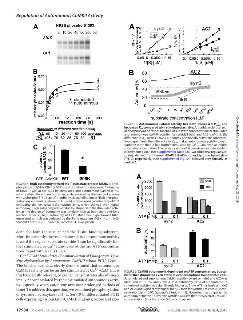
dent, for both the regular and the T-site binding substrate.More importantly, the results showed that autonomous activitytoward the regular substrate syntide-2 can be significantly fur-ther stimulated by Ca2�/CaM even at the mM ATP concentra-tions found within cells (Fig. 6).Ca2�/CaM Stimulates Phosphorylation of Endogenous Tyro-
sine Hydroxylase by Autonomous CaMKII within PC12 Cells—The biochemical data clearly demonstrated that autonomousCaMKII activity can be further stimulated by Ca2�/CaM. But isthis biologically relevant, or are cellular substrates alreadymax-imally phosphorylated by the unstimulated autonomous activ-ity, especially when autonomy acts over prolonged periods oftime? To address this question, we examined phosphorylationof tyrosine hydroxylase (TH) at Ser-19 in differentiated PC12cells expressing variousGFP-CaMKIImutants, before and after
FIGURE 5. Autonomous CaMKII activity has both decreased Vmax andincreased Km compared with stimulated activity. A, double reciprocal plotof phosphorylation rate as function of substrate concentration for stimulatedand autonomous CaMKII activity, for syntide2 (left) and AC2 (right). B, thedifference in Km makes CaMKII autonomy additionally substrate concentra-tion-dependent. The difference in Vmax makes autonomous activity towardsyntide2 more than 2-fold further stimulated by Ca2�/CaM even at infinitesubstrate concentration. The curve for syntide2 is based on four independentexperiments as in A (see supplemental Table S2). Two additional regular sub-strates, derived from human AKAP79 (hAKb-nn) and tyrosine hydroxylase(TH19), respectively, (see supplemental Fig. S4) behaved very similarly assyntide2.
FIGURE 6. CaMKII autonomy is dependent on ATP concentration, but canbe further stimulated even at the mM concentrations found within cells.A, stimulated and autonomous CaMKII activity toward syntide2 and AC2 wasmeasured at 0.1 mM and 2 mM ATP. B, autonomy (ratio of autonomous tostimulated activity) was significantly higher at 2 mM ATP for both syntide2and AC2 (and significantly higher for AC2 than for syntide2 at each ATP con-centration) (p � 0.01, Student’s t test; n 5). However, most importantly,autonomy at for the R-substrate syntide2 was less than 40% even at 2 mM ATPconcentration. Error bars show S.E. in both panels.
FIGURE 4. High autonomy toward the T-substrate protein NR2B. A, phos-phorylation of GST-NR2B-c (a GST fusion protein with cytoplasmic C terminusof NR2B; 1 �M) at Ser-1303 by stimulated and autonomous CaMKII (5 nM)activity after different reaction times, as determined by Western blot analysiswith a phospho-S1303 specific antibody. B, quantification of NR2B phosphor-ylation experiments (as shown in A; n 6) show an average autonomy of 81%(excluding the less reliable 5-s reaction time, which showed even higherautonomy). High autonomy was not due to saturation of the stimulated activ-ity, as the degree of autonomy was similarly high at both short and longreaction times. C, high autonomy of GFP-CaMKII wild type toward NR2B(measured as in B) was reduced by the T-site mutation I205K (*, p � 0.05;Student’s t test; n 3). Error bars indicate S.E. in all panels.
Regulation of Autonomous CaMKII Activity
17934 JOURNAL OF BIOLOGICAL CHEMISTRY VOLUME 285 • NUMBER 23 • JUNE 4, 2010
by guest on May 25, 2018
http://ww
w.jbc.org/
Dow
nloaded from
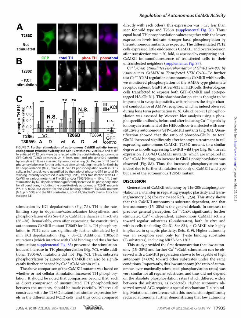
stimulation by KCl depolarization (Fig. 7A). TH is the rate-limiting step in dopamine/catecholamine biosynthesis, andphosphorylation of its Ser-19 by CaMKII enhances TH activity(36–38). Remarkably, even after expressing the constitutivelyautonomous CaMKII mutant T286D for 24 h, TH phosphory-lation in PC12 cells was significantly further stimulated by 5min KCl depolarization (Fig. 7, A–C). Additional T305/6Dmutations (which interfere with CaM binding and thus furtherstimulation; supplemental Fig. S5) prevented the stimulation-induced increase in TH phosphorylation (Fig. 7C), while addi-tional T305/6A mutations did not (Fig. 7C). Thus, substratephosphorylation by autonomous CaMKII can also be signifi-cantly further enhanced by Ca2�/CaM within cells.
The above comparison of the CaMKII mutants was based onwhether or not cellular stimulation increased TH phosphory-lation. It should be noted that comparison beyond that, suchas direct comparison of unstimulated TH phosphorylationbetween the mutants, should be made carefully. Whereas allconstructs with the T286D mutant had similar expression lev-els in the differentiated PC12 cells (and thus could compared
directly with each other), this expression was �1/3 less thanseen for wild type and T286A (supplemental Fig. S6). Thus,equal basal TH phosphorylation values together with the lowerexpression levels indicate stronger basal phosphorylation bythe autonomousmutants, as expected. The differentiated PC12cells expressed little endogenous CaMKII, and overexpressionafter transfection was�20-fold, as assessed by comparing anti-CaMKII immunofluorescence of transfected cells to theiruntransfected neighbors (supplemental Fig. S7).Ca2�/CaM Stimulates Phosphorylation of GluR1 Ser-831 by
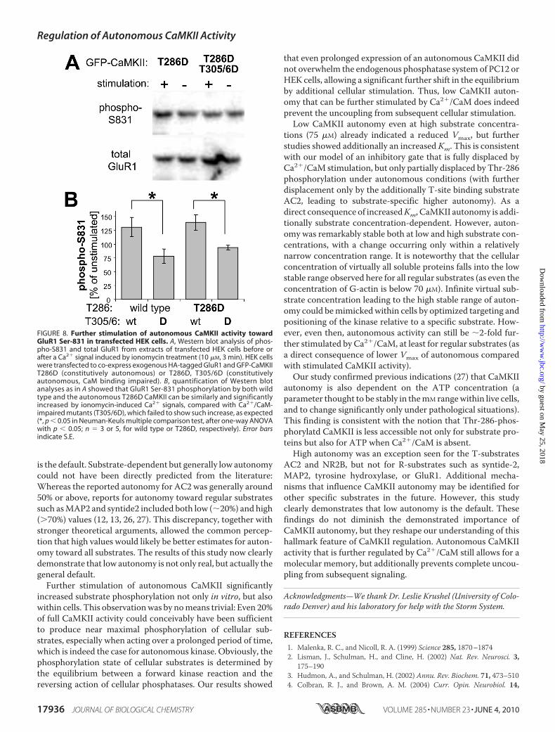
Autonomous CaMKII in Transfected HEK Cells—To furthertest Ca2�/CaM regulation of autonomous CaMKII within cells,we monitored phosphorylation of the AMPA-type glutamatereceptor subunit GluR1 at Ser-831 in HEK cells (heterologouscells transfected to express both GFP-CaMKII and epitope-tagged HA-GluR1). This phosphorylation site is thought to beimportant in synaptic plasticity, as it enhances the single chan-nel conductance of AMPA receptors, which is indeed observedduring long term potentiation (8, 9). GluR1 Ser-831 phosphor-ylation was assessed by Western blot analysis using a phos-phospecific antibody, before and after inducing Ca2� signals byionomycin treatment of the HEK cells co-transfected with con-stitutively autonomous GFP-CaMKII mutants (Fig. 8A). Quan-tification showed that the ratio of phospho-GluR1 to totalGluR1 increased significantly after ionomycin treatment in cellexpressing autonomous CaMKII T286D mutant, to a similardegree as in cells expressing CaMKII wild type (Fig. 8B). In cellexpression T305/6D CaMKII mutants, which are impaired inCa2�/CaM binding, no increase in GluR1 phosphorylation wasobserved (Fig. 8B). Thus, the increased phosphorylation wasindeed due to further stimulation not only of CaMKII wild typebut also of the autonomous T286D mutant.
DISCUSSION
Generation of CaMKII autonomy by Thr-286 autophosphor-ylation is a vital step in regulating synaptic plasticity and learn-ing/memory (15) (for review see Refs. 1,2,4). This study showsthat this CaMKII autonomy is substrate-dependent, and thatlow autonomy (15–25%) is the general default. In contrast toprevious general perception, Ca2�/CaM significantly furtherstimulated Ca2�-independent, autonomous CaMKII activitytoward regular substrates (R-substrates), both in vitro andwithin cells (including GluR1 Ser-831, a CaMKII site highlyimplicated in synaptic plasticity; Refs. 8, 9). Higher autonomywas an exception seen only for T-site binding substrates(T-substrates), including NR2B Ser-1303.This study provided the first demonstration that low auton-
omy (15–25%) and further Ca2�/CaM-stimulation can be ob-served with a CaMKII preparation shown to be capable of highautonomy (�60%) toward other substrates under the sameconditions. Importantly, this low autonomy (the ratio of auton-omous over maximally stimulated phosphorylation rates) wasvery similar for all regular substrates, and thus did not dependon the absolute phosphorylation rates (which differed widelybetween the substrates, as expected). Higher autonomy ob-served toward AC2 required a special mechanism: T-site bind-ing. Mutational interference with this mechanism significantlyreduced autonomy, further demonstrating that low autonomy
FIGURE 7. Further stimulation of autonomous CaMKII activity towardendogenous tyrosine hydroxylase Ser-19 within PC12 cells. A and B, dif-ferentiated PC12 cells were transfected with the constitutively autonomousGFP-CaMKII T286D construct. 24 h later, total and phospho-S19 tyrosinehydroxylase (TH) was assessed by immunostaining (A). Degree of TH Ser-19phosphorylation was further enhanced after stimulating the cells for 5 min byKCl depolarization (B). C, relative TH Ser-19 phosphorylation levels in PC12cells, as in A and B, were quantified by the ratio of phospho-S19 to total THstaining intensity (expressed in arbitrary units), after transfection with GFP-CaMKII or various mutants at Thr-286 and/or T305/306 (n 10 to 14). 5 minstimulation by KCl depolarization significantly increased TH phosphorylationfor all conditions, including the constitutively autonomous T286D mutants(**, p � 0.05), but except for the CaM binding-deficient T305/6D mutants(N.S., p � 0.38) and the GFP control (n.s., p � 0.28; Student’s t tests). Error barsindicate S.E.
Regulation of Autonomous CaMKII Activity
JUNE 4, 2010 • VOLUME 285 • NUMBER 23 JOURNAL OF BIOLOGICAL CHEMISTRY 17935
by guest on May 25, 2018
http://ww
w.jbc.org/
Dow
nloaded from
is the default. Substrate-dependent but generally low autonomycould not have been directly predicted from the literature:Whereas the reported autonomy for AC2 was generally around50% or above, reports for autonomy toward regular substratessuch asMAP2 and syntide2 included both low (�20%) and high(�70%) values (12, 13, 26, 27). This discrepancy, together withstronger theoretical arguments, allowed the common percep-tion that high values would likely be better estimates for auton-omy toward all substrates. The results of this study now clearlydemonstrate that low autonomy is not only real, but actually thegeneral default.Further stimulation of autonomous CaMKII significantly
increased substrate phosphorylation not only in vitro, but alsowithin cells. This observationwas by nomeans trivial: Even 20%of full CaMKII activity could conceivably have been sufficientto produce near maximal phosphorylation of cellular sub-strates, especially when acting over a prolonged period of time,which is indeed the case for autonomous kinase. Obviously, thephosphorylation state of cellular substrates is determined bythe equilibrium between a forward kinase reaction and thereversing action of cellular phosphatases. Our results showed
that even prolonged expression of an autonomous CaMKII didnot overwhelm the endogenous phosphatase system of PC12 orHEK cells, allowing a significant further shift in the equilibriumby additional cellular stimulation. Thus, low CaMKII auton-omy that can be further stimulated by Ca2�/CaM does indeedprevent the uncoupling from subsequent cellular stimulation.Low CaMKII autonomy even at high substrate concentra-
tions (75 �M) already indicated a reduced Vmax, but furtherstudies showed additionally an increased Km. This is consistentwith our model of an inhibitory gate that is fully displaced byCa2�/CaM stimulation, but only partially displaced by Thr-286phosphorylation under autonomous conditions (with furtherdisplacement only by the additionally T-site binding substrateAC2, leading to substrate-specific higher autonomy). As adirect consequence of increasedKm, CaMKII autonomy is addi-tionally substrate concentration-dependent. However, auton-omy was remarkably stable both at low and high substrate con-centrations, with a change occurring only within a relativelynarrow concentration range. It is noteworthy that the cellularconcentration of virtually all soluble proteins falls into the lowstable range observed here for all regular substrates (as even theconcentration of G-actin is below 70 �M). Infinite virtual sub-strate concentration leading to the high stable range of auton-omy could bemimickedwithin cells by optimized targeting andpositioning of the kinase relative to a specific substrate. How-ever, even then, autonomous activity can still be �2-fold fur-ther stimulated by Ca2�/CaM, at least for regular substrates (asa direct consequence of lower Vmax of autonomous comparedwith stimulated CaMKII activity).Our study confirmed previous indications (27) that CaMKII
autonomy is also dependent on the ATP concentration (aparameter thought to be stably in themM rangewithin live cells,and to change significantly only under pathological situations).This finding is consistent with the notion that Thr-286-phos-phorylatd CaMKII is less accessible not only for substrate pro-teins but also for ATP when Ca2�/CaM is absent.
High autonomy was an exception seen for the T-substratesAC2 and NR2B, but not for R-substrates such as syntide-2,MAP2, tyrosine hydroxylase, or GluR1. Additional mecha-nisms that influence CaMKII autonomy may be identified forother specific substrates in the future. However, this studyclearly demonstrates that low autonomy is the default. Thesefindings do not diminish the demonstrated importance ofCaMKII autonomy, but they reshape our understanding of thishallmark feature of CaMKII regulation. Autonomous CaMKIIactivity that is further regulated by Ca2�/CaM still allows for amolecular memory, but additionally prevents complete uncou-pling from subsequent signaling.
Acknowledgments—We thank Dr. Leslie Krushel (University of Colo-rado Denver) and his laboratory for help with the Storm System.
REFERENCES1. Malenka, R. C., and Nicoll, R. A. (1999) Science 285, 1870–18742. Lisman, J., Schulman, H., and Cline, H. (2002) Nat. Rev. Neurosci. 3,
175–1903. Hudmon, A., and Schulman, H. (2002) Annu. Rev. Biochem. 71, 473–5104. Colbran, R. J., and Brown, A. M. (2004) Curr. Opin. Neurobiol. 14,
FIGURE 8. Further stimulation of autonomous CaMKII activity towardGluR1 Ser-831 in transfected HEK cells. A, Western blot analysis of phos-pho-S831 and total GluR1 from extracts of transfected HEK cells before orafter a Ca2� signal induced by ionomycin treatment (10 �M, 3 min). HEK cellswere transfected to co-express exogenous HA-tagged GluR1 and GFP-CaMKIIT286D (constitutively autonomous) or T286D, T305/6D (constitutivelyautonomous, CaM binding impaired). B, quantification of Western blotanalyses as in A showed that GluR1 Ser-831 phosphorylation by both wildtype and the autonomous T286D CaMKII can be similarly and significantlyincreased by ionomycin-induced Ca2� signals, compared with Ca2�/CaM-impaired mutants (T305/6D), which failed to show such increase, as expected(*, p � 0.05 in Neuman-Keuls multiple comparison test, after one-way ANOVAwith p � 0.05; n 3 or 5, for wild type or T286D, respectively). Error barsindicate S.E.
Regulation of Autonomous CaMKII Activity
17936 JOURNAL OF BIOLOGICAL CHEMISTRY VOLUME 285 • NUMBER 23 • JUNE 4, 2010
by guest on May 25, 2018
http://ww
w.jbc.org/
Dow
nloaded from
318–3275. Malinow, R., Schulman, H., and Tsien, R.W. (1989) Science 245, 862–8666. Silva, A. J., Stevens, C. F., Tonegawa, S., andWang, Y. (1992) Science 257,
201–2067. Hayashi, Y., Shi, S. H., Esteban, J. A., Piccini, A., Poncer, J. C., andMalinow,
R. (2000) Science 287, 2262–22678. Benke, T. A., Luthi, A., Isaac, J. T., and Collingridge, G. L. (1998) Nature
393, 793–7979. Derkach, V., Barria, A., and Soderling, T. R. (1999) Proc. Natl. Acad. Sci.
U.S.A. 96, 3269–327410. Kang, H., and Schuman, E. M. (1995) Science 267, 1658–166211. Zhou, Z., Hong, E. J., Cohen, S., Zhao,W. N., Ho, H. Y., Schmidt, L., Chen,
W.G., Lin, Y., Savner, E., Griffith, E. C., Hu, L., Steen, J. A.,Weitz, C. J., andGreenberg, M. E. (2006) Neuron 52, 255–269
12. Miller, S. G., and Kennedy, M. B. (1986) Cell 44, 861–87013. Lou, L. L., Lloyd, S. J., and Schulman,H. (1986) Proc. Natl. Acad. Sci. U.S.A.
83, 9497–950114. Schworer, C. M., Colbran, R. J., and Soderling, T. R. (1986) J. Biol. Chem.
261, 8581–858415. Giese, K. P., Fedorov,N. B., Filipkowski, R. K., and Silva, A. J. (1998) Science
279, 870–87316. Soderling, T. R., and Stull, J. T. (2001) Chem. Rev. 101, 2341–235217. Nolen, B., Taylor, S., and Ghosh, G. (2004)Mol. Cell 15, 661–67518. Bayer, K. U., De Koninck, P., Leonard, A. S., Hell, J. W., and Schulman, H.
(2001) Nature 411, 801–80519. Rosenberg, O. S., Deindl, S., Sung, R. J., Nairn, A. C., and Kuriyan, J. (2005)
Cell 123, 849–86020. Hanson, P. I., Meyer, T., Stryer, L., and Schulman, H. (1994) Neuron 12,
943–95621. Rich, R. C., and Schulman, H. (1998) J. Biol. Chem. 273, 28424–28429
22. Rosenberg, O. S., Deindl, S., Comolli, L. R., Hoelz, A., Downing, K. H.,Nairn, A. C., and Kuriyan, J. (2006) FEBS. J. 273, 682–694
23. De Koninck, P., and Schulman, H. (1998) Science 279, 227–23024. Bayer, K. U., De Koninck, P., and Schulman, H. (2002) EMBO J. 21,
3590–359725. Meyer, T., Hanson, P. I., Stryer, L., and Schulman, H. (1992) Science 256,
1199–120226. Erickson, J. R., Joiner,M. L., Guan, X., Kutschke,W., Yang, J., Oddis, C. V.,
Bartlett, R. K., Lowe, J. S., O’Donnell, S. E., Aykin-Burns, N., Zimmerman,M. C., Zimmerman, K., Ham, A. J., Weiss, R. M., Spitz, D. R., Shea, M. A.,Colbran, R. J.,Mohler, P. J., andAnderson,M. E. (2008)Cell 133, 462–474
27. Smith, M. K., Colbran, R. J., Brickey, D. A., and Soderling, T. R. (1992)J. Biol. Chem. 267, 1761–1768
28. Yang, E., and Schulman, H. (1999) J. Biol. Chem. 274, 26199–2620829. Vest, R. S., Davies, K. D., O’Leary, H., Port, J. D., and Bayer, K. U. (2007)
Mol. Biol. Cell 18, 5024–503330. Bayer, K. U., LeBel, E.,McDonald, G. L., O’Leary, H., Schulman,H., andDe
Koninck, P. (2006) J. Neurosci. 26, 1164–117431. O’Leary, H., Lasda, E., and Bayer, K. U. (2006) Mol. Biol. Cell 17,
4656–466532. Colbran, R. J., and Soderling, T. R. (1990) J. Biol. Chem. 265, 11213–1121933. Hanson, P. I., and Schulman, H. (1992) J. Biol. Chem. 267, 17216–1722434. Lu, C. S., Hodge, J. J., Mehren, J., Sun, X. X., and Griffith, L. C. (2003)
Neuron 40, 1185–119735. Schulman, H. (1984) J. Cell Biol. 99, 11–1936. Yamauchi, T., and Fujisawa, H. (1981) Biochem. Biophys. Res. Commun.
100, 807–81337. Griffith, L. C., and Schulman, H. (1988) J. Biol. Chem. 263, 9542–954938. Bobrovskaya, L., Dunkley, P. R., and Dickson, P. W. (2004) J. Neurochem.
90, 857–864
Regulation of Autonomous CaMKII Activity
JUNE 4, 2010 • VOLUME 285 • NUMBER 23 JOURNAL OF BIOLOGICAL CHEMISTRY 17937
by guest on May 25, 2018
http://ww
w.jbc.org/
Dow
nloaded from
Ulrich BayerSteven J. Coultrap, Isabelle Buard, Jaqueline R. Kulbe, Mark L. Dell'Acqua and K.
/Calmodulin2+CaMKII Autonomy Is Substrate-dependent and Further Stimulated by Ca
doi: 10.1074/jbc.M109.069351 originally published online March 30, 20102010, 285:17930-17937.J. Biol. Chem.
10.1074/jbc.M109.069351Access the most updated version of this article at doi:
Alerts:
When a correction for this article is posted•
When this article is cited•
to choose from all of JBC's e-mail alertsClick here
Supplemental material:
http://www.jbc.org/content/suppl/2010/03/30/M109.069351.DC1
http://www.jbc.org/content/285/23/17930.full.html#ref-list-1
This article cites 38 references, 22 of which can be accessed free at
by guest on May 25, 2018
http://ww
w.jbc.org/
Dow
nloaded from


![Decision Trees - start [Auton Lab] Trees - start [Auton Lab] ... a](https://img.pdfslide.us/doc/110x75/5abccf487f8b9ab1118ea4fb/decision-trees-start-auton-lab-trees-start-auton-lab-a.jpg)