Embed Size (px)
Citation preview
V o l . 2 1 N o . 1 2 6 0 1
T R E N D SI M M U N O L O G Y TO D AY
0167-5699/00/$ – see front matter © 2000 Elsevier Science Ltd. All rights reserved. PII: S0167-5699(00)01752-7
D E C E M B E R 2 0 0 0
he UK adhesion group is aninformal network of scientistsworking on cell adhesion. Ithas been running for five years
and meets twice a year within the UK. Thelatest meeting, a joint meeting with Spanishadhesion groups, brought together immu-nologists and cell biologists with interests incell adhesion and migration to discuss theirrole in physiological responses and howthey contribute to the pathology of cancer,angiogenesis and inflammation.
Should I stay or should I go?Chemokines capture the headlinesCell adhesion is essential for a properly func-tioning immune system. Unlike sedentarycells, leukocytes display an impressivesophistication in the way they regulate theiradhesion and migration. The discovery of alarge family of molecular signposts for theimmune system (chemokines) has revolu-tionized our understanding of how appro-priate immune cells meet up in the rightplace at the right time. Like integrins,chemokines and their receptors displayremarkable redundancy1. The expression ofboth types of receptors on leukocytes israpidly regulated and reflects both the stateof activation and the stage of differentiationof leukocytes. It is this remarkable ability tochange receptors upon activation that allowsleukocytes to traffic efficiently to lymphoidtissue as naive cells or to inflamed tissue asantigen-experienced effector cells.
T. Williams (London, UK) reviewed therole of chemokines as morphogens in theimmune system, with particular emphasison the role of eotaxin within lung tissue.How chemokines are inactivated, removedand cleared in vivo remains unclear. Williamspresented some exciting in vivo studiesshowing that high levels of eotaxin arefound in the blood following an initial peakof production in the lung2. The biologicalcorrelate of this increase in blood eotaxin isincreased release of eosinophils from thebone marrow, leading to blood eosinophilia.
Williams suggested that this might lead toenhanced recruitment of eosinophils into thelung as well as acting as an effective mecha-nism by which supply might be matched todemand. Thus, a single chemokine (eotaxin)appears to act as a messenger between dis-tinct anatomical sites, functioning as a re-cruiting agent in one tissue (lung) and areleasing agent at another (bone marrow).
Differential chemokine production byendothelial cells and stromal microenviron-ments underlies the differential recruitmentof leukocyte subsets to different anatomicallocations. R. Philips (London, UK) illus-trated how discrete subsets of chemokines(SDF-1, SLC and ELC) presented by highendothelial cells in lymph nodes regulateselective T-cell trans-endothelial migration.Once within tissues, leukocytes have to integrate signals from antigen receptors and growth factors to decide whether they should remain or re-circulate to blood via lymphatic vessels. A. Carolos-Martinez(Madrid, Spain) presented a novel expla-nation for how such ‘stop–go’ signals are me-diated at a molecular level. Similar to othermembers of the G-protein-coupled family ofreceptors, chemokine receptors can undergohomo- as well as heterodimerization. Usingthe example of monocyte chemotactic protein 1 (MCP-1) and RANTES binding to CCR5 and CCR2, Carolos-Martinez suggested that while homodimerizationbetween receptors leads preferentially to amigratory response in leukocytes, hetero-dimerization of CCR5 and CCR2 receptorsleads to an adhesive signal and the arrest ofleukocytes within tissue. Furthermore, thedistribution of chemokine receptors withindistinct micro-domains and rafts within theplasma membrane appears to have impor-tant functional consequences, and provides
a mechanism for efficient cross-talk betweengrowth factors, chemokines and integrins3.
Degradomics: metalloproteinasesat the cutting edgeProteolysis is frequently employed as aneffective method of post-translational modi-fication of proteins. Until quite recently therole of extracellular proteinases in refashion-ing the cell surface and surrounding tissuehas remained relatively understudied. Thathas all changed with the discovery of a largeand expanding family of extracellular pro-teinases belonging to the matrix metallo-proteinase (MMP) and ADAM (for ‘a dis-integrin and metalloproteinase’) families4.G. Murphy (Norwich, UK) outlined the roleof MMPs in regulating cell migration. A strik-ing example of how a proteinase can changethe properties, and therefore the function, ofa protein occurs with the processing oflaminin-5 by MMP-2 or the membrane typematrix metalloproteinase 1 (MT-MMP-1).While intact laminin promotes cell adhesion,laminin that has been processed by MMPspreferentially promotes cell migrationowing to exposure of a cryptic epitope. Bycontrast, some MMPs (e.g. MMP-2) appearto be able to dampen inflammatory responsesby cleaving and inactivating certainchemokines (such as MCP-1). This suggeststhat MMPs can act both as effectors and regulators of inflammatory responses5.
In addition to their role in remodeling theextracellular matrix, MMPs play an impor-tant role in releasing cell-membrane andmatrix-bound growth factors and cytokinesvia a process known as ectodomain shed-ding. The use of knockout mice has beenparticularly helpful to study this process. J. Arribas (Barcelona, Spain) reviewed the roleof the tumour necrosis factor a (TNF-a) con-vertase (TACE or ADAM 17) in ectodomainshedding. A surprising finding in TACE-knockout mice is that, in addition to theirinability to process TNF-a, a wide range ofproteins are also not processed, includingcytokines, growth factors and adhesion
Sticky moments with sticky moleculesChristopher D. Buckley and David L. Simmons
A recent meeting* illustrated the
extensive cross-talk that exists
between adhesion receptors,
G-protein-coupled chemokine
receptors, growth factor receptors
and cell surface proteinases.
T
*A meeting of the Joint UK and Spanish adhesion groups was held at ImperialCollege School of Medicine, London, UK, on 4–5 September 2000.
receptors, as well as proteins involved in cell fate determination (Notch) and disease(amyloid precursor protein). A current chal-lenge for the field is to determine which pro-teinases modify and process which candi-date substrates in a physiologically relevant
manner – a process that has been termeddegradomics6.
How the proteolytic activity of MMPsand ADAMs are activated in vivo remainsunclear, but D. Nath (Norwich, UK) illus-trated the role that intracellular signalling
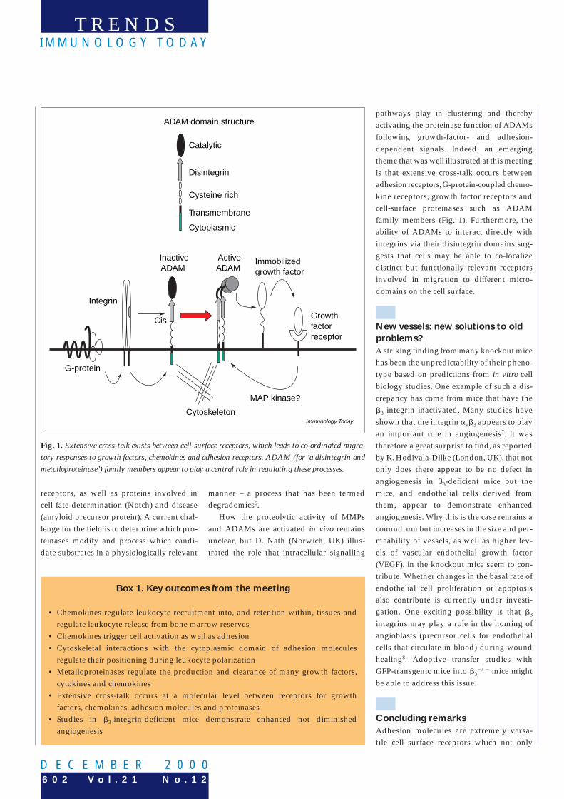
pathways play in clustering and therebyactivating the proteinase function of ADAMsfollowing growth-factor- and adhesion-dependent signals. Indeed, an emergingtheme that was well illustrated at this meetingis that extensive cross-talk occurs betweenadhesion receptors, G-protein-coupled chemo-kine receptors, growth factor receptors andcell-surface proteinases such as ADAM family members (Fig. 1). Furthermore, theability of ADAMs to interact directly withintegrins via their disintegrin domains sug-gests that cells may be able to co-localize distinct but functionally relevant receptorsinvolved in migration to different micro-domains on the cell surface.
New vessels: new solutions to oldproblems?A striking finding from many knockout micehas been the unpredictability of their pheno-type based on predictions from in vitro cellbiology studies. One example of such a dis-crepancy has come from mice that have theb3 integrin inactivated. Many studies haveshown that the integrin avb3 appears to playan important role in angiogenesis7. It wastherefore a great surprise to find, as reportedby K. Hodivala-Dilke (London, UK), that notonly does there appear to be no defect inangiogenesis in b3-deficient mice but themice, and endothelial cells derived fromthem, appear to demonstrate enhancedangiogenesis. Why this is the case remains aconundrum but increases in the size and per-meability of vessels, as well as higher lev-els of vascular endothelial growth factor(VEGF), in the knockout mice seem to con-tribute. Whether changes in the basal rate ofendothelial cell proliferation or apoptosisalso contribute is currently under investi-gation. One exciting possibility is that b3
integrins may play a role in the homing ofangioblasts (precursor cells for endothelialcells that circulate in blood) during woundhealing8. Adoptive transfer studies withGFP-transgenic mice into b3
2/2 mice mightbe able to address this issue.
Concluding remarksAdhesion molecules are extremely versa-tile cell surface receptors which not only
T R E N D SI M M U N O L O G Y TO D AY
6 0 2 V o l . 2 1 N o . 1 2D E C E M B E R 2 0 0 0
Fig. 1. Extensive cross-talk exists between cell-surface receptors, which leads to co-ordinated migra-tory responses to growth factors, chemokines and adhesion receptors. ADAM (for ‘a disintegrin andmetalloproteinase’) family members appear to play a central role in regulating these processes.
Box 1. Key outcomes from the meeting
• Chemokines regulate leukocyte recruitment into, and retention within, tissues andregulate leukocyte release from bone marrow reserves
• Chemokines trigger cell activation as well as adhesion• Cytoskeletal interactions with the cytoplasmic domain of adhesion molecules
regulate their positioning during leukocyte polarization• Metalloproteinases regulate the production and clearance of many growth factors,
cytokines and chemokines• Extensive cross-talk occurs at a molecular level between receptors for growth
factors, chemokines, adhesion molecules and proteinases• Studies in b3-integrin-deficient mice demonstrate enhanced not diminished
angiogenesis
Immunology Today
G-protein
Integrin
InactiveADAM
ActiveADAM
Cytoskeleton
Growthfactorreceptor
Immobilizedgrowth factor
Cis
Catalytic
Disintegrin
Cysteine rich
Transmembrane
Cytoplasmic
ADAM domain structure
MAP kinase?
he mannan binding lectin (MBL)pathway is activated by bind-ing of the C1q-like moleculeMBL, followed by activation of
at least two MBL-associated serine proteases(MASP-1 and MASP-2), leading directly toactivation of C4 and C2. The complete MBLcomplex comprises MBL, MASP-1, MASP-2,together with a third MASP (MASP-3)(J.C. Jensenius, Aarhus, Denmark; T. Fujita,Fukushima, Japan) and a small MBL-associ-ated protein (SMAP; a 19 kDa molecule ofunknown function). MASP-2 can directlycleave C4 and C2, and MASP-1 activates C3 directly; the function of MASP-3 is un-known. In MASP-1-knockout mice, MBLpathway activity is reduced but not absent,supporting the observation that MASP-2 ac-tivates C3 independently of MASP-1 (Fujita).
Complement and autoimmunityDeficiencies in the classical complementpathway in man predispose to lupus-like
syndromes – the risk being greater for earliercomponents (i.e. C1q .C4 .C2 .C3). C1q-deficient (C1q2/2) mice develop autoantibod-ies and a type of lupus that has been linkedto reduced clearance of apoptotic cells1. De-velopment of lupus-like symptoms is strictlydependent on the genetic background of themice (M. Botto, London, UK). Work fromseveral groups further implicates C1q and
other complement components as central tothe removal of apoptotic cells. C1q2/2 and, toa lesser extent, C42/2 mice, are inefficient at clearing injected apoptotic cells from the peritoneum. In addition, peritonealmacrophages from these mice phagocytosefar fewer cells compared with those fromwild-type mice – evidence that the early complement components are required for efficient clearance of apoptotic cells (Botto).
M. Loos (Mainz) demonstrated that bonemarrow transplantation reconstitutedplasma C1q levels in C1q2/2 mice, confirm-ing that C1q is made in bone marrow. Bonemarrow transplantation should be consid-ered in C1q-deficient humans, who other-wise have a .90% chance of developing alupus-like syndrome associated with a highmorbidity and mortality.
Several studies addressed the mechanismby which apoptotic cells react with C1q, andsubsequently interact with receptors, pre-sumably specific for C1q, on macrophages.The complement regulators CD55 and CD46
V o l . 2 1 N o . 1 2 6 0 3
T R E N D SI M M U N O L O G Y TO D AY
0167-5699/00/$ – see front matter © 2000 Elsevier Science Ltd. All rights reserved. PII: S0167-5699(00)01742-4
D E C E M B E R 2 0 0 0
stick cells together, but also provide bio-chemical and biophysical signals thatregulate many aspects of cell behavior. Bycontrolling the membrane organization ofsignalling receptors, imposing spatial or-ganization and regulating the local con-centration of both intracellular and extra-cellular adapter proteins and enzymes, celladhesion molecules directly contribute to the extensive multi-protein complexes thatoccur at the cell surface. These dynamic con-versations between cell surface receptors are now enabling scientists from differentdisciplines to provide a unified molecularexplanation for the long-observed, butpoorly understood, requirement that a num-ber of seemingly distinct cell surface mol-ecules be engaged in order for efficient cellproliferation, differentiation and migrationto occur.
To join the UK adhesion network, please contact
David Simmons ([email protected]).
Christopher Buckley ([email protected])is at the Division of Immunity and Infection,Rheumatology Research Group, University ofBirmingham, Birmingham, UK B15 2TT; DavidSimmons ([email protected]) is atNeuroscience Discovery, SmithKline BeechamR&D, New Third Avenue, Harlow, Essex, UKCM19 5AW.
References1 Rossi, D. and Zlotnik, A. (2000) The biology
of chemokines and their receptors. Annu.Rev. Immunol. 18, 217–242
2 Palframan, R.T. et al. (1998) Mechanisms ofacute eosinophil mobilization from thebone marrow stimulated by interleukin 5:the role of specific adhesion molecules andphosphatidylinositol 3-kinase. J. Exp. Med.188, 1621–1632
3 Sánchez-Madrid, F. and del Pozo, M.A. (1999) Leukocyte polarization in cellmigration and immune interactions.EMBO J. 18, 501–511
4 Primakoff, P. and Myles, D.G. (2000) TheADAM gene family: surface proteins withan adhesion and protease activity packedinto a single molecule. Trends Genet. 16,83–87
5 Blobel, C.P. (2000) Remarkable roles ofproteolysis on and beyond the cell surface.Curr. Opin. Cell Biol. 12, 606–612
6 McQuibban, G.A. et al. (2000) Inflammationdampened by gelatinase A cleavage ofmonocyte chemoattractant protein-3. Science289, 1202–1206
7 Stromblad, S. and Cheresh, D.A. (1996) Celladhesion and angiogenesis. Trends Cell Biol.6, 462–468
8 Asahara, T. et al. (1999) Bone marrow originof endothelial progenitor cells responsiblefor postnatal vasculogenesis inphysiological and pathologicalneovascularization. Circ. Res. 85, 221–228
Into the third century of complement researchB. Paul Morgan, Moh Daha, Seppo Meri and Anne Nicholson-Weller
The complement system is
established as a major homeostatic
system and a factor in diverse
pathologies. Some 500 delegates
attended a recent meeting* to discuss
the latest findings in complement
research, ranging from studies in
sea squirts through to therapeutic
trials of inhibitors. Here, we review
some of the key topics discussed at
the workshop.
T
* The XVIII International Complement Workshop was held atSnowbird, UT, USA, on 23–27 July 2000.