Embed Size (px)
Citation preview
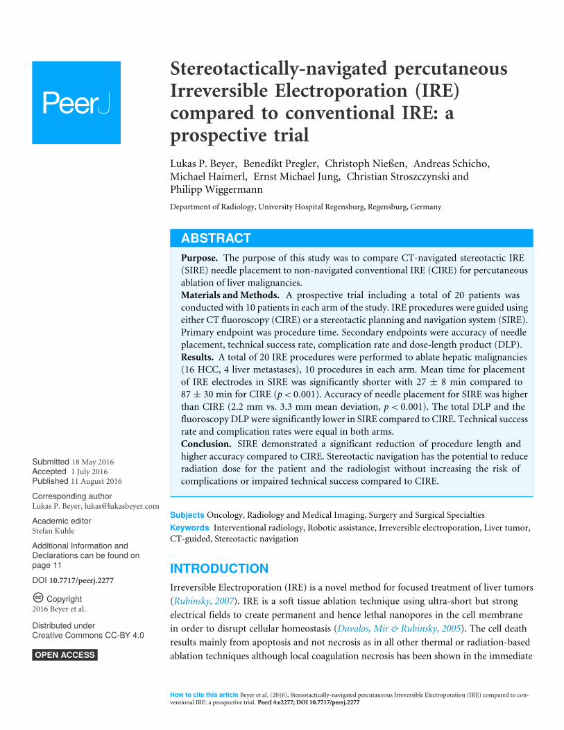
Submitted 18 May 2016Accepted 1 July 2016Published 11 August 2016
Corresponding authorLukas P. Beyer, [email protected]
Academic editorStefan Kuhle
Additional Information andDeclarations can be found onpage 11
DOI 10.7717/peerj.2277
Copyright2016 Beyer et al.
Distributed underCreative Commons CC-BY 4.0
OPEN ACCESS
Stereotactically-navigated percutaneousIrreversible Electroporation (IRE)compared to conventional IRE: aprospective trialLukas P. Beyer, Benedikt Pregler, Christoph Nießen, Andreas Schicho,Michael Haimerl, Ernst Michael Jung, Christian Stroszczynski andPhilipp WiggermannDepartment of Radiology, University Hospital Regensburg, Regensburg, Germany
ABSTRACTPurpose. The purpose of this study was to compare CT-navigated stereotactic IRE(SIRE) needle placement to non-navigated conventional IRE (CIRE) for percutaneousablation of liver malignancies.Materials andMethods. A prospective trial including a total of 20 patients wasconducted with 10 patients in each arm of the study. IRE procedures were guided usingeither CT fluoroscopy (CIRE) or a stereotactic planning and navigation system (SIRE).Primary endpoint was procedure time. Secondary endpoints were accuracy of needleplacement, technical success rate, complication rate and dose-length product (DLP).Results. A total of 20 IRE procedures were performed to ablate hepatic malignancies(16 HCC, 4 liver metastases), 10 procedures in each arm. Mean time for placementof IRE electrodes in SIRE was significantly shorter with 27 ± 8 min compared to87 ± 30 min for CIRE (p< 0.001). Accuracy of needle placement for SIRE was higherthan CIRE (2.2 mm vs. 3.3 mm mean deviation, p< 0.001). The total DLP and thefluoroscopy DLP were significantly lower in SIRE compared to CIRE. Technical successrate and complication rates were equal in both arms.Conclusion. SIRE demonstrated a significant reduction of procedure length andhigher accuracy compared to CIRE. Stereotactic navigation has the potential to reduceradiation dose for the patient and the radiologist without increasing the risk ofcomplications or impaired technical success compared to CIRE.
Subjects Oncology, Radiology and Medical Imaging, Surgery and Surgical SpecialtiesKeywords Interventional radiology, Robotic assistance, Irreversible electroporation, Liver tumor,CT-guided, Stereotactic navigation
INTRODUCTIONIrreversible Electroporation (IRE) is a novel method for focused treatment of liver tumors(Rubinsky, 2007). IRE is a soft tissue ablation technique using ultra-short but strongelectrical fields to create permanent and hence lethal nanopores in the cell membranein order to disrupt cellular homeostasis (Davalos, Mir & Rubinsky, 2005). The cell deathresults mainly from apoptosis and not necrosis as in all other thermal or radiation-basedablation techniques although local coagulation necrosis has been shown in the immediate
How to cite this article Beyer et al. (2016), Stereotactically-navigated percutaneous Irreversible Electroporation (IRE) compared to con-ventional IRE: a prospective trial. PeerJ 4:e2277; DOI 10.7717/peerj.2277
proximity to the electrodes (Ben-David et al., 2013). IRE is used for non-resectable livertumors in the vicinity of vessels (due to its selectivity for tumor tissue while preservingvessel structures as well as the absence of the so-called heat sink effect) (Rubinsky, Onik &Mikus, 2007). IRE has also recently shown therapeutic efficacy and safety in other organslike pancreas and prostate (Martin et al., 2015; Ting et al., 2016). First animal studies haveshown the safety and feasibility of stereotactically delivered IRE for the treatment oftelencephalic gliomas (Rossmeisl et al., 2015).
IRE ablation requires the placement of two or more applicator electrodes between whichthe electrical fields are applied. In order to achieve successful ablation, parallel needleplacement at a pre-defined distance between 1.5 cm and 2 cm is required. Mathematicmodels have shown that parallel placement of IRE electrodes is essential to generate an evendistribution of the electromagnetic field (Edd & Davalos, 2007). This fact was also shown inporcine animal model by Ben-David et al. (2013) who investigated the therapeutic efficacyof IRE with regard to electrode orientation, tissue type and local environment.
Needles are placed under image guidance using ultrasound or computer tomography asimaging methods. Since these methods display one image plane at a time, the realization ofmultiple parallel needle placements can be challenging. Several attempts may be necessaryto achieve the required geometrical configuration of the needle with respect to other needlesand as well as in relation to the anatomical target.
Navigation technology for interventional radiology supports IRE treatments byproviding comprehensive planning of needle configurations using 3D image data andby supporting needle placement through guidance functionality.
This study aims to investigate the potential benefits of CT-navigated stereotactic IRE(SIRE) needle placement compared to non-navigated conventional IRE (CIRE) for ablationof malignant liver lesions.
MATERIALS AND METHODSStudy design, participant selection and patient characteristicsIn a prospective, non-blinded, non-randomized two-armed study carried out between July2015 and February 2016, IRE ablations of malignant liver tumors were performed in 10procedures with stereotactic navigation and 10 cases without. The primary end point ofthe study was the time required until start of the ablation (measured from the time ofthe first CT scan to the start of the ablation). Secondary endpoints included accuracy ofIRE electrode placement (accuracy is measured as lateral deviation of the IRE electrodesto a central reference electrode); overall procedure time; number of needle replacements;radiation dose and number of control scans.
The study has been approved by the Ethics Committee of the University Regensburg(approval number 15-101-0188) and written consent was obtained from all patients. Allprocedures were performed according to the Declaration of Helsinki and the guideline forGood Clinical Practice from the International Conference on Harmonization.
In all cases, indication for percutaneous tumor ablation was determined by aninterdisciplinary tumor board. An IRE was indicated if surgical resection was not possible,
Beyer et al. (2016), PeerJ, DOI 10.7717/peerj.2277 2/13
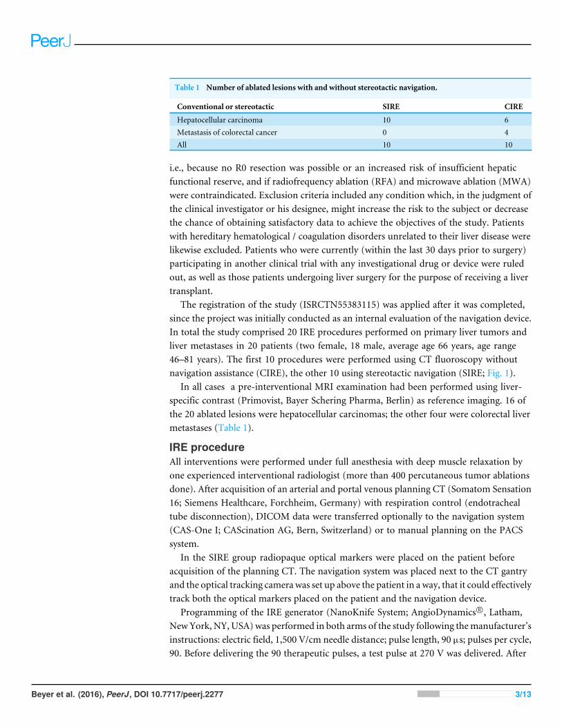
Table 1 Number of ablated lesions with and without stereotactic navigation.
Conventional or stereotactic SIRE CIRE
Hepatocellular carcinoma 10 6Metastasis of colorectal cancer 0 4All 10 10
i.e., because no R0 resection was possible or an increased risk of insufficient hepaticfunctional reserve, and if radiofrequency ablation (RFA) and microwave ablation (MWA)were contraindicated. Exclusion criteria included any condition which, in the judgment ofthe clinical investigator or his designee, might increase the risk to the subject or decreasethe chance of obtaining satisfactory data to achieve the objectives of the study. Patientswith hereditary hematological / coagulation disorders unrelated to their liver disease werelikewise excluded. Patients who were currently (within the last 30 days prior to surgery)participating in another clinical trial with any investigational drug or device were ruledout, as well as those patients undergoing liver surgery for the purpose of receiving a livertransplant.
The registration of the study (ISRCTN55383115) was applied after it was completed,since the project was initially conducted as an internal evaluation of the navigation device.In total the study comprised 20 IRE procedures performed on primary liver tumors andliver metastases in 20 patients (two female, 18 male, average age 66 years, age range46–81 years). The first 10 procedures were performed using CT fluoroscopy withoutnavigation assistance (CIRE), the other 10 using stereotactic navigation (SIRE; Fig. 1).
In all cases a pre-interventional MRI examination had been performed using liver-specific contrast (Primovist, Bayer Schering Pharma, Berlin) as reference imaging. 16 ofthe 20 ablated lesions were hepatocellular carcinomas; the other four were colorectal livermetastases (Table 1).
IRE procedureAll interventions were performed under full anesthesia with deep muscle relaxation byone experienced interventional radiologist (more than 400 percutaneous tumor ablationsdone). After acquisition of an arterial and portal venous planning CT (Somatom Sensation16; Siemens Healthcare, Forchheim, Germany) with respiration control (endotrachealtube disconnection), DICOM data were transferred optionally to the navigation system(CAS-One I; CAScination AG, Bern, Switzerland) or to manual planning on the PACSsystem.
In the SIRE group radiopaque optical markers were placed on the patient beforeacquisition of the planning CT. The navigation system was placed next to the CT gantryand the optical tracking camera was set up above the patient in a way, that it could effectivelytrack both the optical markers placed on the patient and the navigation device.
Programming of the IRE generator (NanoKnife System; AngioDynamics R©, Latham,NewYork, NY, USA) was performed in both arms of the study following themanufacturer’sinstructions: electric field, 1,500 V/cm needle distance; pulse length, 90 µs; pulses per cycle,90. Before delivering the 90 therapeutic pulses, a test pulse at 270 V was delivered. After
Beyer et al. (2016), PeerJ, DOI 10.7717/peerj.2277 3/13
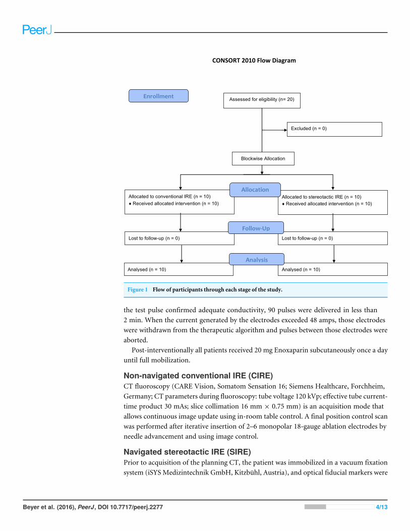
CONSORT2010FlowDiagram
Assessed for eligibility (n= 20)
Excluded (n = 0)
Analysed (n = 10)
Lost to follow-up (n = 0)
Allocated to conventional IRE (n = 10) ♦Received allocated intervention (n = 10)
Lost to follow-up (n = 0)
Allocated to stereotactic IRE (n = 10) ♦Received allocated intervention (n = 10)
Analysed (n = 10)
Allocation
Analysis
Follow-Up
Blockwise Allocation
Enrollment
Figure 1 Flow of participants through each stage of the study.
the test pulse confirmed adequate conductivity, 90 pulses were delivered in less than2 min. When the current generated by the electrodes exceeded 48 amps, those electrodeswere withdrawn from the therapeutic algorithm and pulses between those electrodes wereaborted.
Post-interventionally all patients received 20 mg Enoxaparin subcutaneously once a dayuntil full mobilization.
Non-navigated conventional IRE (CIRE)CT fluoroscopy (CARE Vision, Somatom Sensation 16; Siemens Healthcare, Forchheim,Germany; CT parameters during fluoroscopy: tube voltage 120 kVp; effective tube current-time product 30 mAs; slice collimation 16 mm × 0.75 mm) is an acquisition mode thatallows continuous image update using in-room table control. A final position control scanwas performed after iterative insertion of 2–6 monopolar 18-gauge ablation electrodes byneedle advancement and using image control.
Navigated stereotactic IRE (SIRE)Prior to acquisition of the planning CT, the patient was immobilized in a vacuum fixationsystem (iSYS Medizintechnik GmbH, Kitzbühl, Austria), and optical fiducial markers were
Beyer et al. (2016), PeerJ, DOI 10.7717/peerj.2277 4/13
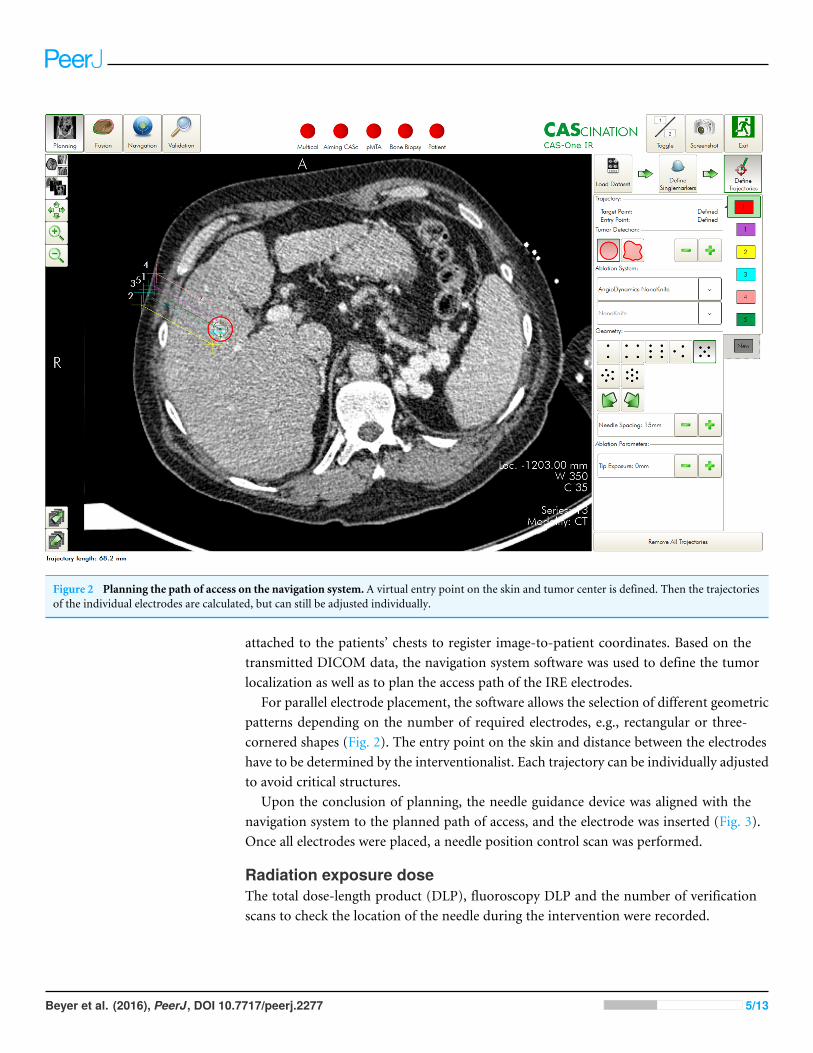
Figure 2 Planning the path of access on the navigation system. A virtual entry point on the skin and tumor center is defined. Then the trajectoriesof the individual electrodes are calculated, but can still be adjusted individually.
attached to the patients’ chests to register image-to-patient coordinates. Based on thetransmitted DICOM data, the navigation system software was used to define the tumorlocalization as well as to plan the access path of the IRE electrodes.
For parallel electrode placement, the software allows the selection of different geometricpatterns depending on the number of required electrodes, e.g., rectangular or three-cornered shapes (Fig. 2). The entry point on the skin and distance between the electrodeshave to be determined by the interventionalist. Each trajectory can be individually adjustedto avoid critical structures.
Upon the conclusion of planning, the needle guidance device was aligned with thenavigation system to the planned path of access, and the electrode was inserted (Fig. 3).Once all electrodes were placed, a needle position control scan was performed.
Radiation exposure doseThe total dose-length product (DLP), fluoroscopy DLP and the number of verificationscans to check the location of the needle during the intervention were recorded.
Beyer et al. (2016), PeerJ, DOI 10.7717/peerj.2277 5/13
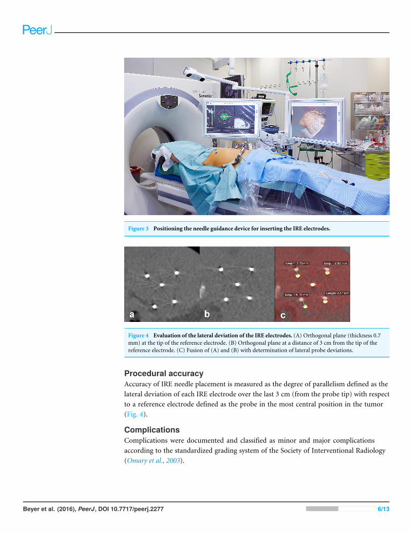
Figure 3 Positioning the needle guidance device for inserting the IRE electrodes.
Figure 4 Evaluation of the lateral deviation of the IRE electrodes. (A) Orthogonal plane (thickness 0.7mm) at the tip of the reference electrode. (B) Orthogonal plane at a distance of 3 cm from the tip of thereference electrode. (C) Fusion of (A) and (B) with determination of lateral probe deviations.
Procedural accuracyAccuracy of IRE needle placement is measured as the degree of parallelism defined as thelateral deviation of each IRE electrode over the last 3 cm (from the probe tip) with respectto a reference electrode defined as the probe in the most central position in the tumor(Fig. 4).
ComplicationsComplications were documented and classified as minor and major complicationsaccording to the standardized grading system of the Society of Interventional Radiology(Omary et al., 2003).
Beyer et al. (2016), PeerJ, DOI 10.7717/peerj.2277 6/13
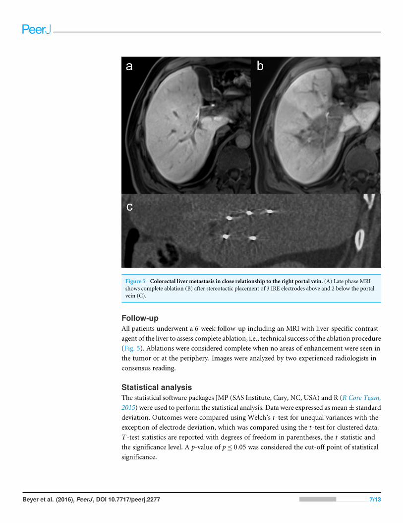
Figure 5 Colorectal liver metastasis in close relationship to the right portal vein. (A) Late phase MRIshows complete ablation (B) after stereotactic placement of 3 IRE electrodes above and 2 below the portalvein (C).
Follow-upAll patients underwent a 6-week follow-up including an MRI with liver-specific contrastagent of the liver to assess complete ablation, i.e., technical success of the ablation procedure(Fig. 5). Ablations were considered complete when no areas of enhancement were seen inthe tumor or at the periphery. Images were analyzed by two experienced radiologists inconsensus reading.
Statistical analysisThe statistical software packages JMP (SAS Institute, Cary, NC, USA) and R (R Core Team,2015) were used to perform the statistical analysis. Data were expressed as mean± standarddeviation. Outcomes were compared using Welch’s t -test for unequal variances with theexception of electrode deviation, which was compared using the t -test for clustered data.T -test statistics are reported with degrees of freedom in parentheses, the t statistic andthe significance level. A p-value of p≤ 0.05 was considered the cut-off point of statisticalsignificance.
Beyer et al. (2016), PeerJ, DOI 10.7717/peerj.2277 7/13
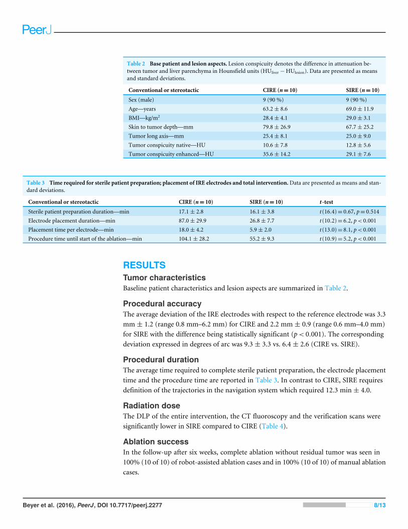
Table 2 Base patient and lesion aspects. Lesion conspicuity denotes the difference in attenuation be-tween tumor and liver parenchyma in Hounsfield units (HUliver −HUlesion). Data are presented as meansand standard deviations.
Conventional or stereotactic CIRE (n= 10) SIRE (n= 10)
Sex (male) 9 (90 %) 9 (90 %)Age—years 63.2± 8.6 69.0± 11.9BMI—kg/m2 28.4± 4.1 29.0± 3.1Skin to tumor depth—mm 79.8± 26.9 67.7± 25.2Tumor long axis—mm 25.4± 8.1 25.0± 9.0Tumor conspicuity native—HU 10.6± 7.8 12.8± 5.6Tumor conspicuity enhanced—HU 35.6± 14.2 29.1± 7.6
Table 3 Time required for sterile patient preparation; placement of IRE electrodes and total intervention.Data are presented as means and stan-dard deviations.
Conventional or stereotactic CIRE (n= 10) SIRE (n= 10) t -test
Sterile patient preparation duration—min 17.1± 2.8 16.1± 3.8 t (16.4)= 0.67, p= 0.514Electrode placement duration—min 87.0± 29.9 26.8± 7.7 t (10.2)= 6.2, p< 0.001Placement time per electrode—min 18.0± 4.2 5.9± 2.0 t (13.0)= 8.1, p< 0.001Procedure time until start of the ablation—min 104.1± 28.2 55.2± 9.3 t (10.9)= 5.2, p< 0.001
RESULTSTumor characteristicsBaseline patient characteristics and lesion aspects are summarized in Table 2.
Procedural accuracyThe average deviation of the IRE electrodes with respect to the reference electrode was 3.3mm ± 1.2 (range 0.8 mm–6.2 mm) for CIRE and 2.2 mm ± 0.9 (range 0.6 mm–4.0 mm)for SIRE with the difference being statistically significant (p< 0.001). The correspondingdeviation expressed in degrees of arc was 9.3 ± 3.3 vs. 6.4 ± 2.6 (CIRE vs. SIRE).
Procedural durationThe average time required to complete sterile patient preparation, the electrode placementtime and the procedure time are reported in Table 3. In contrast to CIRE, SIRE requiresdefinition of the trajectories in the navigation system which required 12.3 min ± 4.0.
Radiation doseThe DLP of the entire intervention, the CT fluoroscopy and the verification scans weresignificantly lower in SIRE compared to CIRE (Table 4).
Ablation successIn the follow-up after six weeks, complete ablation without residual tumor was seen in100% (10 of 10) of robot-assisted ablation cases and in 100% (10 of 10) of manual ablationcases.
Beyer et al. (2016), PeerJ, DOI 10.7717/peerj.2277 8/13
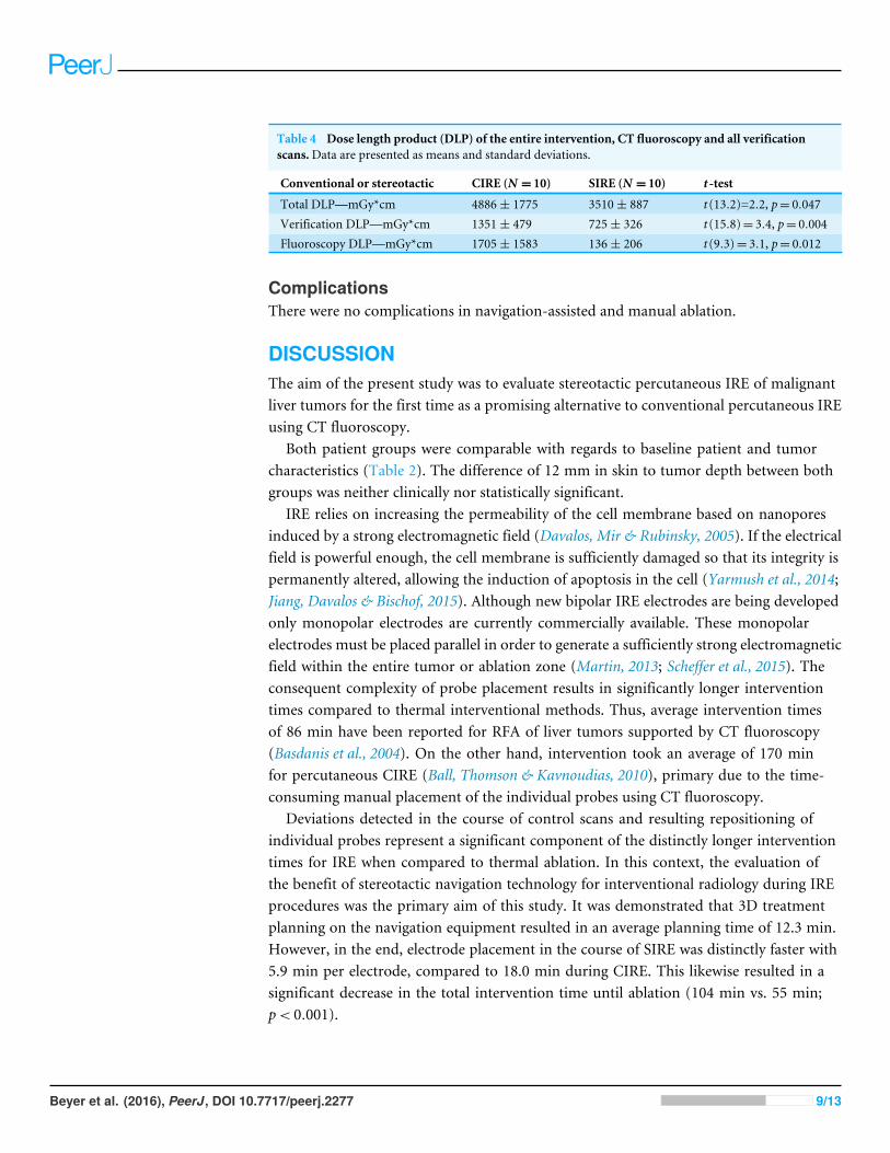
Table 4 Dose length product (DLP) of the entire intervention, CT fluoroscopy and all verificationscans.Data are presented as means and standard deviations.
Conventional or stereotactic CIRE (N = 10) SIRE (N = 10) t -test
Total DLP—mGy*cm 4886± 1775 3510± 887 t (13.2)=2.2, p= 0.047Verification DLP—mGy*cm 1351± 479 725± 326 t (15.8)= 3.4, p= 0.004Fluoroscopy DLP—mGy*cm 1705± 1583 136± 206 t (9.3)= 3.1, p= 0.012
ComplicationsThere were no complications in navigation-assisted and manual ablation.
DISCUSSIONThe aim of the present study was to evaluate stereotactic percutaneous IRE of malignantliver tumors for the first time as a promising alternative to conventional percutaneous IREusing CT fluoroscopy.
Both patient groups were comparable with regards to baseline patient and tumorcharacteristics (Table 2). The difference of 12 mm in skin to tumor depth between bothgroups was neither clinically nor statistically significant.
IRE relies on increasing the permeability of the cell membrane based on nanoporesinduced by a strong electromagnetic field (Davalos, Mir & Rubinsky, 2005). If the electricalfield is powerful enough, the cell membrane is sufficiently damaged so that its integrity ispermanently altered, allowing the induction of apoptosis in the cell (Yarmush et al., 2014;Jiang, Davalos & Bischof, 2015). Although new bipolar IRE electrodes are being developedonly monopolar electrodes are currently commercially available. These monopolarelectrodes must be placed parallel in order to generate a sufficiently strong electromagneticfield within the entire tumor or ablation zone (Martin, 2013; Scheffer et al., 2015). Theconsequent complexity of probe placement results in significantly longer interventiontimes compared to thermal interventional methods. Thus, average intervention timesof 86 min have been reported for RFA of liver tumors supported by CT fluoroscopy(Basdanis et al., 2004). On the other hand, intervention took an average of 170 minfor percutaneous CIRE (Ball, Thomson & Kavnoudias, 2010), primary due to the time-consuming manual placement of the individual probes using CT fluoroscopy.
Deviations detected in the course of control scans and resulting repositioning ofindividual probes represent a significant component of the distinctly longer interventiontimes for IRE when compared to thermal ablation. In this context, the evaluation ofthe benefit of stereotactic navigation technology for interventional radiology during IREprocedures was the primary aim of this study. It was demonstrated that 3D treatmentplanning on the navigation equipment resulted in an average planning time of 12.3 min.However, in the end, electrode placement in the course of SIRE was distinctly faster with5.9 min per electrode, compared to 18.0 min during CIRE. This likewise resulted in asignificant decrease in the total intervention time until ablation (104 min vs. 55 min;p< 0.001).
Beyer et al. (2016), PeerJ, DOI 10.7717/peerj.2277 9/13
In the course of a prospective study of 70 patients, Mbalisike et al. (2014) determinedthat robot-supported percutaneous microwave ablation provides very high precision.According to their measurements there was a minor average deviation of the active centerof the microwave probe (1.9 mm) compared to the center of the tumor. Our own studiesusing robot-assistedMWAof a total of 64 liver tumors found a comparablyminor deviationof 1.3 mm (Beyer et al., 2015).
The distance of the electrodes from the tumor center is not a suitable measure fordetermining the accuracy of the placement of the IRE electrodes, since they have to beinserted in the periphery of the tumor as well as in the tumor center. Unlike thermalablation procedures, exactly parallel insertion of the IRE probes into the liver tissue isdecisive for therapeutic success. Therefore, in the course of our study, parallelism of theinserted electrodes was evaluated as such rather than their relation to the tumor center. Asa result, for SIRE our study indicated an average deviation of 2.2 mm compared to 3.3 mmfor insertion of electrodes using CT fluoroscopy.
Systematic errors, i.e., deviation of all probes in the same direction, cannot be ruled outwith certainty by determining parallelism. Therefore, we view the short-term follow-up(after 6 weeks) as the best measure of the success of ablation. In all 20 cases, technicallysuccessful complete ablation was accomplished by both CIRE and SIRE.
In our study of percutaneous microwave ablation of malignant liver tumors, we showedthat robot support significantly reduced radiation exposure (Beyer et al., 2015). This appliesto SIRE all the more as not only one, but several probes have to be inserted. Particularlygreat differences are seen in the DLP resulting from the use of CT fluoroscopy as well as inthe total radiation exposure of the patient during the course of intervention (Table 4).
During SIRE, the time-consuming manual placement of the ablation electrodes isomitted. Instead, by using an aiming device in the specified insertion position, supportedby 3D planning and patient co-registration, the ablation electrodes could be inserted insitu as one step.
In near future SIRE is probably not only a suitable method just for IRE of hepaticmalignancies, but also for treatment of deep-seated tumors in other anatomical regions.For example one study has reported substantially prolonged survival for IRE as part ofmultimodal treatment of locally advanced pancreatic cancer (Martin et al., 2015). Theplacement of IRE electrodes in the pancreas is highly challenging because of the anatomicalcharacteristics, especially the long access path and immediate proximity to large vesselswhich are respected by the IRE procedure. Therefore, we think that SIRE might be of highvalue for treatment of pancreatic cancer, especially because of the high accuracy and fastelectrode placement, supported by strong 3D planning tools. Further studies should beconducted to evaluate the benefits of SIRE for different anatomical regions.
This study has some limitations. The single-center setup and the low number ofprocedures limits generalization of our results. In particular, reported results for the CIREare very operator dependent and may vary accordingly in different centers.
Beyer et al. (2016), PeerJ, DOI 10.7717/peerj.2277 10/13
ConclusionIn summary, SIRE may be associated with a marked reduction of procedure length andhigh accuracy compared to CIRE. Stereotactic navigation has the potential to reduceradiation dose for the patient without increasing the risk of complications or impairedtechnical success compared to CIRE. Due to the high accuracy and focal nonthermalablation mechanism, SIRE might have the potential to be translated into the treatment ofdeep-seated tumors in other anatomical regions, e.g., pancreatic cancer, in a near future.
ADDITIONAL INFORMATION AND DECLARATIONS
FundingThe authors received no funding for this work.
Competing InterestsThe authors declare there are no competing interests.
Author Contributions• Lukas P. Beyer and Philipp Wiggermann conceived and designed the experiments,performed the experiments, analyzed the data, wrote the paper, prepared figures and/ortables, reviewed drafts of the paper.• Benedikt Pregler conceived and designed the experiments, wrote the paper, revieweddrafts of the paper.• Christoph Nießen and Ernst Michael Jung contributed reagents/materials/analysis tools,reviewed drafts of the paper.• Andreas Schicho and Michael Haimerl prepared figures and/or tables.• Christian Stroszczynski performed the experiments, contributed reagents/materials/-analysis tools, reviewed drafts of the paper.
Clinical Trial EthicsThe following information was supplied relating to ethical approvals (i.e., approving bodyand any reference numbers):
Ethics Committee of the University Regensburg: Approval number 15-101-0188.
Data AvailabilityThe following information was supplied regarding data availability:
The raw data has been supplied as Supplemental Dataset.
Clinical Trial RegistrationThe following information was supplied regarding Clinical Trial registration:
ISRCTN55383115.
Supplemental InformationSupplemental information for this article can be found online at http://dx.doi.org/10.7717/peerj.2277#supplemental-information.
Beyer et al. (2016), PeerJ, DOI 10.7717/peerj.2277 11/13
REFERENCESBall C, Thomson KR, Kavnoudias H. 2010. Irreversible electroporation: a new challenge
in ‘‘out of operating theater’’ anesthesia. Anesthesia and Analgesia 110:1305–1309DOI 10.1213/ANE.0b013e3181d27b30.
Basdanis G, Michalopoulos A, Papadopoulos V, Tzeveleki I, Efthimiadis C, KosmidisC, Mekras D, Harlaftis N. 2004. Clinical short-term results of radiofrequencyablation in patients with liver metastases from colorectal cancer. Techniques inColoproctology 8(Suppl 1):s187–s189 DOI 10.1007/s10151-004-0152-7.
Ben-David E, AhmedM, Faroja M, Moussa M,Wandel A, Sosna J, Appelbaum L,Nissenbaum I, Goldberg SN. 2013. Irreversible electroporation: treatment effectis susceptible to local environment and tissue properties. Radiology 269:738–747DOI 10.1148/radiol.13122590.
Beyer LP, Pregler B, Niessen C, Dollinger M, Graf BM,Müller M, Schlitt HJ,Stroszczynski C,Wiggermann P. 2015. Robot-assisted microwave thermoablationof liver tumors: a single-center experience. International Journal of Computer AssistedRadiology and Surgery 11:253–259 DOI 10.1007/s11548-015-1286-y.
Davalos RV, Mir ILM, Rubinsky B. 2005. Tissue ablation with irreversible electropora-tion. Annals of Biomedical Engineering 33:223–231.
Edd JF, Davalos RV. 2007.Mathematical modeling of irreversible electroporation fortreatment planning. Technology in Cancer Research and Treatment 6:275–286DOI 10.1177/153303460700600403.
Jiang C, Davalos RV, Bischof JC. 2015. A review of basic to clinical studies of irreversibleelectroporation therapy. IEEE Transactions on Bio-medical Engineering 62:4–20DOI 10.1109/TBME.2014.2367543.
Martin RCG. 2013. Irreversible electroporation of locally advanced pancreatichead adenocarcinoma. Journal of Gastrointestinal Surgery 17:1850–1856DOI 10.1007/s11605-013-2309-z.
Martin RCG, Kwon D, Chalikonda S, Sellers M, Kotz E, Scoggins C, McMasters KM,Watkins K. 2015. Treatment of 200 locally advanced (stage III) pancreatic adeno-carcinoma patients with irreversible electroporation: safety and efficacy. Annals ofSurgery 262:486–494; discussion 492–494 DOI 10.1097/SLA.0000000000001441.
Mbalisike EC, Vogl TJ, Zangos S, Eichler K, Balakrishnan P, Paul J. 2014. Image-guidedmicrowave thermoablation of hepatic tumours using novel robotic guidance: anearly experience. European Radiology 25(2):454–462 DOI 10.1007/s00330-014-3398-0.
Omary RA, BettmannMA, Cardella JF, Bakal CW, Schwartzberg MS, Sacks D, RhollKS, Meranze SG, Lewis CA. 2003. Quality improvement guidelines for the reportingand archiving of interventional radiology procedures. Journal of Vascular andInterventional Radiology 14:S293–S295.
R Core Team. 2015. R: a language and environment for statistical computing. Vienna: RProject for Statistical Computing. Available at http://www.r-project.org (accessed on12 June 2016).
Beyer et al. (2016), PeerJ, DOI 10.7717/peerj.2277 12/13
Rossmeisl JH, Garcia PA, Pancotto TE, Robertson JL, Henao-Guerrero N, Neal RE, EllisTL, Davalos RV. 2015. Safety and feasibility of the NanoKnife system for irreversibleelectroporation ablative treatment of canine spontaneous intracranial gliomas.Journal of Neurosurgery 123:1008–1025 DOI 10.3171/2014.12.JNS141768.
Rubinsky B. 2007. Irreversible electroporation in medicine. Technology in CancerResearch and Treatment 6:255–260.
Rubinsky B, Onik G, Mikus P. 2007. Irreversible electroporation: a new ablationmodality–clinical implications. Technology in Cancer Research and Treatment6:37–48.
Scheffer HJ, Melenhorst MCAM, Vogel JA, Van Tilborg AAJM, Nielsen K, Kazemier G,MeijerinkMR. 2015. Percutaneous irreversible electroporation of locally advancedpancreatic carcinoma using the dorsal approach: a case report. Cardiovascular andInterventional Radiology 38:760–765 DOI 10.1007/s00270-014-0950-x.
Ting F, TranM, BöhmM, Siriwardana A, Van Leeuwen PJ, Haynes AM, DelpradoW, Shnier R, Stricker PD. 2016. Focal irreversible electroporation for prostatecancer: functional outcomes and short-term oncological control. Prostate Cancer andProstatic Diseases 19:46–52 DOI 10.1038/pcan.2015.47.
YarmushML, Golberg A, Serša G, Kotnik T, Miklavčič D. 2014. Electroporation-basedtechnologies for medicine: principles, applications, and challenges. Annual Review ofBiomedical Engineering 16:295–320 DOI 10.1146/annurev-bioeng-071813-104622.
Beyer et al. (2016), PeerJ, DOI 10.7717/peerj.2277 13/13