Embed Size (px)
Citation preview

REVIEW
Stereotactic Radiosurgery for Benign (World HealthOrganization Grade I) Cavernous SinusMeningiomas—International StereotacticRadiosurgery Society (ISRS) Practice Guideline: ASystematic Review
Cheng-Chia Lee, MD, PhD∗§§§Daniel M. Trifiletti, MD‡
Arjun Sahgal, MD§
Antonio DeSalles, MD¶
Laura Fariselli, MD||
Motohiro Hayashi, MD#
Marc Levivier, MD∗∗LijunMa, PhD‡‡
Roberto Martínez Álvarez, MD§§
Ian Paddick, PhD¶¶
Jean Regis, MD||||
Samuel Ryu, MD##
Ben Slotman, MD, PhD∗∗∗Jason Sheehan, MD, PhD‡‡‡
∗Department of Neurosurgery,Neurological Institute, Taipei VeteransGeneral Hospital, Taipei, Taiwan, Republicof China; ‡Department of RadiationOncology, University of Virginia,Charlottesville, Virginia; §Department ofRadiation Oncology, University ofToronto, Sunnybrook Odette CancerCentre, Toronto, Ontario, Canada;¶Department of Neurosurgery, Universityof California Los Angeles, Los Angeles,California; ||Radiotherapy unit,Radiosurgery dep Istituto neurologicoCarlo Besta Foundation, Milan, Italy;#Department of Neurosurgery, TokyoWomen’s Medical University, Toyko,Japan; ∗∗Neurosurgery Service andGamma Knife Center, Centre HospitalierUniversitaire Vaudois, Lausanne,Switzerland; ‡‡Division Physics,Department of Radiation Oncology,University of California San Francisco, SanFrancisco, California; §§Department ofRadiosurgery and FunctionalNeurosurgery, Ruber InternationalHospital, Madrid, Spain; ¶¶DivisionPhysics, National Hospital for Neurologyand Neurosurgery, London, UK;||||Department of FunctionalNeurosurgery, Timone UniversityHospital, Aix-Marseille University,Marseille, France; ##Department ofRadiation Oncology, Stony BrookUniversity, Stony Brook, New York;∗∗∗Department of Radiation Oncology,VU University Medical Center,Amsterdam, The Netherlands;‡‡‡Department of Neurological Surgery,University of Virginia, Charlottesville, VA,USA; §§§School of Medicine, NationalYang-Ming University, Taipei, Taiwan
Correspondence:Cheng-chia Lee, MD,Department of Neurosurgery,Neurological Institute,Taipei Veterans General Hospital,17F, No. 201, Shih-Pai Road,Sec. 2, Beitou, Taipei 11217,Taiwan, Republic of China.E-mail: [email protected]
Received, August 21, 2017.Accepted, January 5, 2018.
Copyright C© 2018 by theCongress of Neurological Surgeons
BACKGROUND: Stereotactic radiosurgery (SRS) has become popular as a standardtreatment for cavernous sinus (CS) meningiomas.OBJECTIVE: To summarize the published literature specific to the treatment of CS menin-gioma with SRS found through a systematic review, and to create recommendations onbehalf of the International Stereotactic Radiosurgery Society.METHODS: Articles published from January 1963 to December 2014 were systemicallyreviewed. Three electronic databases, PubMed, EMBASE, and The Cochrane CentralRegister of Controlled Trials, were searched. Publications in Englishwith at least 10 patients(each arm) were included.RESULTS: Of 569 screened abstracts, a total of 49 full-text articles were included in theanalysis. All studies were retrospective. Most of the reports had favorable outcomes with5-yr progression-free survival (PFS) rates ranging from 86% to 99%, and 10-yr PFS ratesranging from 69% to 97%. The post-SRS neurological preservation rate ranged from 80%to 100%. Resection can be considered for the treatment of larger (>3 cm in diameter) andsymptomatic CSmeningioma in patients both receptive to andmedically eligible for opensurgery. Adjuvant or salvage SRS for residual or recurrent tumor can be utilized dependingon factors such as tumor volume and proximity to adjacent critical organs at risk.CONCLUSION: The literature is limited to level III evidence with respect to outcomes ofSRS in patients with CSmeningioma. Based on the observed results, SRS offers a favorablebenefit to risk profile for patients with CS meningioma.
KEYWORDS: Cavernous sinus, Meningioma, Stereotactic radiosurgery, Systematic review, Practice guidelines
Neurosurgery 0:1–14, 2018 DOI:10.1093/neuros/nyy009 www.neurosurgery-online.com
A lthough most meningiomas are benignlesions, classified as World HealthOrganization (WHO) grade I tumors,
their recurrence rates differ based on anatomiclocation. Meningiomas that invade the medialsphenoid wing, clinoidal region, and cavernous
ABBREVIATIONS: CS, cavernous sinus; GK, GammaKnife; ICA, internal carotid artery; IMRT, intensity-modulated radiation therapy; ISRS, InternationalStereotactic Radiosurgery Society; LINAC, linearaccelerator;MRI,magnetic resonance imaging; PFS,progression-free survival; SRS, stereotactic radio-surgery;SRT, stereotactic radiotherapy;WHO,WorldHealth Organization
sinus (CS) have a relatively higher recurrencerate.1 The natural history of CS meningiomasis, however, not fully defined, but early and latetumor progression along with accompanyingmorbidity is not infrequent.1 To decrease therecurrence rate, aggressive and radical surgicalremoval has become a principle of CS menin-gioma surgery.2,3 However, surgery for tumorsin this location is associated with a relativelyhigh incidence of cranial nerve morbidity.In 1993, Duma reported the first series
of patients with CS meningioma treated withstereotactic radiosurgery (SRS).4 In the followingyears, several SRS studies on CS meningiomawere reported.5-10 Although debate continues
NEUROSURGERY VOLUME 0 | NUMBER 0 | 2018 | 1
Downloaded from https://academic.oup.com/neurosurgery/advance-article-abstract/doi/10.1093/neuros/nyy009/4938063by University of Toronto useron 29 March 2018

LEE ET AL
on the optimal management of meningioma involving the CS,SRS has gradually become accepted as a standard treatment forCSmeningiomas typically less than 3 cm diameter. More recently,the combination of microsurgery and SRS has been adopted inseveral centers as a means to reduce the morbidity of surgery whileachieving the goals of decompression and tumor debulking forlarge meningiomas in critical locations, including the CS.5,10,11Fractionated stereotactic radiation, delivered over 25 to 30 treat-ments, has also been a long standing therapy for patients with CSmeningioma, especially those that are large, adjacent to criticalstructures and nonoperable. The focus of this review is on single-fraction SRS; however, discussion of stereotactic radiotherapy(SRT) will be provided as comparative analyses based on thereported literature.Under the auspices of the International Stereotactic Radio-
surgery Society (ISRS) Guideline Committee, we reviewed andsummarized current literature specific to SRS for CS menin-gioma. This aim of this review was to determine the treatmentefficacy of SRS specific to CS meningioma, and the identificationof risk factors in relation to treatment response.
METHODS
Article SelectionThe clinical practice guideline taskforce members of the ISRS
conducted a systematic review of the literature relevant to themanagement of CS meningioma. During the development process, thepanel participated in a series of committee conferences. The panel,through an iterative process, conducted a written review.
Articles were included when they met inclusion criteria. PRISMAguidelines were followed for the analysis (PRISMA 2009). Articles thatdo not meet the following criteria are, for the purposes of this evidence-based clinical practice guideline, not considered appropriate evidence forthis systematic review.
PubMed, EMBASE, and Cochrane Library Search Terms included(“meningioma”[Majr]) OR (“cavernous sinus”[Majr]), (meningioma∗[Title/Abstract]) AND (cavernous sinus OR sella∗ OR parasella∗ ORskull base∗ [Title/Abstract]), (1 or 2) and (radiosurgery[Mesh] OR radio-therapy[Mesh] OR Gamma Knife OR LINAC OR Cyberknife ORproton) AND ((cavern∗ OR sella∗) OR sinus∗), and Limit to Englishand Humans.
To be included in this research, a study had to be an investigationthat investigated human patients suspected of having a mass in the CS,allowance for mixed indications with the caveat that they reported resultsspecific to the CS cohort or a cohort such that ≥50% of the sample wereCS meningioma, enrolled a minimum of 10 patients, contained patients≥18 yr of age. Exclusion criteria included review of meeting abstracts,historical articles, editorials, letters, commentaries, systematic reviews, ormeta-analyses. If a prospective case series, reporting of baseline values,had to be stipulated, no case series with nonconsecutive enrollment ofpatients were permitted.
Literature ReviewThe articles published from January 1966 to December 2014 had
been searched from 3 major databases: EMBASE, PubMed, and The
Cochrane Central Register of Controlled Trials. The policy for searchingthese electronic databases was constructed by the evidence-based clinicalpractice guideline taskforce members, and the authors used previouslypublished search strategies to identify relevant studies. Figure 1 demon-strated the process of the selection via criteria set listed above.
Of 569 screened publications, the search resulted in 120 articles, 71were excluded based on the inclusion and exclusion criteria mentionedabove. The remaining 49 articles included Gamma Knife (GK; ElektaAB, Stockholm, Sweden) SRS series (n= 32), linear accelerator (LINAC)SRS series (n = 6), a proton series (n = 1), SRT series (n = 8), andcomparison between GK SRS and SRT (n = 2).
Ranking the Evidence QualityThe evidence quality was ranked by applying an evidence hierarchy
developed by the ISRS Guidelines Committee for various study types;diagnostic, prognostic, therapeutic, and decisionmodeling. Themethod-ology used to conduct quality evaluations of the evidence can be locatedby using the following link: https://www.cns.org/guidelines/guideline-procedures-policies/guideline-development-methodology. The classifi-cation of published reports was performed according to the scheme listedin Table 1.
Strength of Recommendation Rating SchemeLevel I: high degree of clinical certainty (class I evidence or
overwhelming class II evidence).Level II: clinical certainty (class II evidence or a strong consensus of
class III evidence).Level III: clinical uncertainty (inconclusive or conflicting evidence or
opinion).
RESULTS
The Effect of SRS on aMeningioma Involving the CSSRS is usually delivered in a single fraction, but it may be
delivered in up to 5 fractions in recent SRS models.12 In thisreview, all series consisted of single-fraction SRS. Except forcobalt-based SRS devices such as the GK (Elekta AB), thereexist on the market a number of LINAC-capable SRS modal-ities, for example, Varian’s Edge (Palo Alto, California), Elekta’sVersa HD (Elekta AB), Tomotherapy C© Hi-Art C© (Accuray R© Inc,Sunnyvale, California), the Cyberknife (Accuray R© Inc), andNovalis (BrainLab, München, Germany). In addition, chargedparticle SRS (eg, proton beam radiosurgery) is emerging in theliterature as a viable SRS technology.13Since Duma’s first publication on SRS for CS meningiomas,8
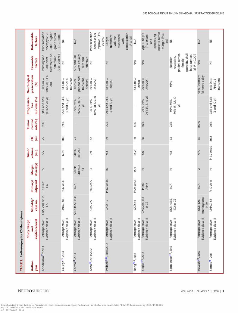
several series emerged and most of them report favorableoutcomes with 5-yr progression-free survival (PFS) rates rangingfrom 86% to 99%, and 10-yr PFS rate ranging from 69%to 97%.7,14-16 Although studies with long-term follow-up arerelatively scant, 2 reports have reported 15- and 20-yr PFS ratesranging from 87% to 92%.17,18 In a multi-institutional seriesof 4565 patients with intracranial meningiomas treated withSRS (1272 with CS meningiomas), Santacroce et al14 demon-strated 5- and 10-yr PFS rates of 95.2% and 88.6%, respectively,
2 | VOLUME 0 | NUMBER 0 | 2018 www.neurosurgery-online.com
Downloaded from https://academic.oup.com/neurosurgery/advance-article-abstract/doi/10.1093/neuros/nyy009/4938063by University of Toronto useron 29 March 2018
SRS FOR CAVERNOUS SINUS MENINGIOMA: ISRS PRACTICE GUIDELINE
FIGURE 1. Flow diagram of search process for identifying the final number of eligible studies.
NEUROSURGERY VOLUME 0 | NUMBER 0 | 2018 | 3
Downloaded from https://academic.oup.com/neurosurgery/advance-article-abstract/doi/10.1093/neuros/nyy009/4938063by University of Toronto useron 29 March 2018

LEE ET AL
TABLE 1. Level of Evidence
Level I evidence Evidence from 1 or more well-designed, randomized controlled clinical trials, including overviews of such trials.Level II evidence Evidence from 1 or more well-designed comparative clinical studies, such as nonrandomized cohort studies, case-control studies,
and other comparable studies, including less well-designed randomized controlled trials.Level III evidence Evidence from case series, comparative studies with historical controls, case reports, and expert opinion, as well as significantly
flawed randomized controlled trials.
and improved tumor control for skull base tumors compared toconvexity tumors.Two large single-institutional series have also been reported
demonstrating favorable outcomes. The first is from Pollocket al,15 which evaluated 115 patients with CS meningiomasdemonstrating a 5- and 10-yr local tumor control rate of 99%and 93%, respectively. The second series is from the Universityof Pittsburgh, which published on 272 patients treated withSRS, demonstrated a 5- and 10-yr PFS rate of 94% and 86%.16Interestingly, their data also demonstrated that patients whounderwent prior microsurgical procedures were less likely todemonstrate improvement in pre-existing cranial neuropathies,compared to those treated with SRS alone.16
Statistically significant (P < .05) factors associated withimproved SRS local control outcomes were identified andincluded higher marginal dose, small- to medium-sized tumors,WHO grade I, upfront SRS (irradiated tumor without surgicalresection), early SRS (cranial deficits < 1 yr), female sex, youngerage, and less conformal plans (Table 2). The incidence of neuro-logical deterioration, or development of new neurological deficitsin those series with long-term follow-up, has been relatively low.Approximately 80% to 100% of patients preserve neurologicalfunctions (Table 2).
The median marginal dose of single-fraction SRS ranged from11 to 19 Gy (Table 2). Data are conflicting with earlier seriessupporting a single fraction dose of >14 Gy as a significantpredictor of local control9,19 (level III evidence), while moremodern data support lower marginal doses ranging from 11 to 12Gy6,20-22 (level III evidence). If we consider all types of menin-gioma in aggregate, a series from Mayo Clinic23 showing that16 Gy delivered to the tumor margin provides long-term localcontrol (25 yr follow-up). Another series from Kuhn et al24suggests that 12 Gy was the minimum sufficient margin dosefor the treatment of meningiomas. Below 12 Gy is as low asone should consider using for meningiomas as there is probablyworsening of local control below this dose. However, theseconstitute level III evidence.
Primary or Adjuvant SRSAdvancements in neuroimaging improved the diagnosis of CS
meningioma. As a result, there are increasing reports of SRS forCS meningioma based on a magnetic resonance imaging (MRI)-based diagnosis. Kano et al16 evaluated the cranial nerve outcomesin patients who underwent SRS for CS meningiomas with or
without prior microsurgery. They observed improvement ratesspecific to cranial nerve deficits after SRS of 20% at 1 yr, 34% at2 yr, 36% at 3 yr, and 39% at 5 yr. Patients who hadnot undergone prior microsurgery had significantly higherimprovement rates of pre-existing cranial nerve symptoms andsigns (P = .001), suggesting that microdissection increases therisk of persistent cranial nerve dysfunction.16
The indications for SRS with or without prior resection varyappreciably from center to center. However, considering thegenerally benign nature of a CS meningioma and the hightumor control rate after SRS alone, clinician treatment preferenceleads to variations in treatment practices and inherent biases inthe published series detailing these treatment outcomes. In thisregard, further well-designed clinical trials would be necessary toascertain the treatment priority.
Radiation Tolerance of Optic Apparatus, Cranial Nerves,and Internal Carotid Artery Abutting CSMeningiomasThe proximity of CS meningioma to the optic apparatus,
cranial nerves, and internal carotid artery (ICA) needs specificconsideration when using solitary high doses of radiation to CStumors.25 Although CS tumors frequently abut the optic nerveand/or chiasm, Tishler et al26 reported that, with a dose tothe optic nerve and chiasm of 8 Gy or less, none of their 35patients developed a radiation-related optic neuropathy. Similarly,CS tumors typically encase adjacent cranial nerves. More recentstudies reported that marginal radiation doses of 10 to 14 Gy(maximum doses of 20-28) were well tolerated and had a lowrisk of radiation-related optic neuropathy.27-32 When prescribinga dose to a CS meningioma, one should be mindful of the opticapparatus and keep the radiation exposure less than 10 Gy.33 Theother cranial nerves of the CS seem to have greater tolerance forirradiation, and the treatment volume could include the majorityof the CS.33 For tolerance of other cranial nerves within theCS, there is no clear evidence showing the maximal radiationtolerance. The risk of permanent radiation-induced cranial nerveinjury is rare, and the incidence is less than 1% in the most seriesthat we collected (Tables 2 and 3). Tishler et al26 found that a doseup to 40 Gy seems to be tolerated when treating lesions involvingthe II and IV cranial nerves. The new onset motor cranial nervedeficits in most series accompany tumor progression and thusare not generally occurring as a radiation-induced deficit. In theabsence of tumor growth, sensory or motor cranial nerve palsies in
4 | VOLUME 0 | NUMBER 0 | 2018 www.neurosurgery-online.com
Downloaded from https://academic.oup.com/neurosurgery/advance-article-abstract/doi/10.1093/neuros/nyy009/4938063by University of Toronto useron 29 March 2018
SRS FOR CAVERNOUS SINUS MENINGIOMA: ISRS PRACTICE GUIDELINE
TABL
E2.
Radios
urge
ryforC
SMen
ingiom
a
Autho
r,Stud
yde
sign
and
Mod
ality/
Prim
ary/
Margin
Tumor
volume
FUTu
mor
control
Prog
ressive-
free
Neu
rological
preserva
tion
Favo
rable
Unfav
orab
leye
arev
iden
celeve
lcase
no.
adjuva
ntdo
se(Gy)
(mL)
(m)
rate
(%)
survival
(%)
(%)
factors
factors
Kond
ziolka
17,a2014
Retrospe
ctive,
Eviden
ceclassIII
GKS
:290
,86in
CSP:154A:
136
155.5
7591%
88%
and87%
(10an
d20
yr)
80%
(n=
188/234)3.1%
ARE
Prim
aryan
dad
juvant
treatm
ent:no
diffe
rence
(93%
vs89
%)
Priorrad
iatio
ntherap
y(P
<
.000
1),highe
rgrad
etumor
(P<.000
1)Gallego
45,2014
Retrospe
ctive,
Eviden
ceclassIII
LINAC
:82
P:47
A:35
1417.96
100
89%
95%
and84
%(5an
d10
yr)
83%
(n=
68/82),4
tran
sien
t
Nil
Nil
Correa
18,2014
Retrospe
ctive,
Eviden
ceclassIII
SRS:36
SRT:38
N/A
SRS:14
SRT:50.4-
54
SRS:6
SRT:23.6
73–
99%,92%
,92%
(5,10,15
yr)
100%
(9pa
tient
had
tran
sien
tde
ficits)
SRSan
dSR
Twereeq
ually
safean
deff
ectiv
e
N/A
Kano
16,2013/2012
Retrospe
ctive,
Eviden
ceclassIII
GKS
:272
P:173A:99
137.9
62–
96%,94%
,86
%(at3,5,10
yr)
89%
(n=
243/272)
Nil
Priorresectio
nde
crease
CNim
prov
e(14%
vs37%)
Pollo
ck15,23 ,2013/2012
Retrospe
ctive,
Eviden
ceclassIII
GKS
:115
P:69
A:46
169.3
8995%
99%
and93%
(5an
d10
yr)
88%
(n=
101/115)
Nil
Larger
treatm
ent
volume
associated
with
complication
(P=.01)
Rong
46b,2013
Retrospe
ctive,
Eviden
ceclassIII
GKS
:84
P:26
A:58
15.4
25.2
4081%
–83%
(n=
70/83)
N/A
N/A
Starke
47a ,2012
Retrospe
ctive,
Eviden
ceclassIII
GKS
:255,138
inCS
P:109
A:14
614
5.0
7886
%99
%,96%
,79%(3,5,10yr)
90%
(n=
230/255)
N/A
Age
>65
y/o
(P=0.00
1)an
dde
creasing
dose
totumor
margin(P
=.05)
Santacroce
14a ,2012
Retrospe
ctive,
Eviden
ceclassIII
GKS
:4565,
1272
inCS
N/A
144.8
6393%
95%,91%
,89
%(5,7.5,10
yr)
93%
Noprior
resection,
grad
eItum
or,
female,
sporad
ic,sku
llba
setumors
(allP
<0.00
1)
Nil
Hayashi
2b,2012
Retrospe
ctive,
Eviden
ceclassIII
GKS
:120,
men
ingiom
ain
19
N/A
12N/A
55100%
–95%
(transient
VIne
rvepa
lsy)
N/A
N/A
Santos
48,2011
Retrospe
ctive,
Eviden
ceclassIII
LINAC
:88
P:47
A:41
14P:3.7A:5.9
86.8
90%
93%
and83%
(5an
d10
yr)
81%
(n=
71/88),6
tran
sien
t
Nil
Nil
NEUROSURGERY VOLUME 0 | NUMBER 0 | 2018 | 5
Downloaded from https://academic.oup.com/neurosurgery/advance-article-abstract/doi/10.1093/neuros/nyy009/4938063by University of Toronto useron 29 March 2018

LEE ET ALTA
BLE2–continue
d
Autho
r,Stud
yde
sign
and
Mod
ality/
Prim
ary/
Margin
Tumor
volume
FUTu
mor
control
Prog
ressive-
free
Neu
rological
preserva
tion
Favo
rable
Unfav
orab
leye
arev
iden
celeve
lcase
no.
adjuva
ntdo
se(Gy)
(mL)
(m)
rate
(%)
survival
(%)
(%)
factors
factors
Williams10 ,2011
Retrospe
ctive,
Eviden
ceclassIII
GKS
:138
P:54
A:84
13.7
7.5
8486
%95%
and69
%(5an
d10
yr)
90%
(n=
124/138)
Youn
gera
geha
dbe
tter
tumor
control
(P=0.022)
New
CNpa
lsy
isrelatedto
tumor
increase
(P<
.001)
Skeie19 ,2010
Retrospe
ctive,
Eviden
ceclassIII
GKS
:100
P:40
A:60
12.4
7.4
8284
%99
%,94%
,92%
(1,5,10yr)
94%
Highe
rmargindo
seAtypical
tumor
Spiege
lman
n49 ,2010/200
2Re
trospe
ctive,
Eviden
ceclassIII
LINAC
:102
P:69
A:33
13.5
767
98%
–95%
(n=
97/102)
Early
radiosurge
ry(<
1yrC
Nde
ficiton
set)
Nil
Takana
shi50
,200
9Re
trospe
ctive,
Eviden
ceclassIII
GKS
:38
P:19
A:19
14.8
8.6
5296
%–
100%
N/A
N/A
Kimba
ll5,1,200
9Re
trospe
ctive,
Eviden
ceclassIII
LINAC
:49
N/A
12.5
5.9
5098
%100%
and98
%(5an
d10
yr)
98%
N/A
N/A
Han
52,200
8Re
trospe
ctive,
Eviden
ceclassIII
GKS
:63,12in
CSP:43
A:20
12.7
6.5
7790
%90
%(5yr)
85%
Nil
Age
>70
y/o
isun
favo
rable
fora
CNim
prov
emen
tKo
ndziolka
53a ,2008
Retrospe
ctive,
Eviden
ceclassIII
GKS
:972,308
inCS
P:536A:
424
147.4
48P:97%
A:
93%
GrII:
50%
GrIII:
17%
At10yr
P:95%
A:91%
92%
Nil
Highe
rgrade
oftumor,
larger
tumor
volume,an
dmultip
lemen
ingiom
aHaseg
awa5,200
7Re
trospe
ctive,
Eviden
ceclassIII
GKS
:115
P:49
A:66
1314
62–
94%
and92%
(5an
d10
yr)
89%
(n=
97/109
)Sm
allto
med
ium-sized
tumors.No
prior
resectionha
dsign
ificant
post-GK
improv
emen
t
Nil
Zach
enho
fer54 ,2006
Retrospe
ctive,
Eviden
ceclassIII
GKS
:36
P:11A:25
P:19
A:13
Mixed
:17P:16
A:24
Mixed
:20
103
94%
–96
%(n
=1/36)
N/A
N/A
Metellus37 ,2005
Retrospe
ctive,
Eviden
ceclassIII
GKS
:36FR
T:38
P:23GK/18FR
A:
13GK/20FR
GK:15FR
:53
GK:5.2FR
:13.5
GK:64
FR:89
GK:94
.4FR
:94.7
–GK:100%
FR:
97.4%
GKS
radiosurge
ryprov
ides
better
radiolog
ical
respon
se(P
=0.03).No
sign
ificance
betw
eenp
anda
treatm
ent
N/A
6 | VOLUME 0 | NUMBER 0 | 2018 www.neurosurgery-online.com
Downloaded from https://academic.oup.com/neurosurgery/advance-article-abstract/doi/10.1093/neuros/nyy009/4938063by University of Toronto useron 29 March 2018

SRS FOR CAVERNOUS SINUS MENINGIOMA: ISRS PRACTICE GUIDELINE
TABL
E2–continue
d
Autho
r,Stud
yde
sign
and
Mod
ality/
Prim
ary/
Margin
Tumor
volume
FUTu
mor
control
Prog
ressive-
free
Neu
rological
preserva
tion
Favo
rable
Unfav
orab
leye
arev
iden
celeve
lcase
no.
adjuva
ntdo
se(Gy)
(mL)
(m)
rate
(%)
survival
(%)
(%)
factors
factors
Pollo
ck55,200
5Re
trospe
ctive,
Eviden
ceclassIII
GKS
:49
P:49
A:0
15.9
10.2
58100%
a(2
resections
fors/s)
98%,85%
,80
%(1,3,7
yr)
86%
(n=
42/49)
Nil
Nil
Liu5
6b,200
5Re
trospe
ctive,
Eviden
ceclassIII
GKS
:175,
men
ingiom
ain
88
P:85
A:90
12.1
6.6
32.5
94%
––
N/A
N/A
Kreil20
a ,2005
Retrospe
ctive,
Eviden
ceclassIII
GKS
:200
P:101A
:99
126.5
9598
%99
%an
d97%
(5an
d10
yr)
95%
(n=
191/200)
N/A
N/A
Kuo5
7b,200
4Re
trospe
ctive,
Eviden
ceclassIII
GKS
:139,
men
ingiom
ain
57
P:20
A:119
153.4
4297%
–98
%N/A
N/A
Maruy
ama5
8 ,2004
Retrospe
ctive,
Eviden
ceclassIII
GKS
:40
P:17A:23
165.4
47–
94%
(5yr)
75%
(n=
30/40)
prim
aryCS
men
ingiom
astumors
compressing
thebrainstem
orsm
aller
than
10cm
3
Dibiase
59a ,2004
Retrospe
ctive,
Eviden
ceclassIII
GKS
:137,CSin
29P:85
A:52
144.5
5491.7%
86.2at
5yr
N/A
Less
conformal
plan
sto
cover
thedu
raltail
malepa
tients,
patie
ntswith
aCI
<1.4
,and
tumor
size
greatertha
n10
ccIwai6 ,2003/200
1Re
trospe
ctive,
Eviden
ceclassIII
GKS
:43
P:21A:22
1114.7
5091%
92%
(5yr)
91%
(n=
39/43)
N/A
N/A
Flicking
er60
a ,2003
Retrospe
ctive,
Eviden
ceclassIII
GKS
:219,CSin
75P:219A:0
145
2997%
93%
(5an
d10
yr)
91%
(5an
d10
yr)
Stereo
tactic
MRan
dlow
dose
tend
toha
velower
rate
ofsequ
elae
N/A
Lee7,200
2Re
trospe
ctive,
Eviden
ceclassIII
GKS
:159
P:83
A:76
136.5
3594
%A:79%
at5yr
P:97%
at5yr
91%
(n=
145/159)
Averag
etumor
diam
eter
<3
cmor
volume
<15cm
3
Priorresectio
n
Nicolato6
,1,200
2/2002
Retrospe
ctive,
Eviden
ceclassIII
GKS
:138
P:68
A:70
14.8
8.1
4897%
96%
(5yr)
96.5%
(n=
107/111)
Noprior
resectionha
dbe
tter
neurolog
ical
recovery
N/A
Villaviscen
cio6
2a,200
1Re
trospe
ctive,
Eviden
ceclassIII
LINAC
:56,CS
in12
P:20
A:36
1560
.426
95%
–100%
N/A
N/A
NEUROSURGERY VOLUME 0 | NUMBER 0 | 2018 | 7
Downloaded from https://academic.oup.com/neurosurgery/advance-article-abstract/doi/10.1093/neuros/nyy009/4938063by University of Toronto useron 29 March 2018

LEE ET AL
TABL
E2–continue
d
Autho
r,Stud
yde
sign
and
Mod
ality/
Prim
ary/
Margin
Tumor
volume
FUTu
mor
control
Prog
ressive-
free
Neu
rological
preserva
tion
Favo
rable
Unfav
orab
leye
arev
iden
celeve
lcase
no.
adjuva
ntdo
se(Gy)
(mL)
(m)
rate
(%)
survival
(%)
(%)
factors
factors
Shin
9 ,2001
Retrospe
ctive,
Eviden
ceclassIII
GKS
:40
P:12,A
:28
184.3
42Margindo
se>14
Gy,0%
recur,Margin
dose
10-12Gy:
20%
recur.No
SRS:100%
recur
86%
and82%
(3an
d10
yr)
80%
(n=
32/40;mild
tran
sien
tin7
pts)
margindo
se>14
Gy
Histological
maligan
ancy
and
partialtreatmen
t,suprasellar
extension,
extensionin
>3
directionou
tside
CS,
Chen
63b,200
1Re
trospe
ctive,
Eviden
ceclassIII
GKS
:69,
men
ingiom
ain
35
P:22
A:47
154.7
30–
–100%
Radiation
expo
seis
relatedto
distan
cebe
twee
nCN
2an
dtumor,
notT
V
N/A
Roch
e8,64 ,2000
Retrospe
ctive,
Eviden
ceclassIII
GKS
:80
P:50
A:30
145.8
3195%
93%
(5yr)
94%
(n=
75/80)
N/A
N/A
Marita
64a ,1999
Retrospe
ctive,
Eviden
ceclassIII
GKS
:88,CS
in32
P:39
A:49
1610
3598
%95%
(5yr)
88%
(n=
78/88)
N/A
N/A
Liscak
22,199
9Re
trospe
ctive,
Eviden
ceclassIII
GKS
:67
P:43
A:24
127.8
19100%
–96
%N/A
N/A
Pend
l65 ,1998
Retrospe
ctive,
Eviden
ceclassIII
GKS
:43
P:17A:24
13.2
15.4
39100%
–100%
N/A
N/A
Chan
g66 ,1998
Retrospe
ctive,
Eviden
ceclassIII
LINAC
:24
–17.7
6.8
45.6
100%
100%
(2yr)
92%
(n=
22/24),the
othe
r4tran
sien
tCN
deficits
N/A
N/A
Kurita6
7 ,1997
Retrospe
ctive,
Eviden
ceclassIII
GKS
:18
P:3A:15
17Diameter:
23mm
35–
86%
(5yr)
94%
(n=
17/18)
N/A
N/A
Dum
a4,199
3Re
trospe
ctive,
Eviden
ceclassIII
GKS
:34
P:6A:28
10-20
Diameter
<35mm
26100%
–94
%(n
=2/34,
othe
r2tran
sien
t)
N/A
N/A
A:adjuv
ant,CI:con
form
ityinde
x,CN
:cranialne
rve,CS
:caverno
ussinu
s,FR
T:fractio
natedradiothe
rapy,G
KS:G
ammaKn
iferadiosurge
ry,G
y:gray,LINAC
:Linearp
artic
leaccelerator,N/A:not
available,P:prim
ary,pt:
patie
nt,SRS
:stereotactic
radiosurge
ry,M
R:mag
netic
resona
nce;SR
T:stereo
tacticradiothe
rapy,yr:year
a Who
lemen
ingiom
agrou
presult,no
tonlyforC
Smen
ingiom
a.bWho
leCS
tumor
grou
presult,no
tonlyforC
Smen
ingiom
a.
8 | VOLUME 0 | NUMBER 0 | 2018 www.neurosurgery-online.com
Downloaded from https://academic.oup.com/neurosurgery/advance-article-abstract/doi/10.1093/neuros/nyy009/4938063by University of Toronto useron 29 March 2018

SRS FOR CAVERNOUS SINUS MENINGIOMA: ISRS PRACTICE GUIDELINE
TABL
E3.
Fraction
ated
Radiothe
rapy
forC
SMen
ingiom
a
Autho
r,Stud
yde
sign
and
Mod
ality/
Prim
aryvs
Dos
eTu
mor
FUTu
mor
control
Prog
ressive-
free
Cran
ialn
erve
preserva
tion
Favo
rable
Unfav
orab
leye
arev
iden
celeve
lcase
no.
adjuva
nt(Gy)
volume
(mon
ths)
rate
(%)
survival
(%)
(%)
factors
factors
Correa,20141
8Re
trospe
ctive,
Eviden
ceclassIII
SRS:36
SRT:38
N/A
SRS:14
SRT:50.4-54
SRS:6
SRT:23.6
73–
99%,92%
,92%
(5,10,15yr)
100%
(9pa
tient
hadtran
sien
tde
ficits)
SRSan
dSR
Twereeq
ually
safean
deff
ectiv
e
N/A
Combs,20133
6Re
trospe
ctive,
Eviden
ceclassIII
FSRT
:376
IMRT
:131
P:238A:269
57.6
53.4
107
–98
%,95%
,94%
,88
%(1yr,3yr,5yr,
10yr)
N/A
benign
histolog
y,female
N/A
Slater,201213
Retrospe
ctive,
Eviden
ceclassIII
Proton
:72
P:51A:21
ForP
:57Fo
rA:59
27.6
7490
%at
5thyear
92%
(68/74),3
optic
,2tempo
rary
ARE
,1transient
diplop
ia.3
panh
ypop
itu-
itarism
N/A
Atypical
histolog
ycause
poor
tumor
control,high
erop
ticradiation
dose
cause
optic
neurop
athy
Metellus,2010
68Re
trospe
ctive,
Eviden
ceclassIII
FCR:53
P:28
A:25
52.9Gyin
29.4
fractio
ns
12.6
8396
%98
%an
d96
%(5
and10
yr)
98%
(perman
ent),
94%
(transient)
N/A
N/A
Litre6
9,200
9Re
trospe
ctive,
Eviden
ceclassIII
FSR:100
P:74
A:26
45Gy
N/A
3397%
94%
(3an
d7yr)
97%
N/A
N/A
Milker-Zab
el70,200
6/2005
Retrospe
ctive,
Eviden
ceclassIII
FSRT
:57
P:29
A:28
57.6
35.2
78100%
–100%
N/A
N/A
Brell.7
12006
/200
3Re
trospe
ctive,
Eviden
ceclassIII
FSRT
:30
P:29
A:28
5211.3
5096
%93%
(4yr)
96%
none
none
Metellus3
7 ,2005
Retrospe
ctive,
Eviden
ceclassIII
GKS
:36FR
:38
P:23GK/18FR
A:
13GK/20FR
GK:15FR
:53
GK:5.2FR
:13.5
GK:64
FR:
89GK:94
.4FR
:94
.7–
GK:100%
FR:
97.4%
GKS
radiosurge
ryprov
ides
better
radiolog
ical
respon
se(P
=0.03).No
sign
ificance
betw
eenpan
datreatm
ent
N/A
Selch7
2 ,2004
Retrospe
ctive,
Eviden
ceclassIII
SRT:45
P:16
A:29
50.4
14.5
3698
%97%
(3yr)
98%
N/A
N/A
Dufou
r73 ,2001
Retrospe
ctive,
Eviden
ceclassIII
RT:31
P:14
A:17
52–
73–
93%
(10yr)
100%
Tumor
control
ratesareeq
ual
with
/with
out
surgical
resection
N/A
Mag
uire
74,199
9Re
trospe
ctive,
Eviden
ceclassIII
RT:28
P:22
A:6
53.1
–41
–81%
(8yr)
Orbita
lsac
fibrosis,an
dde
clineof
cogn
itive
func
tion
Non
eNon
e
A:adjuv
ant,CI:con
form
ityinde
x,CN
:cranialne
rve,CS
:caverno
ussinu
s,FC
R:fractio
natedconformalradiothe
rapy,FRT
:fractiona
tedradiothe
rapy,FSR
T:fractio
natedstereo
tacticradiothe
rapy,G
KS:G
ammaKn
iferadiosurge
ry,G
y:gray,IMRT
:inten
sity-m
odulated
radiationtherap
y,LINAC
:Linearp
artic
leaccelerator,N/A:n
otavailable,P:prim
ary,pt:p
atient,R
T:radiothe
rapy,SRS
:stereotactic
radiosurge
ry,SRT
:stereotactic
radiothe
rapy,yr:year
NEUROSURGERY VOLUME 0 | NUMBER 0 | 2018 | 9
Downloaded from https://academic.oup.com/neurosurgery/advance-article-abstract/doi/10.1093/neuros/nyy009/4938063by University of Toronto useron 29 March 2018

LEE ET AL
SRS-treated CSmeningiomas occur very infrequently when usingcontemporary dose and delivery techniques.14,17
For the ICA, the maximum tolerated dose of radiation iscontroversial. Some rare case reports have described ICA stenosisafter SRS for parasellar, suprasellar, and CS lesions.34,35 However,this clinical question lacks a large and long-term series for analysis.In current radiosurgery practice, the high-dose radiation volumeusually includes the ICA; the risk of long-term changes in thevessel wall of the ICA appears, anecdotally, to be a very rarephenomenon.
Comparing SRS, Proton SRS/Radiotherapy, FractionatedSRT, and Conventional RadiotherapyThere are no level I or II comparisons of SRS, proton
SRS or radiotherapy, SRT, or other sophisticated radiotherapytechniques (eg, intensity-modulated radiation therapy [IMRT])for CS meningioma (Table 3).
Non–case-matched studies18,36,37 from 3 level III studieswith tumor control rates for SRS, or SRT, or IMRT demon-strated that they were similarly safe and efficient techniques intreatment of a CSmeningioma although various methods, variousdoses, various schemes of radiation, various indications, volumes,and prior management. Metellus’ report37 showed neurologicalimprovement for 63% patients who underwent SRT, and for 54%patients who underwent SRS (P > .05). Combs et al,36 in 2013,compared the efficacy and safety between both the groups (105CS meningiomas) who underwent either SRT or IMRT witha median total dose of 57.6 Gy, and they found no significantdifferences. Correa et al,18 in 2014, also published similar resultsin both groups who underwent either SRS or SRT. However,radiologically 29% of patients who underwent SRT, and 53%of patients who underwent SRS, showed tumor shrinkage (P <
.04).37 The result implied that SRS offered a higher rate of tumorshrinkage, but no significance in clinical improvement.Based upon these limited data, high-level evidence is needed
to define the optimal radiation approach for patients with a CSmeningioma. Based upon current available evidence, SRS andSRT confer favorable benefit to risk profiles for most patients withCS meningioma, eligible for either therapy.
DISCUSSION
The clinical management of a patient with a CS menin-gioma is challenging. The fear of causing massive bleeding andcritical neurovascular structural damage has led both surgeonsand patients to proceed cautiously with attempts at resection.SRS and SRT approaches have been proven in many retro-spective studies to have a favorable therapeutic impact with aminimal complication rate. From this systematic review, after SRSmost of the reports had favorable outcomes with 5-yr PFS ratesranging from 86% to 99%, and 10-yr PFS rate ranging from69% to 97%. Post-SRS neurological preservation rates rangedfrom 80% to 100% (Table 2). Median margin dose selection
is dependent on the tumor volume, anatomic relationship tothe adjacent associated neurovascular structures, and physicianpreference. Reported SRS doses generally varied from 11 to 19Gy; however, the optimal single-session SRS dose for a CSmenin-gioma is a subject of debate and requires careful selection on a caseby case basis. Fortunately, more and more modern data supportthat a lower marginal dose ranging from 12 Gy is sufficient fora benign CS meningioma.6,20-22 Furthermore, by using a lowerdose in the CS or by hypofractionating with modern SRS devices,more tumors become eligible for radiosurgery due to limits ofoptic apparatus to SRS.38,39SRS can be delivered as either adjuvant therapy for residual
tumors after subtotal resection, at the time of progression forresidual disease observed, or as primary therapy for unresectabletumors. Little to no substantive differences in local tumor controlor neurological outcome have been reported following SRS forprimary therapy as compared to adjuvant SRS for WHO gradeI meningioma. Many of the published clinical series aggregateresults of patients treated with upfront SRS alone, SRS of aresidual after resection (but before evidence of demonstrablegrowth on serial neuroimaging), and SRS at the time of tumorprogression. In part, the ambiguity pertaining to the naturalhistory of CS meningiomas and the treatment preferences ofclinicians lead to variations in treatment practices and inherentbiases in the published series detailing these treatment outcomes.Patient preference and fitness for a radiation modality will alsonaturally impact the decision to proceed with a primary oradjuvant radiation treatment.With primary SRS for a CS meningioma, histopathologic
evaluation for unfavorable features is unavailable (eg, oratypical/anaplastic meningioma). However, after SRS, the tumorresponses may differ compared to other CS tumors, and arevaluable in confirming the initial radiographic impression.Figure 2 illustrates 3 different response patterns of these CStumors. The tumor volume of a CS meningioma after SRS istypically quite stable, and they rarely regressed within 2 yr. Ifan unpredictable tumor response is noted, we recommend tore-examine the MRI, presentation of clinical course, and re-establishing the pathological diagnosis with biopsy, as necessary.Currently, the optimum follow-up strategy is debated: a
median of 95% and 90% of tumors achieved a steady statein 5 and 10 yr, respectively. It would be reasonable to observethe tumor for the initial several years after radiosurgery, andit provides sufficient evidence regarding its propensity to growand therefore the requirement for further treatment or moreradiological follow-up.40 Routine radiological follow-up could beextended to a longer interval after the tumor has attained a steadystate and clinical follow-up with routine neurological exams andophthalmological assessment should be continued.In this review, the role of hypofractionated SRS and SRT for
a CS meningioma was also explored,18,36,37 particularly thoseexhibiting larger volumes or in close proximity to critical struc-tures. For those CS meningioma patients with a larger volumetumor, a more diffusely infiltrative one, or one with suprasellar or
10 | VOLUME 0 | NUMBER 0 | 2018 www.neurosurgery-online.com
Downloaded from https://academic.oup.com/neurosurgery/advance-article-abstract/doi/10.1093/neuros/nyy009/4938063by University of Toronto useron 29 March 2018

SRS FOR CAVERNOUS SINUS MENINGIOMA: ISRS PRACTICE GUIDELINE
FIGURE 2. The tumor response after SRS in different CS tumors: rapid tumor regression is usually found in a CS hemangioma. A CS meningioma is stable afterSRS, and they rarely regressed with 2 to 3 yr. A radiated neuroma is usually swollen for a period of time (eg, the first 1-2 yr after SRS) and then a noticeable shrinkagecould be seen if follow-up persisted for >2 yr.
brainstem extension, SRT can be considered so as to minimize therisk of complications and optimize tumor coverage.41 For largermeningiomas or for ones with pre-existing edema, hypofrac-tionated SRT may have less likelihood of causing postradiosur-gical edema than single-fraction SRS for meningioma.42 Basedupon the number of publications meeting eligibility criteria forthis guidelines project, contemporary management with SRSrepresents a common approach for management of small tomoderately sized CS meningiomas. In general, clinicians mustselect the approach (SRS, SRT, IMRT, or proton) that permitsa highly targeted irradiation of the CS meningioma while stillachieving a dose considered tolerable to adjacent critical struc-tures based upon radiotoxicity guidelines such as the QuantitativeAnalyses of Normal Tissue Effects in the Clinic studies.43 DeSalles et al,41 in 2002, attempted to develop a grading system toguide treatment selection, either SRS or SRT. They concludedthat a meningioma well contained in the CS, with typical radio-logical characteristics, may be treated successfully with SRS alonewith excellent outcomes. On the other hand, SRTmay be favoredwhen adjuvant treatment is necessary after subtotal resection oftumor encasing eloquent structures, where SRS is not advisable.No matter which radiation modalities clinicians choose, effortshould be made to minimize irradiated volumes to prevent long-term complications, maximize the therapeutic efficacy to target
tissue, and lessen the burden of the procedure(s) for the patientwhenever possible.
Key Areas for Future Investigation• The timing of SRS or SRT after prior resection warrants furtherinvestigation.
• While SRS and radiation therapy are frequently used as anupfront treatment for those with a CS meningioma, there isno level I evidence report of primary SRS as a management fora CS meningioma. Longer follow-up report for this treatmentapproach is warranted.
• The role of hypofractionated SRS for a CSmeningioma, partic-ularly those exhibiting larger volumes or in close proximity tocritical structures, has been explored in limited publications.Optimal dose and fractionation schemes particularly for SRSof a CS meningioma should be explored.
• The neurocognitive effects of SRS and SRT in CS menin-gioma patients warrants further study with the use of validatedneurocognitive tests and appropriate assessment intervals.There are little data on the neurocognitive functions aftertreating neuropathology in the CS. In 1 study of SRS forpituitary adenomas many of which resided in the CS, therewas neurocognitive preservation in the patients after SRS.44Although it is thought that the much lower integral dose of
NEUROSURGERY VOLUME 0 | NUMBER 0 | 2018 | 11
Downloaded from https://academic.oup.com/neurosurgery/advance-article-abstract/doi/10.1093/neuros/nyy009/4938063by University of Toronto useron 29 March 2018

LEE ET AL
TABLE 4. Recommendations for Management of CSMeningioma
Evidence level
Level III SRS/SRT is recommended as a primary/upfront treatment option for an asymptomatic, or mildly symptomatic CS meningioma. Therecurrence rate is not appreciably different between primary or adjuvant therapy for a CS meningioma
Level III Resection should be considered for the treatment of larger and symptomatic CS meningioma in patients both receptive to, and medicallyeligible, for open surgery
Level III SRS/SRT delivered to a CS meningioma has a low risk of complications; most cranial nerve functions are preserved or improved due totumor shrinkage. Carotid artery stenosis after SRS is rare.
Level III When no residual tumor is observed, or only a small tumor lining on the dura of the CS exists postoperatively, serial neuroimaging studiesis not unreasonable. At the time of recurrence or progression of residual tumor, SRS/SRT should be considered
Level III In patients with a CS meningioma that has rapidly and substantially recurred after prior treatment, a subtotal surgical resection or biopsymay be considered. More aggressive features of the tumor (transformation of the tumor fromWHO grade I to a higher grade) should beruled out. These tumors have a predilection for progression and postoperative SRS/SRT with a higher dose should be strongly considered.
Level III The technique for SRS or SRT delivery will depend upon the tumor histology, tumor volume and proximity of the tumor to adjacent criticalstructures (eg, the optic chiasm). SRS using single session marginal doses of 11 to 16 Gy offers a local tumor control rate of 90% or higher at5 yr post-SRS.
SRS probably mitigates neurocognitive deficits as comparedto radiotherapy, more definitive studies are required to studyneurocognition in a prospective fashion in SRS-treated CSmeningioma patients.
• Limits of dose to the optic apparatus also need better clarifi-cation, as it defines the approach, either single or hypofrac-tionated SRS or SRT.
• Higher quality evidence comparing SRS, SRT, IMRT, andcharged particle techniques and devices is needed to help guideclinicians on the specific indications and limitations of eachapproach for patients with a CS meningioma.
• Comparisons of SRS-treated CS meningioma patients to acontrol group of untreated meningioma patients.
• Comparative studies are required of specific radiosurgeryand radiation therapy devices to assess for differences inoutcome. Similarly, comparative studies evaluating the effectsof proficiency and volume of a center on outcome should beperformed.
CONCLUSION
SRS plays an important role in the management of patientswith a CS meningioma. SRS is typically performed in patientswith demonstrable residual tumor or tumor recurrence afterresection. However, the upfront treatment using SRS for a CSmeningioma has gained popularity in recent years. For thosewith a radiographically diagnosed CS meningioma, the post-SRS tumor response can reconfirm the diagnosis of menin-gioma. Radiographic signs of progression in the setting of youngerpatients or patients with symptoms attributable to progressionshould be more strongly considered for intervention. Post-SRScranial nerve deterioration is rare, while improvement in cranialnerve function is not uncommon. Longer and more meticulousfollow-up report is warranted.
Higher levels of evidence are needed to define the optimaltreatment approach for patients with CS meningiomas. Basedupon available evidence, SRS and SRT confer favorable risk-benefit profiles than conventional radiotherapy for most patientswith a CSmeningioma. Further insights may be achieved throughprospective radiosurgical registries (Table 4).
DisclosuresThis evidence-based clinical practice guideline was supported by the Interna-
tional Stereotactic Radiosurgery Society. No funding from outside commercialsources to support the development of this work. Dr Martínez Álvarez is aconsultant for Elekta AB. Dr Sahgal has grants from Elekta AB and educationhonoraria from previous education seminars from Elekta AB, Varian MedicalSystems, Accuray, and Medtronic (kyphoplasty division). Dr Slotman has aresearch grant and speaker honorarium from Varian Medical Systems and speakerhonorarium from ViewRay. Dr Paddick is a consultant for and has received aresearch grant from Elekta, AB. The other authors have no personal, financial, orinstitutional interest in any of the drugs, materials, or devices described in thisarticle.
DisclaimerThese guidelines should not be considered inclusive of all methods of care or
exclusive of other methods or care reasonably directed to obtain similar results.The physician must make the ultimate judgment depending on characteristicsand circumstances of individual patients. Adherence to this guideline will notensure successful treatment in every situation. The authors of this guideline andthe International Society of Stereotactic Radiosurgery assume no liability for theinformation, conclusions, and recommendations contained in this report.
REFERENCES1. Mathiesen T, Lindquist C, Kihlstrom L, Karlsson B. Recurrence of cranial base
meningiomas. Neurosurgery. 1996;39(1):2-7; discussion 8-9.2. De Jesus O, Sekhar LN, Parikh HK, Wright DC, Wagner DP. Long-term
follow-up of patients with meningiomas involving the cavernous sinus: recurrence,progression, and quality of life.Neurosurgery. 1996;39(5):915-919; discussion 919-920.
3. DeMonte F, Smith HK, al-Mefty O. Outcome of aggressive removal of cavernoussinus meningiomas. Journal of neurosurgery. 1994;81(2):245-251.
12 | VOLUME 0 | NUMBER 0 | 2018 www.neurosurgery-online.com
Downloaded from https://academic.oup.com/neurosurgery/advance-article-abstract/doi/10.1093/neuros/nyy009/4938063by University of Toronto useron 29 March 2018

SRS FOR CAVERNOUS SINUS MENINGIOMA: ISRS PRACTICE GUIDELINE
4. Duma CM, Lunsford LD, Kondziolka D, Harsh GRt, Flickinger JC. Stereo-tactic radiosurgery of cavernous sinus meningiomas as an addition or alternative tomicrosurgery. Neurosurgery. 1993;32(5):699-704; discussion 704-695.
5. Hasegawa T, Kida Y, Yoshimoto M, Koike J, Iizuka H, Ishii D. Long-termoutcomes of Gamma Knife surgery for cavernous sinus meningioma. Journal ofneurosurgery. 2007;107(4):745-751.
6. Iwai Y, Yamanaka K, Ishiguro T. Gamma knife radiosurgery for the treatment ofcavernous sinus meningiomas.Neurosurgery. 2003;52(3):517-524; discussion 523-514.
7. Lee JY, Niranjan A, McInerney J, Kondziolka D, Flickinger JC, Lunsford LD.Stereotactic radiosurgery providing long-term tumor control of cavernous sinusmeningiomas. Journal of neurosurgery. 2002;97(1):65-72.
8. Roche PH, Regis J, Dufour H, et al. Gamma knife radiosurgery in themanagement of cavernous sinus meningiomas. Journal of neurosurgery. 2000;93Suppl 3:68-73.
9. Shin M, Kurita H, Sasaki T, et al. Analysis of treatment outcome afterstereotactic radiosurgery for cavernous sinus meningiomas. Journal of neurosurgery.2001;95(3):435-439.
10. Williams BJ, Yen CP, Starke RM, et al. Gamma Knife surgery for parasellarmeningiomas: long-term results including complications, predictive factors, andprogression-free survival. Journal of neurosurgery. 2011;114(6):1571-1577.
11. O’Sullivan MG, van Loveren, Tew HR, Jr JM. The surgical resectability of menin-giomas of the cavernous sinus.Neurosurgery. 1997;40(2):238-244; discussion 245-237.
12. Barnett GH, Linskey ME, Adler JR, et al. Stereotactic radiosurgery–an organizedneurosurgery-sanctioned definition. Journal of neurosurgery. 2007;106(1):1-5.
13. Slater JD, Loredo LN, Chung A, et al. Fractionated proton radiotherapy forbenign cavernous sinus meningiomas. International journal of radiation oncology,biology, physics. 2012;83(5):e633-637.
14. Santacroce A, Walier M, Regis J, et al. Long-term tumor control of benignintracranial meningiomas after radiosurgery in a series of 4565 patients. Neuro-surgery. 2012;70(1):32-39; discussion 39.
15. Pollock BE, Stafford SL, Link MJ, Garces YI, Foote RL. Single-fractionradiosurgery of benign cavernous sinus meningiomas. Journal of neurosurgery.2013;119(3):675-682.
16. Kano H, Park KJ, Kondziolka D, et al. Does prior microsurgery improve orworsen the outcomes of stereotactic radiosurgery for cavernous sinus meningiomas?Neurosurgery. 2013;73(3):401-410.
17. Kondziolka D, Patel AD, Kano H, Flickinger JC, Lunsford LD. Long-termOutcomes After Gamma Knife Radiosurgery for Meningiomas. American journalof clinical oncology. 2014.
18. Correa SF, Marta GN, Teixeira MJ. Neurosymptomatic carvenous sinus menin-gioma: a 15-years experience with fractionated stereotactic radiotherapy and radio-surgery. Radiation oncology. 2014;9:27.
19. Skeie BS, Enger PO, Skeie GO, Thorsen F, Pedersen PH. Gamma knifesurgery of meningiomas involving the cavernous sinus: long-term follow-up of 100patients. Neurosurgery. 2010;66(4):661-668; discussion 668-669.
20. Kreil W, Luggin J, Fuchs I, Weigl V, Eustacchio S, Papaefthymiou G. Longterm experience of gamma knife radiosurgery for benign skull base meningiomas.Journal of neurology, neurosurgery, and psychiatry. 2005;76(10):1425-1430.
21. Hayashi M, Chernov M, Tamura N, et al. Gamma knife radiosurgery for benigncavernous sinus tumors: treatment concept and outcomes in 120 cases. Neurologiamedico-chirurgica. 2012;52(10):714-723.
22. Liscak R, Simonova G, Vymazal J, Janouskova L, Vladyka V. Gamma kniferadiosurgery of meningiomas in the cavernous sinus region. Acta neurochirurgica.1999;141(5):473-480.
23. Pollock BE, Stafford SL, Link MJ. Stereotactic radiosurgery of intracranial menin-giomas. Neurosurg Clin N Am. 2013;24(4):499-507.
24. Kuhn EN, Taksler GB, Dayton O, et al. Patterns of recurrence after stereotacticradiosurgery for treatment of meningiomas. Neurosurgical focus. 2013;35(6):E14.
25. Bentzen SM, Constine LS, Deasy JO, et al. Quantitative Analyses of NormalTissue Effects in the Clinic (QUANTEC): an introduction to the scientific issues.International journal of radiation oncology, biology, physics. 2010;76(3 Suppl):S3-9.
26. Tishler RB, Loeffler JS, Lunsford LD, et al. Tolerance of cranial nerves of thecavernous sinus to radiosurgery. International journal of radiation oncology, biology,physics. 1993;27(2):215-221.
27. Jinhu Y, Jianping D, Xin L, Yuanli Z. Dynamic enhancement features ofcavernous sinus cavernous hemangiomas on conventional contrast-enhanced MRimaging. AJNR. American journal of neuroradiology. 2008;29(3):577-581.
28. Leavitt JA, Stafford SL, Link MJ, Pollock BE. Long-term evaluation of radiation-induced optic neuropathy after single-fraction stereotactic radiosurgery. Interna-tional journal of radiation oncology, biology, physics. 2013;87(3):524-527.
29. Leber KA, Bergloff J, Pendl G. Dose-response tolerance of the visual pathwaysand cranial nerves of the cavernous sinus to stereotactic radiosurgery. Journal ofneurosurgery. 1998;88(1):43-50.
30. Mori K, Handa H, Gi H, Mori K. Cavernomas in the middle fossa. Surgicalneurology. 1980;14(1):21-31.
31. Nakamura N, Shin M, Tago M, et al. Gamma knife radiosurgery for cavernoushemangiomas in the cavernous sinus. Report of three cases. Journal of neurosurgery.2002;97(5 Suppl):477-480.
32. Pollock BE, Link MJ, Leavitt JA, Stafford SL. Dose-volume analysis of radiation-induced optic neuropathy after single-fraction stereotactic radiosurgery. Neuro-surgery. 2014;75(4):456-460; discussion 460.
33. Benedict SH, Yenice KM, Followill D, et al. Stereotactic body radiation therapy:the report of AAPM Task Group 101. Med Phys. 2010;37(8):4078-4101.
34. Lombardi D, Giovanelli M, de Tribolet N. Sellar and parasellar extra-axialcavernous hemangiomas. Acta neurochirurgica. 1994;130(1-4):47-54.
35. Abeloos L, Levivier M, Devriendt D, Massager N. Internal carotid occlusionfollowing gamma knife radiosurgery for cavernous sinus meningioma. Stereotacticand functional neurosurgery. 2007;85(6):303-306.
36. Combs SE, Adeberg S, Dittmar JO, et al. Skull base meningiomas: Long-termresults and patient self-reported outcome in 507 patients treated with fractionatedstereotactic radiotherapy (FSRT) or intensity modulated radiotherapy (IMRT).Radiotherapy and oncology : journal of the European Society for Therapeutic Radiologyand Oncology. 2013;106(2):186-191.
37. Metellus P, Regis J, Muracciole X, et al. Evaluation of fractionated radiotherapyand gamma knife radiosurgery in cavernous sinusmeningiomas: treatment strategy.Neurosurgery. 2005;57(5):873-886; discussion 873-886.
38. Adler JR, Jr., Gibbs IC, Puataweepong P, Chang SD. Visual field preser-vation aftermultisession cyberknife radiosurgery for perioptic lesions.Neurosurgery.2006;59(2):244-254; discussion 244-254.
39. Nguyen JH, Chen CJ, Lee CC, et al. Multisession gamma knife radiosurgery:a preliminary experience with a noninvasive, relocatable frame. World Neurosurg.2014;82(6):1256-1263.
40. Cohen-Inbar O, Tata A, Moosa S, Lee CC, Sheehan JP. Stereotactic radio-surgery in the treatment of parasellar meningiomas: long-term volumetric evalu-ation. Journal of neurosurgery. 2017:1-11.
41. De Salles AA, Frighetto L, Grande CV, et al. Radiosurgery and stereotacticradiation therapy of skull base meningiomas: proposal of a grading system. Stereo-tactic and functional neurosurgery. 2001;76(3-4):218-229.
42. Unger KR, Lominska CE, Chanyasulkit J, et al. Risk factors for posttreatmentedema in patients treated with stereotactic radiosurgery for meningiomas. Neuro-surgery. 2012;70(3):639-645.
43. Mayo C, Martel MK, Marks LB, Flickinger J, Nam J, Kirkpatrick J. Radiationdose-volume effects of optic nerves and chiasm. International journal of radiationoncology, biology, physics. 2010;76(3 Suppl):S28-35.
44. Tooze A, Hiles CL, Sheehan JP. Neurocognitive changes in pituitary adenomapatients after gamma knife radiosurgery: a preliminary study. World Neurosurg.2012;78(1-2):122-128.
45. Mari Pascual Gallego JS, Jose Carlos Bustos, Jose Angel Gutierrez Diaz,Marisa Gonzalez, Kita Sallabanda. Long-Term Follow-Up of 82 CavernousSinus Meningiomas Treated with Radiosurgery. Journal of Cancer Therapy. 2014;5:1005-1011.
46. Rong HT, Hui XH, Ju Y, Ma L, Zhang Q, Chen J. Gamma Knife Radio-surgery for Cavernous Sinus Tumors: A Report of 84 Cases.Neurosurgery Quarterly.2013;23(4):239-243.
47. Starke RM, Williams BJ, Hiles C, Nguyen JH, Elsharkawy MY, SheehanJP. Gamma knife surgery for skull base meningiomas. Journal of neurosurgery.2012;116(3):588-597.
48. dos Santos MA, de Salcedo JB, Gutierrez Diaz JA, et al. Long-term outcomes ofstereotactic radiosurgery for treatment of cavernous sinus meningiomas. Interna-tional journal of radiation oncology, biology, physics. 2011;81(5):1436-1441.
49. Spiegelmann R, Nissim O, Menhel J, Alezra D, Pfeffer MR. Linear acceleratorradiosurgery for meningiomas in and around the cavernous sinus. Neurosurgery.2002;51(6):1373-1379; discussion 1379-1380.
50. Takanashi M, Fukuoka S, Hojyo A, Sasaki T, Nakagawara J, Nakamura H.Gamma knife radiosurgery for skull-base meningiomas. Progress in neurologicalsurgery. 2009;22:96-111.
NEUROSURGERY VOLUME 0 | NUMBER 0 | 2018 | 13
Downloaded from https://academic.oup.com/neurosurgery/advance-article-abstract/doi/10.1093/neuros/nyy009/4938063by University of Toronto useron 29 March 2018

LEE ET AL
51. Kimball MM, Friedman WA, Foote KD, Bova FJ, Chi YY. Linear acceleratorradiosurgery for cavernous sinus meningiomas. Stereotactic and functional neuro-surgery. 2009;87(2):120-127.
52. Han JH, Kim DG, Chung HT, et al. Gamma knife radiosurgery for skull basemeningiomas: long-term radiologic and clinical outcome. International journal ofradiation oncology, biology, physics. 2008;72(5):1324-1332.
53. Kondziolka D, Mathieu D, Lunsford LD, et al. Radiosurgery as definitivemanagement of intracranial meningiomas. Neurosurgery. 2008;62(1):53-58;discussion 58-60.
54. Zachenhofer I, Wolfsberger S, Aichholzer M, et al. Gamma-knife radiosurgeryfor cranial base meningiomas: experience of tumor control, clinical course, andmorbidity in a follow-up of more than 8 years. Neurosurgery. 2006;58(1):28-36;discussion 28-36.
55. Pollock BE, Stafford SL. Results of stereotactic radiosurgery for patients withimaging defined cavernous sinus meningiomas. International journal of radiationoncology, biology, physics. 2005;62(5):1427-1431.
56. Liu AL, Wang C, Sun S, Wang M, Liu P. Gamma knife radiosurgeryfor tumors involving the cavernous sinus. Stereotactic and functional neurosurgery.2005;83(1):45-51.
57. Kuo JS, Chen JC, Yu C, et al. Gamma knife radiosurgery for benigncavernous sinus tumors: quantitative analysis of treatment outcomes.Neurosurgery.2004;54(6):1385-1393; discussion 1393-1384.
58. Maruyama K, Shin M, Kurita H, Kawahara N, Morita A, Kirino T. Proposedtreatment strategy for cavernous sinus meningiomas: a prospective study. Neuro-surgery. 2004;55(5):1068-1075.
59. DiBiase SJ, Kwok Y, Yovino S, et al. Factors predicting local tumorcontrol after gamma knife stereotactic radiosurgery for benign intracranial menin-giomas. International journal of radiation oncology, biology, physics. 2004;60(5):1515-1519.
60. Flickinger JC, Kondziolka D, Maitz AH, Lunsford LD. Gamma knife radio-surgery of imaging-diagnosed intracranial meningioma. International journal ofradiation oncology, biology, physics. 2003;56(3):801-806.
61. Nicolato A, Foroni R, Alessandrini F, Maluta S, Bricolo A, Gerosa M. Therole of Gamma Knife radiosurgery in the management of cavernous sinus menin-giomas. International journal of radiation oncology, biology, physics. 2002;53(4):992-1000.
62. Villavicencio AT, Black PM, Shrieve DC, Fallon MP, Alexander E,Loeffler JS. Linac radiosurgery for skull base meningiomas. Acta neurochirurgica.2001;143(11):1141-1152.
63. Chen JC, Giannotta SL, Yu C, Petrovich Z, Levy ML, Apuzzo ML. Radio-surgical management of benign cavernous sinus tumors: dose profiles and acutecomplications. Neurosurgery. 2001;48(5):1022-1030; discussion 1030-1022.
64. Morita A, Coffey RJ, Foote RL, Schiff D, Gorman D. Risk of injury to cranialnerves after gamma knife radiosurgery for skull base meningiomas: experience in88 patients. Journal of neurosurgery. 1999;90(1):42-49.
65. Pendl G, Schrottner O, Eustacchio S, Ganz JC, Feichtinger K. Cavernoussinus meningiomas–what is the strategy: upfront or adjuvant gamma knife surgery?Stereotactic and functional neurosurgery. 1998;70 Suppl 1:33-40.
66. Chang SD, Adler JR, Jr., Martin DP. LINAC radiosurgery for cavernous sinusmeningiomas. Stereotactic and functional neurosurgery. 1998;71(1):43-50.
67. Kurita H, Sasaki T, Kawamoto S, et al. Role of radiosurgery in the managementof cavernous sinus meningiomas. Acta Neurol Scand. 1997;96(5):297-304.
68. Metellus P, Batra S, Karkar S, et al. Fractionated conformal radiotherapy in themanagement of cavernous sinus meningiomas: long-term functional outcome andtumor control at a single institution. International journal of radiation oncology,biology, physics. 2010;78(3):836-843.
69. Litre CF, Colin P, Noudel R, et al. Fractionated stereotactic radiotherapytreatment of cavernous sinus meningiomas: a study of 100 cases. Internationaljournal of radiation oncology, biology, physics. 2009;74(4):1012-1017.
70. Milker-Zabel S, Zabel A, Schulz-Ertner D, Schlegel W, Wannenmacher M,Debus J. Fractionated stereotactic radiotherapy in patients with benign or atypicalintracranial meningioma: long-term experience and prognostic factors. Interna-tional journal of radiation oncology, biology, physics. 2005;61(3):809-816.
71. Brell M, Villa S, Teixidor P, et al. Fractionated stereotactic radiotherapy inthe treatment of exclusive cavernous sinus meningioma: functional outcome, localcontrol, and tolerance. Surgical neurology. 2006;65(1):28-33; discussion 33-24.
72. Selch MT, Ahn E, Laskari A, et al. Stereotactic radiotherapy for treatment ofcavernous sinus meningiomas. International journal of radiation oncology, biology,physics. 2004;59(1):101-111.
73. Dufour H, Muracciole X, Metellus P, Regis J, Chinot O, Grisoli F. Long-termtumor control and functional outcome in patients with cavernous sinus menin-giomas treated by radiotherapy with or without previous surgery: is there an alter-native to aggressive tumor removal? Neurosurgery. 2001;48(2):285-294; discussion294-286.
74. Maguire PD,Clough R, Friedman AH,Halperin EC. Fractionated external-beamradiation therapy for meningiomas of the cavernous sinus. International journal ofradiation oncology, biology, physics. 1999;44(1):75-79.
COMMENTS
T he authors present a thorough review of the use of SRS for cavernoussinus meningiomas. SRS represents a standard treatment option
for cavernous sinus meningiomas and when appropriately used, hassignificant advantages over open surgery and conventionally fractionatedradiotherapy. While the benefit of surgery is the acquisition of tumortissue and appropriate grading of the tumor, surgery in the cavernoussinus has a much higher rate of morbidity than SRS. The disadvantageof conventionally fractionated radiotherapy is the cognitive toxicity giventhe higher integral doses delivered to the brain and the fact that thehippocampus borders the lateral aspect of the cavernous sinus. Futurequestions to be explored include the indications and dose limitations forhypofractionation and the long-term efficacy of SRS when lower doses(eg 12–14 Gy to the tumor margin) are used.
Michael ChanWinston-Salem, North Carolina
T he International Stereotactic Radiosurgery Society was founded in1995 in order to increase the dialog related to the then still emerging
field of radiosurgery. Since that time multidisciplinary meetings and evena journal have emerged to further analyze the role, indications, and resultsof radiosurgery using a variety of platforms to perform the procedure. Ingeneral, such dialog is beneficial to current and future users and can avoidrecreating the same issues or trying to resolve the same problems that havebeen addressed in prior years of meetings and publications.
The present report summarizes publications related to themanagement of cavernous sinus meningiomas using stereotacticradiosurgery. I think it is reasonable to restate what we have learnedover the years for this mostly benign histology tumor that develops in alocation that is not curable by microsurgical or endoscopic techniques.
1. Most are grade 12. Stereotactic Radiosurgery is associated with long-term tumor
control (10-20 years) in >85% of patients.3. Prior partial surgical removal often results in increased cranial nerve
deficits that do not recover in most patients.4. Tumor control can be achieved in such patients if the tumor margin
dose is 12 Gy or greater.5. Patients treated primarily (no prior surgery) have a greater chance
(perhaps 40%) of improved cranial nerve function. In contrast, priorsurgery reduces by half the chance of cranial nerve recovery.Motor cranialnerves have a low risk of worsening unless tumor growth occurs despiteradiosurgery
6. The structure at risk is the optic nerve and efforts to keep the opticapparatus average dose <8 Gy with a maximal dose within the opticsystem of less than 10 provides safety.
14 | VOLUME 0 | NUMBER 0 | 2018 www.neurosurgery-online.com
Downloaded from https://academic.oup.com/neurosurgery/advance-article-abstract/doi/10.1093/neuros/nyy009/4938063by University of Toronto useron 29 March 2018

SRS FOR CAVERNOUS SINUS MENINGIOMA: ISRS PRACTICE GUIDELINE
7. Most outcomes, including those with the longest follow-up havebeen reported after radiosurgery using the Gamma knife (Elekta AB).All technologies are not the same and neither doctors, patients, orinsurance companies should assume that LINAC, Gamma knife, andProton centers have equivalent risks or results. It is also likely that resultsreflect centers of experience, which aremore likely to publish their results.
8. The risk of carotid closure is low even if the tumor envelops theartery. In the rare events where it occurs, collateral flow developmentduring this slow process largely eliminates the risk of a delayed ischemicevent.
9. Twenty years have been spent accumulating data related to singleradiosurgery sessions including tumor response and cranial nerve effects.There is no compelling reason to revert to fractionated or hypofrac-tionated radiation therapy using guidance technologies to improve theresults. It would take a randomized prospective trial with likely 400patients in each arm followed for 20 years to show benefit.
10. Level 1 Data is the goal of insurance companies and is used todeny care not to provide it. We cannot seek to obtain or even careabout gathering Level I data for such rare and difficult tumors. It is FakeNews.
11. Neurosurgeons who ignore their role in the treatment of suchtumors do so at the risk that other fields will gladly pick up the torch.Ask any cardiac surgeon what happened to their field.
L. Dade LunsfordPittsburgh, Pennsylvania
T here had been a lack of evidence-based guidelines to guide safe andeffective practice of stereotactic radiosurgery (SRS) forWorldHealth
Organization (WHO) grade 1 cavernous sinus meningioma. The Inter-national Stereotactic Radiosurgery Society (ISRS) has put together a verycomprehensive systematic review of the literature and has formulated thispractice guideline. The inclusion of illustrations of post-SRS response forcavernous sinus tumors of different histology has further enhanced theusefulness of this guideline, which will be invaluable to neurosurgeonsand radiation oncologists performing SRS for meningiomas. The effortsof the authors of this ISRS guideline are commendable.
Simon S. LoSeattle, Washington
NEUROSURGERY VOLUME 0 | NUMBER 0 | 2018 | 15
Downloaded from https://academic.oup.com/neurosurgery/advance-article-abstract/doi/10.1093/neuros/nyy009/4938063by University of Toronto useron 29 March 2018