Embed Size (px)
Citation preview

,J. Mol. Hiol. (1984) 178, 731-742
Stereochemistry of ATP and GTP Bound to Fish Haemoglobins
A Transferred Nuclear Overhauser Enhancement, 3 ‘P-Nuclear Magnetic Resonance, Oxygen Equilibrium and Molecular Modelling Study
ANGELA M. GRONENBORNP, Cr. MARIUS CLOREJ~
Division of Physical Biochemistry Nationa, Institute for Medical Research, London N W7 1,4A, U.K.
MAIJRIZIO BRUNORI, BRUNO GIARDINA, GIANCARLO FALCIONI
lnxtitu,te of Ch,emistry, I ~Jniversity and Department of Biochemistry, Medicine II CJniversity, Rome, Italy
AND
MAX F. PERUTZ
Medical Resea,rch Council Laboratory of Molecular Biology Cambridge CB2 2&H, TJ.K.
(Received 16 January 1984, and in revised form 14 May 1984)
This study was undertaken in order to test the models of ATP and GTE’ binding to csarp deoxyhaemoglobin proposed by Perutz & Brunori (1982) and to find out why GTP is a more potent allosteric effector than ATP. We have determined the conformations of both nucleoside triphosphates by nuclear magnetic resonance studies and found them to be the same. The purines are in anti conformation about the glycosidic bond that links them to the ribose; the pentose ring is 3’. rndo; the P O5’-C5’-C4’ torsion angle lies in the trans domain (180” k 20”); the P,- O-Pa and I’,-O-P, angles are as in the free nucleotides, i.e. the trinucleotide cahain is fully extended. Models having this conformation were fitted, first manually and then by energy refinement, to the effector site of an atomic model of human deoxyhaemoglobin in which the side-chains in the IVA, EF and H segments had been replaced by those of carp. The results showed the location of the polar groups in carp haemoglobin to be such that (PO,), can accept hydrogen bonds from Val NA1/3, and from Arg H21/Il, while (PO,), and (PO,), can accept hydrogen bonds from Lys EFB/?, and j2. In ATP, the g-amino group of the purine can donate a hydrogen bond to Glu NA2fii. In GTP, the 2-amino group can donate a hydrogen bond to Glu R;A2fi,; in addition, Val Xal/l, can donate a hydrogen bond to 02’ of the ribose. This additional hydrogen bond may explain why in carp haemoglobin GTP is a stronger allosteric effector than ATP. We have found the influence of the two allosteric effecters on the oxygen affinity of trout TV haemoglohin to be the same. even though the only difference in the lining of
t I’wsrnt addrrss- MHX-l’litnck lnstitut fiir I<iovhrmir. O-HO33 Martinsrird hri Miitwhen. F.K.(:
732 A. (:RONESBORN ET AL
the allost,erio effector sites lies in thr replacement of’ C&i ?ria2p in carp by Asp in trout TV haemoglobin. Model building then showed that formation of a hydrogen bond between Asp Na2fi and the S-amino group of guanine precludes formation of a hydrogen bond between Val NAlfl and 02’ of the Chose or ‘vice WWL. which makes the number of hydrogen honds formed between trout, IV haemoglobin and CTI’ the same as those formed with ATP.
1. Introduction
Mammalian erythrocytes contain n-2.3.bisphosphoglycerate in about equimolar proport’ion to haemoglobin; this combines specifically with deoxyhaemoglobin in a cleft, between the t,wo P-chains. The cleft is lined with four pairs of rationic gt”OUpS, which form a constellation of charges that is complement’ary to the anionic charges of DPGt. In oxyhaemoglobin, the cleft closes up. In consequence; DT’G binds to deoxyhaemoglobin about a hundred times more strongly than to oxyhaemoglobin (Imai, 1982), and it acts as an allosteric effeetor that shifts the oxygen equilibrium curve to the right. Erythrocytes of teleost (bony) fish use adenosine triphosphate, guanosine triphosphat,e or inositol pentaphosphate and probably also lactate as allosteric effecters in place of DPG (Gillen & Riggs, 19’77; Isaacks et al., 1977). While in mammalian haemoglobins, DPG lowers the oxygen affinity more than ATP, the opposite holds in teleost fish (Gillen & Riggs, 1971). Moreover, GTP has been found to lower the oxygen affinit,y of carp haemoglobin twice as much as ATP (Weber & Lykkeboe, 1978). So far, no fish haemoglobin structure has been solved by X-ray analysis. so t’hat the methods used to find the conformations and binding sites of DPG and IHP in human haemoglobin cannot be applied there (Arnone, 1972; Arnone 8r Perutz. 1974).
Mammalian haemoglobins whose oxygen afinity is regulated by DPG have a hydrogen donor side-chain in posit’ion NA2g (His, Gln or Asn) and t’hey have His in position H21P. Teleost fish have eit)her Glu or Asp in position NAB/3 and Arg at H21fl. Substitution of those side-chains in the atomic mode1 of human deoxyhaemoglobin produces a constellation of charged groups stereochemically complementary to strain-free ATP. The model suggests that, when ATP is bound. the carboxylate of either Glu or Asp NASP, accepts a hydrogen bond from the N-6 amino group of adenine: the amino group of VallP2 and the guanidinium group of Arg HZlB, each donat,e a hydrogen bond t,o (PO,),, while Lys EFG#l, and p2 may donate hydrogen bonds to (PO,), and (PO,),, thus neutralizing the four negative charges of ATP. Tn this model. the adenine is in the anti position relative to the ribose (Perutz & Brunori, 1982).
Since the model was first proposed. Braunitzer and his colleagues have determined the amino acid sequence of rhinoceros haemoglobin (Mazur et al.. 1982). Its allosteric effector site shows only a single substitution compared with that of human haemoglobin. His NA2fi + Glu, yet ATP lowers its oxygen affinity more than DPG, and GTP lowers it more than ATP, just as in carp (R. Baumann. unpublished result,s). This observation support’s the existence of a hydrogen bond
t ,\l,l,reviatiorls usrd: I)I’(:. o-1,3-hisI)hosph~~l~(!~~~te: IHP. inouitol hexaphosphate; NMR, nuclear rnilynrtic, rrsonan~v2; I), deuterium; SOF:. nuclear Overhauser efkrt: TRNOE. transferred NOE: ~‘.ID m parts per million.
ATI’ ASI) GTI’ UOITXI) TO FISH HiZEhlO~:I,OHISS X3
, I -5 -4 -3
IOQ lpltot (M )
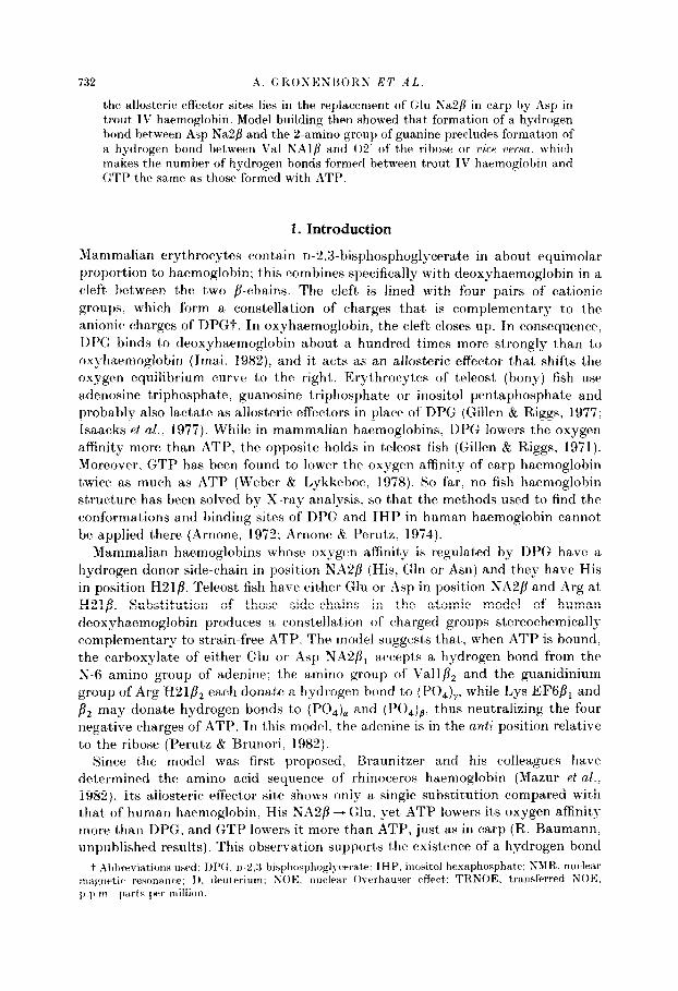
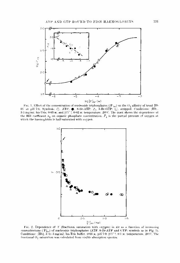
VII:. I 13ffectj of the concentration of nucleoside triphosphates ([ l’],J on the 0, affinity of trout H h I\’ at pH 7.0. Symbols: 0. ATP: 0. 8-Br-,ITP: A, 8-Br-QTP: 0. stripped. (‘onditions: [Hbl, 3.5 mg/ml: his-Tris. OG M: and [Cl- 1. OC2 M: temperature, 20°C’. The inset shows the dependence of the Hill wrtticirnt ?z3 on organic phosphate concentration. Pi is the partial pressure of oxygen at which the harmoglobln is half-saturated with oxygen.
0
/
% A4
A $c. A .OAO 8. cm
1 I 1 I I ̂ r ̂
Cl.3 1.u
[PI,,, (mM) Fro. 2. Dependence of I’ (fractional saturation with oxygen) in air as a function of increasing
concentrations ([PI,,,) of nucleoside triphosphates (QTP. 8-Sr-ATP and GTP: symbols as in Fig. 1). (Conditions: [Hb], 3 to 5 mg/ml; bis-Tris buffer. WO.5 M, pH 7.0: [(‘I-]. 0.1 M; temperature. WV. The fractional 0, saturation was calculated from risible absorption spectra.
734 A. GIi.ONENI~OKN E7’ ..lL
between the purine and 6111 NA28, in fact. it’ can hardly be explained without it, but the stereochemistry of that bond in t’he GTP complex was not clear. A hydrogen bond between the 2-amino group of the guanine and Glu SAdb seemed possible only with the purine in the qn position. but t,here was no reason wh?- this single hydrogen bond should be stronger than that formed by the G-amino group
of adenine, whence this struct’urr failed to account for GTP bring a stronger allosteric effector than ATP. A further doubt arose when it was pointed out to us that Perutz &, Brunori’s (1982) model could bc tested by comparing the affinity of ATP for fish haemoglobin with that of 8-bromo-ATP. in which st)eric hindrance between the bromine and the ribose had been reported to lock the purine in the syn conformation (Bugg Br Thewald. 1969). In t,hat (*onformation, the hydrogen
bond between the S-amino group of adenine and Glu NA2/I cannot be made in a
stereochemically satisfactory way. In consequence, X-bromo-ATP should be a weaker effector than ATP. However. when we csompared the influence of LATP and Sbromo-ATP on the oxygen affinity of trout IV haemoglobin we found them to be equal (Figs 1 and 2). We therefore concluded that Perut)z & Rrunori‘s model must be wrong. even though it looked stereochemically very attractive. These apparent’ contradictions led us to probe the conformations of ATP and GTP bound to carp deoxyhaemoglobin by nuclear magnetic resonance spectroscopy. In particular, we have used the proton-proton transferred nuclear Overhauser effect (Clore & Gronenborn, 1982,1983: Clore d ccl.. 1984; Gronenborn bi Clore, 1982a,b; Gronenborn et al., 1984) to determine the angle of rotat)ion about the glycosidic bond and t)he pucker of the ribose. and employed 31P-XMR spectroscopy to determine the angle of rotation about, bhe (S-06 bond and to obtain qualitative information on the environment of t,he phosphorus atoms.
2. Experimental Procedures
(a) Oxygrn equilibri~cm mmsurentents
(‘omponent, I\: of’ trout haemoglobin (trout Hk IV) was purified from t)rout blood as described by Binotti rt al. (1971). Oxygen equilibrium measurements were obtained by the method of Roasi Fanelli & Ant)onini (1958).
Carp haemoglobin, prepared as described by (‘ondo et al. (1981) was extensively dialvsed against’ D,O. Samples for BMR were prepared in a nitrogen-filled glove box and GMR tubes were sealed with beeswax. Samples for ‘H-NMR, were made up in 99.6% D,O and contained 0.6 mM-carp deoxyhaemoglobin (in tetramer), X.3 rn>f-sodium dithionite. 5.5 mM- nucleoside triphosphate, 288 mM-KC]. 28.8 mnl-pot’assium phosphate, pH 6.7 (met’er reading uncorrect,ed for the isotope effect on thcx glass electrode) and 0.8 mM-EDTA. Samples for 3LP-NMR were made up in 90?,;) H,O/lO?/& D,O and contained 1.4 mm-carp deoxyhaemoglobin (in tetramer), 24 m,n-sodium dithionite. 1.4 or 2.8 mM-nucleoside triphosphate. 100 miv-Tris.HCl. (pH 6.9) and 1 mu-EDTA. ‘H chemical shifts were measured with respect to 2,2 dimethylsilapentane-5-sulphonate as an external standard. 31P chemical shifts were measured relat,ive to Sob orthophosphoric acid as an external standard.
‘H-NMR spectra were obtained at 270 MHz using a Bruker WH-270 spectrometer: 300 transients, obtained by quadrature detection with 4096 data points and a spectral width of 4.2 kHz. were averaged for each spectrum. Prior to Fourier transformation, the free
ATI’ ASD (:TP ISOIJND TO FISH HAEblO(:l~Ol~ISS 735
induction decay was multiplied by an exponential equivalent, to a line broadening of 2 Hz. Thr pulse sequence used in the time-dependent TRKOE measurements was (1, -t, -n/-AT -t3)“, where the selective irradiation at a chosen frequency is applied during the time interval t, (@WI2 to 0.6 s). t, is a short delay (2 ms) to allow for electronic rrc.over?- after the removal of the selective irradiation, AT is the acquisition time (0,437 s), and t, 1s a relaxation delay (3 s) to allow for complete recovery of magnetization of all protons t,o their equilibrium values prior to perturbation by the radiofrequency field. The irracliution power used was suflicient to be in the high power limit. so that saturation is rfliic~tivrly instantaneous whilst select,ivity is preserved so that only a single average resonan(+~’ at, a time was saturated (Dobson et nl.. 1982; Clorr bt Gronenborn, 1983: (:ronrnborn rt CL/.. 1984). As an initial procedure. the spectral region from 3.4 to 7.2 ppm. \vas systematically irradiated at 20 Hz (0.074 p.p.m.) intervals using a 0.4 s pre-saturation pulsr. This region covers all the sugar resonances of the nucleosidr t,riphosphates and the rl and /I (‘H protons of the protein. In this procedure, the selectivity of the TRNOEs is maintained since. in general. t)hc extent of spin-diffusion from indirect, cross-relaxation vi*. prot,ons of the protein is approximately independent of t’he irradiation frequency, providing this is placard within the protein resonance envelope ((Yore & Gronenborn, 1982.1983). All the obsc~rvr~tl TRXOE effects were maximal and centred at the positions of the averaged ligand resonances (note free and bound nucleoside triphosphatc are in fast exchange on the c~hrrnic~al shift scale). indicating that the TRIGOEs arise as a result of direct cross- rc~laxatiorr betwt~en bound ligand prot,ons ((‘lore & Gronenborn, 1982.1983: Gronenborn & (‘lot-~. l!~X%cr.h; (:ronrnborn it al.. 1981).
-“1’-Ni.llIZ sprc+ra were rrcorded at. 81 MHz on a Kruker WM200 spectromet.er with all ac,c{uisition time of 1.024 s (X192 data points and a spectral width of 8 kHz), a pulse-width of 1-5’ and an intrrpulse relaxation delay of 1 s: 6000 transients were averaged for each spchc*trurn. Prior to Fourier transformation. the free induction decay was multiplied by an c~x!)orlr~ntial quivalrnt to a line broadening of 2 Hz.
3. Results and Discussion
(a) JVILI R apPctroscopJ
(Yore & (ironenborn (1983) have described the theory of the time-dependent TRSOE for an exchanging system containing multiple spins as applied to the conformations of ligands bound to proteins. We found that the chemical exchange of nucleosidf~ t,riphosphates with (harp deoxyhaemoglobin is fast on the chemical shift scalr. so t’hat only a single set of exchange-broadened averaged ligand rt:sonanc*es is observed, and that no NOES are observed for the free ligands in the ak)srnc~o of protein at irradiation times of less than one second. ConsequentSly, the initial sloshes of the time-dependent TRNOE of the averaged ligand resonance i following irradiation of the averaged ligand resonance j is simply given by -( 1 -n)cJY. where a is the mole fraction of free ligand and 07 is the cross- rt~laxat~ion rate between t’he bound ligand protons i, and j,. Xote that the (aross- relaxation rates in t.he ligand-prot,ein complex for which COT’, >> I (where w is the IAarmor frequency and Z, the correlation time) are of opposite sign to those in the t’rrfl ligantl for which 0~~ << 1. As a result, the sign of the TRX(>E is negative as opposed to t,he positive steady-state NOE for the free ligand. The (tress-relaxation riLff' o$” is proporti~~nal t.0 ((rtB)-6). where ry is t.he distance between protons iIS and .jB is t hr ligand-protein complex. This relationship has t,wo consequenc~es: (1) tlistanc*e ratios between pairs of bound ligand protons can be determined t1irfhc.i ly and with ease from the initial slopes of the time-dependent TRxOF:s; and (r’) thr value of the cross-relaxation rate decreases rapidly as the interprot,on

tlistjancv2 increases and becomes negligible for ry 2 4 A. Thus. for distanc~es less than 1 .A. the intensity of the averaged ligand resonance on which the TRNOli: is ol~servcd initially decreases wit,h time. while for distances greater than 4 Lq it rxhihits a lag phase.
Figure 3 illustrates a set) of time-tleprndrnt TR~KOE experiments on ATP t)ountl to caarp dec,xvhitrmoglot)in in thr prcsc~ncc~ of a tt.nfold rxc’ess of free ATT’. I)irrc*t TRNOEs arv ohserved on t,he HX rvsonance following irradiation of the Hl’. H2’ and HZ resonances with initial slops of 0.2. I.08 a,ntl 1.05 s-l. rrsptdirely. and on the H 1’ resonance following irradiation of the HZ’ resonancr (initial slope of 1.10 s ‘). Tn cvntrad. lag phases, charac%eristic of indirect TRSOE &+cts, arch observed on t.hr H2 resonance following irradiation of the H 1’. H2’ and H3’ resonances, and on the HI’ resonancr following irradiation of the HY rvsonanct’. Thr same patt.ern of TRNOEs is ohstrred on thtl resonanct’s of (:TP bound t.o carp tleox~harmoglot)in.
Tal)lr I givrs the vomptetc~ set of c,ross-r.el;txi~tiorl ratrs htt wtvn t hr protons of t)ountl ATI’ and GTT’ toget,her with the distanc*e ratios calculated from them. For t)oth ATT’ and (:TT’. the TRSOE data indic~att~ that the tlist,ancrs r”,“,-,,,. ~fii+~.
illltl $F,-tj,, a,rc’ approximately equal. that the dist.anc*e r~~,“,-Hlz is approsimatel?- “5 to 30”,) longer than the r;!-nZ. tlistancv. and that the clistjancatbs rEipH5, antI HH
‘H8-HS” are hot h grvatrr than 4 .q. These clist;rnct~ ratios indicdr a high nrrti c,onfi)f.lnatiori ilt)OUt the glyc*ositlr~ bontl 1 x(04 -( ‘I’ N9--(‘4) - 290 * 20” ] and a pure 5’.rtlclo sugar pu(~kvr for both ATP and (:‘l’f’T. The TRNOE data. howttver. do
+ ‘I’hr pl~voii~li~ houtl torsion anplr x is tlrfiwtl ))y t lw at*mrs 04’ (‘I -XI) (‘4 with WI’O at thr cis position and I)oGtirt ;~nylrs by c~lockwisr rotation oft trcs furthrr pairs of atoms: this is the IWW IYt’.I(’ ~~~~~v~ntion. and thr angle is IX0 tlitkrnt from thr anyk drfinrd by the rarlirr cwnvrntion: i.r
Lx* = xolrl- 1X(1: it,. -iO’ (=ZH) ) x,,, = 1 IO”%,,,.
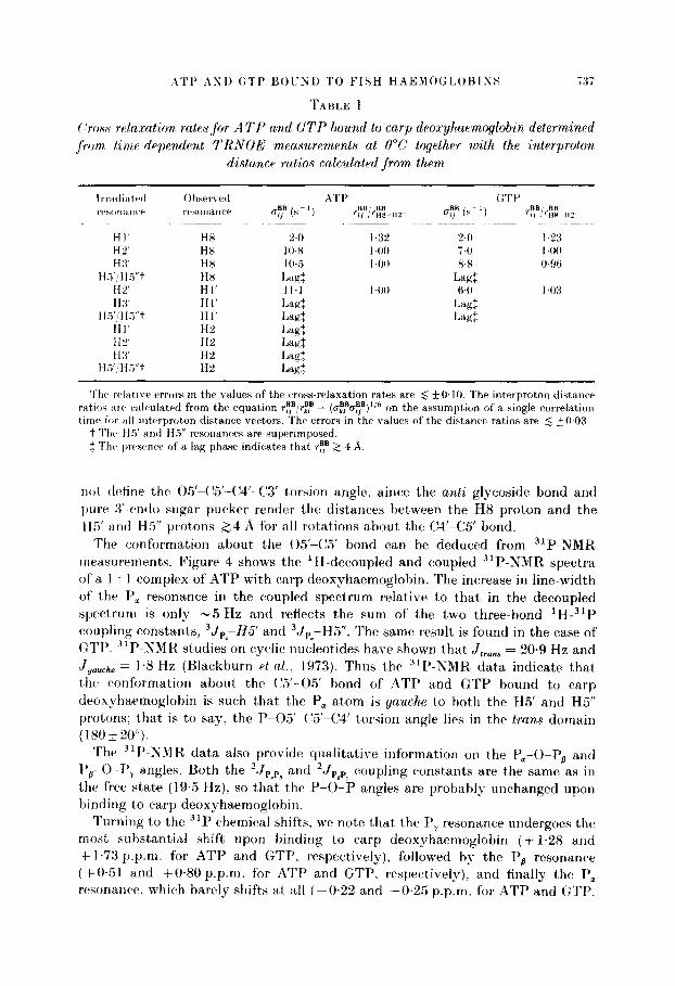
( ‘ross-r~lnx:rction rates for A TY and GTP hound to carp deoxyhaemoglobin determivwd ,from. timp-depen,dent TRNOE measwement~s at 0°C together wvith th,e interproton
distance ratios calculated from them
Irr;tcliatrrl Ohserved A4T I’ GTE’ I’,‘s~,nWn~‘t’ rrw~nancr uy (s- ‘) BB, BB ri, .‘~w-HZ~ CT? (SC’) ry/r;&
HI’ HX 2.0 I.32 24 I.23 HL” HX 10~8 I40 74 I4KJ H 3’ H8 IO.5 14M 8.8 WSfi
H.?‘, HY’t HX Lagf Lag: Hi’ HI’ I I.1 I40 6.0 143 HX HI’ Lap: Lag:
H.i’:H.Yt Hl’ Lag: Lagf HI’ H” Lag: HZ HP Lag: H :I’ HZ Lag:
HT,‘/Hs”t H% I.&
Thr wlatiw error’s in the values of the wowrelaxation rates are 6 +O. 10. The interproton distance ratioh arv calculated from the equation ry,“!rEF = (u~,‘u~)~‘~ on the assumption of a single correlation tirnv for HII interproton distance vectors. The errors in the values of the distance ratios are 5 ~WO3.
t The H5’ and Ha” resonances are superimposed. $ Thv pr’rsenw of’ a lap phase indicates that ry 2 4 ‘9.
not &fine the Ofi’-C!5’--C4’-C3’ torsion angle, since the unti glycoside bond and purr I<‘-condo sugar pucker render the distances between the H8 proton and the H5’ and H5” prot.ons 224 A for all rot,ations about the C4’-C5 bond.
The conformation about t.he 05’-C5’ bond can be deduced from 3iP-XMR, measurements. Figure 4 shows the ‘H-decoupled and coupled 31P-NMR spectra of a 1 : 1 complex of ATP with carp deoxyhaemoglobin. The increase in line-width of the P, resonance in t’he coupled spectrum relative to that in the decoupled spectrum is only -5 Hz and reflects t,he sum of the two three-bond 1H-31P coupling constants, 3J,-115’ and 3Jp - H5”. The same result is found in the case of CTP. 31P-XMR’ studies on cyclic nucieotides have shown that ,I,,,,, = 20.9 Hz and J gauche = 1.8 Hz (Blackburn et al.. 1973). Thus the 31P-~MR data indicate that the conformation about the (,‘5’-05’ bond of ATP and GTP bound to carp deoxyhaemoglobin is such that t’he P, atom is gauche to both the H5’ and H5” protons; that is to say, the P-05’-(‘.5X14’ torsion angle lies in the tran.s domain ( I x0 f 20’).
Thtk 311’-NMR data also provide qualitative information on the Pa-()-P, and F’,-O-P, angles. Both the ‘JpSp, and ‘Jpflp7 coupling constants are the same as in the free state (19.5 Hz), so that the P-0-P angles are probably unchanged upon binding to carp deoxyhaemoglobin.
Turning to the 31P chemical shifts. we note that the P, resonance undergoes the most substantial shift upon binding to carp deoxyhaemoglobin (+ 1.28 and + 1.73 p.p.m. for ATP and GTP, respectively), followed by the P, resonance (+W51 and +W80 p.p.m. for ATP and GTP. respectively), and finally the Per resonance, which barely shifts at all (-0.22 and -0.25 p.p.m. for ATP and GTP.

738 A. GRONENI3OKS EZ’ A I,
FIG. 4. “P-XMK spectra of ATP in a I : I wmplex of’ carp deoxvhaemoglohin with ATP. Trace A. ‘H-decoupled spectrum: trace B, expansion of the P, resonanbr with ‘H-decoupling: trace (‘. expansion of t,he P, resonance without ‘H-decoupling. A stick diagram of’ the ‘H-decoupled spert,rum of free ATP is also shown. The sample temperature was 20°C. The experimental conditions were as given in Experimental Procedures.
respectively). The large downfield shifts experienced by the P, and P, resonances can be explained by the proximity of positively charged amino acid side-chains in the nucleoside triphosphate binding site of carp deoxyhaemoglobin. which polarize electrons surrounding the P, and P, atoms, thereby changing their induced magnetization (i.e. a linear electric field effect as has been proposed for other nucleot,ide-protein complexes; Feeney it ccl.. 1975).
(b) Clonatruction of models
On the basis of the conformations of ATI’ and GTP determined by NMR. we have built molecular models of the interactions between the nucleoside t.riphosphates and deoxyhaemoglobin. Jnitially? we used the Kendrew model of human deoxyhaemoglobin derived from Fermi’s (1975) refinement at’ 2.5 A resolution, in which we merely replaced the side-chain of His NAZ by Glu and that of His H21 by Arg. When satisfactory models had been constructed, their atomic co-ordinates were measured. These rough co-ordinates were then subjected to several cycles of energy refinement by the method of Jack & Levitt (1978); the conformations of the nucleotides were held rigid as determined by NMR, and their positions in the effector site were allowed to vary. To make the calculations more realistic, all the side-chains in the NA, EF and H segments of human haemoglobin were replaced by those of carp.
A’l’t’ AN11 (:TP 13OUNl) TO FISH HAEMOGLOBISS 739
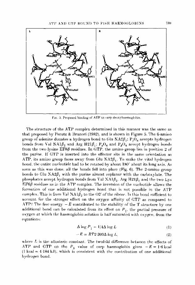
FIG. 5. Proposed binding of ATP to c’cwp droxyhaemoglohin
The structure of the ATP complex determined in this manner was the same as that proposed by Perutz & Brunori (1982)! and is shown in Figure 5. The Camino group of adenine donates a hydrogen bond to Glu KA2/3,; P,O, accepts hydrogen bonds from Val NAl/?, and Arg H21P,; P,O, and P,O, accept hydrogen bonds from the two lysine EF6g residues. In GTP, the amino group lies in position 2 of the purine. If GTP is inserted into the effector site in the same orientation as ATP, its amino group faces away from Glu NABfi,. To make the vital hydrogen bond. the entire nucleotide had to be rotated by about 180” about its long axis. As soon as this was done, all the bonds fell into place (Fig. 6). The S-amino group bonds to Glu r\;A2P1 with the purine almost coplanar with the carboxylate. The phosphates accept hydrogen bonds from \‘a1 NAl/?,, Arg H21B, and the two Lys EFGP residues as in the ATP complex. The inversion of the nucleotide allows t.he formation of one additional hydrogen bond that is not possible in the ATP complex. This is from Val NAl/?, to the 02’ of the ribose. Is this bond sufficient to account for t,he stronger effect on the oxygen affinity of GTP as compared to ATP? The free energy -I3 contributed to the stability of the T structure by one additional bond ca,n be calculated from its effect on P,. the partial pressure of oxygen at which t.he haemoglobin solution is half saturated with oxygen, from the equat)ions:
A log P, = 1/4Alog L (1)
-E = RT2.303A log L. (2)
where L is the allosteric const.ant. The twofold difference between the effects of ATP and GTP on the f’, value of carp haemoglobin gives -E = 1.6 kcal (I kcal = 4.184 kJ). which is consistent with the contribution of one additional hvdrogen bond.
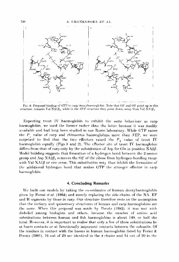
FIG. 6. Proposed binding of GTI’ to carp deox~haemoglobin. Sate that 02 and 03 point up in this st,ructure. t>onards Vnl SAl&. ,h’l w I c in the ATP structure they point down. away from Val NAlfi,.
Expecting trout T\’ haemoglobin to exhibit the same behaviour as carp haemoglobin, we used the former rather than the latt’er because it was readily available and had long been studied in our Rome laboratory. While GTP raises the I’, value of carp and rhinoceros haemoglobins more than ATP. we were surprised to find that the two effecters raised the I’, value of trout TV haemoglobin equally (Figs 1 and 2). The effector site of trout II’ haemoglobin differs from that of carp only by the substitution of Asp for (2111 in position NA2p. Model building suggests that formation of a hydrogen bond between the 2-amino group and Asp NA2f11 removes the 02’ of the ribose from hydrogen-bonding range with Val NAlfl or vicr nersn. This substitution may thus inhibit the formation of the additional hydrogen bond that’ makes GTP the stronger effector in carp haemoglobin.
4. Concluding Remarks
We built our models by taking the co-ordinates of human deoxyhaemoglobin given by Fermi et (11. (1984) and merely replacing the side-chains of the NA, EF and H segments by those in carp. Our struct’ure therefore rests on the assumption that the tertiary and quaternary structures of human and carp haemoglobins are the same. When this proposal was made by Perute (1983). it was met with disbelief among biologists and others. becaause the number of amino acid substitut.ions between human and fish haemoglobins is about 140, or half the total. However, it is important to realise t)hat only a few of these substitutions lie at haem contacts or at functionally important contacts between the subunits. Of the residues in contact with the haems in human haemoglobin listed by Fermi &r Perutz (1981). 16 out of 20 are identical in the a-chains and 14 out of 20 in the

ATI’ ANI) (:TI’ UOI’NL) TO FISH HAEMOULOUINS i4l
fl-chains. At the alp2 contact. which forms the switch between the deoxy and oxy structures. 27 out of 32 residues are identical in the two species. These homologous residues would be stereochemical misfits unless the tertiary and quat,ernary structures superimposed with a standard deviation of less than 1 A.
The a-chain of carp haemoglobin contains one extra residue compared to human, but this lies in the external and non-helical Cl) segment where it can hr
accommodated by only local modifications of structure. Tt has been argued that t hc paramagnetically shifted haem proton resonances of carp deoxyhaemoglobin are diffuse and hear no resemblance to t.hose of human haemoglobin, which suggest,s at, first, sight that the two proteins have different structures, but La Mar and his colleagues have discovered the explanation to be different. Tt appears that in both species bound haems exchange; on rebinding to the globin they may come to lit either side up. In human haemoglobin, over 90?& are one side up, which is the dominant structure found in X-ray and NMR studies. In carp. on the other hand. the two structures are about. equally populated, so that the vinyl and tnet,hyl protons of the porphyrin see two different environments. This multiplicity I)roadens their resonances (La Mar et al., 1984).
How are the equal effects of ATP and Sbromo-ATI’ on the P, value of trout, haemoglobin to be reconciled with our structure? This apparent contradiction has heerr resolved by the discovery by Abdallah rt al. (1975) that steric hindrance between t,hc bromine and the ribose. which seetned to force t’he purine into the syn c.onformation in crystals of 8-bromo-ATP, is relieved when the nucleotide is bound to alcohol dehydrogenase by switching the ribose from the 3’ to the Zendo conformation. thus allowing the purine to take up the anti conformation, as in ATI’.
This work was supported by the Medical Research Council (G.M.(!., A.M.G. and M.F.P.) anti t,he Lister Tnstitute of Preventive Medicine (G.M.C.). G.M.(‘. is a List,er Institut,e Research Frllow. The ‘I P-NMR spectra were recorded on the WM200 spectrometer of the .\ledical Resrarch (council Biomedical NMR Centre at the National Institute for Medical f&sv’itrch. We thank TIr G. Fermi for energy refinement and the computer drawing of Pi~rtrc~ 6
i\bdalfah, M.. Bielmann. ,J. F., Kordstrtim. B. & Brgnden, C.-T. (1!155). Eur. .J. Biochem. 50,
47.5 481. Arnonr. A. (1972). ~Vatwe (London), 237. 146-149. ~~rwtw. .L\. & Perutz, M. F. (1974). Xuturu (London), 249, 34--36. Knotti. I.. Giovenco, S., Giardina. B., Antonini, E., Brunori, M. & Wyman. J. (1971).
Arch. Biochem. Biophys. 142, 274-280. Blackhurn. K. ,J.. Lapper, R. D. & Smith, I. (‘. P. (1973). .J. Am,er. (‘hpm. A’oc. 95. X873-
2878. bugg. (‘. E. & Thewald, U. (1969). Biochem Biophys. Res. Commun. 37. 623.-629. C’lorr, (:. M. & Gronenborn, A. M. (1982). .J. Magn. Reson. 48, 402-417. (‘lorr. G. .Vf. & Gronenborn, A. M. (1983). J. Magn. Reson. 53. 423-442. (‘lorr. G. M.. Gronenborn, A. M. & McLaughlin, L. W. (1984). J. Mol. Biol. 174, 163-173. (‘ondo. S. C:., Biardina, B., Lunadei. M., Frrracin. A. & Brunori. M. (1981). Eur. J.
HiochP’m. 120. 323-327. . *
741 A. GKOSENRORS ET AL.
I)obson, C!. M.. Olejniuxak, E. T., Poulsen, F. M. & Ratcliffe. R. (:. (1982). .J. Mugn. Reson. 48. 87-110.
Feeney. J ., Birdsall. B.. Roberts. G. (“. K. & Burgen. A. S. V. (1975). Xaturr (London), 257. 5644566.
Fermi. G. (1975). ,I. Mol. Biol. 97. 237 256. Fermi. G. & Perutz, M. F. (1981). HaewLoglobin rind Afyoglohin: Atlas of Molecular Structures
in Biology (Phillips. I>. C. & Richards. F. M.. eds). vol. 2. Oxford Cnirersity Press, Oxford.
Fermi. G., Perutz, M. F.. Shaanan. B. & Fourme. R. (1984). J. Mol. Biol. 175. IliY-- 174. Gillen, R. J. & Riggs, A. (1971). C’om~>. Biochrm. Ph,ysiol. 38B, 585-595. Gillen, R. J. bt Riggs. A. (1977). Arch. Biochem. Biophys. 183. 678-685. Gronenborn. A. M. & Clore. G. M. (1982a). .I. Mol. Biol. 157. 155-160. Gronenborn. A. M. & Clore. U. M. (1982b). Biochemistry, 21. 4040-4048. Gronenborn. A. M., Clore, 0. M. & Jeffery. .J. (1984). J. Mol. Biol. 172. 559-572. Tmai, K. (1982). illlostrric Effects in Haemoglobin. (Cambridge University Press. (Cambridge. Isaacks, R. E.. Kim. H. D.. Bartlett. (:. R. 6 Harkness. *J. R. (1977). Li$“c Sci. 20, 987. Jack, A. & Levitt. M. (1978). Acta ~‘rystnllogr. sect. ‘4. 34. 7822791. La Mar, G.. Jue, T.. Hoffmann, B. H. 8r Ir;agai. K. (1984). .I. Mol. Hiol. In the press. Mazur. G., Braunitzer, G. & Wright. P. G. (1982). Hoppe-Seyler’~ Z. Physiol. Chem. 363,
107771086. Rossi Fanelli. A. & Antonini. E. (1958). Arch. Biochewl. Biophys. 77. 478-492. Perutz, M. F. (1983). Mol. Biol. Evol. 1. 1-Z-i. Perutz. M. F. & Brunori. M. (1982). *Vature (London), 299. 421~ 426. Weber, R. E. & Lykkeboe. 0. (1978). J. Pomp. Physiol. 128. 127-137.
Edited by C’. K. (‘antor