Embed Size (px)
Citation preview

Jeffrey J. Popma, MDDirector, Invasive Cardiovascular Services
Caritas Christi Health Care SystemSt. Elizabeth Medical Center
Research AssociateHarvard Medical School
Boston, MA
Stent Fractures:Incidence and Clinical Relevance
AngiographicCore Laboratory

Within the past 12 months, I have had a financial Within the past 12 months, I have had a financial interest/arrangement or affiliation with the organization(s) interest/arrangement or affiliation with the organization(s) listed below.listed below.
Physician Name Company/Relationship
Jeffrey J. Popma, MD Research Grants: Cordis, Boston Scientific,Medtronic, Abbott-Guidant, Biosensors, Radiant, eV3
Medical Advisory Board: Cordis, Boston Scientific, Medtronic
Speaker’s Bureau: Sanofi, BMS, Boston Scientific, Pfizer
Conflict of Interest Statement

StandardizedAcquisitionMethodologyIs Needed To Detect Stent Fractures For PeripheralAnd CoronaryStudies
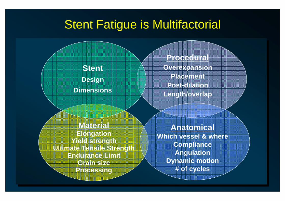
ProceduralOverexpansion
PlacementPost-dilation
Length/overlap
MaterialElongation
Yield strength Ultimate Tensile Strength
Endurance LimitGrain size
Processing
Stent Fatigue is Multifactorial
StentDesign
Dimensions
AnatomicalWhich vessel & where
Compliance Angulation
Dynamic motion# of cycles

• Dynamic loading in a vessel results in the following deformations:– Pulsatile (current fatigue test & FEA) – Bend (static bend modeling in FEA)– Twist– Stretch
• Above are likely a function of species, vessel (RCA, LAD, LCX) and location (proximal, distal)
Preclinical Testing: Dynamic Loading

• 1808 pts with 2920 lesions with angiographic FU in 1491 patients with 2357 lesions (80.7%)
• Definition: Obvious separation of stent• Stent restenosis rate was 11.2%; stent fracture rate
was 3.9% and stent fracture with restenosis was 1.1%• Multivariable predictors for stent fractures with
restenosis: aorto-ostial lesions; severe angulation; SVG and overlapping stents
• Coronary artery motion was also an important predictor of stent fracture with restenosis
SCAIACCI2: Predictors of Stent Fractures
Kadota et al JACC 2008; 51: B7
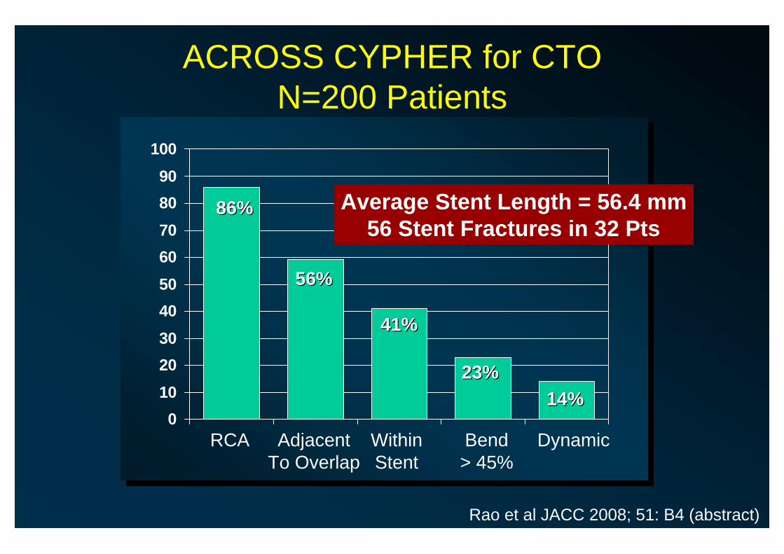
ACROSS CYPHER for CTON=200 Patients
0102030405060708090
100
RCA AdjacentTo Overlap
WithinStent
Bend> 45%
Dynamic
86%86%
56%56%
41%41%
23%23%14%14%
Average Stent Length = 56.4 mm56 Stent Fractures in 32 Pts
Rao et al JACC 2008; 51: B4 (abstract)

ACROSS CYPHEROverall Fracture Rate = 16%
0.097.421.9In-segment BAR. %
0.1603.1Stent Thrombosis, %
0.456.19.4MACE, %
0.425.59.4TLR, %
0.0689.9100Overlapping stents, %
< 0.00145.069.9Stent length, mm
P ValueNo Stent Fracture (N=168)
Stent Fracture (N=32)
Rao et al JACC 2008; 51: B4 (abstract)

BWH Core Lab Definitions for Stent Fracture
-Complete transverse linear type III fracture with stent displacement
Complete transverse stent fracture with abundant movement and displacement of fractured fragments of more than 1 mm during the cardiac cycle
Type IV
Severe –complete separation of stent segments
Multiple single stent fractures resulting in complete transverse linear fracture but without stent displacement
Complete transverse stent fracture without displacement of fractured fragments more than 1 mm during the cardiac cycle
Type III
Moderate –facture >1 strut
Multiple single stent fractures occurring at different sites
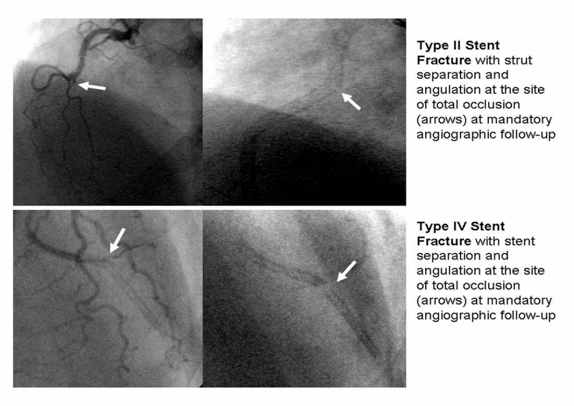
Multiple strut fractures with V-form division of the stent
Type II
Minor –single strut facture
Single strut fracture onlySingle strut fracture or gap between struts greater than 2x normal
Type I
--No strut fractureType 0
Scheinert et al 2Allie et al 1Current ReportClassification
1 Allie et al Endovascular Today 2004; July/August: 22-34 2 Scheinert et al J Am Coll Cardiol 2005; 45:312-315
* Type 5 implies spiral fracture of stent

Single strut fracture Multiple single stent fractures; different sites
Multiple stent fractures; complete
transverse linear fracture
Complete transverse linear Type III fracture with stent displacement
Type I Type II Type III Type IV

349 Patients in the CYPHER arm with follow-up in SIRIUS- 40 Patients with not available CINE films- 2 patients neither of the follow-up CDs can be opened- 2 patients all CD missing305 patients analyzed with 497 follow-up angiograms
4 fractures identified (1.3%),- 3x Fracture Type 1 (0.98%) - 1x Fracture Type 2 (0.33%)
• All fractures occurred with multiple stents near the site of overlap, all vessels calcified including one chronic total occlusion.
• 1 ISR at that site with TLR (Type 1 Fracture – tissue growth)
BWH SIRUS Angiographic AnalysisDid We Miss Something Important?

Coronary Stents:CYPHERTAXUS
Peripheral Stents:
Analysis population
12511312
101CYPHER Angiograms Forwarded to Core Lab
No Fracture By Available PaperworkNo Stent Fracture IdentifiedBx Sonic
51 Cases291
226 Cases
39 Cases
Baseline Angiogram Available 28* 45 stent fractures in 39 patients
Stent Fracture: Review of Adverse Event Reports MAUDE cases between August 2003-July 2006

BWH Angiographic AnalysisAdverse Event Reports (N=39)
13 mm4(10.3%)
4(10.3%)
18 mm
23 mm
28 mm
33 mm
3(7.7%)
13(33.3%)
15 (38.4%)
Type 25 (12.9%)
Type 316
(41.0%)
Type 418
(46.1%)
Fracture Type Stent Length

<0.00117 (3.2)7 (25.0)Total Occlusion<0.00191 (17.1)19 (67.9)Calcification present 0.00228 (5.3)7 (25.0)Proximal Tortuousity< 0.00158 (10.9)13 (46.4)Angulations ≥ 45 degrees0.00282 (15.4)12 (42.9)20 or greater0.001342 (64.6)9 (32.1)10-19.9 mm0.66106 (20.0)7 (25.0)0-9.9 mm<0.00114.4±5.822.1±15.9Lesion Length, mm<0.00110 (1.9)6 (21.4)Ostial Location0.23160 (30.1)12 (42.9)RCA0.27134 (25.2)4 (14.3)LCx0.94234 (44.1)12 (42.9)LAD
Location
P ValueSiriusN = 531, (%)
Stent fractureN = 28, (%)
Variable
Stent Fracture: Baseline Angiographic FindingsStent Fracture: Baseline Angiographic Findings

<0.0015.4±8.214.8±8.9Final % Stenosis<0.0012.67±0.402.33±0.49Final MLD
Within the Stent<0.00116.1±9.724.6±11.1Final % Stenosis<0.0012.38±0.422.06±0.48Final MLD
Within the Segment
N = 531N = 28Final<0.00165.1±12.677.8±15.3% Stenosis<0.0010.97±0.400. 56±0.39MLD, mm0.142.79±0.452.66±0.50RVD, mm
N = 531N = 28BaselineP ValueSiriusStent fractureVariable
Stent Fracture: Baseline Angiographic FindingsStent Fracture: Baseline Angiographic Findings

<0.0012 (0.6)5 (13.2)Aneurysm0.022 (0.6)3 (7.9)Total occlusions0.019.1±5.86.55±5.96ISR Length, mm<0.00111 (3.2)18 (47.4)Restenosis Rate<0.00110.4±16.544.1±28.5Follow-up % Stenosis<0.0010.17±0.440.96±0.71Late Lumen Loss<0.0012.50±0.581.52±0.82Follow-up MLD
Within the Stent<0.00123.6±16.448.6±23.0Follow-up % Stenosis< 0.00131 (8.9)18 (47.4)Restenosis Rate<0.0010.24±0.470.70±0.66Late Lumen Loss<0.0012.15±0.611.41±0.69Follow-up MLD
Within the Segment0.332.79±0.422.72±0.48RVD, mmP ValueN = 350 (%)N = 38 (%)
SiriusStent fractureVariable
Stent Fracture: FollowStent Fracture: Follow--up Findingsup Findings
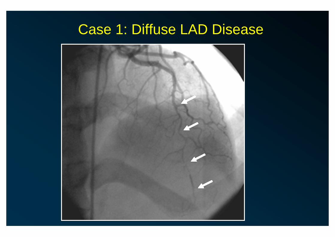
Case 1: Diffuse LAD Disease

Case 1: Diffuse LAD Disease
2.5 mm x 23 mm; 2.5 mm x 33 mm; 3.0 x 18 mm CYPHER stents

Case 1: Diffuse LAD Disease
Final Angiographic Result 3 Month Angiographic Follow-Up

Case 1: Diffuse LAD Disease
Stent Fracture with 3 mm of Stent Overlap
Type 4Stent Fractures
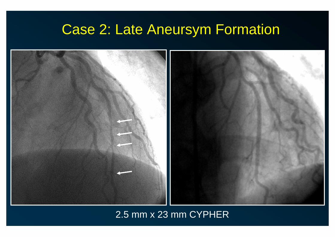
Case 2: Late Aneursym Formation
2.5 mm x 23 mm CYPHER

Case 2: Late Aneursym Formation
3 Month Follow-up

Case 3: Late Aneursym Formation
4 Month Follow-up
Type 4Stent Fracture
4 Month Follow-up

Stent Fracture Evaluated Angiographicallyin >2,300 Stents
Enrolled (TAXUS+BMS):3,787 patients4,509 stents
All patients:2,814 patients3,060 stents
Patients with 9-month angiogram available for analysis:1,864 patients2,342 stents
Patients prospectively assigned to 9-month angiogram:2,877 patients3,553 stents
Assess 9-month fracture
incidence & predictors
Assess all clinical events for association
with fracture
TAXUS Analysis

Per-stent analysis, % (n/N)
NA0% (0)0% (0)0% (0)TAXUS IV
NA0.0% (0)—0.0% (0)†TAXUS ATLAS0.091.1% (10)0.3% (3)0.7% (13)Pooled IV/V/VI1.000.4% (1)0.4% (1)0.4% (2)TAXUS VI0.071.8% (9)0.4% (2/466)1.1% (11)TAXUS V
P valueTAXUSControl*Overall Study
9-Month Incidence of Fracture With Express and Liberté
Among all patients (2,814 patients/3,060 stents; median 1,446 days of follow-up), 4 additional fractures observed beyond 9 months (3 TAXUS Express and 1 TAXUS Liberté)
TAXUS Analysis

Stent Fracture And 9 Month Restenosis
185311Total patients
0.0716.4% (292/1776)40.0% (4/10)In stent binary restenosis, %<0.0127.27±0.5548.85±10.16In stent % Diameter Stenosis
0.020.60±0.011.04±0.21In stent late lumen loss, mm0.032.00±0.021.49±0.29In-stent MLD, mm
P valueNo fractureFractureOutcome
TAXUS Analysis

Stent Fracture and Lesion Complexity
185311Total patients
0.051.32±0.012.27±0.43 (11) Study stents implanted<0.019.83% (181)50.0% (5)Proximal tortuosity, %0.0116.7±0.223.9±3.7Lesion length, mm
0.0137.9% (700)80.0% (8/10)Lesion type C, %
0.0229.4% (545)63.6% (7/11)Previous MI, %
P valueNo fractureFractureCharacteristic
Baseline Characteristics in the Angiographic FU Cohort
TAXUS Analysis

Stent Fracture and 9 M Outcome
1.002.1% (38)0.0% (0)Cardiac death
<0.010.8% (12/1)18.2% (2)ARC Def+prob ST
1.004.7% (94)0.0% (0)All death0.364.0% (74)9.1% (1)Non-Q-wave0.090.8% (15)9.1% (1)Q-wave 0.104.8% (89)18.2% (2)MI (total)0.1311.8% (219)27.3% (3)TLR
185311Total patients
P valueNo fractureat 9 moFracture at 9 mo
Results in the Angiographic Cohort, All Clinical Follow-up
TAXUS Analysis
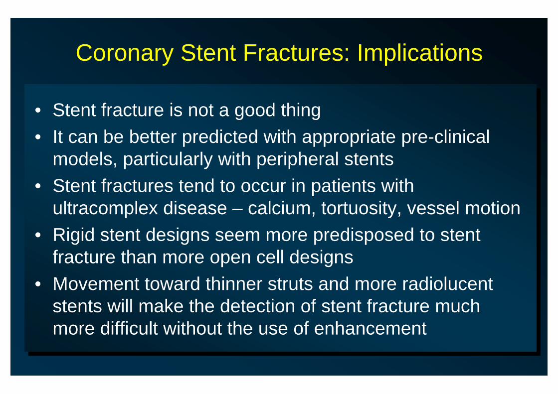
• Stent fracture is not a good thing• It can be better predicted with appropriate pre-clinical
models, particularly with peripheral stents• Stent fractures tend to occur in patients with
ultracomplex disease – calcium, tortuosity, vessel motion• Rigid stent designs seem more predisposed to stent
fracture than more open cell designs• Movement toward thinner struts and more radiolucent
stents will make the detection of stent fracture much more difficult without the use of enhancement
Coronary Stent Fractures: Implications