Embed Size (px)
Citation preview

Page 1/17
Anterior Controllable Antedisplacement and Fusion(ACAF) technique for the treatment of multilevelcervical spondylotic myelopathy with spinalstenosis (MCSMSS): a retrospective study of 45casesXi Luo
Changzheng HospitalKaiqiang Sun
Changzheng HospitalJingchuan Sun
Changzheng HospitalShunmin Wang
Changzheng HospitalYuan Wang
Changzheng HospitalHaisong Yang
Changzheng HospitalJialin Jiang
Changzheng HospitalYongfei Guo
Changzheng HospitalJiangang Shi ( [email protected] )
Changzheng Hospital
Research article
Keywords: cervical spondylotic myelopathy; spinal stenosis; multilevel; anterior controllableantedisplacement fusion (ACAF)
Posted Date: August 9th, 2019
DOI: https://doi.org/10.21203/rs.2.12543/v1

Page 2/17
License: This work is licensed under a Creative Commons Attribution 4.0 International License. Read Full License
Version of Record: A version of this preprint was published at Clinical Spine Surgery: A Spine Publicationon March 17th, 2021. See the published version at https://doi.org/10.1097/BSD.0000000000001144.

Page 3/17
AbstractBackground To investigate the clinical effect of anterior controllable antedisplacement and fusion (ACAF)technique for the treatment of multilevel cervical spondylotic myelopathy with spinal stenosis(MCSMSS), and compare ACAF with hybrid decompression �xation (HDF). Methods A retrospectiveanalysis of 85 cases with MCSMSS was carried out. 45 patients were treated with ACAF, while 40 patientswere treated with HDF. The operation time, intraoperative bleeding volume, postoperative complications,Japanese Orthopaedic Association (JOA) score, Neck Disability Index (NDI) score, ComputedTomography (CT) transverse measurement, cervical curvature and Kang's grade were compared betweentwo groups. Results The patients were followed up for 12 to 17 months. Compared with HDF, ACAF groupachieved better decompression according to CT measurement and Kang’s grade (P < 0.05), and recoveredto a greater cervical Cobb’s angle (P < 0.05). However, JOA score and NDI index showed no signi�cantdifference one year after surgery (P 0.05). Additionally, ACAF presented longer operation time and greaterintraoperative blood loss (P < 0.05). As to complications, ACAF developed less incidences ofcerebrospinal �uid examination (CSF) leakage, neurologic deterioration, epidural hematoma and C5 palsyby comparing with HDF. Conclusions ACAF is an effective method for the treatment of MCSMSS.Compared with HDF, ACAF has the advantages of signi�cant decompression, increasing cervicalcurvature, and reducing the incidences of complications.
BackgroundMultilevel cervical spondylotic myelopathy with spinal stenosis (MCSMSS) is a disorder of spinal corddysfunction characterized by involving three or more cervical segments, which is caused by congenital,developmental or degenerative factors which results in spinal stenosis and compression of spinal cordand its blood vessel at the levels of both intervertebral disc and vertebra[1]. Surgery plays a vitaltreatment part because of the progressive feature of MCSMSS, especially for patients with intolerablesymptoms and suspected neurological damage[2], but considering the multilevel and severecompression, the choice of surgery is controversial[3]. For single-level compression, anterior cervicaldiscectomy and fusion (ACDF) is the “gold standard” for surgical treatment[4, 5]. However, it is di�cult forACDF to achieve complete compression when faced with multilevel lesions. As to the anterior cervicalcorpectomy decompression and fusion (ACCF), although its effect of decompression is signi�cant, theinstability of cervical spine and the high risk of complications cannot be ignored[6].
In order to better solve the clinical problems of MCSMSS, we proposed to utilize a novel treatmentscheme of anterior controllable antedisplacement and fusion (ACAF) on MCSMSS. ACAF is characterizedby hoisting the anterior wall of the spinal canal to make it move forward, thus expanding the volume ofthe spinal canal and relieving the compression. The purpose of this study was to compare ACAF withhybrid decompression �xation (HDF, the combination of ACDF and ACCF) in treatment of MCSMSS[7],and to brie�y introduce ACAF surgical technique, which are reported as follows.
Methods
Page 4/17
1.1. General information
From January 2017 to January 2019, 85 patients diagnosed with MCSMSS and treated with ACAF (45cases) or HDF (40 cases) were selected for the study. X-ray, Computed Tomography (CT) and MagneticResonance Imaging (MRI) of cervical spine were taken before operation. Selection criteria: (1) Con�rmedby cervical spinal stenosis (the sagittal diameter of the spinal canal is less than 12 mm), and theconservative treatment is ineffective; (2) cervical spondylotic myelopathy involving segments≥3.Exclusive criteria: (1) deformity, ankylosing spondylitis, rheumatoid arthritis and other diseases involvingthe cervical spine; (2) cervical spine trauma, surgical history; (3) severe osteoporosis.
All patients agreed to the record of research data, and signed the informed consent.
1.2. Operative methods
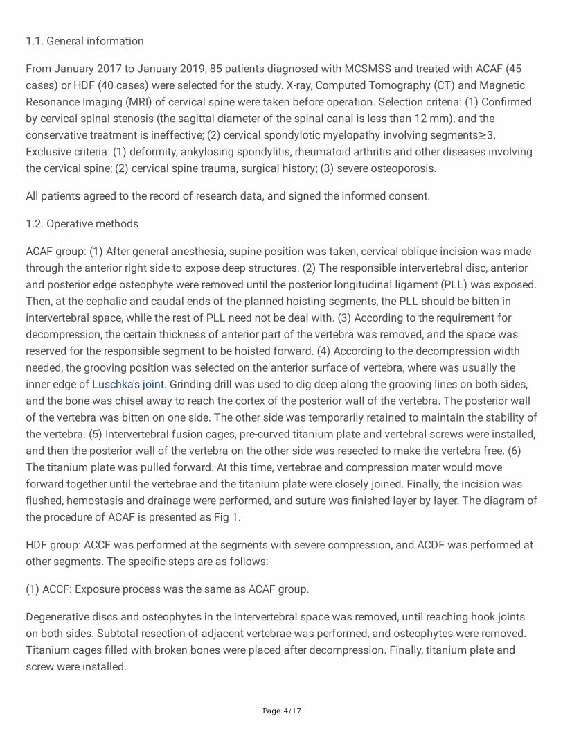
ACAF group: (1) After general anesthesia, supine position was taken, cervical oblique incision was madethrough the anterior right side to expose deep structures. (2) The responsible intervertebral disc, anteriorand posterior edge osteophyte were removed until the posterior longitudinal ligament (PLL) was exposed.Then, at the cephalic and caudal ends of the planned hoisting segments, the PLL should be bitten inintervertebral space, while the rest of PLL need not be deal with. (3) According to the requirement fordecompression, the certain thickness of anterior part of the vertebra was removed, and the space wasreserved for the responsible segment to be hoisted forward. (4) According to the decompression widthneeded, the grooving position was selected on the anterior surface of vertebra, where was usually theinner edge of Luschka's joint. Grinding drill was used to dig deep along the grooving lines on both sides,and the bone was chisel away to reach the cortex of the posterior wall of the vertebra. The posterior wallof the vertebra was bitten on one side. The other side was temporarily retained to maintain the stability ofthe vertebra. (5) Intervertebral fusion cages, pre-curved titanium plate and vertebral screws were installed,and then the posterior wall of the vertebra on the other side was resected to make the vertebra free. (6)The titanium plate was pulled forward. At this time, vertebrae and compression mater would moveforward together until the vertebrae and the titanium plate were closely joined. Finally, the incision was�ushed, hemostasis and drainage were performed, and suture was �nished layer by layer. The diagram ofthe procedure of ACAF is presented as Fig 1.
HDF group: ACCF was performed at the segments with severe compression, and ACDF was performed atother segments. The speci�c steps are as follows:
(1) ACCF: Exposure process was the same as ACAF group.
Degenerative discs and osteophytes in the intervertebral space was removed, until reaching hook jointson both sides. Subtotal resection of adjacent vertebrae was performed, and osteophytes were removed.Titanium cages �lled with broken bones were placed after decompression. Finally, titanium plate andscrew were installed.

Page 5/17
(2) ACDF: Other processes were the same as ACCF. After discectomy, the osteophytes at posterior marginof the vertebra were removed, and the intervertebral space was expanded by distractor to normal height.Intervertebral fusion cage was selected and inserted into the intervertebral space. Titanium plate andscrew �xation was �nally performed.
Postoperative management was as ACAF’s.
All operations were performed by surgeons of the same team. Negative pressure drainage tubes wereplaced and pulled out 24 to 48 hours after operation. All patients were �xed with external cervical bracketfor 3 months.
1.3. Observation Indicators
1.3.1. General indicators
Age, sex, operative levels, operation time, and intraoperative bleeding volume and complications wererecorded.
1.3.2. Functional evaluation
Before and 1 year after operation, neurological function was evaluated by Japanese OrthopaedicAssociation (JOA) score and Neck Disability Index (NDI) score.
1.3.3. CT transverse measurement
By utilizing the measuring tools embedded in software of picture archiving system, 3 parameters weremeasured on CT axis images to evaluate the effects of decompression. The de�nitions of the parameterswere as follows:
(1) Transverse area of spinal canal: the area surrounded by the posterior edge of vertebra (in ACAF) or thetitanium cage (in HDF), the inner side of vertebral plate and the inner side of pedicle.
(2) Decompression width: the distance between the double sides grooves at the anterior wall of spinalcanal
(3) Sagittal diameter of spinal canal: the distance between the posterior edge of vertebra or the titaniumcage, and the base of spinous process
The data was from all the operative segments, and were measured at the middle level of vertebra. Themeasuring diagram is depicted as Fig 2.
1.3.4. C2-7 Cobb’s angle
The a Cobb’s angle method was used to evaluate cervical curvature on the lateral images of X-ray, andthe angle is formed by vertical lines of the upper edge of C2 and the lower edge of C7.

Page 6/17
1.3.5. Kang’s grade
Kang's MRI grading system was used to assess the degree of cervical spinal cord compression[8, 9], andthe speci�c criteria is : grade-0, no spinal canal stenosis; grade-1, subarachnoid compression exceeded50 ; grade-2, spinal cord compression deformed; grade-3, spinal cord T2 weighted signal changes.
All the parameters were measured by two senior spine surgeons independently, and the average value ofeach parameter at each level was taken for independent calculation.
1.4. Statistical methods
SPSS 22.0 was used for statistical analysis. Measurement data were expressed as mean ±standarddeviation ( ). Paired t test was used for intragroup comparison. Two independent samples t-test for inter-group comparison, Chi-square test was for the categorical data comparison. Test level was α=0.05.
Results2.1. Demographics
All the enrolled 85 cases completed the operation successfully, with clinical symptoms relieved andspinal cord function improved, and no incision infection and nerve injury occurred. 85 cases werefollowed up for 12 to 17 months, with an average of 15.6 months.
Among 85 cases, there were 45 cases in the ACAF group, including 33 males and 12 females, aged 40-72(mean, 53.0+10.1) years; 40 cases in the control group, including 22 males and 18 females, aged 38-75(mean, 56.1+9.1) years. There was no signi�cant difference in gender, age and operative levels betweenthe two groups (P > 0.05). The general data is shown in Table 1.
2.2. CT measuring
The preoperative diameter of spinal canal, decompression width and spinal canal area in two groupsshowed no signi�cant differences (P 0.05). After surgery, compared with HDF group, the ACAF grouppresented a better decompression at C3, C4, C5, and C6 levels (P 0.05 ). The data is shown in Table 2.
2.3. Clinical outcomes
According to the data, there was no signi�cant difference in preoperative JOA score, NDI index, Cobb’sangle and Kang's grade between the two groups (P > 0.05). One year after operation, both groups weresigni�cantly improved in terms of JOA score and NDI index (P < 0.05). However, there was no signi�cantdifference in JOA score and NDI index between two groups (P > 0.05). As to postoperative Cobb’ s angleand Kang’ s grade, ACAF revealed its unique superiority, with higher Cobb’ s angle (P 0.05), and lowerKang's grade (P 0.05), comparing to HDF. Additionally, the result showed a higher cost in operative timeand intraoperative bleeding in ACAF group (P 0.05).
Page 7/17
As to the complications, 5 patients (12.5 ) developed cerebrospinal �uid examination (CSF) leakage, 2patients (5.0 ) had neurologic deterioration, 2 patients (5.0 ) presented with epidural hematoma, 2patients (5.0 ) suffered from C5 palsy, and 3 (7.5 ) developed dysphagia in HDF group. Additionally, atthe �nal follow-up, and 4 patients (10.0 ) presented with implant-related complications in HDF group,including 2 cases of cage subsidence and 2 cases of delayed union. In contrast, ACAF groupdemonstrated only 1 case (2.2 ) of CSF leakage and 4 cases (8.8 ) of dysphagia, without othercomplications.
The data is shown in Table 1 and 3.
2.4 Explanatory cases
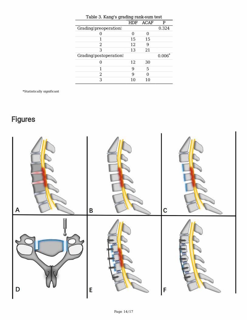
Case 1. Complained of numbness of both arm with gait disturbance for 3 years. The numbness had beenaggravated 2 months before presentation. ACAF was performed at C4 and C5. After the operation, thepatient showed notable relief of neurologic function. Enough decompression of the spinal cord wasachieved according to imaging. The JOA score increased from 7 to 11. There were no complicationsduring follow-up. The imaging examination is shown in Fig 3.
Case 2. Presented with spastic weakness of both hands and gait instability for 2 years. The symptomsprogressively worsen in 3 months before presentation. ACDF was performed at C4-5 level, and ACCF atC5-6 level. After the operation, the patient’s myelopathy showed marked recovery. Preoperative andpostoperative JOA score was 8 and 12, respectively. No complications were observed during follow-up.The imaging examination is shown in Fig 4.
Discussion3.1. Technical features of HDF
The purpose of HDF is to use ACDF in slightly diseased segments and ACCF in the severe[10], so as topreserve the vertebrae, and relieve the compression as much as possible. Compared with ACCF, HDF isassociated with less blood loss, less complications, and higher fusion rate[4, 11]. However, wheninvolving the multilevel compression, in order to avoid dural tear and spinal cord injury, it is sometimesdi�cult to take full advantages of HDF and complete the thorough compression[12], and residualcompressive matter will still produce compression[13]. At the same time, the risk of complications suchas dural tear, spinal cord injury and titanium cage sinking still cannot be ignored in HDF just like inACCF[14].
3.2. Technical features of ACAF
In ACAF, vertebrae-compression complex (VCC) is a vital concept that includes vertebrae of responsiblesegments, osteophytes, proliferative and ossi�ed PLL, which is freed out through grooving at both sidesof vertebrae, and it can be regarded as the anterior wall of the spinal canal. By moving VCC forward cansurgeons expand the volume of the spinal canal and retain more bone of vertebrae simultaneously.
Page 8/17
During the operation, the forward movement of VCC can be monitored by X-ray, so that the whole processof hoisting and spinal cord antedisplacement can be controlled, and the "anatomical reduction" of spinalcord can be achieved. On the other hand, according to experience, the grooving position is roughly at theinner edge of Luschka's Joint, which not only ensures the enough decompression width (the effectiverange almost reaches the medial wall of the pedicle), but also avoids the injury of vertebral artery.Compared with the HDF operated in the anterior median region of cervical spine, ACAF operation mainlyfocuses on the region around Luschka's joint, which is far away from spinal cord and its artery travelingarea, constructing a "safe space" for spinal cord and reduces the risk of spinal cord injury.
3.3. Advantages of ACAF in the treatment of MCSMSS
(1) Good recovery of spinal cord morphology and CSF space
It is observed that the Kang's grade of ACAF decreased signi�cantly, which means the compression ofCSF band and spinal cord is con�rmed on MRI. By pulling the VCC, spinal cord can not only restorenormal anatomical morphology, but also avoid traction caused by backward drift, and the recovery ofCSF space is also bene�cial to the improvement of symptoms after surgery[15, 16].
(2) Fewer complications
1) C5 palsy
Due to the technical limitations of ACCF, the true decompression width of spinal canal is signi�cantlysmaller than the slotting width of the anterior vertebral surface, resulting in incomplete decompression atthe outlet of the nerve root[17]. However, in ACAF, the width of decompression is fully guaranteed byvertical grooving and vertebral hoisting. Anatomically, the Luschka’s joint participates in the formation ofnerve root outlet. So, when VCC moves forward, the space around the outlet of nerve root will be greatlyexpanded to achieve nerve decompression, meanwhile, the risk of nerve root traction is further avoideddue to the recovery of curvature and position of spinal cord.
2) Postoperative hematoma and CSF leakage
When venous plexus and dura mater adhere to surrounding ossi�ed mater, operation is easy likely tocause hematoma and CSF leakage[18]. In ACAF, the operation does not directly involve the dura materand intraspinal venous plexus. When there exists adhesion between dura and ossi�ed mater, after VCC ismoved forward, these structures are still in previous connection relationship, which reduces the risk ofdura tear and hematoma. Furthermore, the forward movement of VCC will pull the dura forward, keep thedura tension to a certain extent, and form a "tent effect", thus blocking the compression of spinal cord byhematoma. So, the ACAF theoretically completely avoid the risk of compression of spinal cord byhematoma after operation.
Limitations

Page 9/17
Due to its retrospective nature and short-time follow-up, the results of this study might be limited. A moreconvincing clinical conclusion would be drawn if a larger asymptomatic cohort is included.
ConclusionsThe results of this study showed that ACAF technique can expand spinal canal volume by completing theanterior displacement of the compressive substance in the treatment of MCSMSS. It can safely andeffectively relieve the problems of spinal cord compression and spinal canal stenosis, signi�cantlyimprove cervical curvature and spinal cord morphology. Additionally, ACAF can reduce the rates ofcomplications such as dura tear and hematoma that often occur in traditional anterior operation. Inconclusion, ACAF is a satisfactory optional surgical method for the treatment of MCSMSS.
DeclarationsEthics approval and consent to participate
All procedures performed in studies involving human participants were in accordance with the ethicalstandards of the Ethics Committee of Changzheng Hospital.
Consent for publication
Informed consent was obtained from all individual participants included in the study.
Availability of data and material
Data sharing is not applicable to this article as no datasets were generated or analysed during the currentstudy.
Competing interests
The authors declare that they have no competing interests.
Funding
This study was funded by

Page 10/17
Innovation Fund for College Students of Second Military Medical University (No. FH2017077) by Xi Luo a‡
Innovation Fund for College Students of Second Military Medical University (No. MS2016045) by Xi Luo a‡
Authors' contributions
"XL analyzed and interpreted the patient data regarding the MCSMSS, and was a major contributor inwriting the manuscript. KS and JS (Jingchuan Sun) performed the examination of the data, andsubstantively revised the manuscript. SW and YW conducted the acquisition of data. HY have helpeddraft the work. JJ and YG proposed the ieda of sutdy design. JS (Jiangang Shi) �nished the �nalassessment of the manuscript. All authors read and approved the �nal manuscript."
Acknowledgements
Not applicable.
References1. Hayashi H, Okada K, Hamada M, Tada K, Ueno R: Etiologic factors of myelopathy. A radiographic
evaluation of the aging changes in the cervical spine. Clin Orthop 1987, 214(214):200-209.
2. Melancia JL, Francisco AF, Antunes JL: Spinal stenosis. Handb Clin Neurol 2014, 119:541-549.
3. Zhang J, Liu H, Bou EH, Jiang W, Zhou F, He F, Yang H, Liu T: Comparative Study Between AnteriorCervical Discectomy and Fusion with ROI-C Cage and Laminoplasty for Multilevel CervicalSpondylotic Myelopathy without Spinal Stenosis. World Neurosurg 2019, 121:e917-e924.
4. Zhu B, Xu Y, Liu X, Liu Z, Dang G: Anterior approach versus posterior approach for the treatment ofmultilevel cervical spondylotic myelopathy: a systemic review and meta-analysis. Eur Spine J 2013,22(7):1583-1593.
5. Li Z, Wang H, Tang J, Ren D, Li L, Hou S, Zhang H, Hou T: Comparison of Three ReconstructiveTechniques in the Surgical Management of Patients With Four-Level Cervical SpondyloticMyelopathy. Spine (Phila Pa 1976) 2017, 42(10):E575-e583.
�. Lao L, Zhong G, Li X, Qian L, Liu Z: Laminoplasty versus laminectomy for multi-level cervicalspondylotic myelopathy: a systematic review of the literature. J Orthop Surg Res 2013, 8(1):45.
7. Ashkenazi E, Smorgick Y, Rand N, Millgram MA, Mirovsky Y, Floman Y: Anterior decompressioncombined with corpectomies and discectomies in the management of multilevel cervicalmyelopathy: a hybrid decompression and �xation technique. J Neurosurg Spine 2005, 3(3):205-209.

Page 11/17
�. Kang Y, Lee JW, Koh YH, Hur S, Kim SJ, Chai JW, Kang HS: New MRI grading system for the cervicalcanal stenosis. AJR Am J Roentgenol 2011, 197(1):W134-140.
9. Harrison DE, Harrison DD, Cailliet R, Troyanovich SJ, Janik TJ, Holland B: Cobb method or Harrisonposterior tangent method: which to choose for lateral cervical radiographic analysis. Spine (Phila Pa1976) 2000, 25(16):2072-2078.
10. Odate S, Shikata J, Kimura H, Soeda T: Hybrid Decompression and Fixation Technique Versus Plated3-Vertebra Corpectomy for 4-Segment Cervical Myelopathy: Analysis of 81 Cases With a Minimum 2-Year Follow-Up. Clinical spine surgery 2016, 29(6):226-233.
11. Sakai K, Okawa A, Takahashi M, Arai Y, Kawabata S, Enomoto M, Kato T, Hirai T, Shinomiya K: Five-year follow-up evaluation of surgical treatment for cervical myelopathy caused by ossi�cation of theposterior longitudinal ligament: a prospective comparative study of anterior decompression andfusion with �oating method versus laminoplasty. Spine (Phila Pa 1976) 2012, 37(5):367-376.
12. Yang H, Lu X, Wang X, Chen D, Yuan W, Yang L, Liu Y: A new method to determine whether ossi�edposterior longitudinal ligament can be resected completely and safely: spinal canal "Rule of Nine" onaxial computed tomography. Eur Spine J 2015, 24(8):1673-1680.
13. Yang H, Lu X, Wang X, Chen D, Yuan W, Yang L, Liu Y: A new method to determine whether ossi�edposterior longitudinal ligament can be resected completely and safely: spinal canal "Rule of Nine" onaxial computed tomography. Eur Spine J 2015, 24(8):1673-1680.
14. Shin JH, Steinmetz MP, Benzel EC, Krishnaney AA: Dorsal versus ventral surgery for cervicalossi�cation of the posterior longitudinal ligament: considerations for approach selection and reviewof surgical outcomes. Neurosurg Focus 2011, 30(3):E8.
15. Yun JB, Lee JW, Lee E, Jin SY, Kim KJ, Kang HS: Cervical compressive myelopathy: �ow analysis ofcerebrospinal �uid using phase-contrast magnetic resonance imaging. Eur Spine J 2016, 26(1):1-9.
1�. Sun K, Wang S, Sun J, Guo Y, Huan L, Xu X, Sun X, Zhang B, Wang Y, Shi J: Analysis of theCorrelation Between Cerebrospinal Fluid Space and Outcomes of Anterior ControllableAntedisplacement and Fusion for Cervical Myelopathy Due to Ossi�cation of the PosteriorLongitudinal Ligament. World Neurosurg 2019, 122:e358-e366.
17. Katsumi K, Yamazaki A, Watanabe K, Ohashi M, Shoji H: Can prophylactic Bilateral C4/5Foraminotomy Prevent Postoperative C5 Palsy After Open. Spine (Phila Pa 1976) 2011, 37(9):748-754.
1�. Tian Y, Yu KY, Wang YP, Qian J, Qiu GX: MANAGEMENT OF CEREBROSPINAL FLUID LEAKAGEFOLLOWING CERVICAL SPINE SURGERY. Chin Med Sci J 2008, 23(2):121-125.
Tables

Page 12/17
Table 1. Demographics and Clinical Outcomes HDF ACAF PNumber of patients 40 45 Gender
Male 22 33 Female 18 12
Age (years) 56.1±9.6 53.0±10.1 0.465Operative level
3-level 17 20 4-level 23 25
Operation duration (min) 143.5±16.1 183.2±14.0 0.000*
Blood loss (ml) 263.3±19.7 320.7±17.3 0.000*
JOA score preoperation 7.2±2.7 8.3±4.0 0.378postoperation 13.3±2.3 14.4±2.6 0.887
NDI index preoperation 17.9±4.1 18.8±5.4 0.625postoperation 6.2±3.4 3.3±0.8 0.155
Cobb's angle (°) preoperation 15.5±8.3 14.0±9.8 0.660postoperation 17.9±4.8 25.3±5.0 0.000*
Complications CSF leakage 5 1
Neurologic deterioration 2 0 Implant complications 4 0
C5 palsy 2 0 Dysphagia 3 4
Epidural hematoma 2 0
Values are presented as mean±SD, or number of patients.
*Statistically significant.

Page 13/17
Table 2. CT measurement HDF ACAF PC3 diameter of spinal canal (mm)
preoperation 8.5±1.3 8.9±1.4 0.062postoperation 10.6±0.9 12.6±1.8 0.001*
C3 decompression width (mm) 15.3±1.0 17.3±1.1 0.000*
C3 spinal canal area (mm2) preoperation 76.5±6.8 73.1±8.2 0.219postoperation 119.8±12.3 139.5±15.5 0.001*
C4 diameter of spinal canal (mm) preoperation 9.6±1.6 8.9±1.6 0.609postoperation 10.2±1.6 12.6±3.1 0.011*
C4 decompression width (mm) 14.9±1.1 16.9±1.1 0.000*
C4 spinal canal area (mm2) preoperation 77.6±9.2 74.8±9.0 0.406postoperation 119.2±11.3 134.5±24.2 0.035*
C5 diameter of spinal canal (mm) preoperation 8.1±1.3 8.7±1.8 0.221postoperation 11.9±1.0 15.7±2.3 0.000*
C5 decompression width (mm) 16.1±1.1 18.1±1.4 0.000*
C5 spinal canal area (mm2) preoperation 76.8±8.4 76.2±11.9 0.869postoperation 126.7±6.9 147.1±23.7 0.003*
C6 diameter of spinal canal (mm) preoperation 10.3±1.5 10.7±1.9 0.358postoperation 13.7±0.9 15.6±2.1 0.006*
C6 decompression width (mm) 16.3±1.2 18.4±1.5 0.000*
C6 spinal canal area (mm2) preoperation 81.9±4.5 77.7±8.3 0.094postoperation 134.1±8.6 147.6±21.1 0.030*
Values are presented as mean±SD.
*Statistically significant.
Page 14/17
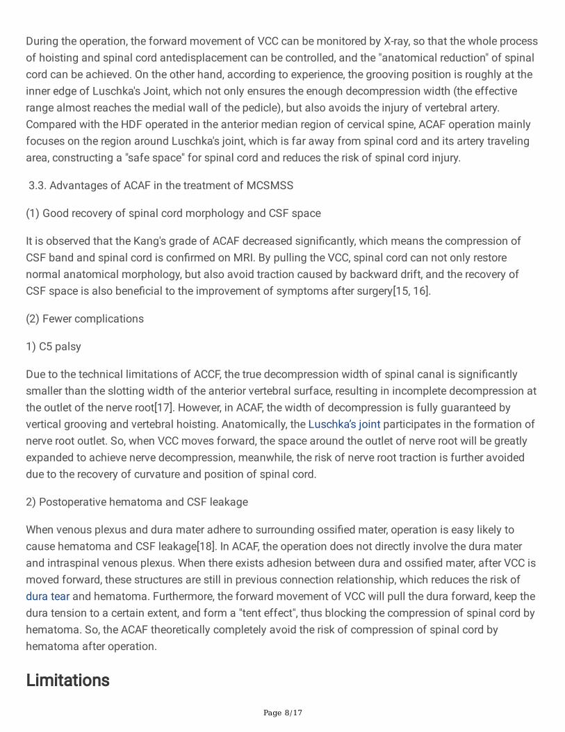
Table 3. Kang's grading rank-sum test HDF ACAF PGrading preoperation 0.324
0 0 0 1 15 15 2 12 9 3 13 21
Grading postoperation 0.006*
0 12 30 1 9 5 2 9 0 3 10 10
*Statistically significant
Figures

Page 15/17
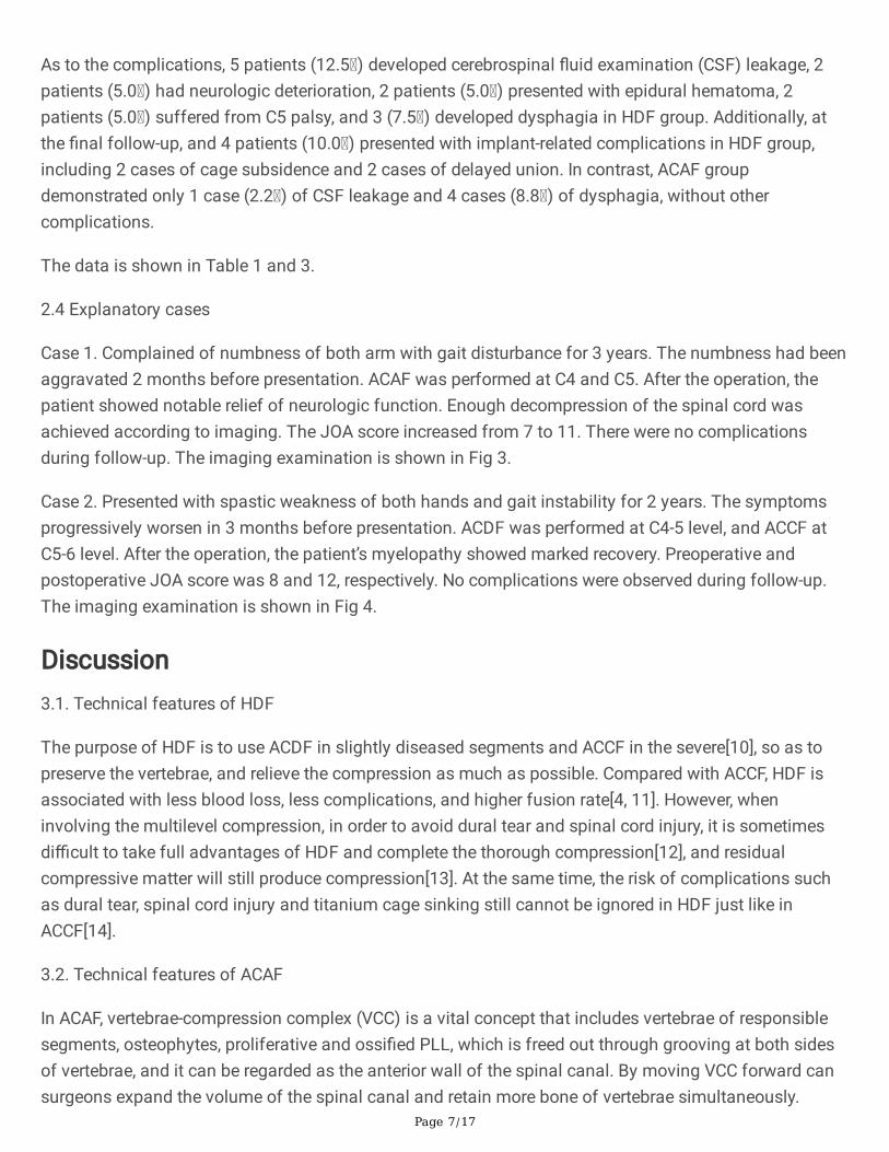
Figure 1
The procedure of ACAF. (A) Expose responsible vertebral discs and vertebrae. (B) Remove the responsibleintervertebral discs and osteophytes of anterior and posterior edge. (C) Perform osteotomy to the anteriorparts of vertebrae. (D) Groove at both sides, but retain the posterior wall of the vertebra at one side. (E)Install titanium plate, vertebral screws and intervertebral fusion cages, and resect the posterior wall of thevertebra at the remaining side to make the vertebra free. (F) Hoist the complex of titanium plate, vertebralscrews, intervertebral fusion cages and vertebrae.
Figure 2
The measuring diagram on CT axis images of HDF (a) and ACAF (b). The red curve shows the transversearea of decompression. The green line and yellow line represent the decompression width and sagittaldiameter of spinal canal, respectively.
Page 16/17
Figure 3
A 57-year-old man being performed ACAF. (A-D) and (E-H) illustrated preoperative and postoperativeconditions, respectively. (A) Lateral X-ray showed the degenerative change and spinal stenosis at C3-5levels. (B) Lateral MRI demonstrated the compression of CSF band at both discs and vertebrae levels. (C)Axis CT depicted spinal stenosis with spinal canal diameter of 9.6 mm before surgery. (D) Axis MRIshowed no CSF signal anterior to the vertebra. (E) Lateral X-ray revealed the hoisting of C4 and C5 aftersurgery (F) Lateral MRI con�rmed that the CSF band reappear at operating level just after surgery. (G)Axis CT depicted the decompression space with spinal canal diameter of 12.5 mm after hoisting. (H) AxisMRI showed the normal CSF signal anterior to the vertebra.

Page 17/17
Figure 4
A-61-year-old man being performed HDF. (A-C) and (D-F) illustrated preoperative and postoperativeconditions, respectively. (A) Lateral X-ray showed the degenerative change and spinal stenosis at C4-6levels. (B) Lateral MRI demonstrated the compression of CSF band at both discs and vertebrae levels. (C)Axis MRI showed CSF signal waned anterior to the vertebra. (D) Lateral X-ray revealed ACDF at C4-5 leveland ACCF at C5-6 level. (E) Lateral MRI con�rmed that the CSF band reappear at operating level. (F) AxisCT depicted the decompression space after corpectomy and titanium mesh cage implantation.