Embed Size (px)
Citation preview
Stem Cells

Stem Cell – Definition
• A cell that has the ability to continuously divide and differentiate (develop) into various other kind(s) of cells/tissues
• Stem cells are different from other cells of the body in that they have the ability to differentiate into other cell/tissue types. This ability allows them to replace cells that have died. With this ability, they have been used to replace defective cells/tissues in patients who have certain diseases or defects.
Stem Cell Characteristics
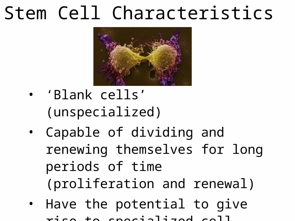
• ‘Blank cells’ (unspecialized)
• Capable of dividing and renewing themselves for long periods of time (proliferation and renewal)
• Have the potential to give rise to specialized cell types (differentiation)

Kinds of Stem CellsKinds of Stem Cells
Stem cell Stem cell typetype DescriptionDescription ExamplesExamples
TotipotentTotipotent Each cell can develop Each cell can develop into a new individualinto a new individual
Cells from early (1-Cells from early (1-3 days) embryos3 days) embryos
PluripotentPluripotent Cells can form any (over Cells can form any (over 200) cell types200) cell types
Some cells of Some cells of blastocyst (5 to 14 blastocyst (5 to 14 days)days)
MultipotentMultipotentCells differentiated, but Cells differentiated, but can form a number of can form a number of other tissuesother tissues
Fetal tissue, cord Fetal tissue, cord blood, and adult blood, and adult stem cellsstem cells
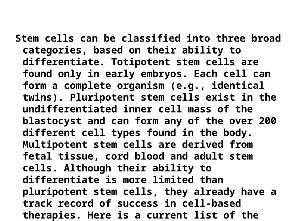
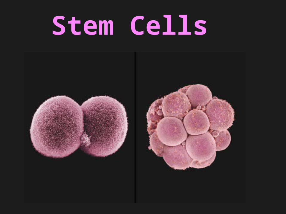
Stem cells can be classified into three broad categories, based on their ability to differentiate. Totipotent stem cells are found only in early embryos. Each cell can form a complete organism (e.g., identical twins). Pluripotent stem cells exist in the undifferentiated inner cell mass of the blastocyst and can form any of the over 200 different cell types found in the body. Multipotent stem cells are derived from fetal tissue, cord blood and adult stem cells. Although their ability to differentiate is more limited than pluripotent stem cells, they already have a track record of success in cell-based therapies. Here is a current list of the sources of stem cells:

1.Embryonic stem cells - are harvested from the inner cell mass of the blastocyst seven to ten days after fertilization.
2.Fetal stem cells - are taken from the germline tissues that will make up the gonads of aborted fetuses.
3.Umbilical cord stem cells - Umbilical cord blood contains stem cells similar to those found in bone marrow.
4.Placenta derived stem cells - up to ten times as many stem cells can be harvested from a placenta as from cord blood.
5.Adult stem cells - Many adult tissues contain stem cells that can be isolated.

This cellCan form the
Embryo and placenta
This cellCan just form the
embryo
Fully mature
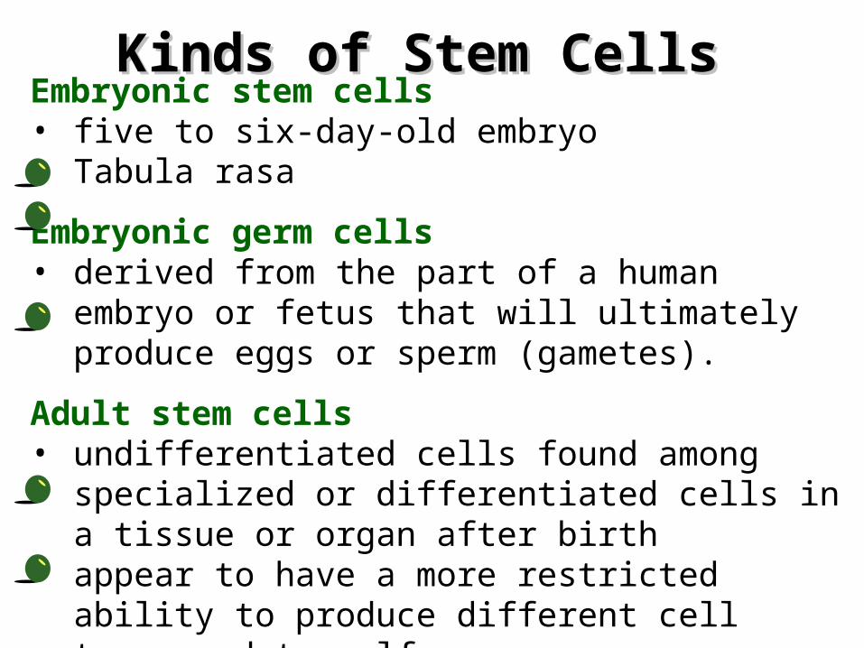
Kinds of Stem CellsKinds of Stem CellsEmbryonic stem cells • five to six-day-old embryo• Tabula rasa
Embryonic germ cells • derived from the part of a human embryo or fetus
that will ultimately produce eggs or sperm (gametes).
Adult stem cells • undifferentiated cells found among specialized or
differentiated cells in a tissue or organ after birth• appear to have a more restricted ability to produce
different cell types and to self-renew.
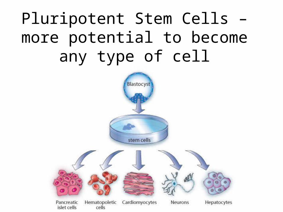
Pluripotent Stem Cells –more potential to become any
type of cell
Multipotent stem cells
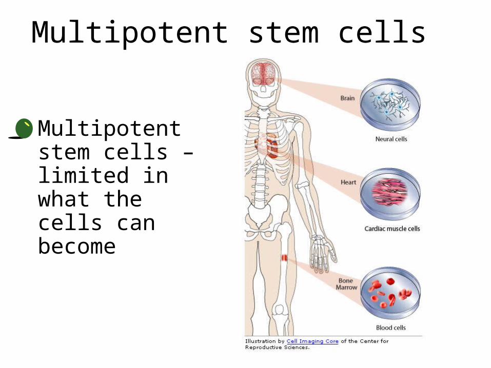
• Multipotent stem cells – limited in what the cells can become
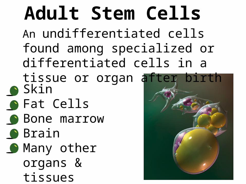
• Skin • Fat Cells• Bone marrow• Brain• Many other organs
& tissues
Adult Stem CellsAn undifferentiated cells found among specialized or differentiated cells in a tissue or organ after birth

Induced Pluripotent Stem Cells
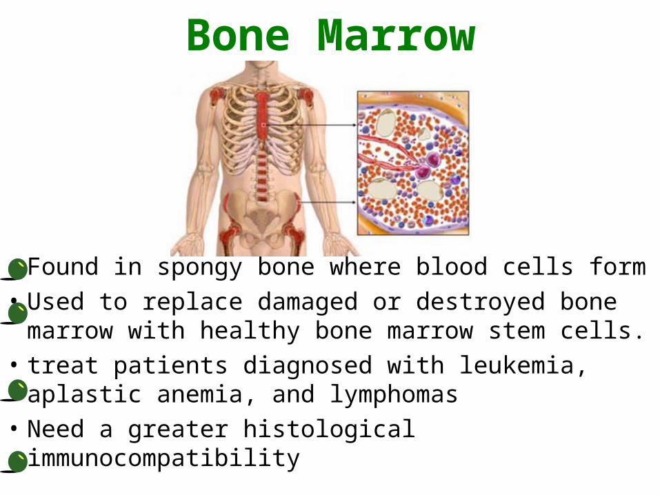
Bone Marrow
• Found in spongy bone where blood cells form• Used to replace damaged or destroyed bone
marrow with healthy bone marrow stem cells.• treat patients diagnosed with leukemia, aplastic
anemia, and lymphomas• Need a greater histological immunocompatibility
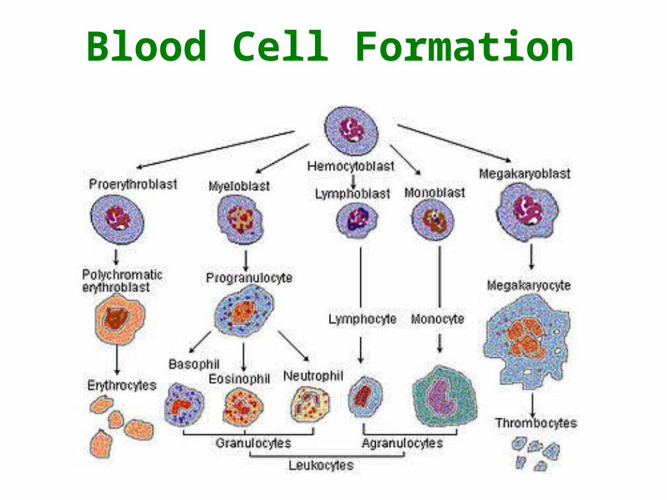
Blood Cell Formation

Umbilical cord stem cells
• Also Known as Wharton’s Jelly• Adult stem cells of infant origin• Less invasive than bone marrow• Greater compatibility• Less expensive

Umbilical cord stem cellsThree important functions:
1. Plasticity: Potential to change into other cell types like nerve cells
2. Homing: To travel to the site of tissue damage
3. Engraftment: To unite with other tissues
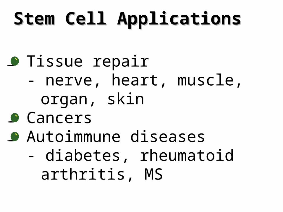
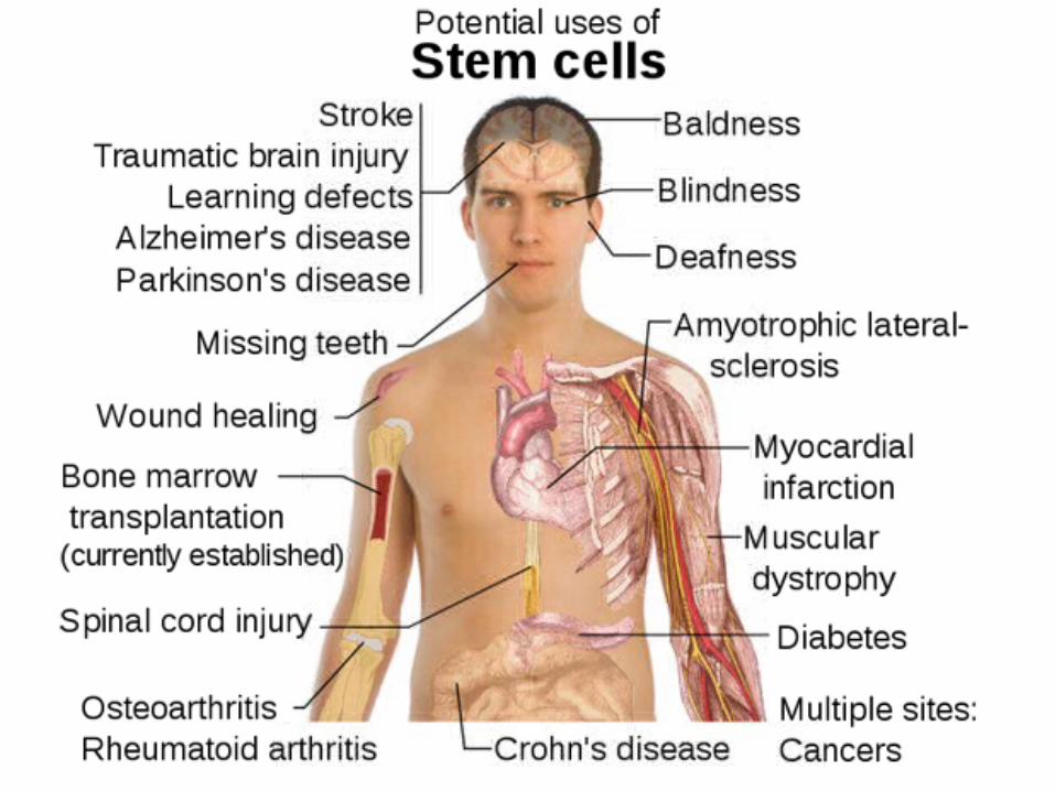
Stem Cell ApplicationsStem Cell Applications
• Tissue repair- nerve, heart, muscle, organ,
skin• Cancers• Autoimmune diseases
- diabetes, rheumatoid arthritis, MS
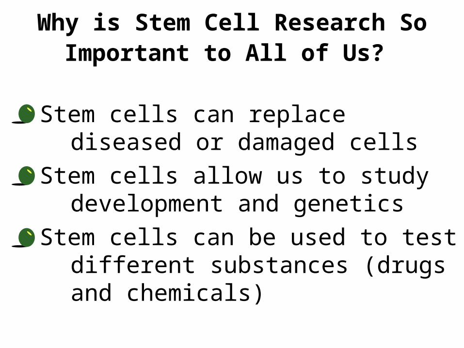
Why is Stem Cell Research So Important to All of Us?
Stem cells can replace diseased or damaged cells
Stem cells allow us to study development and genetics
Stem cells can be used to test different substances (drugs and chemicals)

Terminology:STEM CELL- cell which can make exact copies of
itself indefinitely, can differentiate, and produce specialized cells for various tissues of body.
TOTIPOTENT- can become any kind of cell Early Embryonic SC
PLURIPOTENT- almost any kind of cell Blastocyst Embryonic SC
MULTIPOTENT- limited range of cell typesAdult SC: nerve cells, blood cells, muscle cells,
bone and skin cells.

Potency: A measure of how many types of specialized cell a stem cell can make
Pluripotent Can make all types of specialized cells in the body Embryonic stem cells are pluripotent
Multipotent Can make multiple types of specialized cells, but not all types Tissue stem cells are multipotent

APOPTOSIS

INTRODUCTION
Cell death by injury
-Mechanical damage
-Exposure to toxic chemicals
Cell death by suicide
-Internal signals
-External signals
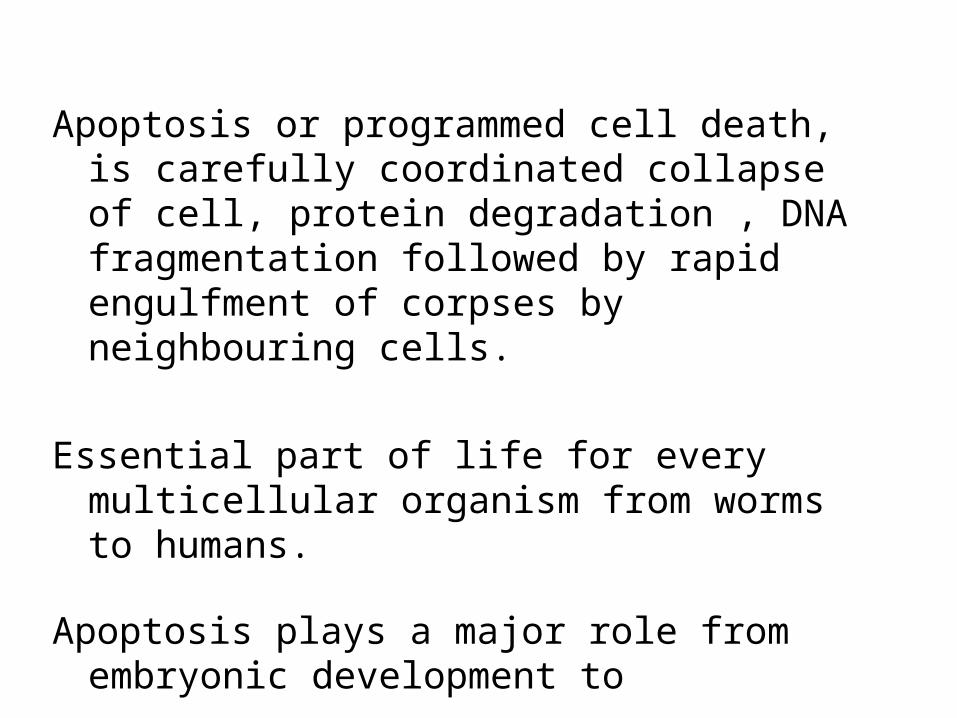
Apoptosis or programmed cell death, is carefully coordinated collapse of cell, protein degradation , DNA fragmentation followed by rapid engulfment of corpses by neighbouring cells.
Essential part of life for every multicellular organism from worms to humans.
Apoptosis plays a major role from embryonic development to senescence.
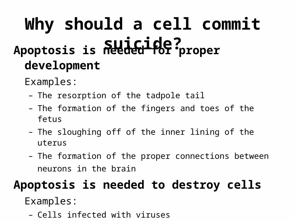
Why should a cell commit suicide?
Apoptosis is needed for proper developmentExamples: – The resorption of the tadpole tail
– The formation of the fingers and toes of the fetus
– The sloughing off of the inner lining of the uterus
– The formation of the proper connections between neurons in the brain
Apoptosis is needed to destroy cells
Examples: – Cells infected with viruses
– Cells of the immune system
– Cells with DNA damage
– Cancer cells
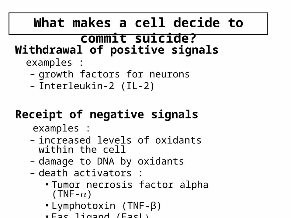
What makes a cell decide to commit suicide?
Withdrawal of positive signalsexamples : – growth factors for neurons – Interleukin-2 (IL-2)
Receipt of negative signals examples : – increased levels of oxidants within the cell – damage to DNA by oxidants – death activators :
• Tumor necrosis factor alpha (TNF-) • Lymphotoxin (TNF-β) • Fas ligand (FasL)

Definition
Apoptosis is a peculiar well controlled individual cell death that is caspase mediated and leads to fragmentation of the cell and organelles into numerous small buds, which are then engulfed by macrophages without surrounding inflammation.

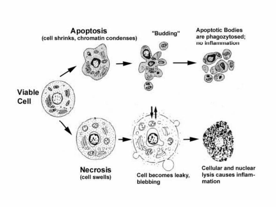
Apoptosis, or programmed cell death, is a normal component of the development and health of multicellular organisms. Cells die in response to a variety of stimuli and during apoptosis they do so in a controlled, regulated fashion. This makes apoptosis distinct from another form of cell death called necrosis in which uncontrolled cell death leads to lysis of cells, inflammatory responses and, potentially, to serious health problems. Apoptosis, by contrast, is a process in which cells play an active role in their own death (which is why apoptosis is often referred to as cell suicide).
Upon receiving specific signals instructing the cells to undergo apoptosis a number of distinctive changes occur in the cell. A family of proteins known as caspases are typically activated in the early stages of apoptosis. These proteins breakdown or cleave key cellular components that are required for normal cellular function including structural proteins in the cytoskeleton and nuclear proteins such as DNA repair enzymes. The caspases can also activate other degradative enzymes such as DNases, which begin to cleave the DNA in the nucleus.

Importance of Apoptosis1) Crucial for embryonic development
-Errors in Apoptosis can lead to Birth Defects2) Important for maintaining homeostasis
- Cell death is balanced with mitosis to regulate cell number.3) Improper regulation contributes to human disease
- Neurodegenerative diseasesParkinson’s Alzheimer’s
- Cancer- Autoimmune diseases e.g. (diabetes type I)- Viral diseases

Characteristics
• It is a process that occurs in almost all living creatures since their early stages of embryological development.
• It is an active cytological process in which energy is consumed (ATP dependent)
• It is programmed or controlled by genetic protocol or program (control of enzymes, cell membrane surface proteins & cytoplasmic molecules, signal transduction, gene expression)
• It may be triggered by intrinsic or extrinsic stimuli
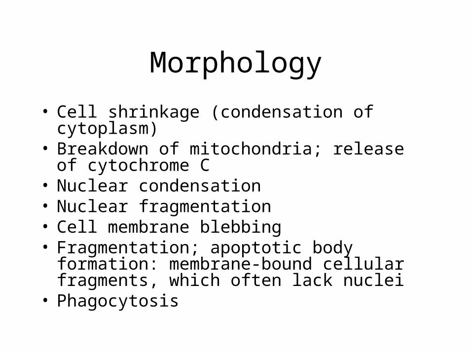
Morphology
• Cell shrinkage (condensation of cytoplasm)• Breakdown of mitochondria; release of
cytochrome C• Nuclear condensation• Nuclear fragmentation• Cell membrane blebbing• Fragmentation; apoptotic body formation:
membrane-bound cellular fragments, which often lack nuclei
• Phagocytosis

Cellular changes associated with apoptosis
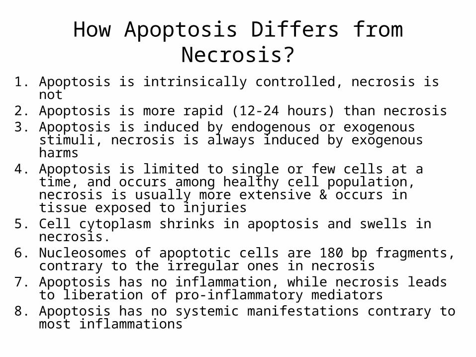
How Apoptosis Differs from Necrosis?
1. Apoptosis is intrinsically controlled, necrosis is not2. Apoptosis is more rapid (12-24 hours) than necrosis3. Apoptosis is induced by endogenous or exogenous stimuli,
necrosis is always induced by exogenous harms 4. Apoptosis is limited to single or few cells at a time, and
occurs among healthy cell population, necrosis is usually more extensive & occurs in tissue exposed to injuries
5. Cell cytoplasm shrinks in apoptosis and swells in necrosis.6. Nucleosomes of apoptotic cells are 180 bp fragments,
contrary to the irregular ones in necrosis7. Apoptosis has no inflammation, while necrosis leads to
liberation of pro-inflammatory mediators8. Apoptosis has no systemic manifestations contrary to most
inflammations
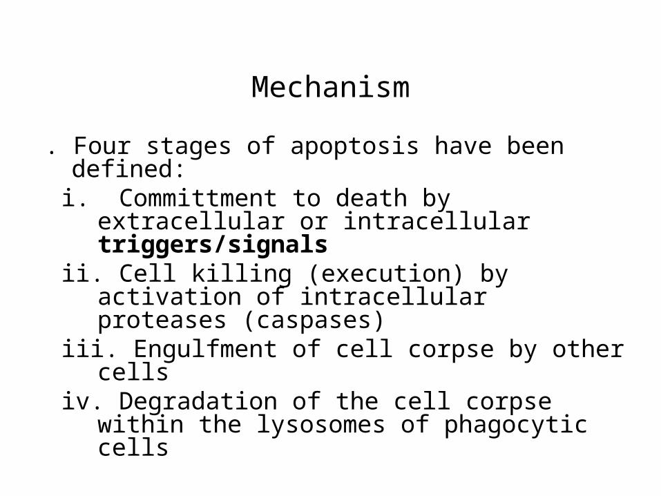
Mechanism
I. Four stages of apoptosis have been defined: i. Committment to death by extracellular or
intracellular triggers/signals ii. Cell killing (execution) by activation of
intracellular proteases (caspases) iii. Engulfment of cell corpse by other cells iv. Degradation of the cell corpse within the
lysosomes of phagocytic cells

II. Stimuli for Apoptotic Cell Death in Mammals i. Growth factor deficiencies ii. Ionizing radiation/ viral infection iii. Free radical toxicity iv. Death receptor activation (such as Fas or
CD95 triggering) v. Metabolic or cell cycle perturbation
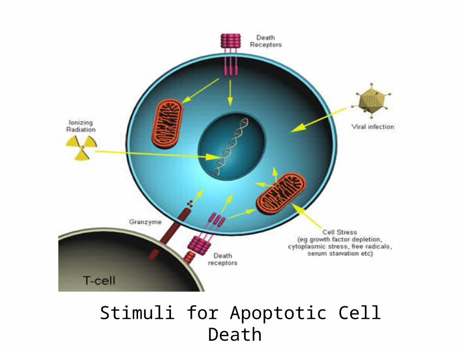
Stimuli for Apoptotic Cell Death
Death Factors
Definition: cytokines that activate an apoptosis program by binding to their specific receptor. Typical examples of death factors are:
1. Fas ligand, 2. TNF (tumor necrosis factor) and 3. TRAIL (TNF-related apoptosis-inducing
ligand).- Apoptosis can also be induced by cytotoxic T-
lymphocytes using the enzyme granzyme.
III. Activation of Caspase cascade i. Various stimuli described above eventually
activate the executioner (caspase) cascade ii. At least 14 different caspases exist in human
cells iii. Caspase cascades are apparently required for
complete execution
40
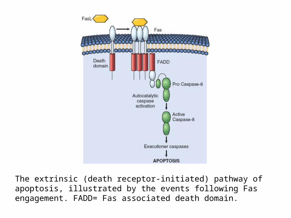
The extrinsic (death receptor-initiated) pathway of apoptosis, illustrated by the events following Fas engagement. FADD= Fas associated death domain.
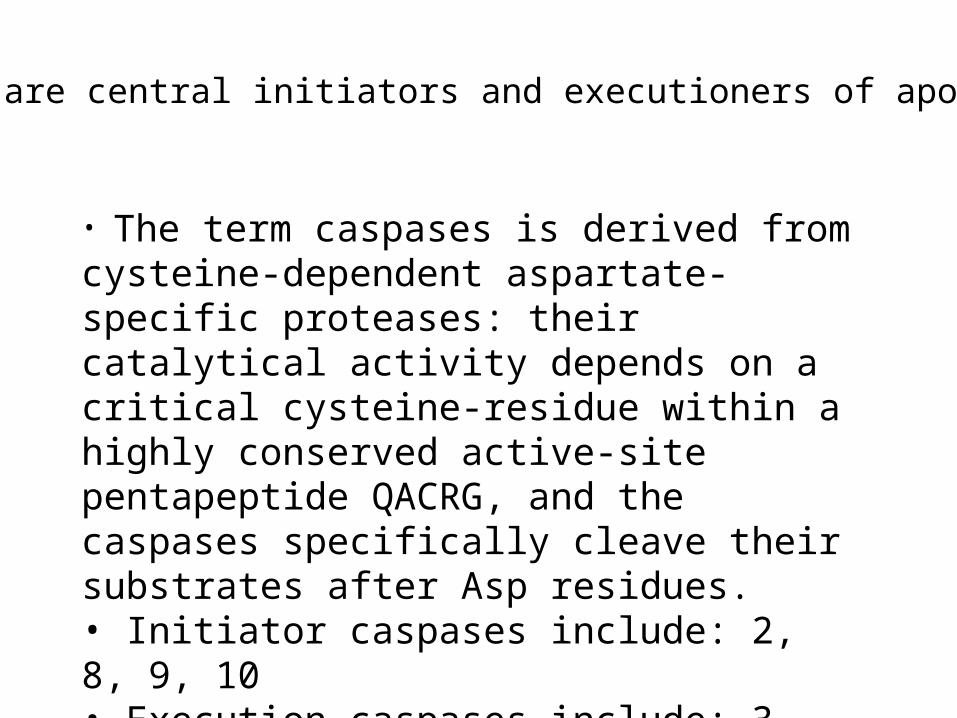
• The term caspases is derived from cysteine-dependent aspartate-specific proteases: their catalytical activity depends on a critical cysteine-residue within a highly conserved active-site pentapeptide QACRG, and the caspases specifically cleave their substrates after Asp residues.• Initiator caspases include: 2, 8, 9, 10• Execution caspases include: 3, 6, 7
Caspases are central initiators and executioners of apoptosis