Embed Size (px)
Citation preview
~ 2343 ~
Journal of Pharmacognosy and Phytochemistry 2020; 9(1): 2343-2352
E-ISSN: 2278-4136
P-ISSN: 2349-8234
www.phytojournal.com
JPP 2020; 9(1): 2343-2352
Received: 24-11-2019
Accepted: 28-12-2019
Nisar Ahmad Dar
Division of Plant Pathology,
Sher-e-Kashmir University of
Agricultural Sciences &
Technology, Shalimar, Srinagar,
Jammu and Kashmir, India
Nisar Ahmad Khan
Division of Plant Pathology,
Sher-e-Kashmir University of
Agricultural Sciences &
Technology, Shalimar, Srinagar,
Jammu and Kashmir, India
Mudasir Ahmad Bhat
Division of Plant Pathology,
Sher-e-Kashmir University of
Agricultural Sciences &
Technology, Shalimar, Srinagar,
Jammu and Kashmir, India
Corresponding Author:
Nisar Ahmad Dar
Division of Plant Pathology,
Sher-e-Kashmir University of
Agricultural Sciences &
Technology, Shalimar, Srinagar,
Jammu and Kashmir, India
Status, symptomatology and partial
characterization of stem bark canker disease of
apple (Malus domestica Borkh.) in Kashmir Valley
Nisar Ahmad Dar, Nisar Ahmad Khan and Mudasir Ahmad Bhat
Abstract
Surveys were conducted to determine the occurrence and distribution of Stem bark canker affecting
Apple in Kashmir valley during 2013 and 2014. Apple orchards were more affected in district Ganderbal
with highest canker incidence (31.33%) and the least in district Shopian (15.33%). Among the villages
surveyed, Watlar of district Ganderbal exhibited the highest canker incidence of 41.00 per cent. Village
Batapora of district Shopian exhibited the least canker incidence of 13.00 per cent. The data over the two
years further revealed that maximum canker incidence of 23.50% was observed on tree trunks followed
by scaffold branches (17.70%) and least on fruiting wood (10.09%). Infected samples were collected
from different localities in four districts of Kashmir valley. Stem bark symptoms mostly characterized
under field conditions by the appearance of small, sunken, reddish brown lesions, which on enlargement
became depressed and developed elliptical cankers with vertical and horizontal slits, partially or
completely girdling the affected trunk or branch. In advanced stages, the cankered surface however,
became was black and brittle which remained studded with numerous black fissures. The fungus isolated
from stem bark canker produced olivaceous to violaceous black fungal colonies with dense aerial
mycelium. The hyphae were smooth, thick walled, septate and dark brown in colour. The pycnidia
formed only in presence of light were globose to sub-globose distributed uniformally over the culture
medium. The conidiogenous cells were smooth, hyaline, sub-cylindrical, swollen at the base, producing
single apical conidium. The conidia were smooth, thin walled, hyaline, unicellular, fusoid to ellipsoidal
with an obtuse apex and truncate or sub-truncate to rounded base. Based on morphological characters
both on host as well as in culture, symptom expression and pathogenicity tests, the fungus causing the
disease was identified as Fusicoccum aesculi Corda.
Keywords: Apple, stem bark canker, incidence, lesions and Fusicoccum aesculi Corda
1. Introduction
Apple (Malus domestica Borkh.) is a premier table fruit of the world and has been under
cultivation since time immemorial. Apple tree owes its origin in South Eastern Europe and
Tien Shan Mountains of Kazakhstan in Asia (Gasteir, 2000) where vast forests of wild apple
trees exist even today. The ten leading apple producing countries contributing to world’s
annual production of 63 million tonnes are USA, China, France, Italy, Turkey, Argentina,
West Germany, Spain, Japan and erstwhile USSR (Snowdon, 1990) [24]. In India, the
commercial cultivation of apple is largely confined to the states of J & K., H.P. and U.K.
which together accounts for 99 per cent of the total production with productivity of 13.07
metric tonnes per hectares in J&K followed by 8.95 metric tonnes per hectares in H.P. and
3.52 metric tonnes per hectares in U.K. (Anonymous, 2011) [3].
In spite of the unique agro climatic conditions of the Kashmir valley being quite conducive for
temperate fruit production, apple productivity per unit area is low owing to many biotic and
abiotic stresses. The major biotic factors inflicting huge economic losses are the fungal
diseases, the predominant among them being scab, powdery mildew, collar rot, Alternaria leaf
blotch, Marssonina blotch, Sooty blotch and stem and branch cankers caused by various fungi
(Bilgrami et al., 1979, Kanwar, 1988; Sharma and Bhardwaj, 1999) [4, 9, 21]. Among these
canker diseases, stem and branch cankers have assumed an alarming proportion and cause
huge economic losses through girdling of branches, limbs, blighting and die-back of twigs
ultimately resulting in the death of whole or part of the plant (Jones and Aldwinkle, 1990) [8].
Apart from girdling of branches, losses also occur through fruit rotting and premature
defoliation (Sharma and Bhardwaj, 1999) [21]. Stem bark canker, Silver leaf, Smoky Canker,
Phomopsis canker, Valsa canker and anthracnose cankers has also been reported from Jammu
& Kashmir state (Malik, 1967; Chib and Andotra, 1985, Khan et al., 2010, 2011 and 2011a) [14, 11].
~ 2344 ~
Journal of Pharmacognosy and Phytochemistry http://www.phytojournal.com Keeping in view the extent of damage inflicted by the canker
diseases, the present study on stem bark apple canker disease
of apple excluding its leaf spot and fruit rot phase, if any,
were, therefore undertaken with main emphasis on status and
symptomatology.
2. Materials and Methods An extensive survey of different Apple (Malus domestica
Borkh.) growing localities in four districts of Kashmir Valley
was conducted during 2013 and 2014 cropping seasons (in the
months of August to September) to determine the distribution
and incidence of Stem bark canker in the state. In each
district, three locations were selected and five orchards in
each of the five villages taken to represent a location. Ten
trees were randomly selected from each orchard for assessing
the incidence of the canker disease. Besides the main trunk,
three scaffold branches (major limbs) and nine branches/twigs
(fruiting wood) from each tree were randomly selected and
examined for the presence of the canker disease and recorded
as per cent canker incidence.
The per cent canker intensity of stem bark canker occurring
only on branches and twigs was calculated after rating the
level of disease on 0-5 scale of Crosse (1957) [5] adopted by
Khan et al. (2011).
Per cent canker intensity was assessed using the formula:
Where,
n = number of branches or twigs in each category;
v = numerical value of each category;
N = number of branches or twigs examined; and
S = the maximum numerical value.
Disease samples from trunks, limbs, branches of apple trees
showing distinct cankerous symptoms, collected during the
course of Survey, were washed with running tap water to
remove the dirt and dust. After removing the bark, the
infected woody tissue was thoroughly sterilized with cotton
swab dipped in absolute alcohol (95%). Small sections of 5
mm² size were cut at the transition zone between healthy and
diseased tissue with a sterilized scalpel and surface sterilized
in 0.1 per cent mercuric chloride solution for 30 seconds. The
sections were then rinsed in distilled water to remove the
traces of mercuric chloride solution, blotted dry and
transferred aseptically onto acidified potato dextrose agar
(PDA) medium contained in sterilized Petri plates and
incubated at 25±1 °C. The culture thus obtained was purified
by hyphal tip method (Pathak, 1972) [16]. Pure cultures
obtained were maintained by repeated sub-culturing at regular
intervals for further studies. The stock cultures were stored in
a refrigerator at 4 °C. The composition of Potato Dextrose
Agar medium was:
1. Peeled potato: 250 grams
2. Dextrose: 20 grams
3. Agar Agar: 20 grams
4. Water: 1000 ml
The pathogenicity test of the isolated pathogen(s) was
performed on healthy one year old potted saplings of apple
cultivar “Red Delicious” as per the technique employed by
Milholland (1972) [15] and Spiers (1977). The saplings were
sprayed with copper oxy-chloride 50 WP @ 0.3% 20 days
before inoculations to exclude any harbouring pathogen. The
pots containing the saplings were kept in diffused sunlight in
polythene chambers, designed for the purpose, maintaining
high humidity inside the chambers by timely irrigating the
pots and intermittently spraying with distilled sterilized water.
On one year old potted apple saplings 5mm vertical and
horizontal incisions of “T” shape were made on the selected
twigs after surface sterilizing the site with absolute alcohol
and a 4 mm test mycelial plug inserted inside the “T” shaped
flap. The inoculated incision was covered with moistened
absorbent cotton and wrapped with paper tape. Incised
inoculated twig with plain PDA medium covered with
moistened cotton and wrapped with paper tape served as
control.
Pathogenicity tests were closely monitored for
symptom/canker development. Re-isolation of the pathogen
from artificially inoculated twigs was carried out and resultant
cultures compared with the original inoculant to satisfy
Koch’s postulates.
The morphological characteristics of the causal organism
were studied taking in pathogen thallus from the host and
after culturing it in the laboratory. The pathogen cultures were
grown on potato dextrose agar medium and the semi-
permanent slides prepared from 7 and 21 days old colonies.
The important characters studied were as under:
Mycelium, width, septation and colour
Fruiting body structure, size and colour
Conidia, shape, size and colour
The diseased branches and twigs were cut off along with
some healthy portion, kept in a humid chamber at room
temperature (20±1°C) and observed for mycelial colour, size
and septation after 72 hours. Diseased branches/twigs were
also observed under stereoscopic microscope for the presence
of fruiting bodies and their morphological characteristics
studied under compound microscope previously calibrated
with the aid of stage and ocular micrometres.
Further the over-wintered branches and twigs collected at
fortnightly intervals were examined under stereoscopic
microscope for the presence of perfect state fruiting bodies.
The morphological details of these fruiting bodies were also
studied using a compound microscope. The morphological
characters of the causal organism studied were compared with
authentic descriptions for their identification and
nomenclature.
3. Results and Discussion
3.1 Survey and Incidence
In the present investigation, an extensive survey of apple
orchards in Anantnag, Kulgam, Ganderbal and Shopian
districts of Kashmir valley during the months of August-
September in 2013 and 2014 was under taken to record the
status of stem bark canker disease. During the surveys stem
bark canker disease was observed on different apple tree parts
with varying degrees of incidence. The cankers were observed
on all the major tree parts like trunks, scaffold branches
(limbs) and fruiting wood (branches and twigs).
The data averaged for two years Table 1; Fig. 1 revealed an
overall total canker incidence of 23.66 per cent throughout the
areas surveyed. The average incidence was maximum on
main trunk (19.64%) followed by that on scaffold branches
(15.14%). The incidence on fruiting wood being the least
~ 2345 ~
Journal of Pharmacognosy and Phytochemistry http://www.phytojournal.com (9.43%). The total canker incidence recorded over the two
years was highest in district Ganderbal (31.33%) followed by
that in district Kulgam (27.00%). However, the incidence was
least (15.33%) in district Shopian.
Among the different locations surveyed, highest canker
incidence was recorded at Watlar (41.00%) followed by Lar
(34.00%) and Devsar (29.00%) while, it was least 13.00 and
14.00 per cent recorded at Batapora and Kapran respectively.
The data presented in Table 3 further reveals that the number
of cankers per tree varied from 1.00 to 2.65 at different
locations surveyed with an average number of 1.75 cankers
per tree. The maximum number of cankers per tree (2.65) was
recorded at Lar in district Ganderbal, while, minimum number
(1.00) was recorded at Wakoora in district Ganderbal.
Table 1: Incidence of stem bark canker on various tree parts of apple at different locations of Kashmir during the year 2013 and 2014
District Location Canker incidence (%)
Number of cankers per tree Trunk* Scaffold branches* Fruiting wood* Total incidence**
Anantnag
Achabal 15.56 12.15 8.76 19.00 1.85
Kellar 18.39 14.45 8.89 21.00 1.84
Kanilwan 21.59 17.30 8.67 23.00 1.81
Mean 18.51 14.63 8.77 21.00 1.83
Kulgam
Devsar 20.14 13.73 9.95 29.00 1.93
D.H.pora 20.71 13.81 10.95 24.00 1.41
Sopat 20.84 14.24 8.19 28.00 1.84
Mean 20.56 13.92 9.70 27.00 1.72
Ganderbal
Wakoora 25.84 11.93 10.84 19.00 1.00
Lar 20.49 22.54 12.47 34.00 2.65
Watlar 24.16 18.65 6.97 41.00 2.11
Mean 23.49 17.70 10.09 31.33 1.92
Shopian
Kapran 21.11 22.96 10.61 14.00 1.55
Batapora 11.25 11.25 8.75 13.00 1.57
Shirmal 15.63 8.70 8.09 19.00 1.52
Mean 16.00 14.30 9.15 15.33 1.55
Grand Mean ± SD 19.64±3.47 15.14±2.71 9.43±1.56 23.66±6.28 1.75
*Figures are the per cent of cankered trees out of the total number of trees examined **Observations based on means of fifty trees recorded in August-September
Fig 1: Incidence of stem bark canker on various tree parts of apple at different locations of Kashmir during the year 2013 & 2014
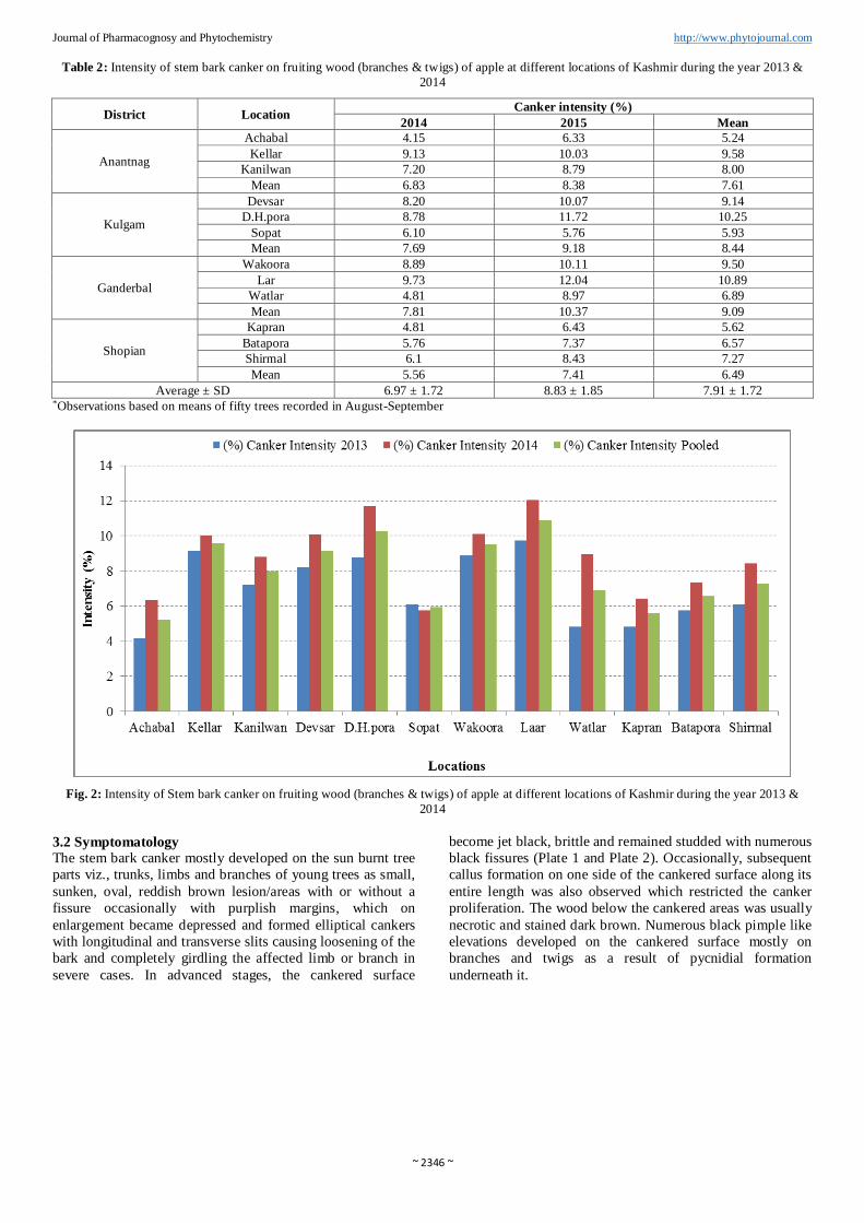
Like canker incidence, the canker intensity on fruiting wood
(branches and twigs) also varied at all the locations surveyed.
The data presented in Table 2; Fig 2 revealed that the canker
intensity ranged between 4.81 to 9.73 and 5.76 to 12.04 per
cent during the years 2013 and 2014 respectively and was
highest in 2014 with 7.41 per cent compared to that 5.56 per
cent in 2013, with an overall canker intensity of 6.49 per cent.
Among the different districts surveyed, district Ganderbal
exhibited the maximum average canker intensity (9.09%)
followed by Kulgam with an average canker intensity
(8.44%). While, minimum canker intensity of 6.49 per cent
was recorded in district Shopian. Among various locations
surveyed the maximum canker intensity of 10.89 per cent was
recorded at Lar of district Ganderbal, while the minimum
canker intensity of 5.24 per cent respectively were recorded at
Achabal of district Anantnag.
~ 2346 ~
Journal of Pharmacognosy and Phytochemistry http://www.phytojournal.com Table 2: Intensity of stem bark canker on fruiting wood (branches & twigs) of apple at different locations of Kashmir during the year 2013 &
2014
District Location Canker intensity (%)
2014 2015 Mean
Anantnag
Achabal 4.15 6.33 5.24
Kellar 9.13 10.03 9.58
Kanilwan 7.20 8.79 8.00
Mean 6.83 8.38 7.61
Kulgam
Devsar 8.20 10.07 9.14
D.H.pora 8.78 11.72 10.25
Sopat 6.10 5.76 5.93
Mean 7.69 9.18 8.44
Ganderbal
Wakoora 8.89 10.11 9.50
Lar 9.73 12.04 10.89
Watlar 4.81 8.97 6.89
Mean 7.81 10.37 9.09
Shopian
Kapran 4.81 6.43 5.62
Batapora 5.76 7.37 6.57
Shirmal 6.1 8.43 7.27
Mean 5.56 7.41 6.49
Average ± SD 6.97 ± 1.72 8.83 ± 1.85 7.91 ± 1.72 *Observations based on means of fifty trees recorded in August-September
Fig. 2: Intensity of Stem bark canker on fruiting wood (branches & twigs) of apple at different locations of Kashmir during the year 2013 &
2014
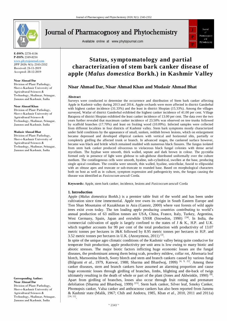
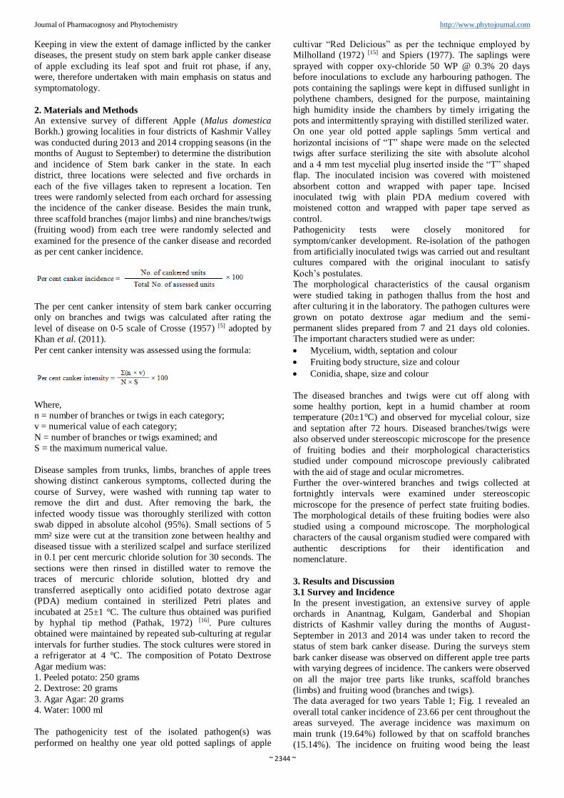
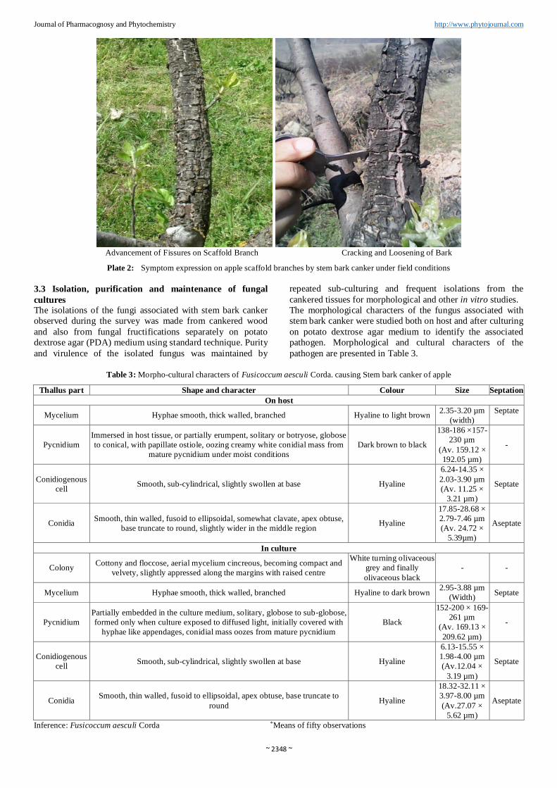
3.2 Symptomatology The stem bark canker mostly developed on the sun burnt tree
parts viz., trunks, limbs and branches of young trees as small,
sunken, oval, reddish brown lesion/areas with or without a
fissure occasionally with purplish margins, which on
enlargement became depressed and formed elliptical cankers
with longitudinal and transverse slits causing loosening of the
bark and completely girdling the affected limb or branch in
severe cases. In advanced stages, the cankered surface
become jet black, brittle and remained studded with numerous
black fissures (Plate 1 and Plate 2). Occasionally, subsequent
callus formation on one side of the cankered surface along its
entire length was also observed which restricted the canker
proliferation. The wood below the cankered areas was usually
necrotic and stained dark brown. Numerous black pimple like
elevations developed on the cankered surface mostly on
branches and twigs as a result of pycnidial formation
underneath it.
~ 2347 ~
Journal of Pharmacognosy and Phytochemistry http://www.phytojournal.com
Initial Lesion on Trunk Horizontal Canker Elongation
Formation of transverse slits Development of fissures/loosening of bark
Plate 1: Symptom expression on apple tree trunks by stem bark canker under field conditions
Canker on Scaffold Branch Development of Transverse Slits/Fissures
~ 2348 ~
Journal of Pharmacognosy and Phytochemistry http://www.phytojournal.com
Advancement of Fissures on Scaffold Branch Cracking and Loosening of Bark
Plate 2: Symptom expression on apple scaffold branches by stem bark canker under field conditions
3.3 Isolation, purification and maintenance of fungal
cultures The isolations of the fungi associated with stem bark canker
observed during the survey was made from cankered wood
and also from fungal fructifications separately on potato
dextrose agar (PDA) medium using standard technique. Purity
and virulence of the isolated fungus was maintained by
repeated sub-culturing and frequent isolations from the
cankered tissues for morphological and other in vitro studies.
The morphological characters of the fungus associated with
stem bark canker were studied both on host and after culturing
on potato dextrose agar medium to identify the associated
pathogen. Morphological and cultural characters of the
pathogen are presented in Table 3.
Table 3: Morpho-cultural characters of Fusicoccum aesculi Corda. causing Stem bark canker of apple
Thallus part Shape and character Colour Size Septation
On host
Mycelium Hyphae smooth, thick walled, branched Hyaline to light brown 2.35-3.20 µm
(width)
Septate
Pycnidium
Immersed in host tissue, or partially erumpent, solitary or botryose, globose
to conical, with papillate ostiole, oozing creamy white conidial mass from
mature pycnidium under moist conditions
Dark brown to black
138-186 ×157-
230 µm
(Av. 159.12 ×
192.05 µm)
-
Conidiogenous
cell Smooth, sub-cylindrical, slightly swollen at base Hyaline
6.24-14.35 ×
2.03-3.90 µm
(Av. 11.25 ×
3.21 µm)
Septate
Conidia Smooth, thin walled, fusoid to ellipsoidal, somewhat clavate, apex obtuse,
base truncate to round, slightly wider in the middle region Hyaline
17.85-28.68 ×
2.79-7.46 µm
(Av. 24.72 ×
5.39µm)
Aseptate
In culture
Colony Cottony and floccose, aerial mycelium cincreous, becoming compact and
velvety, slightly appressed along the margins with raised centre
White turning olivaceous
grey and finally
olivaceous black
- -
Mycelium Hyphae smooth, thick walled, branched Hyaline to dark brown 2.95-3.88 µm
(Width) Septate
Pycnidium
Partially embedded in the culture medium, solitary, globose to sub-globose,
formed only when culture exposed to diffused light, initially covered with
hyphae like appendages, conidial mass oozes from mature pycnidium
Black
152-200 × 169-
261 µm
(Av. 169.13 ×
209.62 µm)
-
Conidiogenous
cell Smooth, sub-cylindrical, slightly swollen at base Hyaline
6.13-15.55 ×
1.98-4.00 µm
(Av.12.04 ×
3.19 µm)
Septate
Conidia Smooth, thin walled, fusoid to ellipsoidal, apex obtuse, base truncate to
round Hyaline
18.32-32.11 ×
3.97-8.00 µm
(Av.27.07 ×
5.62 µm)
Aseptate
Inference: Fusicoccum aesculi Corda *Means of fifty observations
~ 2349 ~
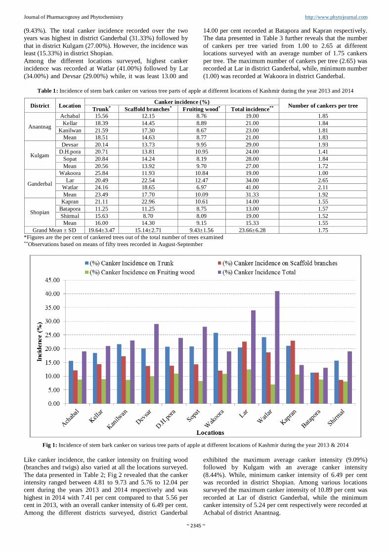
Journal of Pharmacognosy and Phytochemistry http://www.phytojournal.com 3.3.1 In vivo morphology The hyphae were branched, smooth, thick walled, septate,
hyaline to light brown in colour measuring 2.35-3.20 µm in
width. Stereoscope microscopic examination of the thallus
revealed the presence of numerous dark brown to black,
submerged or erumpent pycnidia over cankered branch/twig
surface. These were globose to conical with a papillate ostiole
exuding creamy conidial droplet under moist conditions. The
pycnidial size ranged from 138-186 × 157-230 µm averaging
159.12 × 192.05 µm. The conidiophore reduced to
conidiogenous cells were hyaline, sub-cylindrical, measuring
6.24-14.35 × 2.03-3.90 µm, with an average size of 11.25 ×
3.21 µm, producing a single apical conidium. The conidia
were hyaline, uni-cellular, smooth, ornamented with granular
contents, fusoid to ellipsoidal, somewhat clavate with an
obtuse apex and truncate to rounded base and measured
17.85-28.68 × 2.79-7.46 µm, with an average size of 24.72 ×
5.39 µm.
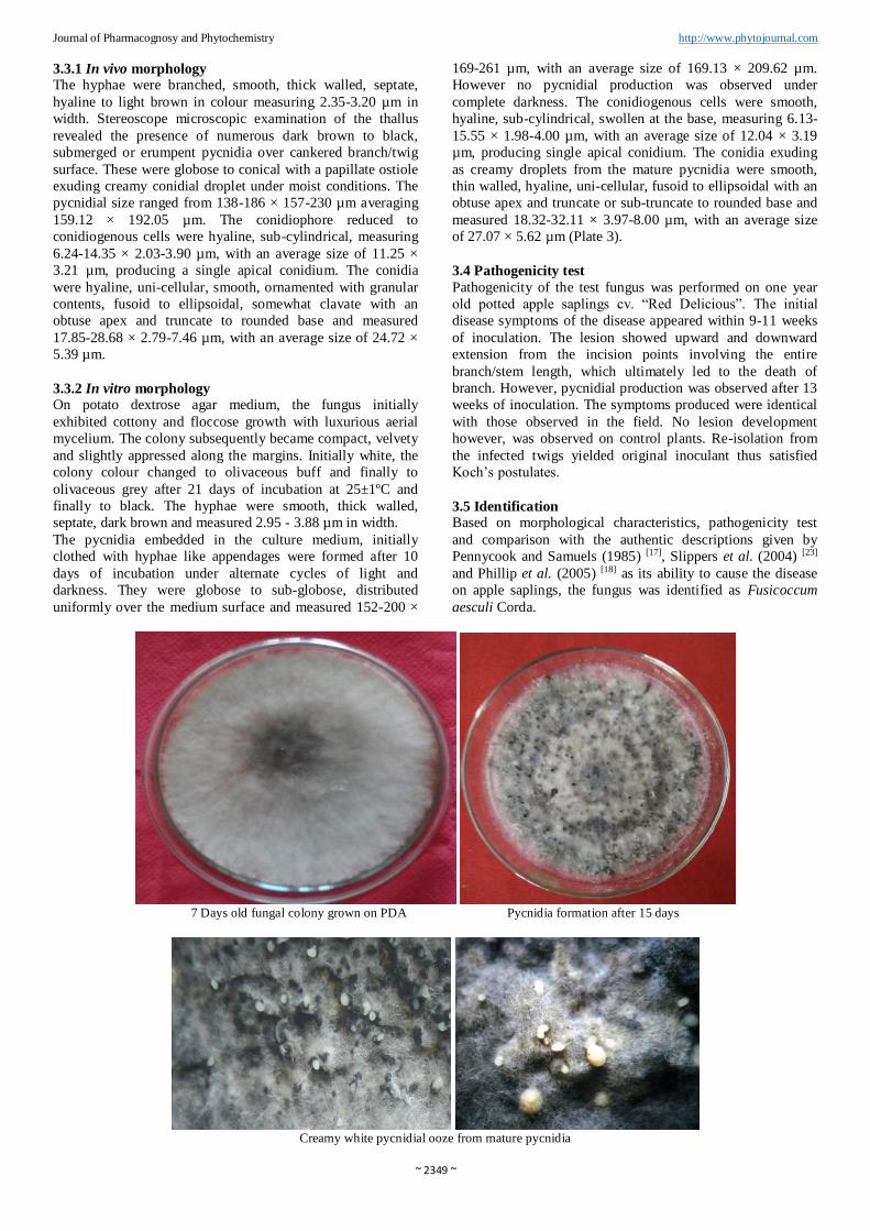
3.3.2 In vitro morphology On potato dextrose agar medium, the fungus initially
exhibited cottony and floccose growth with luxurious aerial
mycelium. The colony subsequently became compact, velvety
and slightly appressed along the margins. Initially white, the
colony colour changed to olivaceous buff and finally to
olivaceous grey after 21 days of incubation at 25±1ºC and
finally to black. The hyphae were smooth, thick walled,
septate, dark brown and measured 2.95 - 3.88 µm in width.
The pycnidia embedded in the culture medium, initially
clothed with hyphae like appendages were formed after 10
days of incubation under alternate cycles of light and
darkness. They were globose to sub-globose, distributed
uniformly over the medium surface and measured 152-200 ×
169-261 µm, with an average size of 169.13 × 209.62 µm.
However no pycnidial production was observed under
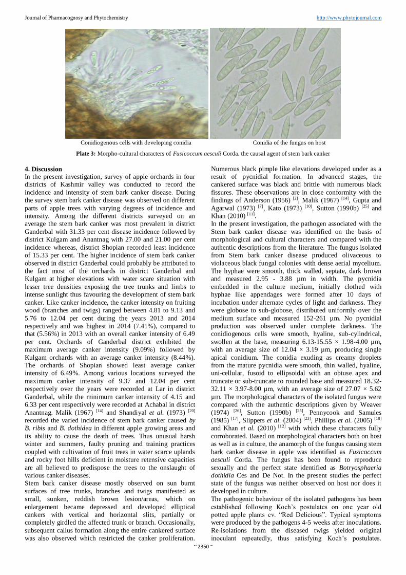
complete darkness. The conidiogenous cells were smooth,
hyaline, sub-cylindrical, swollen at the base, measuring 6.13-
15.55 × 1.98-4.00 µm, with an average size of 12.04 × 3.19
µm, producing single apical conidium. The conidia exuding
as creamy droplets from the mature pycnidia were smooth,
thin walled, hyaline, uni-cellular, fusoid to ellipsoidal with an
obtuse apex and truncate or sub-truncate to rounded base and
measured 18.32-32.11 × 3.97-8.00 µm, with an average size
of 27.07 × 5.62 µm (Plate 3).
3.4 Pathogenicity test
Pathogenicity of the test fungus was performed on one year
old potted apple saplings cv. “Red Delicious”. The initial
disease symptoms of the disease appeared within 9-11 weeks
of inoculation. The lesion showed upward and downward
extension from the incision points involving the entire
branch/stem length, which ultimately led to the death of
branch. However, pycnidial production was observed after 13
weeks of inoculation. The symptoms produced were identical
with those observed in the field. No lesion development
however, was observed on control plants. Re-isolation from
the infected twigs yielded original inoculant thus satisfied
Koch’s postulates.
3.5 Identification Based on morphological characteristics, pathogenicity test
and comparison with the authentic descriptions given by
Pennycook and Samuels (1985) [17], Slippers et al. (2004) [23]
and Phillip et al. (2005) [18] as its ability to cause the disease
on apple saplings, the fungus was identified as Fusicoccum
aesculi Corda.
7 Days old fungal colony grown on PDA Pycnidia formation after 15 days
Creamy white pycnidial ooze from mature pycnidia
~ 2350 ~
Journal of Pharmacognosy and Phytochemistry http://www.phytojournal.com
Conidiogenous cells with developing conidia Conidia of the fungus on host
Plate 3: Morpho-cultural characters of Fusicoccum aesculi Corda. the causal agent of stem bark canker
4. Discussion In the present investigation, survey of apple orchards in four
districts of Kashmir valley was conducted to record the
incidence and intensity of stem bark canker disease. During
the survey stem bark canker disease was observed on different
parts of apple trees with varying degrees of incidence and
intensity. Among the different districts surveyed on an
average the stem bark canker was most prevalent in district
Ganderbal with 31.33 per cent disease incidence followed by
district Kulgam and Anantnag with 27.00 and 21.00 per cent
incidence whereas, district Shopian recorded least incidence
of 15.33 per cent. The higher incidence of stem bark canker
observed in district Ganderbal could probably be attributed to
the fact most of the orchards in district Ganderbal and
Kulgam at higher elevations with water scare situation with
lesser tree densities exposing the tree trunks and limbs to
intense sunlight thus favouring the development of stem bark
canker. Like canker incidence, the canker intensity on fruiting
wood (branches and twigs) ranged between 4.81 to 9.13 and
5.76 to 12.04 per cent during the years 2013 and 2014
respectively and was highest in 2014 (7.41%), compared to
that (5.56%) in 2013 with an overall canker intensity of 6.49
per cent. Orchards of Ganderbal district exhibited the
maximum average canker intensity (9.09%) followed by
Kulgam orchards with an average canker intensity (8.44%).
The orchards of Shopian showed least average canker
intensity of 6.49%. Among various locations surveyed the
maximum canker intensity of 9.37 and 12.04 per cent
respectively over the years were recorded at Lar in district
Ganderbal, while the minimum canker intensity of 4.15 and
6.33 per cent respectively were recorded at Achabal in district
Anantnag. Malik (1967) [14] and Shandiyal et al. (1973) [20]
recorded the varied incidence of stem bark canker caused by
B. ribis and B. dothidea in different apple growing areas and
its ability to cause the death of trees. Thus unusual harsh
winter and summers, faulty pruning and training practices
coupled with cultivation of fruit trees in water scarce uplands
and rocky foot hills deficient in moisture retensive capacities
are all believed to predispose the trees to the onslaught of
various canker diseases.
Stem bark canker disease mostly observed on sun burnt
surfaces of tree trunks, branches and twigs manifested as
small, sunken, reddish brown lesion/areas, which on
enlargement became depressed and developed elliptical
cankers with vertical and horizontal slits, partially or
completely girdled the affected trunk or branch. Occasionally,
subsequent callus formation along the entire cankered surface
was also observed which restricted the canker proliferation.
Numerous black pimple like elevations developed under as a
result of pycnidial formation. In advanced stages, the
cankered surface was black and brittle with numerous black
fissures. These observations are in close conformity with the
findings of Anderson (1956) [2], Malik (1967) [14], Gupta and
Agarwal (1973) [7], Kato (1973) [10], Sutton (1990b) [25] and
Khan (2010) [11].
In the present investigation, the pathogen associated with the
Stem bark canker disease was identified on the basis of
morphological and cultural characters and compared with the
authentic descriptions from the literature. The fungus isolated
from Stem bark canker disease produced olivaceous to
violaceous black fungal colonies with dense aerial mycelium.
The hyphae were smooth, thick walled, septate, dark brown
and measured 2.95 - 3.88 µm in width. The pycnidia
embedded in the culture medium, initially clothed with
hyphae like appendages were formed after 10 days of
incubation under alternate cycles of light and darkness. They
were globose to sub-globose, distributed uniformly over the
medium surface and measured 152-261 µm. No pycnidial
production was observed under complete darkness. The
conidiogenous cells were smooth, hyaline, sub-cylindrical,
swollen at the base, measuring 6.13-15.55 × 1.98-4.00 µm,
with an average size of 12.04 × 3.19 µm, producing single
apical conidium. The conidia exuding as creamy droplets
from the mature pycnidia were smooth, thin walled, hyaline,
uni-cellular, fusoid to ellipsoidal with an obtuse apex and
truncate or sub-truncate to rounded base and measured 18.32-
32.11 × 3.97-8.00 µm, with an average size of 27.07 × 5.62
µm. The morphological characters of the isolated fungus were
compared with the authentic descriptions given by Weaver
(1974) [26], Sutton (1990b) [25], Pennycook and Samules
(1985) [17], Slippers et al. (2004) [23], Phillips et al. (2005) [18]
and Khan et al. (2010) [12] with which these characters fully
corroborated. Based on morphological characters both on host
as well as in culture, the anamorph of the fungus causing stem
bark canker disease in apple was identified as Fusicoccum
aesculi Corda. The fungus has been found to reproduce
sexually and the perfect state identified as Botryosphaeria
dothidia Ces and De Not. In the present studies the perfect
state of the fungus was neither observed on host nor does it
developed in culture.
The pathogenic behaviour of the isolated pathogens has been
established following Koch’s postulates on one year old
potted apple plants cv. “Red Delicious”. Typical symptoms
were produced by the pathogens 4-5 weeks after inoculations.
Re-isolations from the diseased twigs yielded original
inoculant repeatedly, thus satisfying Koch’s postulates.
~ 2351 ~
Journal of Pharmacognosy and Phytochemistry http://www.phytojournal.com Pathogenic nature of canker fungi isolated from apple trees
was proved by various workers (Shandilya, 1971 and Singh,
1985) [19, 22] both in laboratory as an excised twig method as
well as under pot culture and field conditions on injured and
uninjured twigs of young and grown up apple trees by
inoculating culture bits from two weeks old pathogen culture
and covering with moist cotton pads to provide suitable
conditions for growth of the pathogen.
5. Summary and Conclusion The survey of apple orchards in four districts of Kashmir
valley viz; Ganderbal, Kulgam, Anantnag and Shopian
revealed the prevalence of the disease in all the surveyed
districts with an overall canker incidence of 12.67 to 26.00
per cent, during the year 2013 and 18.00 to 36.67 per cent
during 2014. The highest canker incidence (31.33%) was
observed in district Ganderbal and the least in district Shopian
(15.33%). Among the villages surveyed, Watlar of district
Ganderbal exhibited the highest canker incidence of 41.00 per
cent. Village Batapora of district Shopian exhibited the least
canker incidence of 13.00 per cent. The data over the two
years further revealed that maximum canker incidence of
23.50% was observed on tree trunks followed by scaffold
branches (17.70%) and least on fruiting wood (10.09%).
Stem bark symptoms mostly observed on sun burnt surfaces
of trees was characterized by the appearance of small, sunken,
reddish brown lesions, which on enlargement became
depressed and developed elliptical cankers with vertical and
horizontal slits, partially or completely girdling the affected
trunk or branch. In advanced stages, the cankered surface
however, became was black and brittle which remained
studded with numerous black fissures.
The fungus isolated from stem bark canker produced
olivaceous to violaceous black fungal colonies with dense
aerial mycelium. The hyphae were smooth, thick walled,
septate and dark brown in colour. The pycnidia formed only
in presence of light were globose to sub-globose distributed
uniformally over the culture medium. The conidiogenous cells
were smooth, hyaline, sub-cylindrical, swollen at the base,
producing single apical conidium. The conidia were smooth,
thin walled, hyaline, unicellular, fusoid to ellipsoidal with an
obtuse apex and truncate or sub-truncate to rounded base.
Based on morphological characters both on host as well as in
culture, symptom expression and pathogenicity tests, the
fungus causing the disease was identified as Fusicoccum
aesculi Corda.
6. Acknowledgement The first author expresses his heartiest gratitude and thanks to
Dr. N. A. Khan, Associate Professor, Department of Plant
Pathology for his valuable guidance, scientific knowledge,
professional dexterity and constant encouragement to put this
work into present shape and making this study a great
learning experience.
7. References
1. Agarwal RK, Gupta GK. Canker disease complex of
apple trees. In: Second International Symposium on Plant
Pathology, Indian Phytopathological Society, New Delhi,
1971, 160p.
2. Anderson HW. Diseases of Fruit Crops. McGraw Hill
Book Co. Inc, New York, 1956, 501p.
3. Anonymous. Indian Horticulture Database; Area and
Production Statistics. Ministry of Agriculture and
Cooperatives, Government of India, New Delhi, 2017.
(http://nhb.gov.in/area%20_production.html).
4. Bilgrami KS, Jamaluddin, Rizwi MA. Fungi of India Pvt.
Ltd. List and references. Today & Tomorrow’s Printers
& Publishers, New Delhi, 1979, 467p.
5. Crosse JE. Bacterial canker of stone fruits. Annals of
Applied Biology. 1957; 45:19-35
6. Gastier TW. (Ed). Great Moments in Apple History. The
Ohio Fruit ICM News. 2000; 4:24.
7. Gupta GK, Agarwal RK. Canker diseases of apple trees
in Himachal Pradesh. Indian Journal of Mycology and
Plant Pathology. 1973; 3:189-192.
8. Jones AL, Aldwinkle HS. Compendium of Apple and
Pear Diseases. American Phytopathological Society,
USA. APS press, 1990, 100p.
9. Kanwar SM. Apples: Production Technology and
Economics. Tata MacGraw Hill publishing Company
Ltd., New Delhi, 1988, 889p.
10. Kato K. Studies on Physalospora canker of Japanese pear
with special reference to ecology and control. Special
Research Bulletin of the Aichi-Ken Agricultural
Research Centre Nagakute, Aichi, Japan, Series B, 1973,
1-70p.
11. Khan NA. Status and etiology of canker disease of apple
in Kashmir. Ph. D Thesis, Division of Plant Pathology,
SKUAST-K, Shalimar, 2010, 118p.
12. Khan NA, Ahmad M, Ghani MY. Botryosphaeria
dothidea associated with white rot and stem bark canker
of apple in Jammu and Kashmir. Applied Biological
Research. 2010a; 12:69-73.
13. Khan NA, Ahmad M, Ahmad K, Beig MA. Etiology and
occurrence of Valsa apple canker in Jammu and Kashmir
state. Applied Biological Research. 2011b; 13:48-50.
14. Malik AR. The canker that damages apple trees in
Kashmir. Indian Horticulture. 1967; 11:25-26.
15. Milholand RD. Histopathology and pathogenesity of
Botryosphaeria dothidia on blue berry stems.
Phytopathology. 1972; 62:654-660.
16. Pathak VN. Essentials of plant pathology. Prakash
Publishers, Jaipur, India, 1972, 448p.
17. Pennycook SR, Samules GJ. Botryosphaeria and
Fusicoccum species associated with ripe fruit rot of
Actinidia deliciosa (Kiwi fruit) in New Zealand.
Mycotaxon. 1985; 24:445-458.
18. Phillips AJL, Rumbos IC, Alves A, Correia A.
Morphology and Phylogeny of Botryosphaeria dothidea
causing fruit rot of olives. Mycopathologia. 2005;
159:433-439.
19. Shandilya TR. Study of perennial canker of apple (Malus
pumilla Mill.) in Kullu Valley and their control. M. Sc
Thesis, College of Agriculture, Agriculture Complex,
Solan, 1971, 97p.
20. Shandilya TR, Thakur MS, Agarwal RK. Effect of age
and altitude in the incidence of canker disease of apple in
Himachal Pradesh. Indian Journal of Mycology and Plant
Pathology. 1973; 3:102-103.
21. Sharma IM, Bhardwaj SS. Canker and foliar disease of
apple. In: Diseases of Horticultural Crops-fruits (Eds.
Verma, L. R. and Sharma, R. C.). Indus Publishing Co.,
New Delhi, 1999, 15-53p.
22. Singh D. Studies on apple canker caused by Sphaeropsis
malorum Berk, and its control. Ph.D. Thesis, College of
Agriculture, H.P. Krishi Vishva Vidhyala S.N.S. Nagar,
Solan, 1985, 202p.
~ 2352 ~
Journal of Pharmacognosy and Phytochemistry http://www.phytojournal.com 23. Slipers B, Corus PW, Denman S, Coutinho TA,
Wingfield BD, Wingfield MJ. Combined multiple gene
genealogies and phenotypic characters differentiate
several species previously identified as Botryosphaeria
dothidia. Mycologia. 2004; 96:83-101.
24. Snowdon AL. (ed). A colour atlas of postharvest diseases
and disorders of Fruits and Vegetables, Vol. 1and 2. CRC
Press, Boca Raton, Florida, USA, 1990, 302p.
25. Sutton TB. White rot. In: Compendium of Apple and
Pear Diseases (Eds. Jones, A. L. and Aldwinkle, H. S.).
APS Press, St. Paul, Minnesota, 1990b, 16-18p.
26. Weaver DJ. A gummosis disease of peach trees caused
by Botryosphaeria dothidia. Phytopathology. 1974;
64:1429-1432.