Embed Size (px)
Citation preview
Vol. 121 (2012) ACTA PHYSICA POLONICA A No. 4
Proceedings of the 9th National Symposium of Synchrotron Radiation Users, Warsaw, September 26�27, 2011
Status and Vision for Structural Biology
at the Canadian Light SourceP. Grochulskia,b∗, M.N. Fodjea,c and G. Georged
a Canadian Light Source, 101 Perimeter Road, Saskatoon, SK S7N 0X4 Canadab College of Pharmacy and Nutrition, University of Saskatchewan, 110 Science Place,
Saskatoon, SK S7N 5C8, Canadac Department of Biochemistry, University of Saskatchewan, 107 Wiggins Road, Saskatoon SK S7N 5E5, Canada
dDepartment of Geology, University of Saskatchewan, 114 Science Place, Saskatoon, SK S7N 5E2, Canada
The status and vision for Structural Biology at the Canadian Light Source (CLS) is presented. Thebeamlines that have been described in the paper represent a Canadian national resource that is available toscience and industry world-wide. They include state-of-the-art infrastructure and include specialized capabilities,many of which are not available elsewhere, including macromolecular crystallography, biological X-ray spec-troscopy, soft X-ray spectromicroscopy, as well as small angle and wide angle X-ray scattering beamlines. Thevision for Structural Biology at the Canadian Light Source is signi�cantly enhanced by the synergies and col-laborations between the users of the di�erent beamlines and by the strengths of the scienti�c personnel and trainees.
PACS: 87.64.kd, 87.80.Dj
1. Introduction
The Canadian Light Source (CLS) is a 2.9 GeV na-tional synchrotron radiation facility located at the Uni-versity of Saskatchewan in Saskatoon. The CanadianMacromolecular Crystallography Facility (CMCF) is lo-cated at the CLS and consists of the 08ID�1 and 08B1�1 beamlines. The CMCF, together with the BiologicalX-ray Absorption Spectroscopy (BioXAS) and soft X-ray spectromicroscopy (SM) beamlines, form a suite ofbeamlines that allow Canadian researchers to conductcutting-edge research in structural biology at the CLS.This suite will soon be complemented with small- andwide-angle X-ray scattering (SAXS/WAXS) capabilities.
The number of beamlines worldwide that contribute tothe �eld of structural biology is growing and the currentstatistics for mid-energy rings are outlined in Table I. Thebeamlines included in the Table comprise macromolecu-lar crystallography (MX), biological XAS and biologicalSAXS/WAXS beamlines. Some of the biological SAXSbeamlines serve material science as well. The growingneed for biological SAXS beamlines is demonstrated bythe magnitude and sophistication of the SAXS beam-line being built at the PetraIII facility in Hamburg, Ger-many. At all facilities mentioned in Table I, the numberof MX beamlines surpasses that of other techniques, in-dicating that a large number of researchers require highresolution structures of macromolecules to solve their bi-ological problems. Productivity of the MX beamlinesis very easy to monitor due to the BioSync Web initia-tive, where information is tabulated from over 130 MX
∗ e-mail: [email protected]
TABLE I2.5�3.0 GeV synchrotron facilities worldwide and thenumber of MX, BioXAS and BioSAXS/WAXS beamlines
Synchrotron facility MX BioXAS† BioSAXS/WAXS‡
number of beamlinesCLS (CA) 2 3* 1**SLS (CH) 3 0 1SSRL (USA) 5 4 1ALS (USA) 8 0 1#
Diamond (GB) 5 + 1* 0 1*Solei (F) 2 0 1Australian 2 1 1*indicates beamlines being built, **indicates beamlinesin the planning stage, †de�ned as beamlines speci�callydesigned for BioXAS, or for which BioXAS forms a largefraction of the research, ‡de�ned as beamlines designedfor BioSAXS or for which BioSAXS forms large fractionof research, #that beamline is split about 50/50 betweenMX and SAXS.
beamlines at synchrotron radiation facilities worldwide[1]. For example, the CMCF 08ID�1 beamline has beenperforming very well, having had 94 deposited structuresin 2010 and 57 in 2011. At this time, the other syn-chrotron techniques described in this paper do not havea similar formal way of tracking productivity.
2. Experimental Facilities2.1. Canadian Macromolecular Crystallography FacilityThe CMCF consists of the 08ID�1 and 08B1�1 beam-
lines and provides service to more than 60 Principal In-vestigators in Canada and the United States [2]. Up to25% of the beam time is devoted to commercial usersand the general user program is guaranteed up to 55% ofthe useful beam time which is administered under a peer-review proposal system. There are two modes of access to
(866)
Status and Vision for Structural Biology at the Canadian Light Source 867
the CMCF; remote access and on-site data acquisition.In the remote mode, users either control the beamlineremotely from their home facilities, or the CMCF sta�provide �Mail-In� crystallography service. Users with thehighest scored proposals are given priority for the �Mail-In� service.
TABLE IISpeci�cations of the CMCF beamlines
08ID�1 08B1�1Spectral range (keV) 6.0�18.0 4.0�18.0
(Å) 2.1�0.7) 3.1�0.7Energy bandwidth(∆E/E) Si(111) @12keV
1.5× 10⁻⁴ 1.4× 10⁻⁴
Measured Focal Size@ 12 keV (FWHM)(H µm×V µm)
150× 50 210× 190
Flux on the sample @12 keV (250 mA) (pho-tons/s) (from 6th har-monic of the ID)
5× 1012,2× 1012 (100 µm),1× 1012 (50 µm),7× 1011 (20µm),2× 1010 (5 µm)
1.0× 10¹¹
Typical beam size 100 µm 200 µmBeam cross�re atthe sample @12 keV (FWHM)(H mrad×V mrad)
0.9× 0.2 1.8× 0.3
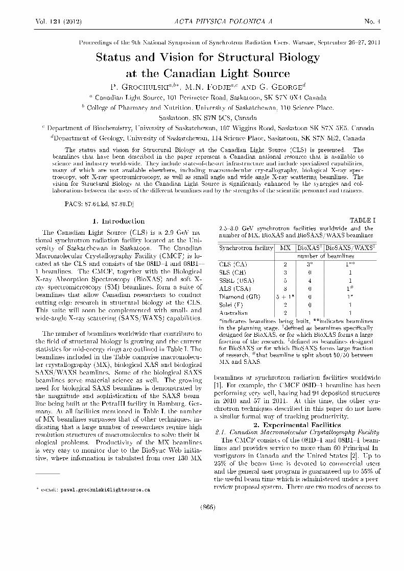
Fig. 1. Overall view of the CMCF endstations; on theleft panel of the 08B1�1 beamline and on the right panelof the 08ID�1 beamline.
Beamline 08ID�1 is an undulator-based beamline ca-pable of measuring data from small crystals and crystalswith large cell dimensions. The 08B1�1 beamline is abend magnet-based beamline which is fully automatedand equipped for remote access. The speci�cations of theCMCF beamlines are shown in Table II and end stationsare shown in Fig. 1. The 08ID�1 beamline is a very brightbeamline allowing selection of beam sizes as small as 5µmwith �ux of 2×10¹⁰ photons/s. However the typical beamsize used at this beamline is 100 µm, delivering a �ux onthe sample of 2 × 10¹² photons/s (Table II). The 08B1�1 beamline is equipped with a mini-Kappa goniometer
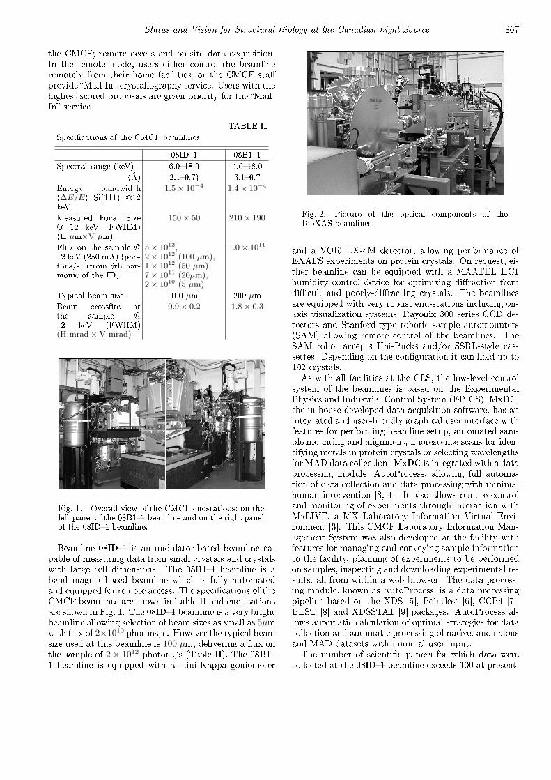
Fig. 2. Picture of the optical components of theBioXAS beamlines.
and a VORTEX-4M detector, allowing performance ofEXAFS experiments on protein crystals. On request, ei-ther beamline can be equipped with a MAATEL HC1humidity control device for optimizing di�raction fromdi�cult and poorly-di�racting crystals. The beamlinesare equipped with very robust end-stations including on-axis visualization systems, Rayonix 300 series CCD de-tectors and Stanford-type robotic sample automounters(SAM) allowing remote control of the beamlines. TheSAM robot accepts Uni-Pucks and/or SSRL-style cas-settes. Depending on the con�guration it can hold up to192 crystals.As with all facilities at the CLS, the low-level control
system of the beamlines is based on the ExperimentalPhysics and Industrial Control System (EPICS). MxDC,the in-house developed data acquisition software, has anintegrated and user-friendly graphical user interface withfeatures for performing beamline setup, automated sam-ple mounting and alignment, �uorescence scans for iden-tifying metals in protein crystals or selecting wavelengthsfor MAD data collection. MxDC is integrated with a dataprocessing module, AutoProcess, allowing full automa-tion of data collection and data processing with minimalhuman intervention [3, 4]. It also allows remote controland monitoring of experiments through interaction withMxLIVE, a MX Laboratory Information Virtual Envi-ronment [3]. This CMCF Laboratory Information Man-agement System was also developed at the facility withfeatures for managing and conveying sample informationto the facility, planning of experiments to be performedon samples, inspecting and downloading experimental re-sults, all from within a web browser. The data process-ing module, known as AutoProcess, is a data processingpipeline based on the XDS [5], Pointless [6], CCP4 [7],BEST [8] and XDSSTAT [9] packages. AutoProcess al-lows automatic calculation of optimal strategies for datacollection and automatic processing of native, anomalousand MAD datasets with minimal user input.The number of scienti�c papers for which data were
collected at the 08ID�1 beamline exceeds 100 at present,

868 P. Grochulski, M.N. Fodje, G. George
including such prestigious journals as Science, Nature,PNAS, J. Mol. Biol. and Cell, the number of PDB en-tries exceeding 220 structures. A very recent exampleof science from the CMCF is the determination of thecrystal structures of the TgAMA1 apo form and complexformed with a RON2 peptide at 2.25 Å and 1.95 Å res-olution, respectively [10]. Toxoplasma and Plasmodiumparasites cause numerous diseases worldwide, includingmalaria and toxoplasmosis. These parasites attack hostcells in a very active manner, providing the receptor forbinding to the host cell. Interaction thus occurs througha protein called AMA1 to a rhoptry neck (RON) com-plex provided by the parasite and injected into the hostcell. Besides providing insights into host cell invasion byapicomplexan parasites, the structure o�ers a basis fordesigning therapeutics targeting these global pathogens.
2.2. Future developmentIn the genomics era, crystals are getting smaller and
smaller, frequently with dimensions less than 10µm. Tobe able to collect data from such small crystals the Hu-ber single axis goniometer of the 08ID�1 beamline willbe upgraded to an air-bearing with a sphere of confusionof less than 1µm. That implementation will allow properutilization of the mini-beam apparatus to deliver a 5µmbeam on a 5µm crystal. To expand the capabilities of theCMCF we are planning to add other spectroscopic tech-niques. For example, Raman, EXAFS and UV-Visibleabsorption spectroscopies can provide information thatcompliments X-ray di�raction data. The 08B1�1 beam-line will therefore be upgraded by enhancing the X-ray-absorption spectroscopy (EXAFS) capabilities with ad-ditional in situ spectroscopic methods such as UV-Visibleabsorption and possibly Raman spectroscopies.
2.3. Biological X-ray Absorption Spectroscopy(BioXAS)
X-ray spectroscopy (XAS) contributes to structural bi-ology in two ways. It can provide information on speciesfor which crystal structures are not available and it canalso provide supplemental information on systems forwhich crystal structures are available. Since about 30%of the human genome is made up of genes encoding met-alloproteins, XAS, but in particular the extended X-rayabsorption �ne structure (EXAFS), is extremely usefulwhen studying the coordination environment of biologi-cal metal ions within a radius of 5 Å. Its major strength isthat very accurate values for average bond lengths can beobtained, typically to better than ±0.02 Å, which is com-parable to accuracies obtained for small-molecule struc-tures [11], although precisions are typically about an or-der of magnitude better than this and relative changesin similar species can be very well de�ned. The EXAFSaccuracy is about ten times better than precisions ob-tained from a typical protein crystallography experimentat a moderate resolution [12] and this may be impor-tant in understanding the detailed chemistry of metalion coordination within a biological system. The EX-AFS bond-length resolution, ∆R, is de�ned as the min-imum distance between similar ligands that can be dis-cerned from analysis of a data set, and this is directly
related to the extent of the data and is approximatelygiven by ∆R ≈ π
/2k, where k is the photo-electron wave
vector and the extent of the data, typically measuredin Å−1. For typical EXAFS data ranges, ∆R is ratherpoor at around 0.15 Å, and this is one major limitationof EXAFS analysis [12]. In some cases this limitationcan be overcome by extending the data range. Whilesimple in principle, collecting EXAFS data at high-k re-quires very stable beamline optics, an excellent signalto noise ratio, and sample limitations in that no ele-ments whose absorption edges might truncate the k-rangecan be present. EXAFS can also determine coordinationnumbers of similarly coordinated ligands, N , and esti-mates of the Debye�Waller factors, σ2, a measure of themean-square-deviation in average bond-length R. BothN and σ2 are typically determined with an accuracy ofaround ±25%. In most cases no direct information on ge-ometry (e.g. bond-angles) is available from EXAFS, butin some experiments signi�cant multiple scattering (MS)is observed. In this case the EXAFS can be sensitive tothe relative arrangement of the atoms providing limitedinformation on bond angles.
The BioXAS facility at the CLS has been designed,all major components have been purchased and it is cur-rently under construction. The facility will be composedof three beamlines powered by two di�erent insertion de-vices, allowing the study of biological and health-relatedmetals. The two spectroscopy BioXAS lines both havean energy range of 5�28 keV (including the K-edges ofV to Cd, and the L-edges of Cs and elements above).The undulator endstation (BioXAS-I) will also go slightlylower � to 4 keV which will allow access to the K-edgeof Calcium and up. The optical hutch of the BioXASbeamlines is shown in Fig. 2. Two of the three beamlineswill be optimized for XAS measurements and powered bya high-�eld �at-top �eld pro�le permanent magnet wig-gler. These beamlines will provide a stable platform forEXAFS studies of metals in biological molecules, witha number of multi-element solid state detectors to allowstudies of dilute systems. The third beamline is an imag-ing endstation with micro-focus capability that will allowmicro-XAS on very small regions and will be powered byan in-vacuum undulator. The imaging end station willbe capable of imaging biological samples by monitoringX-ray �uorescence with three di�erent resolution ranges;macro with a 50�150 µm beam size for large samples(up to 600 mm across), micro for smaller samples witha 3�5 µm beam size, and nano with 150�250 nm beamsize. The BioXAS imaging beamline will have the uniquecapability of being able to rapidly change between thedi�erent resolutions available.
Metal ions are important for some of the most com-plex chemistry carried out by living organisms, and, forexample, are essential components of the limiting en-zymes of the carbon, nitrogen and oxygen cycles. Apartfrom this fundamental importance, metal ions are alsoimportant in numerous diseases. Examples include pro-gressive neurodegenerative diseases such as Alzheimer's
Status and Vision for Structural Biology at the Canadian Light Source 869
disease, Parkinson's disease [13] and multiple sclerosis,diseases of metal dysregulation such as Wilson's [14] andMenke's diseases, and severe fatal complaints such as sul-�te oxidase de�ciency. Metal ions are also importantenvironmental toxins (e.g. mercury and arsenic), andthere is also growing interest in new metal-containingpharmaceuticals, such as platinum and ruthenium-basedchemotherapeutic drugs. The BioXAS infrastructure atCLS will provide world-class capabilities for research inall of these �elds, and will be highly complementary tothe other life-science cluster beamlines.
2.4. Soft X-ray spectromicroscopy
The CLS soft X-ray spectromicroscopy (SM) beamlinecombines X-ray absorption spectroscopy in the 130 eV to2.5 keV energy range with microscopy (2-D or 3-D) ata high spatial resolution of 25�30 nm [15]. It is used tostudy the interactions of proteins, lipids, and metals andto map their distributions. The samples can be studiedwet or dry. An example of investigation done on the SMbeamline is the characterization of magnetism of individ-ual magnetosomes in magnetotactic bacteria [16]. Sincethe late 1960's, scientists have known that some bacte-ria make internal compasses by growing tiny magneticcrystals called magnetosomes. The bacteria use them tonavigate, positioning themselves in environments mostsuited for their survival, with cells of the same speciesgrowing crystals of uniform size, structure and out of thesame magnetic minerals.
2.5. SAXS/WAXS capabilities
The past decade has witnessed tremendous advancesin instrumentation and interpretation of X-ray scatter-ing results from macromolecules in solution. This makespossible the extraction of molecular shapes from analysisof scattering at very small di�raction angles (SAXS) [17,18]. The increasing impact of SAXS is clearly evidencedby the ever-increasing number of publications describingapplications of these techniques. A search of PubMedwith the text `small-angle X-ray scattering AND protein'showed the following number of hits: 367 (2010 to May2011), 397 (2008�2009), 265 (2006�2007), 215 (2004�2005). In addition to the extremely useful informationcontained in the scattering at small angles, there is alsoa wealth of information that can be extracted from thescattering at wide angles (WAXS). These data providemore detailed information, corresponding to higher res-olution and can describe conformational changes in pro-teins during their work cycle (time resolved experiments)[19]. SAXS and WAXS are also important tools for thecharacterization of drug delivery systems (nanomedicine)[20, 21].At present there is no SAXS/WAXS beamline at the
CLS. However, as a part of the Brockhouse X-RayDi�raction and Scattering Sector, a material scienceSAXS/WAXS beamline is being built and potentiallycould be used by structural biologists. A dedicated Bio-logical SAXS beamline is currently in the planning stage.
2.6. Important Synergies
At the University of Saskatchewan there are two HealthResearch Groups that are very closely associated withthe Canadian Light Source: the Molecular Design Re-search Group (MDRG) and the CIHR Training Grant inHealth Research Using Synchrotron Techniques (CIHR-THRUST). The CIHR-THRUST is a cross-disciplinarytraining and mentoring program intended for the devel-opment and use of synchrotron techniques to addresskey questions in Canadian health research. THRUSTwill train more than 50 of the next generation of Cana-dian synchrotron health researchers between 2009 and2015. An example of work from the THRUST programis a publication on the in�uence of prion protein expres-sion levels on local copper, iron and zinc content in themouse brain [22]. The MDRG integrates the capabili-ties of the Canadian Light Source and the SaskatchewanStructural Sciences Centre into research focused on pro-tein structure/function, signalling networks and drug de-velopment. An example of the collaborative work accom-plished by the MDRG is the structural investigation ofmyo-inositol dehydrogenase from Bacillus subtilis [23].
3. Conclusions
The facilities for Structural Biology at the CanadianLight Source represent a Canadian national resource thatis available to science and industry world-wide. Beam-lines include state-of-the-art infrastructure and special-ized capabilities, many of which are not available else-where. The current vision for Structural Biology at theCanadian Light Source is signi�cantly enhanced by thesynergies and collaborations between the users of the dif-ferent beamlines and by the strength of the scienti�c per-sonnel and trainees.
Acknowledgements
The authors would like to thank the whole CMCFsta�, Dr. Chithra Karunakaran for providing informa-tion on X-ray Spectromicroscopy and Dr. Shaun Labiukfor careful reading of the manuscript and valuable sug-gestions. Work in the George laboratory is supportedby a Canada Research Chair, Natural Sciences and En-gineering Research Council of Canada, the Canadian In-stitutes of Health Research and the Saskatchewan HealthResearch Foundation. The Canadian Light Source is sup-ported by the Natural Sciences and Engineering ResearchCouncil of Canada, the National Research Council ofCanada, the Canadian Institutes of Health Research, theProvince of Saskatchewan, Western Economic Diversi�-cation Canada, and the University of Saskatchewan.
References
[1] A. Kuller, W. Fleri, W.F. Bluhm, J.L. Smith, J. West-brook, P.E. Bourne, Trends in Biochemical Sciences23, 213 (2002).
870 P. Grochulski, M.N. Fodje, G. George
[2] P. Grochulski, M.N. Fodje, J. Gorin, S.L. Labiuk,R. Berg, J. Synchrotron Rad. 18, 681 (2011).
[3] M.N. Fodje, K. Janzen, R. Berg, G. Black, S. Labiuk,J. Gorin, P. Grochulski, J. Synchrotron Rad. 19, 274(2012).
[4] M.N. Fodje, R. Berg, G. Black, P. Grochulski,K. Janzen, Automation of the Macromolecular Crys-tallography Beamlines at the Canadian Light Source,PCaPAC, Saskatoon (2010).
[5] W. Kabsch, Journal of Appl. Crystallogr.26, 795(1993).
[6] P.R. Evans, Acta Cryst. D62, 72 (2006).[7] N.4. Collaborative Computational Project, Acta
Cryst. 50, 760 (1994).[8] G.P. Bourenkov, A.N. Popov, Acta Cryst. D62, 58
(2006).[9] K. Diederichs, Acta Cryst. D62, 96 (2006).[10] M.L. Tonkin, M. Roques, M.H. Lamarque, M. Pug-
ni£re, D. Douguet, J. Crawford, M. Lebrun,M.J. Boulanger, Science 333, 463 (2011).
[11] M.A. DePristo, P.I. deBakker, T.L. Blundell, Struc-ture 12, 831 (2004).
[12] M.J. Pushie, G.N. George, Coord. Chem. Rev. 255,1055 (2011).
[13] B.F.G. Popescu, M.J. George, U. Bergmann,A.V. Garachtchenko, M.E. Kelly, R.P.E. McCrea,K. Lüning, R.M. Devon, G.N. George, A.D. Han-son, S.M. Harder, L.D. Chapman, I.J. Pickering,H. Nichol, Phys. Med. Biol. 54, 651 (2009).
[14] L. Zhang, J. Lichtmannegger, K.H. Summer, S. Webb,I.J. Pickering, G.N. George, Biochemistry 48, 891(2009).
[15] K.V. Kaznatcheev, C.Ch. Karunakaran, U.D. Lanke,S.G. Urquhart, M. Obst, A. Hitchcock, Nucl. Inst.Meth. 582, 96 (2007).
[16] K.P. Lam, A.P. Hitchcock, M. Obst, J.R. Lawrence,G.D.W. Swerhone, G.G. Leppard, T. Tyliszczak,C. Karunakaran, J. Wang, K. Kaznatcheev, D.A.Bazylinski, U. Lins, Chemical Geology 270, 110(2010).
[17] G.L Hura, L.A. Menon, M. Hammel, R. Rambo,L.F. Poole II, S.E. Tsutakawa, F. Jenney Jr,S. Classen, K. Frankel, R. Hopkins, S. Yang, J. Scott,B. Dillard, M. Adams, J.A. Tainer, Nature Methods6, 606 (2009).
[18] T.D. Grant, J.R. Luft, J.R. Wol�ey, H. Tsuruta,A. Martel, G.T. Montelione, E. Snell, Biopolymers95 , 517 (2011).
[19] M. Cammarata, M. Levantino, F. Schotte, P.A. An�n-rud, F. Ewald, J. Choi, A. Cupane, M. Wul�, H. Ihee,Nature Methods 5, 881 (2008).
[20] C. Leal, N.F. Bouxsein, K.K. Ewert, C.R. Sa�nya, J.Am. Chem. Soc 132, 16841 (2010).
[21] Y. Dong, B.J. Boyd, Int. J. Pharm. 417, 101 (2011).[22] M.J. Pushie, I.J. Pickering, G.R. Martin, S. Tsutsui,
F.R. Jirik, G.N. George, Metallomics 3, 206 (2011).[23] K.E. van Straaten, H. Zheng, D.R. Sanders,
D. Palmer, Biochem. J. 432, 237 (2010).