Embed Size (px)
Citation preview

u n i ve r s i t y o f co pe n h ag e n
Københavns Universitet
Dairy products viscosity estimated by laser speckle correlation
Postnov, Dmitry D; Moller, Flemming; Sosnovtseva, Olga
Published in:PLOS ONE
DOI:10.1371/journal.pone.0203141
Publication date:2018
Document versionPublisher's PDF, also known as Version of record
Citation for published version (APA):Postnov, D. D., Moller, F., & Sosnovtseva, O. (2018). Dairy products viscosity estimated by laser specklecorrelation. PLOS ONE, 13(9), [e0203141]. https://doi.org/10.1371/journal.pone.0203141
Download date: 27. dec.. 2019

RESEARCH ARTICLE
Dairy products viscosity estimated by laser
speckle correlation
Dmitry D. Postnov1,2*, Flemming Moller3, Olga Sosnovtseva1
1 Department of Biomedical Sciences, Copenhagen University, Copenhagen, Denmark, 2 Department of
Biomedical Engineering, Boston University, Boston, United States of America, 3 Physical Food Science,
DuPont Nutrition Biosciences Aps, Edwin Rahrs Vej 38, DK-8220 Brabrand, Denmark
Abstract
Dairy products exhibit several physical properties that are crucial to define whether we like
the food or not: firmness, creaminess, thickness, or lightness. Viscosity changes the flow
properties of food and influences the appearance and the consistency of a product; this con-
trol variable is important in most production stages—manufacture, processing, and storage.
Viscosity of heterogeneous products at a given temperature depends on its composition
and physical state of its substances. Although rheology provides a method to access the
product viscosity, it lacks non-contact full-field monitoring. We apply a simple correlation
analysis of laser speckle images to evaluate viscosity properties of dairy products. Our
approach ensures robust measurements with high degree of detectability.
Introduction
We often accept food products on the basis of their texture: How spreadable and creamy a
food product is. This affects taste perception [1]. Moreover, products with different viscosity
but with similar palatability, macronutrient composition and energy density significantly dif-
fers in their intake: a case study in obesity [2]. Main factors—used ingredients, quality of raw
material, technological process—can influence rhelogical properties of dairy products. The fat
concentration, physico-chemical properties of fat globules and protein content affect viscosity.
These compounds can be modified during thermal and mechanical operations that can signifi-
cantly change rheological characteristics. For these reasons, food rheology is an important
quality control tool during and after food manufacture and processing.
The market requires to design new food and beverages that are accepted by consumers.
Food industry studies and predicts consumers’ perceptions of food through instrumental mea-
surements [3–6]. There are different rheological tests in the food industry [7, 8]. By applying a
controlled force (or strain) and measuring the resulting force (or strain), it is possible to access
rheological properties of a material. The rheometer is a research-grade analytical instrument
that measures viscous, elastic, and viscoelastic properties of liquids, semisolids, and solids.
Capillary viscometers, falling-ball viscometers, rotational and oscillatory rheometers, among
others, are used to perform rheological measurements [9]. Dairy products are normally physi-
cal homogeneous, but physical or chemical changes (e.g. sedimentation, creaming, wheying
PLOS ONE | https://doi.org/10.1371/journal.pone.0203141 September 7, 2018 1 / 10
a1111111111
a1111111111
a1111111111
a1111111111
a1111111111
OPENACCESS
Citation: Postnov DD, Moller F, Sosnovtseva O
(2018) Dairy products viscosity estimated by laser
speckle correlation. PLoS ONE 13(9): e0203141.
https://doi.org/10.1371/journal.pone.0203141
Editor: Guy Cloutier, University of Montreal,
CANADA
Received: April 18, 2018
Accepted: August 15, 2018
Published: September 7, 2018
Copyright: © 2018 Postnov et al. This is an open
access article distributed under the terms of the
Creative Commons Attribution License, which
permits unrestricted use, distribution, and
reproduction in any medium, provided the original
author and source are credited.
Data Availability Statement: Data is available via
Dryad repository, DOI: doi:10.5061/dryad.b23dd4r.
Funding: DD Postnov’s work was supported by the
grant from Novo Nordisk Foundation
(NNF17OC0025224). DuPont Nutrition Biosciences
Aps provided support in the form of salaries for FM
and providing product samples but did not have
any additional role in development of optical
method and related analysis, decision to publish, or
preparation of the manuscript. The specific roles of
these authors are articulated in the ‘author
contributions’ section.

off) can occur during storage and limit the shelf life. Some defects can be seen and detected
with imaging techniques but viscosity changes due to age gelation or fat crystallization cannot
be visualized with traditional visualization techniques.
Over the last decades, development of fast digital acquisition and data processing technolo-
gies advanced laser speckle techniques for medical applications in the retina [10], skin [11],
mesenterium [12], the kidney [13], and the brain [14]. When coherent light scatters from a
medium, it produces a random interference pattern termed speckle pattern. The intensity of
speckle patterns fluctuates due to movement of scattering particles in the medium. This leads
to a blurring of the speckle pattern, to a reduction in the local speckle contrast and, thus, to a
coding velocity distribution.
Recently, Hajjarian and Nadkarni [15, 16] nailed a new approach—laser speckle rheology—
to assess tissue viscoelasticity by measuring the time constant of fluctuations of laser speckle
intensity. Speckle fluctuations are highly sensitive to Brownian movement of scattering parti-
cles; they are influenced by the viscoelasticity of the medium and are analyzed by means of the
speckle intensity autocorrelation curves.
We applied a simple correlation analysis of laser speckle images to evaluate viscosity prop-
erties of dairy products without direct contact and modifications of the medium. The advan-
tages of the suggested approach over traditional methods include minimal sample preparation,
contact-free assessment, fast acquisition time, and simultaneous characterization of rheologi-
cal properties as well as physical defects.
Materials and methods
Imaging
Dairy product samples were provided by DuPont Nutrition Biosciences Aps, Denmark. The
samples were produced by a combination of 38% fat cream, skimmed milk and viscosity
increasing agent CREMODAN1 719 at different concentrations. Cremodan1 719 is a blend
of food-grade stabilisers of hydrocolloids (E412, E415, E407) in the powder form. The hydro-
colloids increases viscosity with increasing concentrations. In the used levels they should also
prevent protein aggregation and resulting sedimentation. The final set consisted of 27 samples
with 0.2, 1.95, 3.7% of fat and 0.000, 0.015, 0.03, 0.045, 0.06, 0.075, 0.09, 0.105, 0.12% of CRE-
MODAN1 719.
The laser module LDM785 controlled by the diode driver Thorlabs CLD1011LP delivered
coherent infrared light to the sample; the laser power density was about 20 mW/cm2 [17].
Back-scattered light was collected by CMOS camera Basler acA2000-165umNIR that operated
at a rate of 400 (low frequency LF mode) or 2000 (high frequency HF mode) frames per sec-
ond. In the LF mode, an image of 400 × 200 pixels was recorded with 2 ms of exposure time; in
the HF mode, an image of 400 × 25 pixels was recorded with 0.5 ms of exposure time. To
deliver light to the camera, VZM™ 1000i lens (Edmund Optics) were used at 10x magnification.
The field of view and focal distance remained the same during experiments. Temperature of
the samples and in the room was kept constant at about 22 ˚C.
Three imaging sessions were performed in LF mode and one session in HF mode. The sam-
ples were selected randomly. Each recording lasted for one minute; the first 10 seconds were
removed from the analysis as a transient period.
The samples were analysed using a Physica MCR 301 rheometer (Anton Paar, Austria) and
a DG26.7 double gab geometry. A standard viscosity profile was made starting at a shear rate
of 0.1 s−1 and increasing to 200 s−1 using a logarithmic ramp. 25 points were recorded using a
fixed measuring duration of 20 s pr. point. The samples were stored at refrigerated tempera-
ture (app. 5 ˚C) and tempered at 10 ˚C in the measuring system for 5 minutes before
Dairy products viscosity estimated by laser speckle correlation
PLOS ONE | https://doi.org/10.1371/journal.pone.0203141 September 7, 2018 2 / 10
Competing interests: F Moller was supported by
DuPont Nutrition Biosciences Aps. Financial
support includes expenses related to the dairy
samples production and publication fee. This does
not alter our adherence to PLOS ONE policies on
sharing data and materials.

measurements. The analyses were performed at 20 ˚C. The measured data were fitted to the
linear model and shear viscosity μ was extracted assuming that samples are Newtonian fluid at
low fat and CREMODAN1 719 concentrations. R-squared was used as a statistical measure of
how close the data are to the fitted regression line.
Data analysis
Scattering particles—fat globules or large proteins—create a speckle pattern that changes due
to Brownian motion (Fig 1). Speed of particles and, thus, change of the speckle pattern are
mainly defined by temperature, particle size, and medium viscosity. At constant temperature,
particle size remained the same in the samples with the same fat concentration; in the samples
with different CREMODAN1 719 concentrations particle speed was defined only by medium
viscosity. To estimate viscosity variations by means of laser speckle imaging, we suggested two
approaches.
Correlation time. The first approach is based on the analysis of intensity autocorrelation
function for speckle dynamics [14, 18]:
gð2ÞmnðtÞ ¼hImnðtÞImnðt þ tÞi
hImnðtÞi2
; ð1Þ
Fig 1. Speckle images. Two consecutive frames for a sample with 3.7% of fat and 0.12% of CREMODAN1 719. Color
codes intensity of scattered light. Black circles mark exemplary regions where the speckle pattern remains unchanged
and, thus, increases correlation.
https://doi.org/10.1371/journal.pone.0203141.g001
Dairy products viscosity estimated by laser speckle correlation
PLOS ONE | https://doi.org/10.1371/journal.pone.0203141 September 7, 2018 3 / 10

where m, n are pixel indexes, I is speckle intensity, t is time corresponding to the first frame,
and τ is a time lag. g(2) was calculated for samples over 100 time points and 50 lags. In the
absence of static scattering g(2) is related to the field temporal autocorrelation function g(1) [14]
as
gð2ÞðtÞ ¼ 1þ bjgð1ÞðtÞj2; ð2Þ
where β accounts for loss of correlation related to the speckle size and light source properties.
gð1ÞðtÞ ¼ exp �t
tc
� �n� �
; ð3Þ
where n takes values of 0.5, 1 or 2 depending on the scattering regime and the motion proper-
ties [19]. Correlation time τc is a key parameter in laser speckle theory; it is inversely propor-
tional to the speed of scattering particles [14, 20]. We estimate correlation time by substituting
τ with τc and 0 in Eqs 2 and 3 and numerically solving the following equation with respect
to τc:
hgð2ÞðtcÞimn ¼hgð2Þð0Þimn � 1
e2þ 1; ð4Þ
where e is mathematical constant approximately equal to 2.718. As correlation time inversely
proportional to the particle speed, we expect that it will increase with larger viscosity which is
known to decrease particles speed in Brownian motion.
Frames correlation. The second approach is based on 2-dimensional linear correlation
between frames:
Cðt; tÞ ¼P
m
PnðImnðtÞ � �IðtÞÞðImnðt þ tÞ � �Iðt þ tÞÞ
ffiffiffiffiffiffiffiffiffiffiffiffiffiffiffiffiffiffiffiffiffiffiffiffiffiffiffiffiffiffiffiffiffiffiffiffiffiffiffiffiffiffiffiffiffiffiffiffiffiffiffiffiffiffiffiffiffiffiffiffiffiffiffiffiffiffiffiffiffiffiffiffiffiffiffiffiffiffiffiffiffiffiffiffiffiffiffiffiffiffiffiffiffiffiffiffiffiffiffiffiffiffiffiffiffiffiffiffiffiffiffiffi
ðP
m
PnðImnðtÞ � �IðtÞÞ2Þð
Pm
PnðImnðt þ tÞ � �Iðt þ tÞÞ
2Þ
q ; ð5Þ
where �IðtÞ;�Iðt þ tÞ are mean intensities of corresponding frames. Similar to the Pearson
correlation coefficient, C(t, τ) shows degree of linear correlation and is equal to 1 if I(t) and
I(t + τ) are linearly related and to 0 if there is no correlation. Since motion of particles causes
random changes in speckle pattern, correlation reflects relative number of particles moved
between two frames. This metric depends on the time between frames τ. If it is too long, the
frames become independent of each other; if it is too short, there might be not enough motion
to change the speckle pattern. For recordings in LF mode, we used only consecutive frames,
thus, τ was fixed at the value of 2.5 ms. For recordings in HF mode the time lag τ was varied
from 0.5 ms to 25 ms to obtain linear correlation curves similar to g(2). The linear correlation
was calculated for all possible pairs of frames in each recording. Values above and below one
standard deviation from the median of the linear correlation were rejected to avoid imaging
artifacts such as skipped frames. Remaining values were averaged to provide a reliable estima-
tion of linear correlation. Resulting average takes values from 0 to 1 (negative linear correlation
is unlikely for random changes of the speckle pattern): 0 corresponds to high particles mobility
(low viscosity) and 1 to low particles mobility (high viscosity). We call it viscosity index:
Vt ¼ CtðjCtðtÞ � Mtj < jMt � stjÞ; ð6Þ
where Mτ and στ are median value and standard deviation for all correlation coefficients calcu-
lated at the selected τ. Cτ(|Cτ(i) − Mτ|< |Mτ − στ|) denotes all correlation coefficients Cτ at τthat satisfy this filtering conditions. Viscosity indexes calculated from data at LF mode were
analyzed for significant difference between samples with different CREMODAN1 719
Dairy products viscosity estimated by laser speckle correlation
PLOS ONE | https://doi.org/10.1371/journal.pone.0203141 September 7, 2018 4 / 10

concentrations. Unlike correlation time (Eq 4), the linear frame correlation (Eq 5) does not
directly depend on particle speed, but shows stability degree of speckle pattern that is linked to
particle mobility.
Robustness analysis. We used Student’s test for independent samples to detect a minimal
difference in CREMODAN1 719 concentrations with 95% confidence. To estimate robustness
of the identified changes we calculated detectability index [17]:
D ¼ j�vðc1Þ � �vðc2Þj=ð0:5� ðsvðc1Þ þ svðc2ÞÞÞ; ð7Þ
where �v and σv are mean and standard deviation of an analyzed variable. c1 or c2 are CREMO-
DAN1 719 concentrations to be compared. Analyzed variable can be either τc (Eq 4) or V(Eq 6).
Results
Fig 2 depicts correlation time and viscosity indexes calculated for the data recorded in LF
mode. CREMODAN1 719 increases viscosity of dairy products: the larger concentration of
CREMODAN1 719, the stronger correlation between the frames, longer correlation time (A),
and higher viscosity index (B). Variations of the correlation time within the same sample
increase with increasing CREMODAN1 719 concentration while curves are well separated for
different fat concentrations. Variations of the viscosity index are low and decrease with
increasing fat concentration. Increasing fat concentration results in increased number of large,
slow moving particles; this makes viscosity estimation more precise and robust.
We introduced detectability index (Eq 7) to evaluate how reliable detected differences are.
Table 1 compares detectability index for τc (Eq 4) or V (Eq 6) for all CREMODAN1 719 con-
centrations. One can see that Dv tends to increase with increasing both fat and CREMODAN1
719 concentrations; such increase is observed in 81% and 71% of compared pairs, respectively.
Fig 2. Characterization of samples. Correlation time τc (A) and viscosity index V (B) calculated for all samples in LF mode. Both characteristics
increase with increasing CREMODAN1 719. This is related to the rise of viscosity.
https://doi.org/10.1371/journal.pone.0203141.g002
Dairy products viscosity estimated by laser speckle correlation
PLOS ONE | https://doi.org/10.1371/journal.pone.0203141 September 7, 2018 5 / 10

Dtcdemonstrates similar behavior with increasing fat concentration (75% of compared pairs)
but does not show steady improvement with increase of CREMODAN1 719 concentration
(48% of compared pairs). This indicates that autocorrelation function g(2) (Eq 1) is more vul-
nerable to acquisition noise.
Table 1. Detectability index. It estimates reliable differences that the proposed approaches can distinguish.
CREMODAN1 719, % fat 0.2% fat 1.95% fat 3.17%
Dv DtcDv Dtc
Dv Dtc
0.000-0.015 0.42 0.94 2.25 2.24 2.54 7.30
0.015-0.030 0.87 1.09 2.49 1.47 3.19 6.53
0.030-0.045 0.93 0.78 2.51 2.46 4.58 5.48
0.045-0.060 1.16 0.93 3.61 1.98 6.68 3.63
0.060-0.075 2.17 1.33 3.50 2.05 2.29 1.26
0.075-0.090 2.43 0.89 4.98 2.65 4.25 2.21
0.090-0.105 3.20 1.43 4.04 2.51 5.08 1.47
0.105-0.120 2.99 1.80 2.75 1.32 4.82 2.13
https://doi.org/10.1371/journal.pone.0203141.t001
Fig 3. Robustness of the method. Color codes confidence with which different CREMODAN1 719 concentrations can be distinguished in the samples with the
same fat concentration. Confidence was calculated on the basis of Student’s t-test for τc (top panel) and V (bottom panel). Dark red and magenta colors correspond
to the confidence above 95%. Viscosity index allows for 95% confidence detection in a wider range of concentrations.
https://doi.org/10.1371/journal.pone.0203141.g003
Dairy products viscosity estimated by laser speckle correlation
PLOS ONE | https://doi.org/10.1371/journal.pone.0203141 September 7, 2018 6 / 10

To estimate robustness and precision of the method, we calculated confidence at which one
can distinguish different CREMODAN1 719 concentrations by means of correlation time and
viscosity index (Fig 3). In most cases variations are low enough to assess 0.015% changes in
CREMODAN1 719 concentration with 95% confidence. The most difficult case is to make
precise estimations at low concentration of fat and CREMODAN1 719 (left panel). This limi-
tation is related to the fast movements and low number of large particles.
To validate the method we compared viscosity estimated from laser speckle measurements
with shear viscosity. Shear viscosity μ was obtained by linear fitting of shear stress (measured
by rheometer) to shear rate (Fig 4A). Shear viscosity was measured for samples with 0.2 and
3.7% fat concentrations and 0.000, 0.03, 0.06, 0.09, 0.12% CREMODAN1 719 concentrations.
Pearson correlation coefficient between μ and τc was 0.9856 and 0.9947 at 0.2% and 3.7% fat
concentrations, respectively; for viscosity index V they were 0.9975 and 0.9716. This points to
almost linear relation between laser speckle and traditional rheology measurements (Fig 4B
and 4C). Non-linear dependency between shear viscosity and laser speckle measurements is
expected at higher values of fat or CREMODAN1 719 concentrations as non-Newtonian
properties of the dairy product are more pronounced (Fig 4A).
Correlation time τc is one of the parameters that define the shape of autocorrelation curve
g(2). Correlaion time corresponds to a specific time lag ((Eq 4)) while viscosity index V depends
on time lag (sampling frequency). To show how V changes for different sampling frequencies
and how well τc describes the shape of g(2), we used the data obtained in HF mode. Fig 5 repre-
sents corresponding correlation curves. Both g(2) and V reach their minimum around a time
lag τ of 5–10 ms. This implies that a sampling frequency below 200 frames per second can not
capture observed variations of viscosity; the consecutive frames are not correlated. A better
separation of samples is possible if the correlation is close to one; this will require much higher
frame rate (about 20000 frames per second), higher laser power and low signal-to-noise ratio.
Another observation is that the form of curves slightly varies for different CREMODAN1 719
concentrations (e.g, some curves cross each other). In such cases τc or V are not sufficient to
estimate viscosity. But this effect is mainly determined by the presence of noise; longer record-
ings or appropriate method to compensate the acquisition noise could solve the problem.
Fig 4. Viscosity estimation. (A) Shear viscosity μ obtained by a linear fit of rheometery measurements (left axis) and R2 goodness of fit (right axis). With increasing
CREMODAN1 719 and fat concentrations the fit quality decreases because of non-Newtonian properties of the diary product. τc (B) and V (C) as functions of μ show
almost linear correlation—particularly for low-fat samples. Solid lines correspond to 0.2% fat concentration, dashed lines to 3.7%.
https://doi.org/10.1371/journal.pone.0203141.g004
Dairy products viscosity estimated by laser speckle correlation
PLOS ONE | https://doi.org/10.1371/journal.pone.0203141 September 7, 2018 7 / 10

Conclusion
We showed that variations of dairy product viscosity can be estimated by simple laser speckle
analysis—without special high frequency cameras and frequency response analysis. To esti-
mate viscosity we suggested two approaches:
• Estimation of correlation time τc (Eq 4) from speckle autocorrelation function (Eq 1) gives
linear relation to viscosity for Newtonian liquids;
• Estimation of viscosity index V (Eq 6) from frame-to-frame linear correlation (Eq 5) evalu-
ates a degree of stability of speckle patterns that is related to particle mobility.
Correlation analysis of laser speckle data is sufficient for robust measurements of viscosity
variations; it detects 0.015% changes in CREMODAN1 719 concentration with 95% confi-
dence in most cases (44% for 0.2% fat concentration, 88% for 1.95% and 3.7% fat concentra-
tions). Precision of the method rises with increasing fat concentration of the product.
We should mention several benefits of frame-to-frame linear correlation versus speckle
autocorrelation: (i) computational implementation is simpler and less time consuming, (ii) its
value does not dependent on the speckle contrast, and (iii) it is less vulnerable to acquisition
noise and temporal artifacts. This results in better separation of viscosity variations. Simple
modification of experimental protocol—increasing length of recording or imaging speed—can
significantly improve detectability of viscosity changes.
Fig 5. Correlation curves. Autocorrelation g(2) (top panel) and viscosity index V (bottom panel) versus time lag. Time lag is defined as a delay between frames. Color
codes CREMODAN1 719 concentrations. Although curves have slightly different forms, they reach minimum within 5–10 ms.
https://doi.org/10.1371/journal.pone.0203141.g005
Dairy products viscosity estimated by laser speckle correlation
PLOS ONE | https://doi.org/10.1371/journal.pone.0203141 September 7, 2018 8 / 10
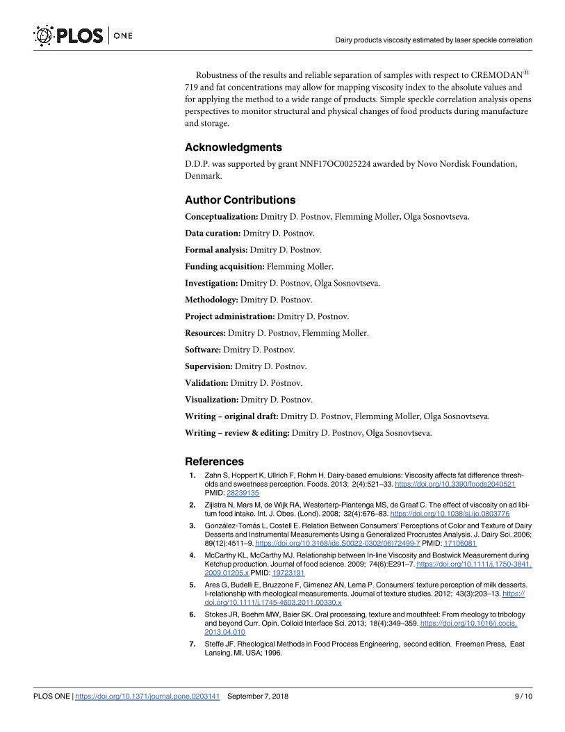
Robustness of the results and reliable separation of samples with respect to CREMODAN1
719 and fat concentrations may allow for mapping viscosity index to the absolute values and
for applying the method to a wide range of products. Simple speckle correlation analysis opens
perspectives to monitor structural and physical changes of food products during manufacture
and storage.
Acknowledgments
D.D.P. was supported by grant NNF17OC0025224 awarded by Novo Nordisk Foundation,
Denmark.
Author Contributions
Conceptualization: Dmitry D. Postnov, Flemming Moller, Olga Sosnovtseva.
Data curation: Dmitry D. Postnov.
Formal analysis: Dmitry D. Postnov.
Funding acquisition: Flemming Moller.
Investigation: Dmitry D. Postnov, Olga Sosnovtseva.
Methodology: Dmitry D. Postnov.
Project administration: Dmitry D. Postnov.
Resources: Dmitry D. Postnov, Flemming Moller.
Software: Dmitry D. Postnov.
Supervision: Dmitry D. Postnov.
Validation: Dmitry D. Postnov.
Visualization: Dmitry D. Postnov.
Writing – original draft: Dmitry D. Postnov, Flemming Moller, Olga Sosnovtseva.
Writing – review & editing: Dmitry D. Postnov, Olga Sosnovtseva.
References1. Zahn S, Hoppert K, Ullrich F, Rohm H. Dairy-based emulsions: Viscosity affects fat difference thresh-
olds and sweetness perception. Foods. 2013; 2(4):521–33. https://doi.org/10.3390/foods2040521
PMID: 28239135
2. Zijlstra N, Mars M, de Wijk RA, Westerterp-Plantenga MS, de Graaf C. The effect of viscosity on ad libi-
tum food intake. Int. J. Obes. (Lond). 2008; 32(4):676–83. https://doi.org/10.1038/sj.ijo.0803776
3. Gonzalez-Tomas L, Costell E. Relation Between Consumers’ Perceptions of Color and Texture of Dairy
Desserts and Instrumental Measurements Using a Generalized Procrustes Analysis. J. Dairy Sci. 2006;
89(12):4511–9. https://doi.org/10.3168/jds.S0022-0302(06)72499-7 PMID: 17106081
4. McCarthy KL, McCarthy MJ. Relationship between In-line Viscosity and Bostwick Measurement during
Ketchup production. Journal of food science. 2009; 74(6):E291–7. https://doi.org/10.1111/j.1750-3841.
2009.01205.x PMID: 19723191
5. Ares G, Budelli E, Bruzzone F, Gimenez AN, Lema P. Consumers’ texture perception of milk desserts.
I-relationship with rheological measurements. Journal of texture studies. 2012; 43(3):203–13. https://
doi.org/10.1111/j.1745-4603.2011.00330.x
6. Stokes JR, Boehm MW, Baier SK. Oral processing, texture and mouthfeel: From rheology to tribology
and beyond Curr. Opin. Colloid Interface Sci. 2013; 18(4):349–359. https://doi.org/10.1016/j.cocis.
2013.04.010
7. Steffe JF. Rheological Methods in Food Process Engineering, second edition. Freeman Press, East
Lansing, MI, USA; 1996.
Dairy products viscosity estimated by laser speckle correlation
PLOS ONE | https://doi.org/10.1371/journal.pone.0203141 September 7, 2018 9 / 10

8. Melito HS, Daubert CR. Rheological innovations for characterizing food material properties. Annual Rev
Food Sci Technol. 2011; 2:153–79. https://doi.org/10.1146/annurev-food-022510-133626
9. Tabilo-Munizaga G, Barbosa-Canovas GV. Rheology for the food industry. J. Food Engineering 2005;
67:147–156. https://doi.org/10.1016/j.jfoodeng.2004.05.062
10. Srienc AI, Kurth-Nelson ZL, Newman EA. Imaging retinal blood flow with laser speckle flowmetry. Front.
Neuroenergetics. 2010; 2(128):1–10.
11. Mahe G, Humeau-Heurtier A, Durand S, Leftheriotis G, Abraham P. Assessment of skin microvascular
function and dysfunction with laser speckle contrast imaging. Circulation: Cardiovascular Imaging.
2012; 5:155–163. https://doi.org/10.1161/CIRCIMAGING.111.970418 PMID: 22253335
12. Nyvad J, Mazur A, Postnov DD, Straarup MS, Soendergaard AM, Staehr C, et al. Intravital investigation
of rat mesenteric small artery tone and blood flow. The Journal of physiology. 2017. https://doi.org/10.
1113/JP274604 PMID: 28568894
13. Postnov DD, Holstein-Rathlou NH, Sosnovtseva O. Laser speckle imaging of intra organ drug distribu-
tion. Biomedical optics express. 2015; 6(12):5055–62. https://doi.org/10.1364/BOE.6.005055 PMID:
26713217
14. Boas DA, Dunn AK. Laser speckle contrast imaging in biomedical optics. J Biomed Opt. 2010; 15(1):
011109. https://doi.org/10.1117/1.3285504 PMID: 20210435
15. Hajjarian Z, Nadkarni SK. Evaluating the Viscoelastic Properties of Tissue from Laser Speckle Fluctua-
tions. Scientific Reports. 2012; 2–316.
16. Hajjarian Z, Nadkarni SK. Evaluation and Correction for Optical Scattering Variations in Laser Speckle
Rheology of Biological Fluids. PLoS One. 2013; 8(5):e65014. https://doi.org/10.1371/journal.pone.
0065014 PMID: 23705028
17. Postnov DD, Sosnovtseva O, Tuchin VV. Improved detectability of microcirculatory dynamics by laser
speckle flowmetry. Journal of biophotonics. 2015; 8(10):790–794. https://doi.org/10.1002/jbio.
201500152 PMID: 26110702
18. Hajjarian Z, Nia HT, Ahn S, Grodzinsky AJ, Jain RK, Nadkarni SK. Laser Speckle Rheology for evaluat-
ing the viscoelastic properties of hydrogel scaffolds. Scientific reports. 2016; 6:37949. https://doi.org/
10.1038/srep37949 PMID: 27905494
19. Davis MA, Gagnon L, Boas DA, Dunn AK. Sensitivity of laser speckle contrast imaging to flow perturba-
tions in the cortex. Biomedical optics express. 2016; 7(3):759–75. https://doi.org/10.1364/BOE.7.
000759 PMID: 27231587
20. Duncan DD, Kirkpatrick SJ. Can laser speckle flowmetry be made a quantitative tool?. JOSA A. 2008;
25(8):2088–94. https://doi.org/10.1364/JOSAA.25.002088 PMID: 18677371
Dairy products viscosity estimated by laser speckle correlation
PLOS ONE | https://doi.org/10.1371/journal.pone.0203141 September 7, 2018 10 / 10