Embed Size (px)
Citation preview
u n i ve r s i t y o f co pe n h ag e n
Københavns Universitet
Purification, crystal structure determination and functional characterization of type IIIantifreeze proteins from the European eelpout Zoarces viviparusWilkens, Casper; Poulsen, Jens-Christian Navarro; Ramløv, Hans; Lo Leggio, Leila
Published in:Cryobiology
DOI:10.1016/j.cryobiol.2014.07.003
Publication date:2014
Document VersionEarly version, also known as pre-print
Citation for published version (APA):Wilkens, C., Poulsen, J-C. N., Ramløv, H., & Lo Leggio, L. (2014). Purification, crystal structure determinationand functional characterization of type III antifreeze proteins from the European eelpout Zoarces viviparus.Cryobiology, 69(1), 163-168. https://doi.org/10.1016/j.cryobiol.2014.07.003
Download date: 01. maj. 2019
1
Purification, crystal structure determination and functional characterization of type III 1
antifreeze proteins from the European eelpout Zoarces viviparus 2
Casper Wilkensa,c, Jens-Christian N. Poulsen,a Hans Ramløvb and Leila Lo Leggioa1 3
a Department of Chemistry, University of Copenhagen, Universitetsparken 5, DK-2100 4
Copenhagen, Denmark. 5
b Department of Science, Systems and Models, Universitetsvej 1, Roskilde University, DK-6
4000, Roskilde, Denmark 7
c current address: DTU Systems Biology, Technical University of Denmark, Soltofts Plads 8
224, DK-2800, Kgs. Lyngby, Denmark 9
10
1 Corresponding author: Leila Lo Leggio: Department of Chemistry, University of 11
Copenhagen, Universitetsparken 5, DK-2100 Copenhagen, Denmark. Tel: +45 35320295. 12
Fax: +45 35320322. E-mail: [email protected]. 13
E-mails: Casper Wilkens, [email protected]; Hans Ramløv, [email protected]; Jens-Christian N. 14
Poulsen, [email protected] 15
Abstract 16
Antifreeze proteins (AFPs) are essential components of many organisms adaptation to cold 17
temperatures. Fish type III AFPs are divided into two groups, SP isoforms being much less 18
active than QAE1 isoforms. Two type III AFPs from Zoarces viviparus, a QAE1 (ZvAFP13) 19
and an SP (ZvAFP6) isoform, are here characterized and their crystal structures determined. 20
We conclude that the higher activity of the QAE1 isoforms cannot be attributed to single 21

2
residues, but rather a combination of structural effects. Furthermore both ZvAFP6 and 22
ZvAFP13 crystal structures have water molecules around T18 equivalent to the tetrahedral-23
like waters previously identified in a neutron crystal structure. Interestingly, ZvAFP6 forms 24
dimers in the crystal, with a significant dimer interface. The presence of ZvAFP6 dimers was 25
confirmed in solution by native electrophoresis and gel filtration. To our knowledge this is 26
the first report of dimerization of AFP type III proteins. 27
Keywords: Antifreeze protein, Zoarces viviparus, crystal structure, dimerization, protein 28
purification 29
Abbreviations: TH (thermal hysteresis), Thf (hysteresis freezing point), Tm (melting point), 30
Tf (freezing point), AFP (antifreeze protein), IBS (ice binding site) 31
32
33
34
35
3
36
1. Introduction 37
Ectothermic animals that are frequently exposed to temperatures below the melting point 38
(Tm) of their body fluids must either avoid freezing of their body fluids and survive the low 39
temperatures or be able to tolerate ice formation in their tissues [22]. The body fluids of fish 40
living in ice laden waters have a temperature similar to that of the surrounding water. The Tm 41
of the body fluids is higher than that of the sea water, hence the fish are supercooled, and 42
should in principle freeze when they ingest or touch an ice crystal [9]. 43
Antifreeze proteins (AFPs) are an essential component of the adaptations many of these 44
animals have evolved to survive low temperatures [7,9]. AFPs inhibit the growth of ice 45
crystals to a certain extent. The inhibition of the ice growth depresses the temperature at 46
which already present ice crystals grow, the hysteresis freezing point (Thf), without changing 47
the melting point (Tm) that is predicted by Raoult´s law (the colligative freezing point 48
depression). This separation of the Thf and the Tm is termed thermal hysteresis (TH) or 49
antifreeze activity. It is still debated how AFPs inhibit ice crystal growth; however, the 50
general consensus is that the AFPs recognize and bind to various ice surface planes. Ice 51
growth is restricted to the regions between the adsorbed AFPs causing an increase in local 52
curvature which makes it less favorable for water molecules to join the ice crystal eventually 53
leading to an arrest in its growth [24,30]. The morphology of the ice crystals usually changes 54
to bipyramidal when fish AFPs are absorbed at the ice crystal’s surface [9]. 55
Type III AFPs have so far been found in fish belonging to the Zoarcoidei suborder (two 56
Antarctic and five Northern hemisphere species). Type III AFPs are divided into two groups 57
designated SP and QAE after their ability to bind to the ion-exchange matrices SP- and QAE-58
sepharose, respectively. The two groups share ~50% sequence identity, while within groups 59
the sequence identity is ~90% for the SP group and ~75% for the QAE group. An alignment 60
4
of representative type III AFP sequences is shown in Figure 1. Several reports have shown 61
that the QAE isoforms are more active in terms of TH compared to SPs, which are inactive in 62
this respect. A QAE subgroup (QAE2) is also impaired in terms of antifreeze activity. 63
However, SPs, as QAEs, induce the characteristic bipyramidal ice crystal morphology [2,6]. 64
The role of the SPs is still unknown, but in vitro the QAE1s and SPs from Zoarces elongatus 65
have been shown to co-operate and thereby increase the TH activity to the levels of TH found 66
in vivo [21]. 67
The AFPs investigated in this study originate from Zoarces viviparus caught in Roskilde 68
fjord. Previous studies have shown by sequence analysis that the AFPs from Z. viviparus 69
belong to type III [1,27]. NMR studies of one of them, ZvAFP13, have shown secondary 70
structure elements similar to other type III AFPs [1]. 71
Here we report the expression, purification and X-ray crystal structures for two type III AFPs 72
from Z. viviparus, ZvAFP13 representing the QAE1 and ZvAFP6 representing the SP 73
isoform classes, respectively. 74
75
2. Materials and methods 76
Unless otherwise stated, general laboratory chemicals were from Sigma-Aldrich, vectors and 77
strains from Novagen, and enzymes for molecular biology from Fermentas. 78
Genes, cloning and expression: cDNA had been reverse transcribed from mRNA encoding 79
for ZvAFP6 and ZvAFP13 that was isolated from Z. viviparus, ligated into pGEM-T Easy 80
vector (Promega) and transformed into JM109 Escherichia coli cells (Promega). JM109 E. 81
coli cells (Promega) with pGEM-T Easy vector (Promega) carrying the genes encoding for 82
ZvAFP6 (KC622345) and ZvAFP13 (ABN42205) were amplified from the vector by PCR. 83
The mature genes were ligated with T4 DNA ligase into the pET-26b vector after digestion 84
with NdeI and XhoI. After transformation into the E. coli BL21 (DE3), cells were grown at 85
5
28°C in LB medium supplemented with 50 µg/ml Kanamycin until cell growth reached 86
OD600 0.7-0.8. To induce expression, 0.5 mM isopropyl thio-β-d-galactoside was added to the 87
LB medium, and the cultures were grown at 28°C for further 16-18 hours. The cells were 88
then pelleted (5000g for 20 min at 4°C), resuspendend in 1/10 volume of 50 mM sodium 89
acetate at pH 8 and lysed by sonication. 90
Purification: the pH of the supernatant obtained after sonication was adjusted to 4 using 99% 91
acetic acid in order to precipitate most of the non AFPs. The samples were centrifuged 92
(12000g for 20 min at 4°C) and the supernatant applied to a 5 ml HiTrap SP HP cation-93
exchange column (GE Healthcare) at a flow rate of 0.5 ml/min with a linear NaCl gradient 94
(0–1 M) in 50 mM NaOAc buffer pH 4. The peak fractions were checked for TH and 95
bipyrimidal ice crystal formation. The fractions containing the AFP were pooled and stored at 96
-20°C. The purity was checked on 15% SDS/PAGE gels. The pooled fractions were 97
concentrated using Amicon Ultra 15 centrifugal filter devices with a molecular cut off at 3 98
kDa (Millipore). The concentration of the protein samples was measured by the BCA Protein 99
Assay Kit (Thermo Fischer Scientific). 100
Activity: TH was determined as described in Nishimiya et al. [21] and Ramløv [23] using a 101
Nanoliter Osmometer (Otago Osmometers). A cooling rate of 1°C pr. min was used and an 102
annealing time of 1 minute. The ice crystals were as small as possible while still being visible 103
under the microscope. 104
Native MW estimation: Gel filtration was carried out at 4°C in a 50 mM NaOAc buffer at pH 105
4.0 containing 0.3 M NaCl using a Superdex 75 column (10/300 GL) and the following 106
proteins as MW standards: aldolase (158 kDa), ovalbumin (43 kDa), carbonic anhydrase (29 107
kDa) and ribonuclease A (13.7 kDa). Detection of ZvAFP6 was carried out at A214 due to the 108
low abundance of aromatics. 109

6
For native PAGE 12% gels were used. The gels were run at RT at 150 V for 1.5 h (XCell 110
SureLock© Mini-Cell system; Invitrogen) in 30 mM MES, 30mM histidine, pH 6.1 and with 111
reversed cathode and anode. Ribonuclease A was used as standard. 112
Crystallization: Initial screenings were setup at RT with an Oryx 8 crystallization robot 113
(Douglas Instruments) in MRC 2 sitting drop plates (Douglas Instruments). Initial conditions 114
were optimized in hanging drops in 24 well VDX plates (Hampton Research) with a drop 115
volume of 4 µl and a reservoir volume of 1 ml. ZvAFP13 crystals grew in 2.5 M (NH4)2SO4, 116
0.1 M citric acid, pH 4.5 with a protein stock concentration of 2mg/ml. ZvAFP6 crystals 117
grew in similar conditions; however, the pH of the citric acid was 4.0 and the protein 118
concentration was 10 mg/ml. 119
Data collection and processing: A ZvAFP13 and a ZvAFP6 crystal were flash frozen in 120
liquid nitrogen and X-ray data were collected at beamline 911-2 at Maxlab, Lund, Sweden at 121
100 K with a maximum resolution of 1.45 Å for ZvAFP13 and 1.2 Å for ZvAFP6. Data were 122
processed using XDS [15]. For ZvAFP13 the space group was determined to be P212121 with 123
1 molecule in the asymmetric unit. For ZvAFP6 the space group was determined to be C2221 124
with 2 molecules in the asymmetric unit. Data collection statistics are shown in Table 1. 125
Structure determination and refinement: the structures of ZvAFP6 and ZvAFP13 were 126
determined by the molecular replacement method with PDB ID 1OPS and 4MSI respectively 127
as search models using Molrep [28]. Cycles of refinement using Refmac5 [20] were 128
alternated with cycles of manual model building in Coot [8]. In the last rounds of refinements 129
the structures were refined anisotropically. The final structures were evaluated with several 130
validation tools including Molprobity [3]. Refinement statistics are in Table 1. 131
132
7
Bioinformatics: For the sequence analysis the type III AFPs sequences deposited at the 133
National Center for Biotechnology Information (NCBI), U.S. National Library of Medicine 134
(http://www.ncbi.nlm.nih.gov/genbank/) were retrieved and aligned using the program 135
MAFFT [16]. The alignments were visualized using BOXSHADE 136
(http://www.ch.embnet.org/software/BOX_form.html). Pymol was used to visualize 3D-137
structures (Schrödinger), while the PISA server was used to analyze the interfaces [18]. 138
3. Results 139
Purification: The main purification step in the final procedure was a pH precipitation. The 140
ion-exchange step originally carried out after the pH precipitation actually decreases the 141
purity of ZvAFP13 with respect to higher MW contaminants; therefore this step was later 142
omitted. Furthermore, difficulties were encountered in concentrating the fractions from the 143
ion-exchange step, while the protein could be brought without difficulty to the concentrations 144
necessary for crystallization by concentrating the supernatant from the pH precipitation step. 145
Activity: Both isoforms induced the previously observed bipyramidal ice crystal morphology. 146
The QAE1 isoform ZvAFP13 had a TH of 0.38±0.03ºC at a concentration of 1 mg/ml and of 147
0.96±0.01ºC at a concentration of 5 mg/ml. This activity is comparable or higher than the 148
activity reported for other QAE1 isoforms produced recombinantly, as for example in [21] or 149
purified from fish [29]. The SP isoform ZvAFP6 was completely inactive on its own up to a 150
concentration of 10 mg/ml. However, added to ZvAFP13 with both proteins at a 151
concentration of 1 mg/ml nearly doubled the activity (0.75±0.02ºC). 152
Crystal structures: Well diffracting crystals were obtained for both ZvAFP6 and ZvAFP13. 153
ZvAFP13 crystallizes isomorphously to many other reported type III AFPs [5,13,17], while 154
ZvAFP6 crystallizes, as the first reported type III AFP, in the space group C2221 with two 155
molecules in the asymmetric unit forming a dimer in the crystal (see Discussion). 156
8
The crystal structure of ZvAFP13 was determined to a 1.45Å resolution. The Cα rmsd with 157
the MR model (PDB ID 4MSI) was 0.384 Å over 52 residues aligned. The structure of the SP 158
isoform, ZvAFP6, was determined to 1.2 Å resolution, representing the first high resolution 159
structure of a SP isoform. The Cα rmsd with the MR model (PDB ID 1OPS) was 0.295 Å for 160
the A chain of ZvAFP6 and 0.268 Å for the B chain, after omitting the two last C-terminal 161
residues of the A chain which are clearly in a different conformation and are not modeled in 162
the B chain. The fold of ZvAFP13 and ZvAFP6 (Figure 2a) is very similar to all other type 163
III AFPs and comprises a compact, globular, single domain. 164
Overall the electron density is well defined for both proteins, except for the termini. In 165
ZvAFP13, the first three N-terminal residues (M0, N1 and Q2) and the last C-terminal 166
residue modeled (P65) have poor electron density. Alternate conformations are noted in 167
Table 1. All other side chains had excellent electron density showing a single conformation. 168
In ZvAFP6 the N-terminal M could not be modeled, either due to disorder or protease activity 169
found in E. coli [19]. Disorder was also observed at the C-terminus, where P65 and Y63 are 170
the last modeled residues for chains A and B respectively, with poor density for the two last 171
residues modeled. In chain B only the side chain density is less defined for the stretch K25-172
S30. E36 (both chains) and M56A show poor side chain density. Alternate conformations are 173
given in Table 1. P29 is a cis-proline in both structures. Additional refinement and 174
geometrical quality information are in Table 1. Final R-factors/R-frees were 13.4%/17.6% for 175
ZvAFP13 and 17.9%/19.5% for ZvAFP6 respectively after anisotropic refinement. 176
The rmsd for Cα atoms between ZvAFP6 (chain A) and ZvAFP13 was 0.63 Å. Between the 177
A and B chain of ZvAFP6 it was 0.49 Å for all atoms and 0.20 for Cα atoms only. 178
Native oligomeric state of ZvAFP6: in order to see if the dimeric state of ZvAFP6 in the 179
crystals is also present in solution, gel filtration was carried out. Comparison with standards 180

9
shows a MW of around 11 kDa most consistent with a dimer for the most prominent peak 181
(Figure 3A). Native PAGE cannot be run under standard conditions for ZvAFP6 because of 182
its high pI, however by reversing the current it can be electrophoresed at pH 6.1. ZvAFP6 183
runs close to a ribonuclease A standard, which has approximately the same pI (9.5 vs 9.3 as 184
calculated for ZvAFP6) and double the MW (13.7 kDa) of a ZvAFP6 monomer (Figure 3B). 185
Similar experiments carried out for ZvAFP13 were not conclusive and the MW in solution 186
could not be clearly established. 187
4. Discussion 188
Expression and purification: A modification of the protocol by Nishimiya et al. [21] for 189
expression of soluble type III AFPs in E. coli was followed for the Z. viviparus AFPs. Since 190
type III AFPs are still active at low pH [4], the pH of the lysate could be lowered to pH 4, 191
where most E. coli proteins precipitated, and an almost pure AFP preparation was obtained, 192
which was suitable for structural studies without further purification. 193
Activity: Nishimiya et al. [21] previously showed that SP and QAE1 type III AFPs from Z. 194
elongatus act in a cooperative manner. Z. viviparus produces both QAE1s and SPs AFPs, and 195
when combined they also act in a cooperative manner. 196
Structural determinants for QAEs and SPs differences in activities: The hydration layer of 197
type III AFPs has been subject of many investigations [14,25,26] since the original studies 198
showing that at the ice binding site (IBS) of HPLC12 from M. americanus the water structure 199
is ice-like [12,31]. Howard et al. [14] found a cluster of four water molecules, one with 200
weaker density that was close to a tetrahedral geometry in the vicinity of T18, which was 201
used to build a model for the ice face. Equivalents of these waters could be identified in both 202
ZvAFP13 and ZvAFP6, though they are not present in the previously published SP structure 203
10
(PDB ID 1OPS) [32], so no obvious difference can be seen in the water structure organization 204
near the IBS for QAE1 and SP variants according to the structures presented here. 205
The involvement of residues Q9, L10, I13, N14, T15, A16, T18, L19, V20, M21, V41 and 206
Q44 at the IBS has been verified by mutational analysis of the QAE1 isoform HPLC12 from 207
M. americanus [2,6,13]. Most of these residues are very well conserved in both QAE and SP 208
proteins, with some variation at Q9 (V in QAE2), I13 (sometimes M in SPs), L19 (V in 209
QAE2s and mostly P in SPs), V20 (G in QAE2s and mostly A in SPs), M21 (one exception), 210
V41 (sometimes an I in QAEs). Recently an inactive QAE2 protein (nfeAFP11) was 211
conferred active similar to an active QAE1 variant through triple or quadruple mutations 212
(V9Q/V19L/G20V and V9Q/V19L/G20V/I41V), underlying the importance of these residues 213
[10]. 214
The most consistent differences between QAE1s and SPs are L19, which in SPs is mostly P, 215
and V20, which in SPs is mostly an A. These two residues were shown to be important for 216
the differences between QAE1 and SP in an investigation by Granham et al. [11] who 217
produced the P19L/A20V variant of a SP isoform (nfeAFP6) from Z. elongatus improving its 218
ability to slow down the ice crystal growth 30-fold, however without conferring the ability to 219
arrest ice growth completely. Clearly these residues, while important, do not present the full 220
story, as for example the QAE1 isoform AB1 from Austrolycicthys brachycephalus is active 221
(1.27 °C at 2.9mM) and contains P19/A20 [4] like the SP isoforms generally do. Another 222
QAE1, AM1 from Anarhichas minor, has P19/I20 at this position. P19/A20 is also not fully 223
conserved in SPs: the Uniprot sequence associated with PDB 1OPS has P19/V20, HPLC1 224
from M. americanus has P19/V20, nfeAFP1, 3 and 4 have L19/A20. All QAE2s have 225
V19/G20. 226

11
We note that the nature of the residue at position 37 (I in most QAEs and M in many SPs) is 227
correlated with position 19. AB1, the fully active QAE1 protein mentioned above, which 228
unusually has a P at position 19 also has a M at position 37. Am1 that contains P19 has I37 229
though, but the activity of Am1 is unknown. As exemplified by the superposition of the 230
ZvAFP13 and ZvAFP6 structures (Figure 2D), L19 and I37 are within contact distance (3.8 Å 231
from L CD2 to I CG2) and it maybe that I at this position is necessary to stabilize the V 232
position. Furthermore if residue 37 were a M it could be expected to destabilize V19, as it 233
would result in contacts as short as 2.5 Å, if it were in the same position as in the ZvAFP6 234
structure. Thus it could be that an I at position 37 is necessary to fully reap the benefits of L 235
at position 19. 236
K61 is also a residue that has been discussed in the literature. Mutation of K61 to I affects ice 237
growth inhibition activity and the residue is thought to position N14 correctly, while R47 and 238
D58 form a salt bridge stabilizing the loop on which K61 resides [13]. Since SP isoforms 239
usually lack R47 and/or D58 these residues could play a role in the difference from QAEs as 240
suggested in [11]. However we observe that in both molecules of ZvAFP6 and in PDB ID 241
1OPS, K61 and N14 are positioned as in ZvAFP13 and other QAEs even though the SPs lack 242
the salt bridge forming residues. 243
Dimerization of ZvAFP6: The most interesting feature in the ZvAFP6 structure is that the 244
protein forms non-covalent dimers in the crystal as detected by analysis with the PISA server 245
[18]. The interface in the dimers covers an area of approximately 500 Å2 solvent accessible 246
surface per monomer (compared to about 3500 Å2 of total accessible surface for each 247
monomer). The dimer is formed by protein molecules related crystallographically by a two 248
fold axis. The observation of dimers formed by two separate polypeptide chains is to our 249
knowledge unprecedented for type III AFPs, and according to the PISA server prediction the 250
interaction observed in the crystal is strong enough to be of biological relevance in solution. 251

12
Gel filtration and native PAGE analysis further supported the formation of dimers in solution 252
for ZvAFP6. Since these techniques are affected by the molecular shape, the shape of the 253
ZvAFP6 dimer observed in the crystal was compared to the crystal structure of bovine 254
ribonuclease A (PDB code 5RSA). Both molecules had slightly elongated shapes. 255
Ribonuclease A has dimensions of about 38 Å in the longest dimension and 20-25 Å in the 256
shortest dimensions (excluding a small 2 residues N-terminal protrusion). The dimer of 257
ZvAFP6 formed in the crystal has also a longer dimension of about 38-39 Å and shorter 258
dimensions around 23-25 Å. Thus it seems reasonable to assume that the results of gel 259
filtration and the native PAGE are reliable. 260
It is interesting to note that ZvAFP13 is crystallized under similar high sulphate conditions, 261
but does not show dimerization in the crystals; unfortunately, determination of the MW in 262
solution was not conclusive. Two of the residues discussed up to now and which tend to be 263
different in SPs and QAEs, P19 and M37, are very important in dimerization (Figure 2C). 264
F34, fully conserved among the SPs and not found in any of the QAEs, seems also to be 265
essential in forming the interface (Figure 2C). 266
In the only other available SP crystal structure (PDB ID 1OPS), dimers are not formed. This 267
is hard to rationalize in terms of the few residues that are different between ZvAFP6 and 268
1OPS at the interface, but may be due to differences in the crystallization conditions for 269
1OPS, perhaps destabilizing dimerization. 270
Dimers as observed in the ZvAFP6 crystals could affect ice binding (either favourably or 271
unfavourably), since when the IBS in one of the monomers is bound, the N- and C-terminal 272
tail of the other monomer in the dimer as well as loop 26-35 would protrude towards the ice 273
face. While the biological relevance of dimerization is at this moment in time highly 274
speculative, the demonstration of dimers both in the crystal and in solution for the SP type 275
13
ZvAFP6 opens a new dimension for research in the structure-function relationships of AFP 276
type III. 277
5. Acknowledgments 278
Dorthe Boelskifte (University of Copenhagen) for technical assistance, the staff at MAXLAB 279
for help with data collection, DANSCATT for travel support, and Thomas F. Sørensen for the 280
genes. 281
6. References 282
[1] C.N. Albers, M. Bjorn-Mortensen, P.F. Hansen, H. Ramløv, T.F. Sorensen, Purification 283
and structural analysis of a type III antifreeze protein from the European eelpout Zoarces 284
viviparus, CryoLett. 28 (2007) 51-60. 285
[2] J. Baardsnes, P.L. Davies, Contribution of hydrophobic residues to ice binding by fish 286
type III antifreeze protein. Biochimi. Biophys. Acta 1601 (2002) 49-54. 287
[3] V.B. Chen, B. Arendall III, J.J. Headd, D.A. Keedy, R.M. Immormino, G.J. Kapral, L.W. 288
Murray, J.S. Richardson, D.C. Richardson, MolProbity: all-atom structure validation for 289
macromolecular crystallography, Acta Cryst. D66 (2010) 12-21. 290
[4] C.-H.C. Cheng, , A.L. DeVries, Structures of antifreeze peptides from the Antarctic eel 291
pout, Austrolycicthys brachycephalus, Biochim. Biophys. Acta 997 (1989) 55-64. 292
[5] C.I. DeLuca, P.L. Davies, Q. Ye, Z. Jia, The effects of steric mutations on the structure of 293
type III antifreeze protein and its interaction with ice, J. Mol. Biol. 275 (1998) 515-525. 294
[6] C.I. DeLuca, H. Chao, F.D. Sönnichsen, B.D. Sykes, P.L. Davies, Effect of type III 295
antifreeze protein dilution and mutation on the growth inhibition of ice, Biophys. J. 71 (1996) 296
2346-2355. 297
[7] J.G. Duman, Antifreeze and ice nucleator proteins in terrestrial arthropods, Annu. Rev. 298
Physiol. 63 (2001) 327-357. 299
14
[8] P. Emsley, B. Lohkamp, W.G. Scott, K. Cowtan, Features and development of Coot, 300
Acta Cryst. D53 (2010) 486-501. 301
[9] G.L. Fletcher, C.L. Hew, P.L. Davies, Antifreeze proteins of teleost fishes, Annu. Rev. 302
Physiol. 63 (2001) 359-390. 303
[10] C.P. Garnham, Y. Nishimiya, S. Tsuda, P.L. Davies, Engineering a naturally inactive 304
isoform of type III antifreeze protein into one that stop the growth of ice, FEBS Lett. 586 305
(2012) 3876-3881. 306
[11] C.P. Garnham, A. Natarajan, A.J. Middleton, M.J. Kuiper, I. Braslavsky, P.L. Davies, 307
Compound ice-binding site of an antifreeze protein revealed by mutagenesis and fluorescent 308
tagging, Biochemistry 49 (2010) 9063-9071. 309
[12] K.R. Gallagher, K.A. Sharp, Analysis of thermal hysteresis protein hydration using the 310
random network model, Biophys. Chem. 105 (2003) 195-209. 311
[13] S.P. Graether, C.I. DeLuca, J. Baardsnes, G.A. Hill, P.L. Davies, Z. Jia, Quantitative and 312
qualitative analysis of type III antifreeze protein structure and function, J. Biol. Chem. 274 313
(1999) 11842-11847. 314
[14] E.I. Howard, M.P. Blakeley, M. Haertlein, I. Petit-Haertlein, A. Mitschler, S.J. Fischer, 315
A. Cousido-Siah, A.G. Salvay, A. Popov, C. Muller-Dieckmann, T. Petrova, A. Podjarny, 316
Neutron structure of type-III antifreeze protein allows the reconstruction of AFP-ice 317
interface, J. Mol. Recognit. 24 (2011) 724-732. 318
[15] W. Kabsch, Automatic processing of rotation diffraction data from crystals of initially 319
unknown symmetry and cell constants, J. Appl. Cryst. 26 (1993) 795-800. 320
[16] K. Katoh, M.C. Frith, Adding unaligned sequences into an existing alignment using 321
MAFFT and LAST, Bioinformatics 28 (2012) 3144-3146. 322
[17] T.-P. Ko, H. Robinson, Y.-G. Gao, C.-H.G. Cheng, A.L. DeVries, A.H.-J. Wang, The 323
refined crystal structure of an eel pout type III antifreeze protein RD1 at 0.62-Å resolution 324
15
reveals structural microheterogeneity of protein and salvation, Biophys. J. 84 (2003) 1228-325
1237. 326
[18] E. Krissinel, K. Henrick, Inference of macromolecular assemblies from crystalline state, 327
J. Mol. Biol. 372 (2007) 774-797. 328
[19] J.-Y. Li, Y.-M. Cui, L.-L. Chen, M. Gu, J. Li, F.-J. Nan, Q.-Z. Ye, Mutations at the S1 329
sites of methionine aminopeptidases from Escherichia coli and Homo sapiens reveal the 330
residues critical for substrate specificity, J. Biol. Chem. 279 (2004) 21128-21134. 331
[20] G.N. Murshudov, A.A. Vagin, E.J. Dodson, Refinement of macromolecular structures by 332
the maximum-likelihood method, Acta Cryst. D53 (1997) 240-255. 333
[21] Y. Nishimiya, R. Sato, M. Takamichi, A, Miura, S. Tsuda, Co-operative effect of the 334
isoforms of type III antifreeze protein expressed in Notched-fin eelpout, Zoarces elongatus 335
Kner, FEBS J. 272 (2005) 482-492. 336
[22] H. Ramlov, Aspects of natural cold tolerance in ecothermic animals, Human 337
reproduction 15 (2000) 26-46. 338
[23] H. Ramløv, Measuring antifreeze activity. In Graether, S.P (Ed.), Biochemistry and 339
Function of Antifreeze Proteins, Nova Science Publishers Inc., New York, 2010, pp. 7-42. 340
[24] J.A. Raymond, A.L. DeVries, Adsorption inhibition as a mechanism of freezing 341
resistance in polar fishes, Proc. Natl. Acad. Sci. U. S. A. 74 (1977) 2589-2593. 342
[25] A.B. Siemer, K.-Y. Huamg, A.E. McDermott, Protein-ice interaction of and antifreeze 343
protein observed with solid-state NMR, PNAS 107 (2010) 17580-17585. 344
[26] N. Smolin, V. Dagett, Formation of ice-like water structure on the surface of an 345
antifreeze protein, J. Phys. Chem. B 112 (2008) 6193-6202. 346
[27] T.F. Sorensen, C.H. Cheng, H. Ramlov, Isolation and some characterization of antifreeze 347
protein from European eelpout Zoarces viviparus, CryoLett. 27 (2006) 387-399. 348
16
[28] A. Vagin, A. Teplyakov, MOLREP: an automated program for molecular replacement, J. 349
Appl. Cryst. 30 (1997) 1022-1025. 350
[29] X. Wang, A.L. DeVries, C.-H.C. Cheng, Antifreeze peptide heterogeneity in an 351
Antarctic eel pout includes an unusually large major variant comprised of two 7 kDa type III 352
AFPs linked in tandem, Biochim. Biophys. Acta 1247, 163-172. 353
[30] P.W. Wilson, Explaining thermal hysteresis by the Kelvin effect, CryoLett. 14 (1993) 354
31-36 355
[31] C. Yang, K.A. Sharp, The mechanism of the type III antifreeze protein action: a 356
computational study, Biophys. Chem. 109 (2004) 137-148. 357
[32] D.S. Yang, W.C. Hon, S. Bubanko, Y. Xue, J. Seetharaman, C.L. Hew, F. Sicheri, 358
Identification of the ice-binding surface on a type III antifreeze protein with a "flatness 359
function" algorithm, Biophys. J. 74 (1998) 2142-2151. 360
361
362
363
17
7. Figures and tables 364
Table1: Data collection and refinement statistics. 365
Figure 1: Sequence alignment of type III antifreeze proteins mentioned in the discussion. 366
Asterisks indicate ice binding residues, residues with grey background are the mutations that 367
are mentioned in text, and residues with black background are the ones that are important in 368
dimerization of ZvAFP6. 369
Figure 2a-d: A) Superimposition of ZvAFP6 (orange), ZvAFP13 (green), RD1 from 370
Lycodichthys dearborni (pink) (PDB ID: 1UCS), HPLC12 from Zoarces americanus 371
(yellow) (PDB ID: 4MSI), and HPLC3 from Zoarces americanus (blue) (PDB ID: 1OPS). B) 372
superimposition of presumed ice binding residues of ZvAFP6 (orange) and ZvAFP13 (green). 373
C) The interface between ZvAFP chain a (orange) and chain b (purple). F34 and M37 are 374
presented as sticks and the ice binding site residues are presented as spheres, with carbon 375
atoms colored as the rest of the monomer, sulfur in yellow, oxygen in red and nitrogen in 376
blue. D) Overlay of residues 19 and 37 from ZvAFP6 (orange) and ZvAFP13 (green). 377
Figure 3: Native oligomeric state of ZvAFP6 in solution. A) Gel filtration trace, with 378
Ribonuclease A in black (13.7 KDa) and ZvAFP6 in grey. The major peak corresponds to a 379
size of circa 11 kDa Inset: SDS-PAGE of loaded ZvAFP6 sample (S) and selected fractions 380
(F1-F3, 1 mL fractions between 12-15 mL elution volume). M are the MW markers (97 to 14 381
kDa) ) B: Native PAGE of ZvAFP6 and Ribonuclease A. 382
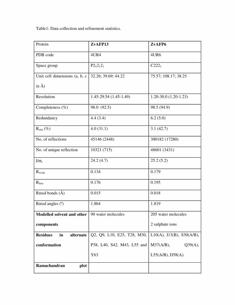
Table1: Data collection and refinement statistics.
Protein ZvAFP13 ZvAFP6
PDB code 4UR4 4UR6
Space group P212121 C2221
Unit cell dimensions (a, b, c
in Å)
32.26; 39.69; 44.22 75.57; 108.17; 38.25
Resolution 1.45-29.54 (1.45-1.49) 1.20-30.0 (1.20-1.23)
Completeness (%) 98.0 (92.5) 98.5 (94.9)
Redundancy 4.4 (3.4) 6.2 (5.0)
Rrim (%) 4.0 (31.1) 3.1 (42.7)
No. of reflections 45146 (2448) 300182 (17280)
No. of unique reflection 10321 (715) 48601 (3431)
I/σI 24.2 (4.7) 25.2 (5.2)
Rwork 0.134 0.179
Rfree 0.176 0.195
Rmsd bonds (Å) 0.015 0.018
Rmsd angles (º) 1.864 1.819
Modelled solvent and other
components
90 water molecules 205 water molecules
2 sulphate ions
Residues in alternate
conformation
Q2, Q9, L10, E25, T28, M30,
P38, L40, S42, M43, L55 and
Y63
L10(A), I13(B), S30(A/B),
M37(A/B), Q39(A),
L55(A/B), D58(A).
Ramachandran plot

(Molprobity)
Residues in favored regions
(%)
98.4 100.0
Residues in allowed regions
(%)
100.0 100.0
Outliers None None
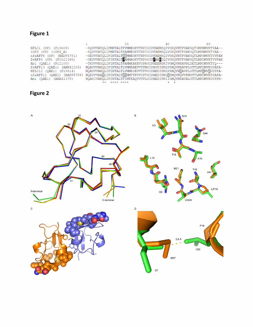
Figure 1
Figure 2
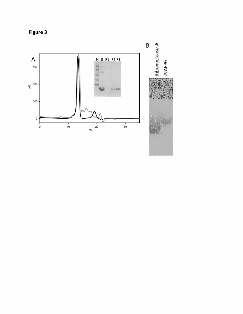
Figure 3