Embed Size (px)
Citation preview
METHODS ARTICLEpublished: 24 November 2011doi: 10.3389/fphar.2011.00076
State-of-the-art automated patch clamp devices: heatactivation, action potentials, and high throughput in ionchannel screeningSonja Stoelzle1*, Alison Obergrussberger 1, Andrea Brüggemann1, Claudia Haarmann1, Michael George1,
Ralf Kettenhofen2 and Niels Fertig1
1 Nanion Technologies GmbH, Munich, Germany2 Axiogenesis AG, Cologne, Germany
Edited by:
Juha Kammonen, Pfizer, UK
Reviewed by:
Christian Wahl-Schott,Ludwig-Maximilian University,GermanyMichel Vivaudou, Institut de BiologieStructurale, FranceLishuang Cao, Pfizer, UK
*Correspondence:
Sonja Stoelzle, Nanion TechnologiesGmbH, Gabrielenstrasse 9, 80636Munich, Germany.e-mail: [email protected]
Ion channels are essential in a wide range of cellular functions and their malfunction under-lies many disease states making them important targets in drug discovery. The availabilityof standardized cell lines expressing ion channels of interest lead to the development ofdiverse automated patch clamp (APC) systems with high-throughput capabilities. Thesesystems are now available for drug screening, but there are limitations in the applicationrange. However, further development of existing devices and introduction of new systemswiden the range of possible experiments and increase throughput. The addition of wellcontrolled and fast solution exchange, temperature control and the availability of the cur-rent clamp mode are required to analyze standard cell lines and excitable cells such asstem cell-derived cardiomyocytes in a more physiologically relevant environment. Here wedescribe two systems with different areas of applications that meet the needs of drugdiscovery researchers and basic researchers alike. The here utilized medium throughputAPC device is a planar patch clamp system capable of recording up to eight cells simultane-ously. Features such as temperature control and recordings in the current clamp mode aredescribed here. Standard cell lines and excitable cells such as stem cell-derived cardiomy-ocytes have been used in the voltage clamp and current clamp modes with the view tofinding new drug candidates and safety testing methods in a more physiologically relevantenvironment. The high-throughput system used here is a planar patch clamp screeningplatform capable of recording from 96 cells in parallel and offers a throughput of 5000 datapoints per day. Full dose response curves can be acquired from individual cells reducingthe cost per data point.The data provided reveals the suitability and relevance of both APCplatforms for drug discovery, ion channel research, and safety testing.
Keywords: automated patch clamp, electrophysiology, temperature control,TRPV3, hERG, nAChR, current clamp,
ion channels
INTRODUCTIONIon channels are pore-forming proteins that catalyze the pas-sive transport of ions through biological membranes. Given theimportance of ion channels in many physiological processes, it isno surprise that they are a major therapeutic target (Hille, 1992;Ashcroft, 2000). To date over 13% of currently known drugs havebeen shown to act primarily on ion channels, a multi-billion dollarindustry (Clare, 2010).
Patch clamp electrophysiology remains the gold standard forstudying ion channels since it was first described in the 1970s(Neher and Sakmann, 1976). However, conventional patch clampis notoriously low throughput and technically demanding. Thus,automation of the patch clamp technique increases throughputand ease of use (Farre et al., 2009), bringing patch clamp to awider audience.
Over the last decade, several automated patch clamp (APC)devices became available and are now commonplace in many lab-oratories (for recent reviews see Dunlop et al., 2008; Farre et al.,
2008). These devices provide information rich data whilst increas-ing throughput, which is essential for both drug discovery andsafety testing. Such APC devices are not limited to the phar-maceutical industry. Universities and research institutions havealso started to implement the use of higher throughput APCdevices in their laboratories (Milligan et al., 2009; Balansa et al.,2010). National screening centers and high-throughput core facil-ities are appearing worldwide, combining increased throughput,sophistication, and high quality.
However, the providers of APC devices are pushing the bound-aries by continually improving existing platforms and creatingnew and innovative devices, which have higher throughput andnew features. Such features are introduced in this paper.
MATERIALS AND METHODSSYSTEM COMPONENTSThe medium throughput patch clamp device used here was aPatchliner System (Nanion Technologies, Germany). It includes
www.frontiersin.org November 2011 | Volume 2 | Article 76 | 1
Stoelzle et al. State-of-the-art automated patch clamp
four (Patchliner Quattro) or eight (Patchliner Octo) amplifierchannels (EPC-10 Quadro amplifiers, HEKA Elektronik, Ger-many), PatchControlHT software, integrated with PatchMaster(HEKA Elektronik, Germany), for acquisition of data, and onlineanalysis of the recorded data. The data analysis software for visu-alization of traces and results of data analysis is IGOR Pro-based(WaveMetrics, USA). The data output and compound informa-tion is compatible with most database formats. A graphical userinterface is used to program and execute up to 48 unattendedrecordings, approximately 500 data points per day can be gener-ated. User intervention is possible at any time during an experi-ment. The software offers a detailed user control in combinationwith versatile experimental possibilities. In comparison to otheravailable APC devices the Patchliner is equipped with temperaturecontrol, continuous waste removal allowing an unlimited numberof compound additions, and the ability to perform current clamprecordings.
The high-throughput system utilized here was the SyncroPatch96 (Nanion Technologies, Germany), a screening platform capa-ble of recording from 96 cells in parallel. Full dose responsecurves can be acquired from individual cells with giga-ohm (GΩ)seals, generation of up 5000 data points per day is possible. Theplatform has built-in amplifiers (Triton+, Tecella LLC, USA),its own dedicated software (PatchControl96, Nanion Technolo-gies, Germany), and a data analysis package. Temperature con-trol or current clamp are not integrated features. Data outputand compound information is compatible with most databaseformats.
For both systems, planar borosilicate-glass patch clamp chipsare used (Farre et al., 2009).
CELL CULTURE FOR PLANAR PATCH CLAMPFor planar patch clamp, a wide range of different cell suspensionscan be used (Bruggemann et al., 2008; Li et al., 2008; Milliganet al., 2009). Cell suspensions for APC devices utilizing planarpatch clamp chips should be homogeneous (Milligan et al., 2009),since the cell capture is blind. The cells are ideally single. Cell clus-ters, cell fragments, or debris in the suspension decrease the cellcapture rate and, thus, the overall success rate.
Standard cell linesThe confluency of the cells should be in the range of 50–80%. Forculturing cells for use on the Patchliner and SyncroPatch 96, T75flasks are typically used. For harvesting the cells for patch clampexperiments, no significant difference in percentage of giga-ohmseals could be found for using Trypsin, accutase, or phosphate-buffered saline ethylenediaminetetraacetic acid (PBS–EDTA) forlifting the cells.
Cell lines used in this paper:
hERG stably expressed in HEK cells (Millipore, USA).hTRPV3 stably expressed in HEK cells (Millipore, USA).hNav1.5 stably expressed in HEK cells (Millipore, USA).P2X2/3 receptors stably expressed in 1321N1 cells (Evotec,Germany).HEK cells stably expressing homomeric human nAChR α7subunits (Galantos Pharma GmbH, Germany).
GABAA subunits α1β2γ2 stably expressed in HEK cells (sourceanonymous).
Harvesting protocol for cell lines. Amounts are listed for a T75flask exemplarily:
• Wash twice with 10 ml PBS (without Ca2+ and Mg2+).• Add 2 ml of detacher (PBS–EDTA 2 mM, Trypsin/EDTA, Accu-
tase etc.).• Incubate approximately 3 min in a humidified incubator at 37˚C
and 95% O2/5% CO2.• Check the detachment of cells using a microscope. Move the
plate or flask gently to detach all cells from the bottom (do nothit the flask, tap gently if necessary).
• Add 10 ml of cell culture medium (room temperature, RT),according to the cells used.
• Pipette the cells gently up and down with a 10-ml pipette.• After pipetting five times, look at the cells under a microscope.
If the cells are single ( 80–90%), no further pipetting is needed.• If cells still form clusters, gently pipette cells another 10 times.
Repeat this step until cells are single ( 80–90%).• Centrifuge the cells (2 min, 100 g).• Discard the supernatant.• Resuspend the cells in external recording solution resulting in a
cell density of approximately 1 × 10⁶ cells/ml.• A visual control of the cell suspension under the microscope
should reveal single, round cells with smooth membrane edgesand few cell clusters.
• Transfer cells to the cell hotel of the Patchliner where cells arecontinuously pipetted up and down to maintain single cells andviability.
Stem cell-derived cardiomyocytesDifferentiation of genetically modified mouse embryonic stemcells (mESC) and the antibiotic selection of the mESC-derivedcardiomyocytes have been described previously in detail (Kolossovet al., 2006).
Vials of at least one or five million viable mESC-derivedcardiomyocytes (Cor.At cells, Lonza, Walkersville, USA, cata-log numbers XCAC-1010 or XCAC-1050, respectively) obtaineddirectly from the manufacturer (Axiogenesis, Cologne, Germany)were thawed as described in the distributors technical man-ual (https://www.lonza.com/go/literature/). For long-term stor-age, the cells are frozen as single cell suspensions in liquid nitrogenor −150˚C freezers. When cultured overnight at an appropriatecell density, the thawed cardiomyocytes form spontaneously andsynchronously contracting monolayers.
Cells were seeded at a density of 105 viable cells/cm2 culturearea in one T25 cell culture flask with 5 ml Cor.At CompleteCulture Medium when a vial with one million viable mESC-derived cardiomyocytes was used. Two T75 cell culture flasks eachwith 10 ml Cor.At Complete Culture Medium were used whena vial containing five million mESC-derived cardiomyocytes wastaken.
iCell cardiomyocytes (Cellular Dynamics International, Madi-son, WI, USA) were plated on T25 culture flasks coatedwith 0.1% gelatin as per manufacturers’ instructions. A vial
Frontiers in Pharmacology | Pharmacology of Ion Channels and Channelopathies November 2011 | Volume 2 | Article 76 | 2
Stoelzle et al. State-of-the-art automated patch clamp
containing > 1.5 × 10⁶ platable cells (catalog number CMC-100-110-001) was plated on one T25 flask, a vial containing > 7.5 × 10⁶
platable cells (catalog number CMC-100-110-005) was platedon five T25 flasks. For each 1 × 10⁶ viable cells, 9 ml cold iCellCardiomyocytes Plating Media (4˚C) was used. After 24 h themedia was exchanged for iCell Cardiomyocytes MaintenanceMedia.
Harvesting protocol for mouse stem cell-derived Cor.At car-diomyocytes. Amounts are listed for a T75 flask exemplarily:
• Cells must be cultured for at least 2–4 days before patch clampexperiments are performed.
• Wash twice with 10 ml 4˚C cold PBS (without Ca2+/Mg2+)/EDTA(2 mM).
• Incubate at 4˚C for 15 min.• Remove PBS/EDTA.• Add 5 ml pre-warmed (RT) Trypsin 0.05%/EDTA 0.02% in PBS
solution.• Incubate 4–5 min in a humidified incubator at 37˚C and 95%
O2/5% CO2.• Check the detachment of cells under a microscope. Move the
plate or flask gently to detach all cells from the bottom (do nothit the flask).
• Add 10 ml of Cor.At Complete Culture Medium (RT).• Centrifuge the cells (2 min, 100 g).• Discard the supernatant.• Add external recording solution resulting in a cell density of
approximately 1 × 10⁶- 5 × 10⁷/ml. Carefully resuspend thecells by gentle pipetting (two times max.).
• A visual control of the cell suspension under the microscopeshould reveal single, round cells with smooth membrane edgesand no cell clusters.
• Transfer cells to the cell hotel of the Patchliner where cells arecontinuously pipetted up and down to maintain single cells andviability.
Harvesting protocol for human iPS cell-derived iCell cardiomy-ocytes. Amounts are listed for a T25 flask exemplarily:
• Cells should be cultured for at least 2–4 days before patch clampexperiments are performed.
• Wash twice with 5 ml 4˚C cold PBS (without Ca2+/Mg2+)/EDTA(2 mM).
• Add 5 ml 4˚C cold PBS (without Ca2+/Mg2+)/EDTA (2 mM).• Incubate at 4˚C for 15 min.• Remove PBS/EDTA.• Add 2 ml Trypsin 0.05%/EDTA 0.02% in PBS solution.• Rock the dish from side to side to ensure an even distribution
of Trypsin.• Immediately remove the Trypsin (before cells start to detach).
Despite removing the liquid, there is still enough Trypsin on thesurface to detach the cells.
• Incubate the flask for 3–8 min at 37˚C and 95% O2/5% CO2.Check the detachment of cells under a microscope after 3 min.Move the plate or flask gently to detach all cells from the bot-tom (do not hit the flask, tap gently if required). If cells are notdetached return the flask to the incubator.
• Once cells start to detach, add 1 ml media and 1 ml exter-nal recording solution. Carefully resuspend the cells by gentlepipetting (two times max.).
• Transfer cells to the cell hotel of the Patchliner where cells arecontinuously pipetted up and down to maintain single cells andviability.
Patch clamp solutions. Internal solution: 50 mM KCl, 10 mMNaCl, 60 mM KF, 20 mM EGTA, 10 mM HEPES/KOH, pH 7.2 (forFigures 1, 2, 5, 6, and 8) or 50 mM CsCl, 10 mM NaCl, 60 mM
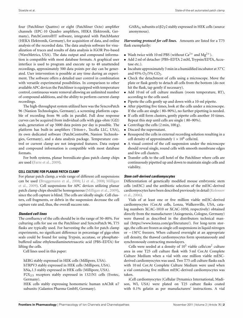
FIGURE 1 | Effect of erythromycin on hERG-mediated currents at (A)
room temperature and (B) 35˚C. The graphs are shown on the same scale.Hundred micromolar erythromycin significantly blocked hERG currents at35˚C but had little effect on hERG currents at RT, n = 1 each.
FIGURE 2 | Concentration response curves for erythromycin at room
temperature (n = 12) and at 35˚C (n = 18). Erythromycin is approximately10-fold more potent at 35˚C compared with room temperature. An overlayof the concentration response curves at room temperature and 35˚C clearlyshows the shift in potency.
www.frontiersin.org November 2011 | Volume 2 | Article 76 | 3
Stoelzle et al. State-of-the-art automated patch clamp
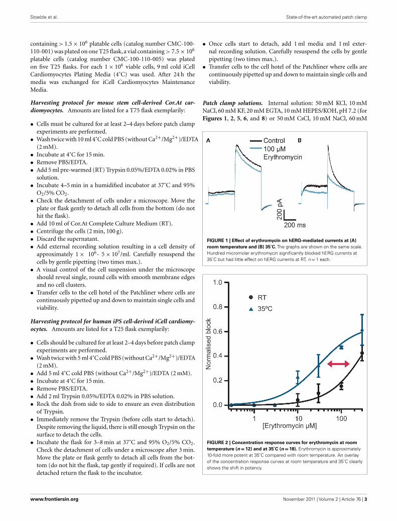
FIGURE 3 | (A) Currents elicited using a voltage ramp protocol from −100to 100 mV over 200 ms and activation of TRPV3 receptors by increasingconcentrations of 2-APB. (B) Concentration response curve for n = 11 cellsrevealing an EC50 for 2-APB of 61.1 ± 7.6 μM.
FIGURE 4 | Current traces ofTRPV3 activated by heated solution.
External solution was heated to the temperature shown and applied to thecell. Currents started to activate at ≥38˚C.
CsF, 20 mM EGTA, 10 mM HEPES/CsOH (for Figures 3, 4, 9, and10). External solution (except for recordings shown in Figure 9B):140 mM NaCl, 4 mM KCl, 1 mM MgCl2, 2 mM CaCl2, 5 mM d-Glucose monohydrate, 10 mM HEPES/NaOH pH 7.4. Externalsolution for nicotinic α7 channel recordings (Figure 9B): 80 mMNaCl, 3 mM KCl, 45 mM CaCl2, 10 mM HEPES (Na+ salt)/HCl,pH 7.4. Solutions for action potential (AP) recording in the con-ventional patch clamp rig (Figure 5G) were 130 mM KAs, 15 mMKCl, 5.5 mM MgCl2, 5 mM Na2ATP, 5 mM K2 phosphocreatine,
10 mM HEPES, pH 7.25 (internal) and 140 mM NaCl, 4 mM KCl,2 mM CaCl2, 1 mM MgCl2, 10 mM glucose, 10 mM HEPES, pH7.35 (external).
Electrophysiology. Whole cell patch clamp recordings were con-ducted as previously described (Farre et al., 2009; Stoelzle et al.,2011). Currents were elicited using voltage protocols in the voltageclamp mode. For hERG, a voltage step protocol from the holdingpotential (−80 mV) to +40 mV for 500 ms followed by a 500-msstep to −40 mV, repeated every 20 s. Peak amplitude at −40 mVwas used for analysis. For Nav 1.5, currents were elicited using10 ms voltage steps from −120 to 0 mV repeated every 1 s. Forligand-gated experiments, cells were held at a constant holdingpotential of −80 mV and solutions were exchanged within 100 msto activate receptors and minimize ligand exposure time. For heatactivation of TRPV3 channels, external solution was heated to thetemperature indicated and applied to the cell. Alternatively, 2-APBat increasing concentrations at RT was used to activate TRPV3. Avoltage ramp protocol from −100 to 100 mV over 200 ms was usedto record TRPV3 currents. Current amplitude at 90 mV was usedfor analysis. APs were generated using a depolarizing pulse to thethreshold at which an AP was elicited. Membrane potential waskept at −80 to −100 mV (cell dependent). The AP traces shownrepresent an average response of four recorded APs. The APs werenormalized to the time point of the beginning of the upstroke.Equipment: NPC-16 Patchliner Octo (with temperature controloption) and SyncroPatch 96 (Nanion Technologies GmbH, Ger-many). Patch clamp amplifier for the Patchliner: EPC-10 Quadro(HEKA Elektronik GmbH, Germany), patch clamp amplifier forthe SyncroPatch 96: Triton + (Tecella, CA, USA), PatchControlHT and PatchControl 96 software (Nanion Technologies GmbH,Germany). Software for data acquisition (PatchMaster, HEKAElektronik GmbH, Germany; PatchControl 96, Nanion Technolo-gies GmbH, Germany) and analysis (IGOR Pro WaveMetrics Inc.,OR, USA and SyncroPatch Data Analysis Package, Nanion Tech-nologies GmbH, Germany). NPC-16 or NPC-96 chips (single-use,disposable; Nanion Technologies GmbH, Germany) were used.
APPLICATIONS AND NOVEL FEATURESTEMPERATURE REGULATIONExperiments at physiological temperatureCompounds can display different properties or different poten-cies at physiological temperature (35˚C) vs. RT. Therefore, it isa desirable option to be able to study ion channels at elevatedtemperature. To meet this need, several heating elements wereintroduced into the Patchliner. The surrounding of the planarpatch clamp chip can be heated to maintain constant, physiolog-ical temperature. The solution, which is pipetted onto the cell,can also be heated separately. To prevent any degradation of com-pounds inside the pipette due to long heating phases, only thevolume which is required (typically 40–100 μl, depending on theapplication) is heated, directly before application to the chamberof the patch clamp chip. Heating of the solution, which is appliedto the cell takes 23.4 ± 4 s (for temperatures between 30 and 70˚C;data not shown).
One compound, which has been shown to have an increasein potency at physiological temperature, is erythromycin.
Frontiers in Pharmacology | Pharmacology of Ion Channels and Channelopathies November 2011 | Volume 2 | Article 76 | 4
Stoelzle et al. State-of-the-art automated patch clamp
FIGURE 5 | (A) Representative traces of a Cor.At cardiomyocyte recordedin the voltage clamp mode and block by increasing concentrations of TTX.Raw data of Na+ currents in control solution and in the presence ofincreasing TTX concentrations (0.3, 1, 3, 10, and 30 μM), elicited by 10 msvoltage steps to 0 mV from a holding potential of −80 mV, sweep interval2 s. (B) Corresponding concentration response curve with a calculated IC50
of 1.3 ± 0.4 μM (n = 3). (C) Corresponding time plot of the experimentshowing the stability of the recording. Current amplitude was reducedstep by step upon application of the increasing concentrations. A washout
of TTX was performed at the end of experiment. (D) Representative tracesof highly reproducible APs as recorded in the current clamp mode. Sweepinterval between each stimulus was 10 s. Traces were recorded over15 min, the first and the last 4 APs are shown. The same experiment asshown in (A) was performed on the semi-automated patch clamp systemPort-a-Patch (E). The corresponding IC50 was calculated to be 1.1 ± 0.5 μM(n = 4) (F). (G) Comparison of an AP recorded with the Port-a-Patch (toptrace) and using a conventional patch clamp rig (lower trace). Gray lineindicates 0 mV.
Erythromycin is a macrolide antibiotic, which can cause QTprolongation and cardiac arrhythmia. Erythromycin has beenshown to block hERG channels at physiological temperature withan IC50 of approximately 40 μM (Stanat et al., 2003). How-ever, at RT erythromycin is much less potent. At a concentrationof 100 μM, erythromycin causes no significant block of hERGcurrents at RT but significantly blocks currents at physiologicaltemperature (Guo et al., 2005).
Here we present data collected on a Patchliner Octo with tem-perature control at RT and at 35˚C and the effect this has onthe potency of erythromycin. Current responses of two individualcells to 500 ms voltage pulses to +40 mV and then −40 mV in thepresence and absence of 100 μM erythromycin at RT and 35˚Care shown in Figure 1. At RT (Figure 1A) 100 μM erythromycincaused little reduction in current amplitude (at −40 mV; approx.15%) compared with an almost 50% reduction in current ampli-tude at 35˚C (Figure 1B). This is in good agreement with theliterature (Guo et al., 2005).
Typically, single concentrations of erythromycin were appliedto each cell. Figure 2 shows averaged concentration responsecurves for erythromycin at RT and at 35˚C overlaid. At higherconcentrations (300 μM), erythromycin did block hERG currentsat RT by approximately 40% and gave an IC50 of 427.5 μM calcu-lated from the graph (n = 12). At 300 μM, erythromycin blocked
hERG currents at 35˚C by 70% and the IC50 at this tempera-ture was calculated to be 30.7 μM (n = 18). This is in excellentagreement with values reported in the literature (Stanat et al.,2003).
Table 1 shows success rates of reaching a giga-ohm seal andcell parameters for hERG stably expressed in HEK cells (Millipore,USA), n = 35.
Heat activation of TRPV3Transient receptor potential (TRP) channels are an important classof receptors found widely distributed throughout the mammaliancentral and peripheral nervous systems. They have been shownto be activated by many stimuli including temperature, mechano-stimulation, divalent cations, and pH (for review see Clapham,2003). TRP channels are receiving much attention as potentialtargets for the treatment of, for example, chronic pain, asthma,and diabetes insipidus (Clapham,2003; Gudermann and Flockerzi,2005).
We have used the Patchliner to study TRPV3 channels usingeither 2-APB or heat to activate the receptors. High quality datacould be achieved with a high success rate for obtaining seals inthe GΩ range (> 80%). Table 2 shows success rates and cell para-meters for TRPV3 stably expressed in HEK cells (Millipore, USA)activated by either 2-APB or heat.
www.frontiersin.org November 2011 | Volume 2 | Article 76 | 5
Stoelzle et al. State-of-the-art automated patch clamp
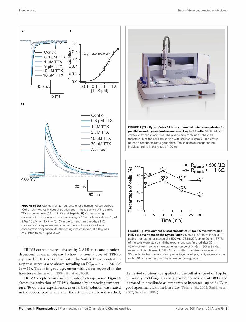
FIGURE 6 | (A) Raw data of Na+ currents of one human iPS cell-derivediCell cardiomyocyte in control solution and in the presence of increasingTTX concentrations (0.3, 1, 3, 10, and 30 μM). (B) Correspondingconcentration response curve for an average of four cells reveals an IC50 of2.5 ± 1.0 μM for TTX (n = 4). (C) In the current clamp mode, a TTXconcentration-dependent reduction of the amplitude as well as aconcentration-dependent AP shortening was observed. The IC50 wascalculated to be 5.6 μM (n = 2).
TRPV3 currents were activated by 2-APB in a concentration-dependent manner. Figure 3 shows current traces of TRPV3expressed in HEK cells and activation by 2-APB. The concentrationresponse curve is also shown revealing an EC50 = 61.1 ± 7.6 μM(n = 11). This is in good agreement with values reported in theliterature (Chung et al., 2004; Hu et al., 2009).
TRPV3 receptors can also be activated by temperature. Figure 4shows the activation of TRPV3 channels by increasing tempera-ture. To do these experiments, external bath solution was heatedin the robotic pipette and after the set temperature was reached,
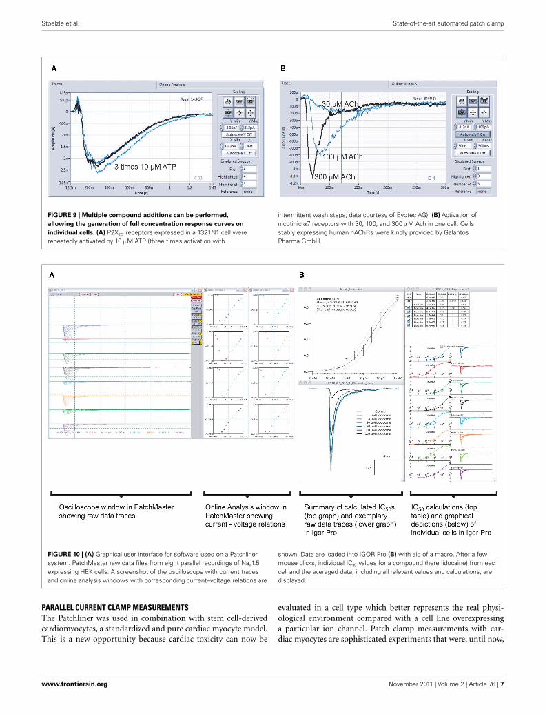
FIGURE 7 |The SyncroPatch 96 is an automated patch clamp device for
parallel recordings and online analysis of up to 96 cells. All 96 cells arevoltage clamped at any time. The pipette arm contains 16 channels,therefore 16 of the cells are served with solution in parallel. The deviceutilizes planar borosilicate-glass chips. The solution exchange for theindividual cell is in the range of 100 ms.
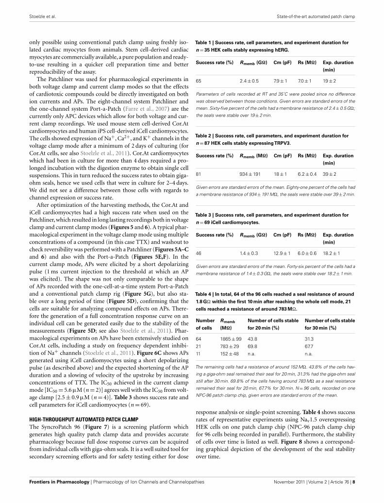
FIGURE 8 | Development of seal stability of 96 Nav1.5 overexpressing
HEK cells over time on the SyncroPatch 96. 69.8% of the cells had astable membrane resistance of >500 MΩ (783 ± 29 MΩ) for 20 min, 67.7%of the cells were stable until the experiment was finished after 30 min.43.8% of cells having a membrane resistance of >1 GΩ (1865 ± 99 MΩ)were stable for 20 min, 31.3% of them still had a stable resistance after30 min. Note the increase of cell percentage developing a higher resistancewithin 10 min after reaching the whole cell configuration.
the heated solution was applied to the cell at a speed of 10 μl/s.Outwardly rectifying currents started to activate at 38˚C andincreased in amplitude as temperature increased, up to 54˚C, ingood agreement with the literature (Peier et al., 2002; Smith et al.,2002; Xu et al., 2002).
Frontiers in Pharmacology | Pharmacology of Ion Channels and Channelopathies November 2011 | Volume 2 | Article 76 | 6
Stoelzle et al. State-of-the-art automated patch clamp
FIGURE 9 | Multiple compound additions can be performed,
allowing the generation of full concentration response curves on
individual cells. (A) P2X2/3 receptors expressed in a 1321N1 cell wererepeatedly activated by 10 μM ATP (three times activation with
intermittent wash steps; data courtesy of Evotec AG). (B) Activation ofnicotinic α7 receptors with 30, 100, and 300 μM Ach in one cell. Cellsstably expressing human nAChRs were kindly provided by GalantosPharma GmbH.
FIGURE 10 | (A) Graphical user interface for software used on a Patchlinersystem. PatchMaster raw data files from eight parallel recordings of Nav1.5expressing HEK cells. A screenshot of the oscilloscope with current tracesand online analysis windows with corresponding current–voltage relations are
shown. Data are loaded into IGOR Pro (B) with aid of a macro. After a fewmouse clicks, individual IC50 values for a compound (here lidocaine) from eachcell and the averaged data, including all relevant values and calculations, aredisplayed.
PARALLEL CURRENT CLAMP MEASUREMENTSThe Patchliner was used in combination with stem cell-derivedcardiomyocytes, a standardized and pure cardiac myocyte model.This is a new opportunity because cardiac toxicity can now be
evaluated in a cell type which better represents the real physi-ological environment compared with a cell line overexpressinga particular ion channel. Patch clamp measurements with car-diac myocytes are sophisticated experiments that were, until now,
www.frontiersin.org November 2011 | Volume 2 | Article 76 | 7
Stoelzle et al. State-of-the-art automated patch clamp
only possible using conventional patch clamp using freshly iso-lated cardiac myocytes from animals. Stem cell-derived cardiacmyocytes are commercially available, a pure population and ready-to-use resulting in a quicker cell preparation time and betterreproducibility of the assay.
The Patchliner was used for pharmacological experiments inboth voltage clamp and current clamp modes so that the effectsof cardiotoxic compounds could be directly investigated on bothion currents and APs. The eight-channel system Patchliner andthe one-channel system Port-a-Patch (Farre et al., 2007) are thecurrently only APC devices which allow for both voltage and cur-rent clamp recordings. We used mouse stem cell-derived Cor.Atcardiomyocytes and human iPS cell-derived iCell cardiomyocytes.The cells showed expression of Na+, Ca2+, and K+ channels in thevoltage clamp mode after a minimum of 2 days of culturing (forCor.At cells, see also Stoelzle et al., 2011). Cor.At cardiomyocyteswhich had been in culture for more than 4 days required a pro-longed incubation with the digestion enzyme to obtain single cellsuspensions. This in turn reduced the success rates to obtain giga-ohm seals, hence we used cells that were in culture for 2–4 days.We did not see a difference between those cells with regards tochannel expression or success rate.
After optimization of the harvesting methods, the Cor.At andiCell cardiomyocytes had a high success rate when used on thePatchliner, which resulted in long lasting recordings both in voltageclamp and current clamp modes (Figures 5 and 6). A typical phar-macological experiment in the voltage clamp mode using multipleconcentrations of a compound (in this case TTX) and washout tocheck reversibility was performed with a Patchliner (Figures 5A–Cand 6) and also with the Port-a-Patch (Figures 5E,F). In thecurrent clamp mode, APs were elicited by a short depolarizingpulse (1 ms current injection to the threshold at which an APwas elicited). The shape was not only comparable to the shapeof APs recorded with the one-cell-at-a-time system Port-a-Patchand a conventional patch clamp rig (Figure 5G), but also sta-ble over a long period of time (Figure 5D), confirming that thecells are suitable for analyzing compound effects on APs. There-fore the generation of a full concentration response curve on anindividual cell can be generated easily due to the stability of themeasurements (Figure 5D; see also Stoelzle et al., 2011). Phar-macological experiments on APs have been extensively studied onCor.At cells, including a study on frequency dependent inhibi-tion of Na+ channels (Stoelzle et al., 2011). Figure 6C shows APsgenerated using iCell cardiomyocytes using a short depolarizingpulse (as described above) and the expected shortening of the APduration and a slowing of velocity of the upstroke by increasingconcentrations of TTX. The IC50 achieved in the current clampmode [IC50 = 5.6 μM (n = 2)] agrees well with the IC50 from volt-age clamp [2.5 ± 0.9 μM (n = 4)]. Table 3 shows success rate andcell parameters for iCell cardiomyocytes (n = 69).
HIGH-THROUGHPUT AUTOMATED PATCH CLAMPThe SyncroPatch 96 (Figure 7) is a screening platform whichgenerates high quality patch clamp data and provides accuratepharmacology because full dose response curves can be acquiredfrom individual cells with giga-ohm seals. It is a well suited tool forsecondary screening efforts and for safety testing either for dose
Table 1 | Success rate, cell parameters, and experiment duration for
n = 35 HEK cells stably expressing hERG.
Success rate (%) Rmemb (GΩ) Cm (pF) Rs (MΩ) Exp. duration
(min)
65 2.4 ± 0.5 7.9 ± 1 7.0 ± 1 19 ± 2
Parameters of cells recorded at RT and 35˚C were pooled since no difference
was observed between those conditions. Given errors are standard errors of the
mean. Sixty-five percent of the cells had a membrane resistance of 2.4 ± 0.5 GΩ;
the seals were stable over 19 ± 2 min.
Table 2 | Success rate, cell parameters, and experiment duration for
n = 87 HEK cells stably expressingTRPV3.
Success rate (%) Rmemb (MΩ) Cm (pF) Rs (MΩ) Exp. duration
(min)
81 934 ± 191 18 ± 1 6.2 ± 0.4 39 ± 2
Given errors are standard errors of the mean. Eighty-one percent of the cells had
a membrane resistance of 934 ± 191 MΩ, the seals were stable over 39 ± 2 min.
Table 3 | Success rate, cell parameters, and experiment duration for
n = 69 iCell cardiomyocytes.
Success rate (%) Rmemb (GΩ) Cm (pF) Rs (MΩ) Exp. duration
(min)
46 1.4 ± 0.3 12.9 ± 1 6.0 ± 0.6 18.2 ± 1
Given errors are standard errors of the mean. Forty-six percent of the cells had a
membrane resistance of 1.4 ± 0.3 GΩ, the seals were stable over 18.2 ± 1 min.
Table 4 | In total, 64 of the 96 cells reached a seal resistance of around
1.8 GΩ within the first 10 min after reaching the whole cell mode, 21
cells reached a resistance of around 783 MΩ.
Number
of cells
Rmemb
(MΩ)
Number of cells stable
for 20 min (%)
Number of cells stable
for 30 min (%)
64 1865 ± 99 43.8 31.3
21 783 ± 29 69.8 67.7
11 152 ± 48 n.a. n.a.
The remaining cells had a resistance of around 152 MΩ. 43.8% of the cells hav-
ing a giga-ohm seal remained their seal for 20 min, 31.3% had the giga-ohm seal
still after 30 min. 69.8% of the cells having around 783 MΩ as a seal resistance
remained their seal for 20 min, 67.7% for 30 min. N = 96 cells, recorded on one
NPC-96 patch clamp chip, given errors are standard errors of the mean.
response analysis or single-point screening. Table 4 shows successrates of representative experiments using Nav1.5 overexpressingHEK cells on one patch clamp chip (NPC-96 patch clamp chipfor 96 cells being recorded in parallel). Furthermore, the stabilityof cells over time is listed as well. Figure 8 shows a correspond-ing graphical depiction of the development of the seal stabilityover time.
Frontiers in Pharmacology | Pharmacology of Ion Channels and Channelopathies November 2011 | Volume 2 | Article 76 | 8
Stoelzle et al. State-of-the-art automated patch clamp
Voltage clamp recordings are continuous during compoundapplication and solution exchange time is in the order of 100 ms.This allows for screening of compound action on ligand- andvoltage-gated ion channels. An APC device with fast solutionexchange is a prerequisite for studying fast desensitizing chan-nels such as nicotinic acetylcholine receptor (nAChR). Enhancersof nAChRs, such as galantamine, may have therapeutic benefitfor Alzheimer’s disease (Samochocki et al., 2003). It is, therefore,essential to be able to search for nAChR agonists and enhancers ata higher throughput.
Figure 9A shows a typical screenshot with recordings of aligand-gated ion channel, P2X2/3. P2X2/3 receptors in a 1321N1cell were repeatedly activated by 10 μM ATP, demonstrating thereliability of repetitive drug applications. nAChR α7 express-ing HEK cells were activated by increasing concentrations ofACh (30, 100, 300 μM; Figure 9B). Given the importance ofthis channel as a potential therapeutic target for conditionssuch as Alzheimer’s disease, schizophrenia, and Parkinson’s dis-ease (for review see Colquhoun et al., 2003; Albuquerque et al.,2009), it is critical that researchers have tools to study themreliably.
The disposable planar borosilicate-glass substrate used for sealformation ensures high quality data and long lasting seals whenusing the Patchliner and SyncroPatch 96. In general, with a suc-cess rate for completed experiments of 51.7 ± 10% (n = 567), theSyncroPatch 96 has a data throughput of approximately 5000 datapoint per day. With a Patchliner Octo, having a success rate ofaround 80%, approximately 500 data points can be generated perday (Farre et al., 2009).
DATA ANALYSISIncreasing the throughput in APC devices results in an increasein data generation and, thereby, necessitates the need for auto-mated data analysis. Figure 10A shows a screenshot of one runon a Patchliner Octo, which records from eight cells at a time.Here, Nav1.5 stably expressing HEK cells were used (Millipore,USA). The raw data are saved as PatchMaster (.dat) files and anadditional Microsoft office excel spreadsheet is generated, whichcan be easily loaded in IGOR Pro (WaveMetrics, USA). We devel-oped a macro which is implemented in IGOR Pro that enables theupload, graphical display of raw data traces and IC50 calculationof individual cells or an average of all cells for each compound insummary (Figure 10B).
For the analysis of concentration response curves, the Syn-croPatch 96 software provides an intuitive graphical user interface(Figure 11). The recording sites are color coded according to userdefined success criteria (membrane resistance, access resistance,current amplitude etc.). The user can generate individual IC50
values or an average IC50 of the recorded cells. Current–voltagerelationships, leak-subtraction, and time-dependence of recordedvalues can be displayed besides a repertory of other calculations.
DISCUSSION AND OUTLOOKThe APC devices used here for data recordings are equipped forstudying ion channels in various areas of research, drug discov-ery, and safety screening. The data quality and reproducibility ofpharmacological experiments is consistent between the automatedsystems and a manual patch clamp rig or semi-automated patchclamp system like the Port-a-Patch as shown for current clamp
FIGURE 11 | Graphical user interface of the screening and data analysis
software used on the SyncroPatch 96. (A) Screenshot of depiction of rawdata of GABAA subunits α1β2γ2 expressing HEK cells as recorded on oneNPC-96 patch clamp chip. Ninety-six small color coded pictures as seen inthe upper left part display all 96 parallel recordings. Depending on the sealresistance, pictures are green (Rmemb > 100 MΩ), blue (Rmemb = 50–100 MΩ),light blue or gray (Rmemb < 50 MΩ or no cell capture). One highlightedexperiment is displayed below, 16 other selected experiments are displayedon the right. Traces show currents of individual cells which were activated by
six GABA concentrations (0.3, 1, 3, 10, 30, and 100 μM). Intermittent washsteps with control solution were performed. (B) Screenshot showing thesame experiment as in (A), but displaying individual concentration responsecurves. For highlighted experiments (in lower part and right), note thedifferent shades of blue overlaid on the curves, representing the sixincreasing concentrations of GABA. The white color represents theintermittent wash steps. (C) Averaged concentration response curve andrelated parameters of successful experiments (here 70 out of 96) of thesame experiment.
www.frontiersin.org November 2011 | Volume 2 | Article 76 | 9
Stoelzle et al. State-of-the-art automated patch clamp
recordings from pluripotent stem cell-derived cardiomyocytes.The overall instrument performance, success rates, longevity ofrecordings, and compatibility with various cell lines and assaytypes are fundamental for effective screening work in electrophys-iology departments. Effective screening in the broader sense alsomeans cost effectiveness, which is, of course, an essential quali-fication. The combination of an in vitro cardiac cell model withhigher throughput patch clamp screening technology opens up anew way for cardiotoxicity prediction in a physiologically relevantcell system. The ability to perform current clamp recordings anda temperature-regulated cell environment are unique features ofthe Patchliner, currently not available on other higher throughputAPC systems. Furthermore, the temperature control feature of thePatchliner can be used to record ion currents and APs at physiolog-ical temperatures and to activate temperature-regulated receptorssuch as the TRP family of receptors to discover specific inhibitorswith a reduced side-effect profile. The data shown on TRP chan-nels prove not only the versatility of the liquid handling that isneeded to repeatedly activate the cell by perfusing heated solution,but also the exactness of heating and precisely timed exposure ofthe cell to the heated solution.
The Patchliner is compatible with many different cell lines,showing little discrepancy between the cell lines used (datadescribed in this paper and Bruggemann et al., 2006; Farre et al.,2007, 2009). Interestingly, recordings of a variety of primary cellshave also been described using the Patchliner (Milligan et al.,2009). Few, if any,other APC devices have been able to demonstratesuch compatibility with primary cells.
In both academic and pharmaceutical laboratories it is ofadvantage to be able to record ion channels in a more physi-ologically relevant environment, e.g., primary cell cultures, fordrug discovery, biophysical and physiological analysis, and safetypharmacology using an APC system.
Fast external exchange, minimal compound exposure, and con-tinuous current recording make the Patchliner and SyncroPatch96 suited to ligand-gated ion channel measurements. Fast desen-sitizing receptors such as the nicotinic α7 receptor can be reliablyrecorded when taking advantage of the possibility to minimizethe exposure time of the cell to the compound. The applied com-pound solution, e.g., containing the ligand can be replaced by acontrol solution after 200 ms. Volumes of the ligand containingor control solution and speed of application are set by the userto control speed of exchange and ligand exposure time. Settingscan be adapted according to the receptor of interest to optimizereproducibility of current responses.
Providers of APC devices constantly strive to improve andextend the application range of these devices and features. Asdescribed in this manuscript, recordings in the current clampmode and at physiological temperature are relevant featuresand will be of good use in the future in both academic andpharmaceutical research.
ACKNOWLEDGMENTSWe thank Davide Pau, Scottish Biomedical, UK for performing APrecordings on a conventional patch clamp rig and for letting ususe his data.
REFERENCESAlbuquerque, E. X., Pereira, E. F., Alkon-
don, M., and Rogers, S. W. (2009).Mammalian nicotinic acetylcholinereceptors: from structure to func-tion. Physiol. Rev. 89, 73–120.
Ashcroft, F. M. (2000). Ion Channels andDisease: Channelopathies, London:Academic Press.
Balansa, W., Islam, R., Fontaine,F., Piggott, A. M., Zhang, H.,Webb, T. I., Gilbert, D. F., Lynch,J. W., and Capon, R. J. (2010).Ircinialactams: subunit-selectiveglycine receptor modulators fromAustralian sponges of the familyIrciniidae. Bioorg. Med. Chem. 18,2912–2919.
Bruggemann, A., Farre, C., Haar-mann, C., Haythornthwaite, A.,Kreir, M., Stoelzle, S., George, M.,and Fertig, N. (2008). Planar patchclamp: advances in electrophys-iology. Methods Mol. Biol. 491,165–176.
Bruggemann, A., Stoelzle, S., George,M., Behrends, J. C., and Fer-tig, N. (2006). Microchip tech-nology for automated and paral-lel patch-clamp recording. Small 2,840–846.
Chung, M. K., Lee, H., Mizuno,A., Suzuki, M., and Caterina, M.
J. (2004). 2-Aminoethoxydiphenylborate activates and sensitizes theheat-gated ion channel TRPV3. J.Neurosci. 24, 5177–5182.
Clapham, D. E. (2003). TRP chan-nels as cellular sensors. Nature 426,517–524.
Clare, J. J. (2010). Targeting ion chan-nels for drug discovery. Discov. Med.9, 253–260.
Colquhoun, D., Unwin, N., Shelly, C.,Hatton, C., and Sivilotti, L. (2003).“Nicotinic acetylcholine receptors,”in Burger’s Medicinal Chemistry andDrug Discovery: Drug Discovery andDrug Development, eds A. Burgerand D. J. Abraham (New York:Wiley), 357–405.
Dunlop, J., Bowlby, M., Peri, R., Tawa,G., Larocque, J., Soloveva, V., andMorin, J. (2008). Ion channel screen-ing. Comb. Chem. High ThroughputScreen. 11, 514–522.
Farre, C., George, M., Bruggemann, A.,and Fertig, N. (2008). Ion channelscreening – automated patch clampon the rise. Drug Discov. Today Tech-nol. 5, e23–e25.
Farre, C., Haythornthwaite, A., Haar-mann, C., Stoelzle, S., Kreir, M.,George, M., Bruggemann, A.,and Fertig, N. (2009). Port-a-patch and patchliner: high fidelity
electrophysiology for secondaryscreening and safety pharmacology.Comb. Chem. High ThroughputScreen. 12, 24–37.
Farre, C., Stoelzle, S., Haarmann, C.,George, M., Bruggemann, A., andFertig, N. (2007). Automated ionchannel screening: patch clampingmade easy. Expert Opin. Ther. Targets11, 557–565.
Gudermann, T., and Flockerzi, V.(2005). TRP channels as newpharmacological targets. NaunynSchmiedebergs Arch. Pharmacol. 371,241–244.
Guo, J., Zhan, S., Lees-Miller, J. P., Teng,G., and Duff, H. J. (2005). Exag-gerated block of hERG (KCNH2)and prolongation of action poten-tial duration by erythromycin attemperatures between 37 degrees Cand 42 degrees C. Heart Rhythm 2,860–866.
Hille, B. (1992). Ionic Channels ofExcitable Membranes. Sunderland,MA: Sinauer Associates.
Hu, H., Grandl, J., Bandell, M., Petrus,M., and Patapoutian, A. (2009).Two amino acid residues deter-mine 2-APB sensitivity of theion channels TRPV3 and TRPV4.Proc. Natl. Acad. Sci. U.S.A. 106,1626–1631.
Kolossov, E., Bostani, T., Roell, W., Bre-itbach, M., Pillekamp, F., Nygren, J.M., Sasse, P., Rubenchik, O., Fries,J. W., Wenzel, D., Geisen, C., Xia,Y., Lu, Z., Duan, Y., Kettenhofen, R.,Jovinge, S., Bloch, W., Bohlen, H.,Welz, A., Hescheler, J., Jacobsen, S.E., and Fleischmann, B. K. (2006).Engraftment of engineered ES cell-derived cardiomyocytes but not BMcells restores contractile function tothe infarcted myocardium. J. Exp.Med. 203, 2315–2327.
Li, J., Sukumar, P., Milligan, C. J., Kumar,B., Ma, Z. Y., Munsch, C. M., Jiang,L. H., Porter, K. E., and Beech,D. J. (2008). Interactions, functions,and independence of plasma mem-brane STIM1 and TRPC1 in vascularsmooth muscle cells. Circ. Res. 103,e97–e104.
Milligan, C. J., Li, J., Sukumar, P.,Majeed, Y., Dallas, M. L., English,A., Emery, P., Porter, K. E., Smith,A. M., Mcfadzean, I., Beccano-Kelly,D., Bahnasi, Y., Cheong, A., Nay-lor, J., Zeng, F., Liu, X., Gamper,N., Jiang, L. H., Pearson, H. A.,Peers, C., Robertson, B., and Beech,D. J. (2009). Robotic multiwell pla-nar patch-clamp for native and pri-mary mammalian cells. Nat. Protoc.4, 244–255.
Frontiers in Pharmacology | Pharmacology of Ion Channels and Channelopathies November 2011 | Volume 2 | Article 76 | 10
Stoelzle et al. State-of-the-art automated patch clamp
Neher, E., and Sakmann, B. (1976).Single-channel currents recordedfrom membrane of denervatedfrog muscle fibres. Nature 260,799–802.
Peier, A. M., Reeve, A. J., Andersson, D.A., Moqrich, A., Earley, T. J., Her-garden, A. C., Story, G. M., Col-ley, S., Hogenesch, J. B., Mcintyre,P., Bevan, S., and Patapoutian, A.(2002). A heat-sensitive TRP chan-nel expressed in keratinocytes. Sci-ence 296, 2046–2049.
Samochocki, M., Höffle, A., Fehren-bacher, A., Jostock, R., Ludwig, J.,Christner, C., Radina, M., Zerlin,M., Ullmer, C., Pereira, E. F. R.,Lübbert, H., Albuquerque, E. X.,and Maelicke, A. (2003). Galant-amine is an allosterically potenti-ating ligand of neuronal nicotinicbut not of muscarinic acetylcholine
receptors. J. Pharmacol. Exp. Ther.305, 1024–1036.
Smith, G. D., Gunthorpe, M. J., Kelsell,R. E., Hayes, P. D., Reilly, P., Facer, P.,Wright, J. E., Jerman, J. C., Walhin,J. P., Ooi, L., Egerton, J., Charles, K.J., Smart, D., Randall, A. D., Anand,P., and Davis, J. B. (2002). TRPV3is a temperature-sensitive vanilloidreceptor-like protein. Nature 418,186–190.
Stanat, S. J., Carlton, C. G., Crumb,W. J. Jr., Agrawal, K. C., and Clark-son, C. W. (2003). Characteriza-tion of the inhibitory effects of ery-thromycin and clarithromycin onthe HERG potassium channel. Mol.Cell. Biochem. 254, 1–7.
Stoelzle, S., Haythornthwaite, A., Ket-tenhofen, R., Kolossov, E., Bohlen,H., George, M., Bruggemann,A., andFertig, N. (2011). Automated patch
clamp on mESC-derived cardiomy-ocytes for cardiotoxicity prediction.J. Biomol. Screen.
Xu, H., Ramsey, I. S., Kotecha, S. A.,Moran, M. M., Chong, J. A., Law-son, D., Ge, P., Lilly, J., Silos-Santiago,I., Xie, Y., Distefano, P. S., Curtis, R.,and Clapham,D. E. (2002). TRPV3 isa calcium-permeable temperature-sensitive cation channel. Nature 418,181–186.
Conflict of Interest Statement: Theauthors declare that the research wasconducted in the absence of anycommercial or financial relationshipsthat could be construed as a potentialconflict of interest.
Received: 01 August 2011; accepted: 07November 2011; published online: 24November 2011.
Citation: Stoelzle S, Obergrussberger A,Brüggemann A, Haarmann C, GeorgeM, Kettenhofen R and Fertig N (2011)State-of-the-art automated patch clampdevices: heat activation, action poten-tials, and high throughput in ion channelscreening. Front. Pharmacol. 2:76. doi:10.3389/fphar.2011.00076This article was submitted to Frontiersin Pharmacology of Ion Channels andChannelopathies, a specialty of Frontiersin Pharmacology.Copyright © 2011 Stoelzle, Ober-grussberger , Brüggemann, Haarmann,George, Kettenhofen and Fertig . This isan open-access article subject to a non-exclusive license between the authors andFrontiers Media SA, which permits use,distribution and reproduction in otherforums, provided the original authors andsource are credited and other Frontiersconditions are complied with.
www.frontiersin.org November 2011 | Volume 2 | Article 76 | 11