Embed Size (px)
Citation preview
16672 DOI: 10.1021/la103542y Langmuir 2010, 26(22), 16672–16676Published on Web 10/20/2010
pubs.acs.org/Langmuir
© 2010 American Chemical Society
Stamp Wound Assay for Studying Coupled Cell Migration
and Cell Debris Clearance
Jiyeon Lee,† Yu-Lin Wang,‡ Fan Ren,† and Tanmay P. Lele*,†
†Department of Chemical Engineering, University of Florida, Gainesville, Florida 32611, United States, and‡Institute of Nanoengineering and Microsystems, National Tsing Hua University, Hsinchu, Taiwan 30013
Received September 3, 2010. Revised Manuscript Received October 13, 2010
A new method for studying wound healing under realistic conditions in vitro was developed. The method involvescreating defined patterns of damaged cell debris with poly(dimethyl)siloxane (PDMS) stamping. This novel assaypermitted the quantification of wound healing rates in the presence of cell debris. Experimental results with this assaysuggest that cell migration in the presence of cell debris is a two step process requiring (1) non-muscle myosinII-dependent cell clearance followed by (2) cell migration into newly cleared wound areas. The novel stampwound assayallows the study of coupled cell migration and debris clearance and is a more realistic wound healing assay in vitro.
Introduction
Wound healing is a complex process that is critical for pre-serving the integrity of multicellular organisms and tissue homeo-stasis.1 The process involves the migration of cells of differenttypes directed by chemotactic signals into the wound. Traditionalin vitro models of wound healing2-4 involve scratching a con-fluent cell monolayer with a microneedle or micropipet tip, andcapturing the time-dependent closure of the cell-free woundedarea with microscopy. Such studies have allowed the discovery ofkey signaling pathways that control the migration of cells duringwound closure.5-7 One limitation of the scratch wound assay isthat it lacks precision for creating a controlledwound.Alternativeassays to create wounds have been recently reported that uselaser photoablation8 or masks to prevent cell adhesion to defined
“wound” areas.9-11 While these techniques offer more reliablemodels to studywoundhealing, they all share the common featurethat the wound area is devoid of any cells. However, woundhealing in the body involves not only the migration of cells intothe wound but also the simultaneous clearing of cell debris. Theclearing of cell debris is performed by both professional phago-cytes (such as macrophages) and nonprofessional phagocytes(such as epithelial cells).12-16 Realistic assays that allow studyof the coupled process of wound healing and phagocytosis of celldebris are therefore desirable.
Here, we report a new technique to make wounds on anepithelial cell monolayer using a stamping technique. Themethodinvolves the physical contact of a soft mold with raised featuresonto confluent epithelial cells. With this method, we successfullycreated well-defined wounds with dead cell debris in the woundarea. Imaging over several hours showed that the cells migrateinto the wound after first clearing the wound area of cell debris.The clearing process is remarkably efficientwith no trace of debrisdetectable after clearance. The rate of wound closure in thepresence of cell debris was found to be comparable to that inthe absence of cell debris. Interestingly, the inhibition of non-muscle myosin II with blebbistatin slowed the healing of thewound in the presence of cell debris, but was unable to inhibitwound healing without dead cells.
Experimental Methods
Fabrication of Poly(dimethyl)siloxane (PDMS)Molds.Amaster mold was fabricated by a conventional photolithographymethod. Prepolymers of soft PDMS (Sylgard 184, DowCorning,MI) were poured over the photoresist master mold and degassed
*Towhom correspondence should be addressed. E-mail: [email protected]: (352) 392-0317. Fax: (352) 392-9513.(1) Shaw, T. J.; Martin, P. Wound repair at a glance. J. Cell Sci. 2009, 122(Pt
18), 3209–3213.(2) Pinco, K. A.; He,W.; Yang, J. T. alpha4beta1 integrin regulates lamellipodia
protrusion via a focal complex/focal adhesion-independent mechanism. Mol. Biol.Cell 2002, 13(9), 3203–3217.(3) Yarrow, J. C.; Perlman, Z. E.; Westwood, N. J.; Mitchison, T. J. A high-
throughput cell migration assay using scratch wound healing, a comparison ofimage-based readout methods. BMC Biotechnol. 2004, 4, 21.(4) Wang, E.; Zhao,M.; Forrester, J. V.; McCaig, C. D. Electric fields andMAP
kinase signaling can regulate early wound healing in lens epithelium. Invest.Ophthalmol. Visual Sci. 2003, 44(1), 244–249.(5) Rodriguez, L. G.; Wu, X.; Guan, J. L. Wound-healing assay. Methods Mol.
Biol. 2005, 294, 23–29.(6) Lo, C. M.; Keese, C. R.; Giaever, I. Monitoring motion of confluent cells in
tissue culture. Exp. Cell Res. 1993, 204(1), 102–109.(7) van der Meer, A. D.; Vermeul, K.; Poot, A. A.; Feijen, J.; Vermes, I. A
microfluidic wound-healing assay for quantifying endothelial cell migration. Am.J. Physiol. 2009, 298(2), H719–725.(8) Tamada, M.; Perez, T. D.; Nelson, W. J.; Sheetz, M. P. Two distinct modes
of myosin assembly and dynamics during epithelial wound closure. J. Cell Biol.2007, 176(1), 27–33.(9) Block, E. R.; Matela, A. R.; SundarRaj, N.; Iszkula, E. R.; Klarlund, J. K.
Wounding induces motility in sheets of corneal epithelial cells through loss ofspatial constraints: role of heparin-binding epidermal growth factor-like growthfactor signaling. J. Biol. Chem. 2004, 279(23), 24307–24312.(10) van Horssen, R.; Galjart, N.; Rens, J. A.; Eggermont, A. M.; ten Hagen,
T. L. Differential effects ofmatrix and growth factors on endothelial and fibroblastmotility: application of a modified cell migration assay. J. Cell. Biochem. 2006, 99(6), 1536–1552.(11) Poujade, M.; Grasland-Mongrain, E.; Hertzog, A.; Jouanneau, J.; Chavrier,
P.; Ladoux, B.; Buguin, A.; Silberzan, P. Collective migration of an epithelialmonolayer in response to a model wound. Proc. Natl. Acad. Sci. U.S.A. 2007, 104(41), 15988–15993.
(12) Kovalchin, J. T.; Wang, R.; Wagh, M. S.; Azoulay, J.; Sanders, M.;Chandawarkar, R. Y. In vivo delivery of heat shock protein 70 accelerates woundhealing by up-regulating macrophage-mediated phagocytosis. Wound RepairRegener. 2006, 14(2), 129–137.
(13) Witte, M. B.; Barbul, A. General principles of wound healing. Surg. Clin.North. Am. 1997, 77(3), 509–528.
(14) Birge, R. B.; Ucker, D. S. Innate apoptotic immunity: the calming touch ofdeath. Cell Death Differ. 2008, 15(7), 1096–1102.
(15) Parnaik, R.; Raff, M. C.; Scholes, J. Differences between the clearance ofapoptotic cells by professional and non-professional phagocytes. Curr. Biol. 2000,10(14), 857–60.
(16) Fullard, J. F.; Kale, A.; Baker, N. E. Clearance of apoptotic corpses.Apoptosis 2009, 14(8), 1029–1037.
DOI: 10.1021/la103542y 16673Langmuir 2010, 26(22), 16672–16676
Lee et al. Letter
for 20min in a vacuum and then cured at 60 �C in an oven for 2 h.After peel off, the PDMS mold was sterilized with 70% ethanoland washed several times with sterilizedDI water. Any remainingsolvent and prepolymer was removed by baking the PDMSmoldattached to a glass slide at 120 �C in an oven for 2 h.
Cell Culture and Soft Imprinting with the PDMS Mold.Human esophageal epithelial cells (Het1A) were cultured inLHC-9 medium (Invitrogen, Eugene, OR) supplemented with5% donor bovine serum (DBS) (Gibco, Grand Island, NY).Cells were passed to glass bottom dishes (MatTek, Ashland,MA). When cells were confluent, the fabricated PDMS moldwas placed on the cell monolayer with an 86.3 g weight on top.After 20 min, the weight and the PDMS mold were carefullyremoved from the cell culture dish.
Time-Lapse Microscopy. After imprinting the cell mono-layer with a PDMS mold, cell culture dishes were washed withphosphate-buffered saline (PBS) once for removing nonadherentcells andnewmediawas added to the dish. Phase contrast imagingwas performed for 18 h on the Nikon TE 2000 microscope with ahumidified incubator (In Vivo Scientific, St. Louis, MO). Imageswere collected every 5 or 10 min using 10�, 20�, and 60�objectives.
Cell Viability Assay. The live/dead viability/cytotoxicity kitfor mammalian cells (Invitrogen, Eugene, OR) was used forobserving live and dead cells. Stamped cells were washed withPBS once and incubated with 4 μM calcein AM and 4 μMethidium homodimer-1 (EthD-1) for 30-45 min. After washing
with PBS, new media was added to the dish. Cells were imagedwith time lapse microscopy as described above.
Stress Fiber and Nuclei Staining. Cells were stained forF-actin and the nucleus using previously published methods.17
Briefly, cells were fixed with 4% paraformaldehyde and permea-bilized with 0.2%Triton X-100. F-actin was stained with phalloi-din conjugated with Alexa Fluor 594 (Invitrogen, Eugene, OR),and the nucleus was stained with 40-6-diamidino-2-phenylindole(DAPI) (Sigma Aldrich, St. Louis, MO).
Blebbistatin Assay. Imprinted cells were washed with PBSonce, and 5 μMblebbistatin (Calbiochem, San Diego, CA) in cellmediawas added to the dishes. Imageswere collected every 10minusing a 10� objective for 18 h on the Nikon TE 2000microscope.The area of the wound was measured with Image J, and the time-dependent wound closure ratio was pooled and averaged.
Results
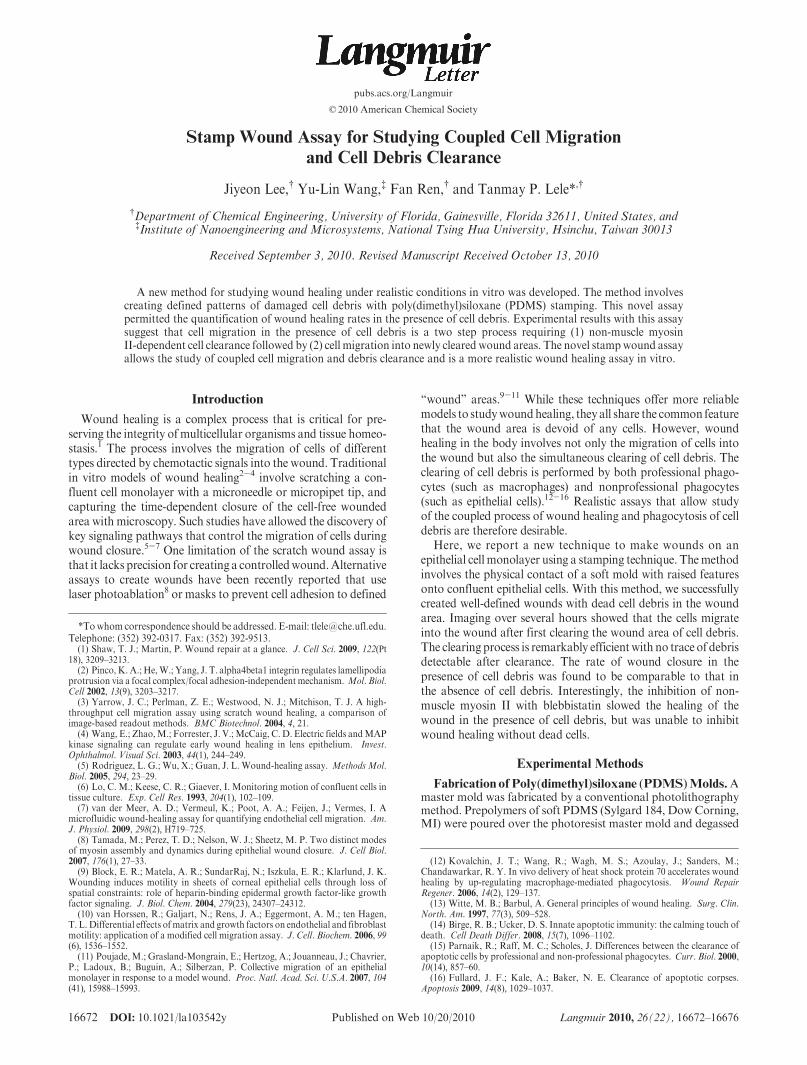
We utilized the soft imprint technology to stamp a wound on aconfluent monolayer of cultured cells. Figure 1 shows the experi-mental scheme. Briefly, PDMS prepolymers were poured on thephotoresist master mold and then cured. The raised features onthe PDMS were square in shape with a size of 250 μm. Briefimprinting of the PDMS mold on the confluent human epithelialcell monolayer caused cells to have a flattened morphology inthe stamped area (Figure 2). On tracking the wounded areasfor several hours, cells were observed to heal the wound at 15 h(Figure 2).
We next stained cells to measure viability of the stamped cells.All the cells in the stamped area were found to be dead with
Figure 1. Schematic diagram of stamp wound assay.
Figure 2. Time lapse images of stampedHet1A cells. Neighboring cells (outside of dashed line) started migrating into patterned cells (insideof dashed line) after removing the stamp. Stamping caused flattened cells (stamped wound).
(17) Lee, J.; Chu, B. H.; Chen, K. H.; Ren, F.; Lele, T. P. Randomly oriented,upright SiO2 coated nanorods for reduced adhesion of mammalian cells.Biomaterials 2009, 30(27), 4488–4493.
16674 DOI: 10.1021/la103542y Langmuir 2010, 26(22), 16672–16676
Letter Lee et al.
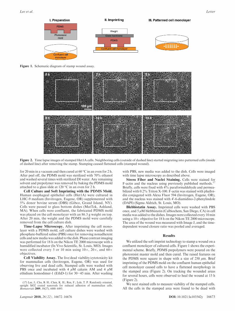
predominant nuclei, suggesting that the brief stamping causedsubstantial damage to the cell (red fluorescent nuclei inFigure 3A).Cells in the stamped area did not display significant F-actinstaining although they had prominentmisshapen nuclei (Figure 1Sin the Supporting Information). This suggests that the stampingprocess caused substantial damage to the cytoplasm and greatlydeformed the nucleus. Time lapse images further revealed that thedead cell debris (inside of dashed line in Figure 3B) was “cleared”by inwardly migrating cells. Cells were observed to clear dead celldebris first and then migrate further into the wound (Figure 3B).The clearance process was remarkably efficient with no traces ofthe debris visible on the dish (see the movie in the SupportingInformation). Overlaid images of phase contrast and fluorescentimages in Figure 3C show that cleared dead cells were engulfed byneighboring live cells.
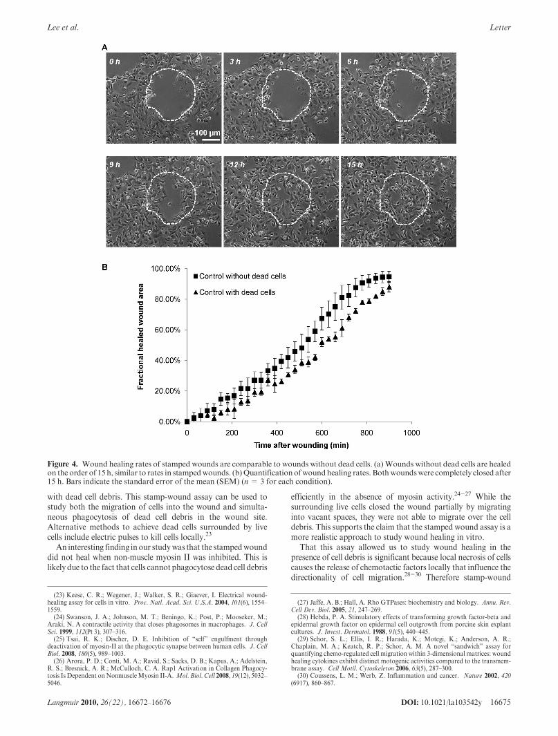
To create “empty” wounds, the PDMSmold was imprinted onthe cell monolayers, shifted laterally while maintaining contactwith the cells and then peeled off from the confluent cells. Onlateral shifting, the stamped cells were found to be removed andclean wounds were created (Figure 4A). The rate of woundclosure in wounds without dead cells was comparable to thatmeasured in the “stamped wound” with dead cells (Figure 4B).
Given that wound closure in the stamp-wound assay is accom-panied by the clearance of dead cell debris, we hypothesized thatthis process may require contractile forces generated by the
inwardly migrating cells. We therefore stamped cells and thenadded blebbistatin (5 μM) which inhibits non-muscle myosin IIspecifically.18 Upon addition, blebbistatin was not washed out toprevent possible recovery of myosin II activity during the slowhealing process. Interestingly, cells were not cleared after blebbis-tatin treatment and the wound remain unhealed even after 15 h(Figure 5). However, wound healing without dead cells was onlyslightly slowed in the presence of blebbistatin (Figure 6). Theseresults suggest a fundamental requirement for actomyosin con-tractility in wound closure in the “stamped wound” assay incontrast to the conventional wound healing assay.
Discussion
Soft imprinting technology is widely used for fabricating patternsat the nano- and micrometer scale.19 This method has been used topattern extracellular matrix proteins (such as fibronectin) on thesubstrate andconfine cell adhesionon individualprotein islands.20-22
Here, we used a similar soft imprinting method to create wounds
Figure 3. Epithelial cells migrate to the wound after first clearing dead cells. (a) Overlaid image of phase contrast and fluorescent microscopicimages (red fluorescence indicates dead cells; green fluorescence indicates live cells). (b) Phase contrast images of healing of stampedHet1A cells.Live cells canbe seen toclearneighboringdeadcells.Dashed line indicates theboundaryof stampedcells. (c)Overlaid imagesofphase contrast andfluorescent microscopic images during the healing process. White arrows mark live cells ingesting red fluorescent, dead cell debris.
(18) Russell, R. J.; Xia, S. L.; Dickinson, R. B.; Lele, T. P. Sarcomere mechanicsin capillary endothelial cells. Biophys. J. 2009, 97(6), 1578–1585.
(19) Xia, Y. N.; Whitesides, G. M. Soft lithography. Angew. Chem., Int. Ed.1998, 37(5), 551–575.
(20) Brock, A.; Chang, E.; Ho, C. C.; LeDuc, P.; Jiang, X. Y.; Whitesides,G. M.; Ingber, D. E. Geometric determinants of directional cell motility revealedusing microcontact printing. Langmuir 2003, 19(5), 1611–1617.
(21) Kane, R. S.; Takayama, S.; Ostuni, E.; Ingber, D. E.; Whitesides, G. M.Patterning proteins and cells using soft lithography. Biomaterials 1999, 20(23-24),2363–2376.
(22) Whitesides, G. M.; Ostuni, E.; Takayama, S.; Jiang, X.; Ingber, D. E. Softlithography in biology and biochemistry. Annu. Rev. Biomed. Eng. 2001, 3, 335–373.
DOI: 10.1021/la103542y 16675Langmuir 2010, 26(22), 16672–16676
Lee et al. Letter
with dead cell debris. This stamp-wound assay can be used tostudy both the migration of cells into the wound and simulta-neous phagocytosis of dead cell debris in the wound site.Alternative methods to achieve dead cells surrounded by livecells include electric pulses to kill cells locally.23
An interesting finding inour studywas that the stampedwounddid not heal when non-muscle myosin II was inhibited. This islikely due to the fact that cells cannot phagocytose dead cell debris
efficiently in the absence of myosin activity.24-27 While thesurrounding live cells closed the wound partially by migratinginto vacant spaces, they were not able to migrate over the celldebris. This supports the claim that the stamped wound assay is amore realistic approach to study wound healing in vitro.
That this assay allowed us to study wound healing in thepresence of cell debris is significant because local necrosis of cellscauses the release of chemotactic factors locally that influence thedirectionality of cell migration.28-30 Therefore stamp-wound
Figure 4. Wound healing rates of stamped wounds are comparable to wounds without dead cells. (a) Wounds without dead cells are healedon the order of 15 h, similar to rates in stampedwounds. (b)Quantificationofwoundhealing rates. Bothwoundswere completely closed after15 h. Bars indicate the standard error of the mean (SEM) (n= 3 for each condition).
(23) Keese, C. R.; Wegener, J.; Walker, S. R.; Giaever, I. Electrical wound-healing assay for cells in vitro. Proc. Natl. Acad. Sci. U.S.A. 2004, 101(6), 1554–1559.(24) Swanson, J. A.; Johnson, M. T.; Beningo, K.; Post, P.; Mooseker, M.;
Araki, N. A contractile activity that closes phagosomes in macrophages. J. CellSci. 1999, 112(Pt 3), 307–316.(25) Tsai, R. K.; Discher, D. E. Inhibition of “self” engulfment through
deactivation of myosin-II at the phagocytic synapse between human cells. J. CellBiol. 2008, 180(5), 989–1003.(26) Arora, P. D.; Conti, M. A.; Ravid, S.; Sacks, D. B.; Kapus, A.; Adelstein,
R. S.; Bresnick, A. R.; McCulloch, C. A. Rap1 Activation in Collagen Phagocy-tosis Is Dependent onNonmuscleMyosin II-A. Mol. Biol. Cell 2008, 19(12), 5032–5046.
(27) Jaffe, A. B.; Hall, A. Rho GTPases: biochemistry and biology. Annu. Rev.Cell Dev. Biol. 2005, 21, 247–269.
(28) Hebda, P. A. Stimulatory effects of transforming growth factor-beta andepidermal growth factor on epidermal cell outgrowth from porcine skin explantcultures. J. Invest. Dermatol. 1988, 91(5), 440–445.
(29) Schor, S. L.; Ellis, I. R.; Harada, K.; Motegi, K.; Anderson, A. R.;Chaplain, M. A.; Keatch, R. P.; Schor, A. M. A novel “sandwich” assay forquantifying chemo-regulated cell migration within 3-dimensional matrices: woundhealing cytokines exhibit distinct motogenic activities compared to the transmem-brane assay. Cell Motil. Cytoskeleton 2006, 63(5), 287–300.
(30) Coussens, L. M.; Werb, Z. Inflammation and cancer. Nature 2002, 420(6917), 860–867.
16676 DOI: 10.1021/la103542y Langmuir 2010, 26(22), 16672–16676
Letter Lee et al.
healing may occur not only because of the disruption of cell-celljunctions but also because of a local burst in chemotactic factors.We found that the rate of wound closure in the “stampedwound”with dead cells was comparable to that measured in the conven-tional wound without dead cells. This is remarkable given that, inthe stamped wound, the cells have to clear dead cell debris whichincludes significantly large objects (on the size scale of the cell).We speculate that chemotactic factors released in the wounded
area as a result of stamping may accelerate the migration ofsurrounding live cells into the wound, thereby resulting in only asmall decrease in wound healing rates (despite the additionalclearance step). The efficiency with which cells clear cell debris issurprising: no structures are evident on the dish surface onceclearance occurs (movie in the Supporting Information). Theclearance process itself appears similar to phagocytosis, and it isremarkable that cells continue to migrate and close the woundswith this extra payload at rates only slightly slower than woundassays without dead cells (Figure 4B). These results suggest thatthis assay could be very useful in studying phagocytosis bymacrophages in a co-culture assay with wounded epithelial cells.
Conclusions
A new method for creating realistic wounds in adherent cellmonolayers was proposed. The results suggest that cell migrationin the stamped-wound assay may involve a complex interplaybetween chemotaxis, migration due to disruption of cell-celljunctions, and myosin-dependent cell clearance. This novel assayis expected to greatly improve our understanding of the process ofwound healing.
Acknowledgment.WethankDr.AnandGupte for providingusHet1A cells. T.P.L. acknowledges support fromAHA 0735203N,NSF CMMI-0954302 and NSF CMMI-0927945.
Supporting Information Available: Fluorescent images ofF-actin and nuclei stained cells in the stamped wound. Phasecontrast time lapse movie (avi format) of wound healing inthe stampedwound assay (total time of 15 h). This material isavailable free of charge via the Internet at http://pubs.acs.org.
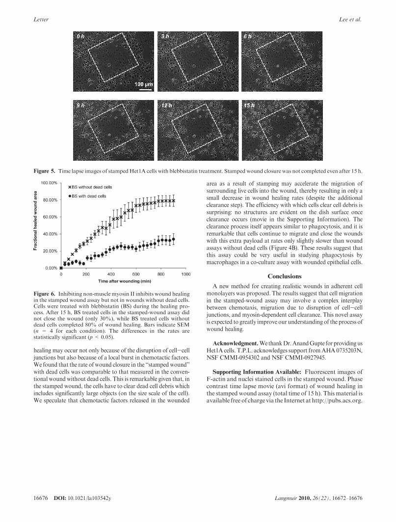
Figure 5. Time lapse images of stampedHet1A cells with blebbistatin treatment. Stampedwound closurewas not completed even after 15 h.
Figure 6. Inhibiting non-muscle myosin II inhibits wound healingin the stamped wound assay but not in wounds without dead cells.Cells were treated with blebbistatin (BS) during the healing pro-cess. After 15 h, BS treated cells in the stamped-wound assay didnot close the wound (only 30%), while BS treated cells withoutdead cells completed 80% of wound healing. Bars indicate SEM(n = 4 for each condition). The differences in the rates arestatistically significant (p< 0.05).