Embed Size (px)
Citation preview

Stabilized micelles as delivery vehicles for paclitaxel
Krassimira Yoncheva1, Patricia Calleja2, Maite Agüeros2, Petar Petrov3, Ivanka Miladinova1,
Christo Tsvetanov3, Juan M. Irache2
1 Department of Pharmaceutical Technology and Biopharmacy, Faculty of Pharmacy, 2
Dunav Str., 1000 Sofia, Bulgaria
2 Department of Pharmacy and Pharmaceutical Technology, University of Navarra, 31080
Pamplona, Spain
3 Institute of Polymers, Bulgarian Academy of Sciences, 103 Akad. G. Bonchev Str., 1113
Sofia, Bulgaria
Corresponding author:
K. Yoncheva, PhD
Dept. Pharmaceutical Technology and Biopharmaceutics
Faculty of Pharmacy
2 Dunav Str.
1000 Sofia
Bulgaria
Tel. (+359)29236527
Fax (+359)29879874
e-mail: [email protected]

2
Abstract
Paclitaxel is an antineoplastic drug used against a variety of tumors, but its low aqueous
solubility and active removal caused by P-glycoprotein in the intestinal cells hinder its oral
administration. In our study, new type of stabilized Pluronic micelles were developed and
evaluated as carriers for paclitaxel delivery via oral or intravenous route. The pre-stabilized
micelles were loaded with paclitaxel by simple solvent/evaporation technique achieving high
encapsulation efficiency of 70 %. Gastrointestinal transit of the developed micelles was
evaluated by oral administration of rhodamine-labeled micelles in rats. Our results showed
prolonged gastrointestinal residence of the marker encapsulated into micelles, compared to a
solution containing free marker. Further, the oral administration of micelles in mice showed
high area under curve of micellar paclitaxel (similar to the area of i.v. Taxol), longer mean
residence time (9-times longer than i.v. Taxol) and high distribution volume (2-fold higher
than i.v. Taxol) indicating an efficient oral absorption of paclitaxel delivered by micelles.
Intravenous administration of micelles also showed a significant improvement of
pharmacokinetic parameters of micellar paclitaxel vs. Taxol, in particular higher area under
curve (1.2-fold), 5-times longer mean residence time and lower clearance, indicating longer
systemic circulation of the micelles.
Keywords: stabilized micelles; paclitaxel; Pluronic; gastrointestinal transit; oral absorption

3
Graphical abstract
Schematic presentation describing the improved pharmacokinetic parameters achieved by
administration of paclitaxel-loaded stabilized Pluronic micelles (PTX-PM). For comparison,
the same parameters calculated after administration of paclitaxel in the form of commercial
solution (Taxol).

4
1. Introduction
Polymeric micelles present a great potential as drug delivery systems for compounds that
are hydrophobic and exhibit poor bioavailability. The unique core-shell structure of micelles
enables incorporation of poorly soluble drugs into the inner hydrophobic core thus improving
their stability and bioavailability. The hydrophilic shell plays an important role for the in vivo
behavior of the micelles, in particular their steric stabilization and ability to interact with the
cells. According to some references polymeric micelles could facilitate drug transport by fluid
phase endocytosis as well as to provide receptor mediated transport by functionalization of
the shell (Torchilin, 2004; Mathot et al., 2007).
The micelles of poly(ethylene oxide)-b-poly-(propylene oxide)-b-poly(ethylene oxide)
(PEO–PPO–PEO) amphiphilic triblock copolymers are evaluated as drug and gene delivery
systems, as well as in diagnostic imaging as carriers for various contrasting agents (Kabanov
et al., 2002). PEO–PPO–PEO micelles can be prepared from commercially available
copolymers (Pluronics) and combine several attractive properties, including resistance of the
PEO shell to protein adsorption, ability of the temperature responsive PPO core to solubilize
water-insoluble compounds and availability of hydroxyl groups to which receptor-specific
ligands can be attached (Alexandridis and Hatton, 1995). Since the formation of PEO–PPO–
PEO micelles is a dynamic phenomenon, one possible drawback may be associated with
micellar disaggregation upon dilution at physiological conditions. Rapoport and co-workers
proposed different strategies for stabilization of PEO–PPO–PEO micelles, in particular
radical cross-linking of micelle core using benzoyl peroxide as radical initiator, introduction
of vegetable oil aiming to increase hydrophobic interactions within the core, or formation of
hydrogel inside the micelle core (Plurogel®) (Rapoport, 1999). More recently, an efficient
method for the stabilization of PEO-PPO-PEO micelles by UV-induced formation of an
interpenetrating network of poly(pentaerythritol tetraacrylate), in which the polyether chains
5
were physically entrapped, has been reported (Petrov et al., 2006). The stabilized polymeric
micelles (SPM) resisted changes in concentration and solvent and maintained their structure
and size even when ultrasound irradiated at 20 kHz.
Paclitaxel (PTX) is an antineoplastic drug successfully used against a variety of tumors
including ovarian, breast and non-small cell lung tumors (Gregory and DeLisa, 1993; Chen et
al., 2003). In clinics, paclitaxel is intravenously administered as infusion (i.e. Taxol®). Due
to its limited aqueous solubility (0.3 μg/ml), the commercial injectable formulations use a
mixture of Cremophor EL and ethanol (1:1 v/v) (Singla et al., 2002). Apart from other
problems, the presence of Cremophor EL results in hypersensitivity reactions, nephrotoxicity,
and neurotoxicity (Onetto et al., 1993). In addition, this pharmaceutical excipient can also
modify the pharmacokinetics of PTX (Sparreboom et al., 1999). To overcome these problems,
several alternative pharmaceutical carriers have been developed for PTX delivery including
liposomes (Wang et al., 2010; Zhao et al., 2011), polymeric micelles (Hamaguchi et al., 2005;
Forrest at el., 2008) and nanoparticles (Koziara et al., 2006; Win and Feng, 2006). In the last
years, different paclitaxel formulations have been clinically studied such as Opaxio®
(formerly Xyotax®) and Abraxane®. The former is a polymer conjugate between paclitaxel
and poly(glutamate) (Boddy et al., 2005) whereas the latter, which was recently
commercialised, is based on the incorporation of paclitaxel in human serum albumin
nanoparticles (Roy et al., 2009).
On the other hand, a number of different attempts have been performed to develop oral
formulations of paclitaxel. Nevertheless, apart from the low aqueous solubility, oral treatment
with paclitaxel is hampered by the fact that the drug is substrate of the P-glycoprotein and
cytocrome P-450 (Letchford et al., 2009). As a result the oral bioavailability of paclitaxel is
extremely low (Malingre et al., 2001). In the last years, different approaches have been
proposed to solve this problem including the co-administration of pharmaceutical excipients

6
with capability to disturb the effect of intestinal P-gp and/or cytocrome P450. In this context,
the combination between cyclodextrins and poly(anhydride) nanoparticles yielded oral
bioavailability up to 85 % in rat. In this approach, the cyclodextrin permits to promote the
encapsulation of the lipophilic drug into the nanoparticles and to inhibit the activity of the P-
gp, whereas the function of poly(anhydride) nanoparticles is to develop bioadhesive
interactions within the gastrointestinal mucosa and control the release of their content
(Agüeros et al., 2010).
The aim of this work was to prepare, characterize and evaluate the pharmacokinetics of
paclitaxel incorporated in stabilized Pluronic micelles. The stabilization of micelles by cross-
linking of their core was performed aiming to prevent disaggregation of micelles upon
dilution in physiological fluids. On the other hand, drug encapsulation in micelles can
diminish drug extravasation to normal tissues and provide a passive drug targeting to tumors
via the enhanced permeability and retention effect (EPR) (Maeda et al., 2000). This increased
permeability is favorised by the prolonged circulation of micelles and lack of renal clearance
(Han et al., 2006; Batrakova et al., 2008). On the other hand, Pluronic copolymers (as other
pharmaceutical excipients) may inhibit the activity of drug efflux transporters such as P-gp,
MRPs and BCRP (Kabanov et al., 2002; Thomas et al., 2003; Huang et al., 2008). Thus, the
combination of these properties with the fact that the shell of polymeric micelles could
establish interactions with the cells may be an adequate strategy to (i) increase the oral
bioavailability and/or (ii) promote the efficacy of the loaded drug.
2. Materials and methods
2.1. Materials
PEO43PPO15PEO43 (Pluronic F-38; MW 4700 gmol-1) were kindly donated by BASF
Pentaerythritol tetraacrylate (PETA) and rhodamine B were purchased from Aldrich.

7
Paclitaxel (USP 26 grade >99.5%) was supplied by 21CEC (London, United Kingdom).
Docetaxel (Taxotere) was provided by Sanofi-Aventis (Paris, France). Taxol® was
purchased from Bristol-Myers Squibb (New York, USA). All other solvents and reagents
were of analytical grade.
2.2. Preparation of stabilized Pluronic micelles (PM)
PEO43PPO15PEO43 (5 g) was dissolved in 250 ml double distilled water in a glass vessel
under stirring. Acetone solution of PETA (0.05g/ ml) was added and the temperature of the
solution was increased to 60 oC. Argon was bubbled through the solution for 45 min, followed
by irradiation with a full spectrum UV light (TQ 150 Original Hanau high-pressure 150 W
mercury lamp provided with a quartz tube and a cooling quartz jacket) for 45 min. The
stabilized polymeric micelles were purified by dialysis against water using a cellulose
membrane (Sigma, cutoff 12,000 gmol-1) for 14 days. Finally, the micelles dispersion was
frozen and subsequently lyophilized.
2.3. Loading of pre-stabilized Pluronic micelles with rhodamine (R-PM)
The pre-stabilized micelles were fluorescently labelled with rhodamine B by solvent
evaporation technique. For this purpose, 10 ml ethanol solution of rhodamine (concentration
0.1 mg/ml, 0.15 mg/ml or 0.3 mg/ml) was added to 10 ml of aqueous dispersion of the
stabilized micelles (3 mg/ml) and the compounds were incubated for 24h. The ethanol was
evaporated under reduced pressure (Buchi-144, Switzerland) and dialysis of the aqueous
dispersion towards distilled water (Sigma, cutoff 12,000 g mol-1) was performed in order to
eliminate the free rhodamine. Finally, the purified aqueous dispersion containing rhodamine
loaded micelles was lyophilized (Genesis 12EL, Virtis, USA) using sucrose as cryoprotector
(5 % w/v).

8
2.4. Loading of pre-stabilized Pluronic micelles with paclitaxel (PTX-PM)
Ten ml of ethanol solution of paclitaxel (concentration 0.1 mg/ml, 0.15 mg/ml or 0.3
mg/ml) was added to 10 ml of aqueous dispersion of the pre-stabilized micelles (3 mg/ml) and
the compounds were incubated for 24h. The ethanol was evaporated under reduced pressure
(Buchi-144, Switzerland) and the resulted micelle dispersion was filtered (0.22 μm). The filter
was rinsed with ethanol and these fractions were determined for non-encapsulated paclitaxel.
The aqueous dispersion containing paclitaxel loaded micelles was lyophilized (Genesis 12EL,
Virtis, USA) using sucrose as cryoprotector (5 % w/v).
2.5. Characterisation of the micelles
Size and zeta-potential were determined by photon correlation spectroscopy and
electrophoretic laser doppler anemometry using a Zetamaster analyzer (Malvern Instruments,
UK). Samples were dispersed in either distilled water or saline (0.154 M and 0.308 M NaCl
solution) and measured at 25oC with a scattering angle of 90o.
2.6. Determination of drug loading
The amount of rhodamine incorporated into the R-PM was determined by
spectrofluorimetry at wavelength λex =540nm and λem =580nm (GENios, Austria). The marker
loading was calculated as a difference between its initial concentration and the concentration
found in the aqueous fractions collected after the dialysis procedure. For the calculations
standard curve of rhodamine B in distilled water was prepared in the concentration range of
0.2-4.0 μg/ml (r>0.9978).
The amount of paclitaxel incorporated into the PTX-PM was calculated as a difference
between its initial concentration and the concentration found in the ethanol rinsing fractions.
Paclitaxel was determined by HPLC method, in particular the chromatographic system was an

9
Agillent 1100 series (Waldbornn, Germany), coupled with a UV diode array detection system
(25). Data were analyzed using the Chemstation G2171 program (B.01.03). The separation of
PTX was carried out at 30ºC on a reversed-phase 150 x 3 mm C18 Phenomenex Gemini
column (particle size 5 m). The mobile phase, pumped at 0.5 ml/min, was 50:50 acetonitrile
- phosphate buffer (0.02 M, pH=2.0) and effluent was monitored with UV detection at 228
nm. For the calculations standard curve of paclitaxel was prepared in the concentration range
of 1.25-80.0 μg/ml (r>0.9991). The limit of quantification was calculated as 40 ng/ml with a
relative standard deviation of 5.2 %.
The equations used for calculation of theoretical drug loading, actual drug loading and
encapsulation efficiency are given bellow:
2.7. Distribution of R-PM within the gut
The study was performed in compliance with the regulations of the responsible committee
of the University of Navarra in line with the European legislation on animal experiments
(86/609/EU). Rhodamine B labelled micelles were administered in the form of aqueous
suspensions (10 mg/ml) to fasted Wistar rats orally. The animals were sacrificed by cervical
dislocation at 1, 3 and 8 h post administration. The abdominal cavity was opened and GIT
was removed and divided into three regions: stomach, small intestine and caecum. Each
segment was opened lengthwise along the mesentery and rinsed with phosphate saline buffer
Theoretical drug loading (%) = Weights of drug and stabilized micelles
Weight of drug initially added X 100
Actual drug loading (%) = Weight of drug loaded micelles
Weight of drug incorporated into micelles X 100
Encapsulation efficiency (%) = Theoretical drug loading
Actual drug loading X 100

10
(pH 7.4) in order to eliminate the lumen contents. Further, each segment was cut into portions
of 2 cm length and digested in 1 ml of 3N NaOH for 24 h (Arbos et al., 2002). Rhodamine
was extracted with 2 ml methanol, vortexed for 1 min and centrifuged at 4000 rpm for 10
min. Aliquots (1 ml) of the supernatants were assayed for rhodamine by spectrofluorimetry
(GENios, Austria) to estimate the fraction of the micelles within gastrointestinal mucosa.
Calculations were made using standard curves of rhodamine prepared by addition of
rhodamine solutions in 3N NaOH (0.5-10 g/ml) to tissue segments following the same
treatment steps.
Control rhodamine solution (119 μg/ml) was administered to rats and the presence of
rhodamine within gastrointestinal mucosa was determined following the same procedures as
above.
2.8. Pharmacokinetic study in mice
Animal experiments were performed in compliance with regulations of the responsible
Ethical Committee of the University of Navarra (protocol number 076-06) in strict accordance
with the European legislation in animal experiments. Female C57BL/6J mice (average weight
20 g) (Harlan, Spain) were housed under normal conditions with free access to food and
water. The animals were placed in metabolic cages and fasted overnight to prevent
corpophagia but allowing free access to water.
For the pharmacokinetic study, the mice were divided at random into 4 groups (n=5). The
first two groups received orally a single dose (10 mg paclitaxel/kg) of either (i) commercial
Taxol or (ii) paclitaxel-loaded micelles (PTX-PM). Besides, Taxol and PTX-PM were also
intravenously administered to the other two groups of mice at the same dose (10 mg/kg).
All the formulations were administered dispersed or dissolved in 1 ml of water (oral) or
saline (intravenous). Blood samples of 200 l were collected at different times in tubes

11
containing EDTA (Microvette® EDTA tripotassium salt; Sarstedt, Numbrecht, Germany).
The volemia was recovered via intraperitoneal injection of an equal volume of normal saline
solution preheated at body temperature. Blood samples were centrifuged for 10 min at 10,000
rpm and the supernatant plasma fractions were stored at -80ºC until HPLC analysis.
2.9. HPLC quantification of paclitaxel in plasma samples
The amount of paclitaxel was determined in plasma by HPLC as described above.
Calibration curves were used for the conversion of the PTX/DCX chromatographic area to the
concentration. Calibrator and quality control samples were prepared by adding appropriate
volumes of standard PTX ethanol solution to drug free plasma. Calibration curves were
designed over the range of 40–3200 ng/ml (r2>0,999). An aliquot (100 l) of plasma sample
was mixed with 25 l of internal standard solution (docetaxel, 4 g/ml in methanol,
previously evaporated). After vortex mixing, liquid-liquid extraction was accomplished by
adding 4 ml of tert-buthylmethylether following vortex gentle agitation (1 min). The mixture
was centrifuged for 10 min at 5000 rpm, the organic layer was transferred to a clean vial and
evaporated until dry (Savant, Barcelona, Spain). Finally, the residue was dissolved in 125 l
of reconstitution solution (acetonitrile/0,01M phosphate buffer, 50/50 v/v, pH=2.0) and
transferred to auto-sampler vials, capped and placed in the HPLC auto sampler. A 100 l
aliquot of each sample was injected onto the HPLC column.
Under these experimental conditions the run time was 14 min. The limit of quantification
was calculated as 80 ng/ml with a relative standard deviation of 5.2 %. Accuracy values
during the same day (intra-day assay) at low, medium and high concentrations of PTX was
always within the acceptable limits (-1.81 and 3.49 %) at all concentrations tested.
2.9. Pharmacokinetic data analysis

12
The pharmacokinetic analysis of concentration-time data, obtained after the administration
of the different PTX formulations, were analyzed using a noncompartimental model using
Phoenix WinNonlin 6.0 software (Pharsight Corporation, Mountain View, EEUU). The
pharmacokinetic parameters estimated were: the peak of maximun concentration (Cmax), time
to peak concentration (Tmax), the half-life of the terminal phase (t1/2), the area under the
concentration-time curve from time 0 to 8 or 24 hours (AUC0-8, AUC0-24), the mean residence
time (MRT) and the total body clearance (Cl). The mean residence time (MRT) was
calculated as the AUMC (area under the concentration-time curve at the first moment) divided
by AUC. The absolute availability (F) was calculated according to:
F= AUCoral / AUCi.v.
where AUCoral and AUCi.v. were the area under the concentration-time curve after the oral and
i.v. administration, respectively.
2.10. Statistical analysis
The results were expressed as mean values SD. The Mann-Withney U-test was used to
investigate statistical differences. In all cases, p < 0.05 was considered to be significant. All
calculations were performed using SPSS® statistical software program (SPSS® 14, Microsoft,
USA).
3. Results
3.1. Characterization of the micelles
The main physico-chemical properties of the micelles are presented in Table 1. The
resulted stabilized micelles have an average diameter less than 200 nm, narrow distribution
and negative zeta-potential values. As shown, drug-loaded micelles did not differ in size

13
compared to the non-loaded micelles. The latter is due to the fact that the dense poly-PETA
network locks the micellar structure and does not allow any re-arrangement and expansion.
Physical stability of the stabilized micelles was evaluated by observation of their size and
polydispersity with time. The size was measured in aqueous media with increasing salt
concentration aiming to investigate the influence of the ionic strength. The ratio between the
size observed after 24 hours in different media (aqueous dispersion, 0.154 M and 0.308 M
NaCl solution) to initial size measured in distilled water was calculated (Figure 1). Since the
size ratios were in a narrow range, the micelles were considered stable.
In order to evaluate the influence of the initial drug/stabilized micelles ratio on drug
loading degree, different concentrations of rhodamine/ paclitaxel with respect to the micelle
concentration were varied. The results demonstrated that the increase of the initial
concentration from 1 to 1.5mg increased loading degree for both molecules (Figure 2).
However, further increase of initial paclitaxel concentration decreased encapsulation
efficiency suggesting that the optimal ratio between initial paclitaxel and stabilized micelles
was 1:20.
3.2. Gastrointestinal transit of the stabilized micelles
The study was performed using the stabilized micelles loaded with rhodamine (R-PM) as
described above (an initial ratio 1:20). The study included oral administration of micelles and
evaluation of the localization of rhodamine in the gastrointestinal mucosa. In parallel, control
solution of rhodamine with a concentration similar to the rhodamine concentration into the
micelles was administered. The results showed that the total amount of rhodamine within the
gastrointestinal mucosa decreased with time (Figure 3). In fact, one hour post administration
about 40 % of the given dose was found in the mucosa (mainly in the stomach) whereas, 2
hours later, only 21 % of the rhodamine dose remained within the mucosa. The rhodamine

14
dose remained 8 h post-administration (18 %) was similar to that found 3 h post-
administration, which clearly demonstrated the prolonged transit of micellar rhodamine. In
any case, these percentages were significantly higher (p<0.05) than those observed for the
control solution of rhodamine (Figure 4).
3.3. Pharmacokinetic study of paclitaxel when loaded in stabilized Pluronic micelles
For the pharmacokinetic study, Taxol and PTX-PM were intravenously or orally
administered to mice. Figure 5 illustrates plasma profiles of PTX after i.v. administration of
PTX-loaded micelles or Taxol to mice. As shown, plasma concentrations of both
formulations (micelles and commercial) were similar during the first hour post-
administration. Then, plasma curve of PTX-PM was characterised by a plateau in the
concentration during at least 2 hours followed by a low decrease with time. On the contrary,
for Taxol®, the plasma curve was characterised by a rapid decline in the paclitaxel plasma
levels during the first 2 h after administration followed by a slow fall of drug concentrations
till the end of the experiment (8 hours). Under these circumstances, the micelles provided
higher AUC (1.2-fold) compared to Taxol (Table 2). Further, when commercial Taxol was
administered to mice by the oral route, no PTX plasma levels were found (Figure 6). On the
contrary, when PTX was included into micelles, these formulations displayed drug levels in
plasma for at least 8 h. The plasma curve was characterised by a high Cmax 30 min post-
administration, only 3-times lower than that obtained with the same formulation i.v.
administered (Table 2), followed by a slow and almost constant decline during the following
8 hours. The oral bioavailability of paclitaxel when loaded into the stabilized Pluronic
micelles was calculated to be about 0.9.
4. Discussion

15
Dynamic core-shell micelles of PEO43PPO15PEO43 copolymer were stabilized by UV –
induced crosslinking of tetrafunctional acrylate, PETA, within the micelles according to
procedure described elsewhere (Petrov et al., 2008). Pentaerythritol tetraacrylate was applied
as cross-linking agent because of its ability to form an interpenetrating network in which the
polyether chains are physically entrapped. Previous studies have shown that the micelles
obtained with large amount of PETA (10–50 mass% with respect to PPO fraction) are
extremely stable and do not disaggregate upon dilution in water and organic solvents and
maintain their integrity even when irradiated with ultrasound (Petrov et al., 2006). In the case
of aggregates stabilized with smaller amount of PETA (3 mass %), copolymer molecules can
dissociate from the polymer network resulting in aggregates dissociation (Li et al., 2007).
Thus, one may play with the long-term stability of the micelles by tuning the density of poly-
PETA network. Taking into account the above considerations, the cross-linking agent was
used in a concentration of 30 % with respect to PPO fraction. This network does not decrease
noticeably the loading capacity of PPO core and, on the other hand, does not influence the
flexibility of extended hydrophilic PEO-chains of resulted stabilized micelles. The latter is
very important taking into account that the mobility of hydrophilic chains on the periphery of
drug carriers determines both the stability and the in vivo distribution of the particles. In this
view, our observations showed that the size and polydispersity of the stabilized Pluronic
micelles did not change even in the presence of electrolyte. This fact suggested that the PEO
chains are extended and prevent any agglomeration of the micelles in the presence of
electrolytes.
Two methods have been reported for loading of polymeric micelles with drug molecules,
in particular physical and chemical loading (Jones and Leroux, 1999). The second method
includes formation of chemical bonds between drug molecule and polymeric carrier, usually
during the process of micelles formation. In the present study the first method was applied.

16
The pre-stabilized micelles were loaded with fluorescent marker rhodamine B or with
paclitaxel by solvent evaporation technique. Comparing both molecules, higher loading and
encapsulation efficiency was achieved with paclitaxel. The latter fact was explained with the
more hydrophobic nature of paclitaxel (logP of 3.5) than rhodamine (logP of 1.95).
Gastrointestinal transit of the stabilized micelles was evaluated by oral administration to
rats. The most important finding was that the transit of the rhodamine-loaded micelles was
prolonged compared to the control solution of free rhodamine. Thus, 8 hours after
administration only 6 % of rhodamine applied in the form of solution was spread on the whole
GIT versus 18 % for stabilized micelles (Figure 4). Another interesting point was that the
micelles were concentrated in the small intestine of animals after oral administration (I3 and
I4 portions in Figure 3). All of these results appear to be in line with previous studies about
the interaction of micelles with the intestinal mucosa (Gaucher et al., 2010). In fact, the size
and hydrophilicity of the shell may limit their interaction with the gut contents and would
favour the diffusion of these carriers through the mucus layer. Similar phenomenon has been
previously reported for pegylated poly(anhydride) nanoparticles (Yoncheva et al., 2007). The
authors postulated that the “brush” layer of PEG-chains reduced an interaction between
nanoparticles and mucin, which corresponded to better adhesion and facilitated their proximal
contact with enterocytes. In the present study, the extended flexible PEO-chains in the micelle
shell probably function in a similar way.
Intravenous administration of Taxol and PTX-PM into mice showed a significant
improvement of pharmacokinetic parameters after administration of micellar paclitaxel (Table
2). More important, the higher MRT (about 5-times) and lower Cl values (about 2-fold)
indicated that PTX-PM formulation increased the time of systemic circulation. This fact could
be explained with the protective “stealth” properties of micelle PEO-shell that hinders their
recognition and uptake by the cells of RES. These results are in agreement with previous data

17
published by Kabanov and collaborators (Kabanov et al., 2002). This manuscript studied the
pharmacokinetics and biodistribution of Pluronics after single i.v. administration and reported
that these block copolymers remained in circulation a substantial period of time. The results
obtained after oral administration of our stabilized micelles were even more interesting. The
AUC calculated after oral administration of micelles was similar to that calculated for the i.v.
administration of Taxol (Table 2). On the other hand, the mean residence time of paclitaxel
after oral administration of PTX-PM was almost two-times longer than that achieved after i.v.
administration of micelles and 9-times longer than Taxol. These data together with the low
clearance and high distribution volume (2-fold higher than i.v. Taxol) indicated efficient oral
absorption of PTX. In our opinion, the efficient oral absorption of PTX is associated with the
properties of the stabilized micelles. Based on the results from the biodistribution studies with
rhodamine loaded micelles (R-PM) it could be considered that the flexible PEO-chains in
micelles shell enable micelle penetration through the mucus layer, facilitating their transport
to the absorptive surface of the enterocytes. On the other hand, it has been described that
Pluronics and other non-ionic surfactants inhibit the activity of intestinal Pgp (Batrakova et
al., 1998a; 1998b). This fact has been explained by a combination of decreased ATPase
activity and ATP depletion due to increased membrane fluidity (Batrakova et al., 2004;
Bansal et al., 2009). Nonetheless, studies performed on various cell models have shown that
polymeric micelles could be internalized, with fluid-phase pinocytosis appearing as a major
route (Allen et al., 1999; Luo et al., 2002). More recently, Mathot et al. have found that
micelles obtained from monomethylether poly(ethyleneglycol)(750)-poly(caprolactone-co-
trimethylene carbonate) can cross lipid bilayers via passive diffusion and demonstrate an oral
bioavailability of 40 % in rats (Mathot et al., 2007). Taking into account these reports, the
results in the present study may be explained by an increased absorption of paclitaxel when

18
loaded into the newly developed stabilized micelles, an absorption or translocation of micelles
through the gastrointestinal mucosa or a combination of both phenomena.
In summary, the present study demonstrated the development of new, in particular
stabilized micelles, as paclitaxel delivery system. The extended PEO-chains in the micelle
shell probably enabled interactions between epithelial cells of gastrointestinal mucosa and
micelles, which in turn prolonged gastric residence time of paclitaxel. In vivo study showed
an improvement of pharmacokinetic parameters after intravenous and oral administration of
PTX-loaded stabilized Pluronic micelles. Both, the prolonged gastric residence and the high
physical stability of micelles achieved by cross-linking of micelle core, have been considered
prerequisite for the efficient oral paclitaxel absorption.
Acknowledgements
This work was supported by the Ministry of Science and Innovation (project SAF2008-
02538), Grant PFIS (2008, Instituto de Salud Carlos III) and Foundation “Caja Navarra: Tú
eliges, tú decides” (Nanotecnología y Medicamentos, number 10828) in Spain. P.P.
acknowledges the financial support by the National Science Fund of Bulgaria (National
Centre for New Materials UNION, contract no. DCVP-02/2).
References
Agüeros, M., Zabaleta, V., Espuelas, S., Campanero, M.A., Irache, J.M., 2010. Increased oral
bioavailability of paclitaxel by its encapsulation through complex formation with
cyclodextrins in poly(anhydride) nanoparticles. J. Control. Release 145, 2–8.
Alexandridis, P., Hatton, T.A., 1995. Poly(ethylene oxide)-poly(propylene oxide)-
poly(ethylene oxide) block copolymer surfactants in aqueous solutions and at interfaces:

19
thermodynamics, structure, dynamics, and modeling. Colloids Surf. A: Physicochem. Eng.
Aspects 96, 1–46.
Allen, C., Yu, Y., Eisenberg, A., Maysinger, D., 1999. Cellular internalization of PCL(20)-b-
PEO(44) block copolymer micelles. Biochim. Biophys. Acta 1421, 32-38.
Arbos, P., Arangoa, M.A., Campanero, M.A., Irache, J.M., 2002. Quantification of the
bioadhesive properties of protein-coated PVM/MA nanoparticles. Int. J. Pharm. 242, 129-136.
Bansal, T., Akhtar, N., Jaggi, M., Khar, R.K., Talegaonkar, S., 2009. Novel formulation
approaches for optimising delivery of anticancer drugs based on P-glycoprotein modulation.
Drug Discovery Today 14, 1067-1074.
Batrakova, E.V., Han, H.Y., Alakhov, V., Miller, D.W., Kabanov, A.V., 1998a. Effects of
pluronic block copolymers on drug absorption in Caco-2 cell monolayers. Pharm. Res. 15,
850-855.
Batrakova, E.V., Han, H.Y., Miller, D.W., Kabanov, A.V., 1998b. Effects of pluronic P85
unimers and micelles on drug permeability in polarized BBMEC and Caco-2 cells. Pharm.
Res. 15, 1525-1532.
Batrakova, E.V., Li, S., Li, Y., Alakhov, V.Y., Kabanov, A.V., 2004. Effect of pluronic P85
on ATPase activity of drug efflux transporters. Pharm. Res. 21, 2226-2233.
Batrakova, E.V., Kabanov, A.V., 2008. Pluronic block copolymers: evolution of drug
delivery concept from inert nanocarriers to biological response modifiers. J. Control. Release
130, 98-106.
Boddy, A.V., Plummer, E.R., Todd, R., Sludden, J., Griffin, M., Robson, L., Cassidy, J.,
Bissett, D., Bernareggi, A., Verrill, M., Calvert, A., 2005. A Phase I and pharmacokinetic
study of paclitaxel poliglumex (Xyotax), investigating both 3-weekly and 2-weekly schedules.
Clin. Cancer Res. 11, 7834-7840.

20
Chen, Q., Zhang, Q.Z., Liu, J., Li, L.Q., Zhao, W.H., Wang, Y.J., Zhou, Q.H., Li, L., 2003.
Multi-center prospective randomized trial on paclitaxel liposome and traditional taxol in the
treatment of breast cancer and non-small-cell lung cancer. Chin. J. Oncol. 25, 190–192.
Forrest, M.L., Yanez, J.A., Remsber, C.M., Ohgami, Y., Kwon, G.S., Davies, N.M., 2008.
Paclitaxel prodrugs with sustained release and high solubility in poly(ethylene glycol-b-
poly(ε-caprolactone) micelle nanocarriers: pharmacokinetic disposition, tolerability, and
cytotoxicity. Pharm. Res. 25, 194-206.
Gaucher, G., Satturwar, P., Jones, M.C., Furtos, A., Leroux, J.C., 2010. Polymeric micelles
for oral drug delivery. Eur. J. Pharm. Biopharm. 76, 147-158.
Gregory, R.E., DeLisa, A.F., 1993. Paclitaxel: a new antineoplastic agent for refractory
ovarian cancer. Clin. Pharm. 12, 401–415.
Hamaguchi, T., Matsumura, Y., Suzuki, M., Shimizu, K., Goda, R., Nakamura, I., Nakatomi,
I., Yokoyama, M., Kataoka, K., Kakizoe, T., 2005. NK105, a paclitaxel-incorporating
micellar nanoparticle formulation, can extend in vivo antitumour activity and reduce the
neurotoxicity of paclitaxel. Br. J. Cancer 92, 1240-1246.
Han, L.M., Guo, J., Zhang, L.J., Wang, Q.S., Fang, X.L., 2006. Pharmacokinetics and
biodistribution of polymeric micelles of paclitaxel with Pluronic P123. Acta Pharmacol. Sin.
27, 747-753.
Huang, J., Si, L., Jiang, L., Fan, Z., Qiu, J., Li, G., 2008. Effect of pluronic F68 block
copolymer on P-glycoprotein transport and CYP3A4 metabolism. Int. J. Pharm. 356, 351-353.
Jones, M.C., Leroux, J.C., 1999. Polymeric micelles – a new generation of colloidal drug
carriers. Eur. J. Pharm. Biopharm. 48, 101-111.
Kabanov, A.V., Batrakova, E.V., Alakhov, V.Y., 2002. Pluronic block copolymers for
overcoming drug resistance in cancer. Adv. Drug Deliv. Rev. 54, 759-779.

21
Kabanov, A.V., Batrakova, E.V., Alakhov, V.J., 2002. Pluronic block copolymers as novel
polymer therapeutics for drug and gene delivery. J. Control. Release 82, 189-212.
Koziara, J., Whisman, T., Tseng, M., Mumper, R.J., 2006. In-vivo efficacy of novel paclitaxel
nanoparticles in paclitaxel-resistant human colorectal tumors. J. Control. Release 112, 312-
319.
Letchford, K., Liggins, R., Wasan, K.M., Burt, H., 2009. In vitro human plasma properties of
poly(ethylene glycol)-block-poly(caprolactone) nanoparticles. Eur. J. Pharm. Biopharm. 71,
196–206.
Li, F., Ketelaar, T., Marcelis, A.T.M., Leermakers, F.A.M., Cohen Stuart, M,A,, Sudhölter,
E.J.R., 2007. Stabilization of polymersome vesicles by an interpenetrating polymer network.
Macromolecules 40, 329–333.
Luo, L., Tam, J., Maysinger, D., Eisenberg, A., 2002. Cellular internalization of poly(ethylene
oxide)-b-poly(epsilon-caprolactone) diblock copolymer micelles. Bioconjug. Chem. 13, 1259-
1265.
Maeda, H., Wu, J., Sawa, T., Matsumura, Y., Hori, K., 2000. Tumor vascular permeability
and the EPR effect in macromolecular therapeutics: a review. J. Control. Release 65, 271-284.
Malingre, M.M., Beijnen, J.H., Schellens, J.H., 2001. Oral delivery of taxanes. Invest. New
Drugs 19, 155–162.
Mathot, F., des Rieux, A., Ariën, A., Schneider, Y.J., Brewster, M., Préat, V., 2007. Transport
mechanism of mmePEG750P(CL-co-TMC) polymeric micelles across the intestinal barrier. J.
Control. Release 124, 134-43.
Onetto, N., Canetta, R., Winograd, B., Catane, R., Dougan, M., Grechko, J., Burroughs, J.,
Rozencweig, M., 1993. Overview of Taxol safety. J Natl. Cancer Inst. Monogr. 15, 131–39.
Petrov, P., Bozukov, M., Burkhardt, M., Muthukrishnan, S., Mueller, A.H.E., Tsvetanov, Ch.,
2006. Stabilization of polymeric micelles with a mixed poly(ethylene oxide)/poly(2-

22
hydroxyethyl methacrylate) shell by formation of poly(pentaerythritol tetraacrylate)
nanonetworks within the micelles. J. Mater. Chem. 16, 2192–2199.
Petrov, P., Jiayin, Y., Yoncheva, K., Müller, A., Tsvetanov, Ch., 2008. Wormlike morphology
formation and stabilization of “Pluronic P123” micelles by solubilization of pentaerythritol
tetraacrylate. J. Phys. Chem. B 112, 8879–8883.
Rapoport, N., 1999. Stabilization and activation of Pluronic micelles for tumor-targeted drug
delivery. Colloids Surf. B: Biointerfaces 16, 93-111.
Roy, V., LaPlant, B.R., Gross, G.G., Bane, C.L., Palmieri, F.M., 2009. Phase II trial of
weekly nab (nanoparticle albumin-bound)-paclitaxel (nab-paclitaxel) (Abraxane) in
combination with gemcitabine in patients with metastatic breast cancer (N0531). Ann. Oncol.
20, 449–453.
Singla, A.K., Garg, A., Aggarwal, D., 2002. Paclitaxel and its formulations. Int. J. Pharm.
235, 179–192.
Sparreboom, A., van Zuylen, L., Brouwer, E., Loos, W., de Bruijn, P., Gelderblom, H., Pillay,
M., Nooter, K., Stoter, G., Verweij, J., 1999. Cremophor EL-mediated alteration of paclitaxel
distribution inhumanblood: clinical pharmacokinetic implications. Cancer Res. 59, 1454–
1457.
Thomas, H., Coley, H.M., 2003. Overcoming multidrug resistance in cancer: an update on the
clinical strategy of inhibiting p-glycoprotein. Cancer Control 10, 159-165.
Torchilin, V.P., 2004. Targeted polymeric micelles for delivery of poorly soluble drugs. Cell
Mol. Life Sci. 61, 2549-2559.
Wang, X., Zheng, H., Zhu, Z., Wei, Y., Chen, L., 2010. Clinical pharmacokinetics of
paclitaxel liposome with a new route of administration in human based on the analysis with
ultra performance liquid chromatography. J. Pharm. Sci. 99, 4746-4752.
23
Win, K.Y., Feng, S.S., 2006. In vitro and in vivo studies on vitamin E TPGS-emulsified
poly(D,L-lactic-co-glycolic acid) nanoparticles for paclitaxel formulation. Biomaterials 27,
2285-2291.
Yoncheva, K., Guembe, L., Campanero, M.A., Irache, J.M., 2007. Evaluation of bioadhesive
potential and intestinal transport of pegylated poly(anhydride) nanoparticles. Int. J. Pharm.
334, 156-165.
Zhao, L., Ye, Y., Li, J., Wei, Y.M., 2011. Preparation and the in vivo evaluation of paclitaxel
liposomes for lung targeting delivery in dogs. J. Pharm. Pharmacol. 63, 80-86.

24
Table 1. Physico-chemical properties of non-loaded stabilized Pluronic micelles (PM), rhodamine-loaded (R-PM) and paclitaxel-loaded micelles (PTX-PM). Data expressed as the mean ± SD, n=3.
Sample
Size, nm
PI
Zeta-potential, mV
PM
177.2 ± 8
0.252
-12.4 ± 3
R-PM 165.4 ± 16
0.372 -17.2 ± 3
PTX-PM 180.0 ± 5 0.235 -15.2 ± 2
25
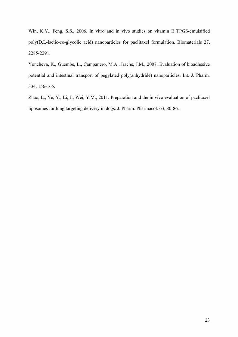
Table2. Plasma pharmacokinetic parameters (mean ± SD) of polymeric micelles in C57BL/6J female mice following a single i.v. and oral administration. Dose PXL= 10 mg/kg.
Sample Route AUC
(µg h/mL) C max (ng)
T
max (h)
T ½ Z (h-1)
MRT (h)
V (mL)
Cl (mL/h)
F
Taxol ® i.v. 90.2±24.5 60.5±6.4 0 2.27±0.5 1.39±0.4 5.8±0.6 2.7±0.3
PTX-PM i.v. 110.0±29.1 68.1±3.8 0 2.2±0.1 5.1±2.4 6.3±0.6 1.3±0.8
Taxol ® Oral ND ND ND ND ND ND ND ND
PTX-PM Oral 99.2±17.3 19.2±3.6 0.5 0.1±0.0 12.7±1.6 12.3±1.5 1.1±0.3 0.9
AUC area under the concentration-time curve from time 0 to 8; Cmax: peak concentration; Tmax: time to peak concentration; MRT: mean residence time; t1/2z: half-life of the terminal phase. F: oral bioavailability.

26
Figure 1. Chart diagrams representing the ratio between the size of the micelles observed after
24h incubation in different media to initial size measured in distilled water.

27
Figure 2. Influence of the initial concentration of rhodamine (a) or paclitaxel (b) on the
loading degree into micelles and encapsulation efficiency. Data expressed as the mean ± SD,
n=3.
0
10
20
30
40
50
60
70
Lo
adin
g, μ
g/m
g m
icel
les
,
0
10
20
30
40
50
60
70
EE
, %
,
Paclitaxel loading
EE
1mg : 30mg 1.5mg : 30mg 3mg : 30mg
0
10
20
30
40
50
60
70
Lo
adin
g, μ
g/m
g m
icel
les
0
10
20
30
40
50
60
70
EE
, %
,
Rhodamine B loading
EE
1mg : 30mg 1.5mg : 30mg 3mg : 30mg

28
StI1
I2I3
I4C
8h
3h
1h0
5
10
15
20F
ract
ion
of
rho
dam
ine
(%)
,
Figure 3. Distribution of the rhodamine in the gastrointestinal segments after oral
administration of rhodamine-loaded stabilized Pluronic micelles (R-PM). St-stomach,
intestinal parts: I1 – duodenum, I2 and I3 - jejunum, I4 representing ileum, C-caecum).

29
Figure 4. Remained rhodamine fractions in the gastrointestinal segments of rats 8 hours after
oral administration of rhodamine-loaded stabilized micelles and control rhodamine solution.

30
Figure 5. Paclitaxel plasmatic levels after intravenous administration (single dose of 10mg/kg)
of either stabilized paclitaxel-loaded micelles (PTX-PM) or Taxol®. Data expressed as mean
± SD (n=5).

31
Figure 6. Paclitaxel plasmatic levels after oral administration (single dose of 10mg/kg) of
either stabilized paclitaxel-loaded micelles (PTX-PM) or Taxol®. Data expressed as mean ±
SD (n=5).


![Original Article Potential biomarkers for paclitaxel ... · Potential biomarkers for paclitaxel sensitivity in ... larynx and oropharynx cancer [5, 15]. ... Biomarkers for paclitaxel](https://img.pdfslide.us/doc/110x75/5af0f1e17f8b9a572b901a03/original-article-potential-biomarkers-for-paclitaxel-biomarkers-for-paclitaxel.jpg)

![Cardiologie francophone - franco 2005.ppt [Lecture seule] · 2007-05-14 · AMG Pico Elite Paclitaxel Artax Paclitaxel Aachen Resonance EuroCor Taxcor Paclitaxel Biolimus A9 Biomatrix](https://img.pdfslide.us/doc/110x75/5e42b3f5800daf02232992fa/cardiologie-francophone-franco-2005ppt-lecture-seule-2007-05-14-amg-pico.jpg)