Embed Size (px)
Citation preview
St. Boniface General Hospital Acute Myocardial Infarction Care Map
Standards Document and Charting Guidelines, Update: April 2008 Table of Contents Acute MI Inclusion and Exclusion Criteria ...............................................................page 2 Acute MI Discharge Criteria ......................................................................................page 2 Medical Standards.......................................................................................................page 2 Nursing Standards ......................................................................................................page 7 Physiotherapy Standards...........................................................................................page 10 Cardiac Activity Step Program ................................................................................page 10 Teaching Standards...................................................................................................page 10 Community Cardiac Educational Referral Process ..................................................page 14 Clinical Psychology Referral Process ......................................................................page 15 Charting Guidelines ..................................................................................................page 16 Acute AMI Indicators ...............................................................................................page 23 Reference List ...........................................................................................................page 24 Appendices ..............................................................................................................page 26 Appendix A Nursing Assessment Parameters Appendix B List of Videos Appendix C Medication Counseling Cards Appendix D WRHA Heparin Nomogram Supporting Documentation ....................................................................................page 43 - WRHA Referral Criteria for Social Work - WRHA Referral Criteria for Spiritual Care - WRHA Referral Criteria for Occupational Therapy - SBGH Referral Criteria for Clinical Dietician - Glycemic control and the MI Care Map ……………………………………… ..page 49 Copy of the Acute MI Care Map System.......................................................….. page 51 - Acute MI Standard Admission Physician Order Form - Acute MI Care Map - Acute MI Patient/Family Care Guide - Coping with Heart Attack How are You Doing Checklist - ACS Heparin Nomogram - Cardiac Rehabilitation Referral Form - Follow up with Your Family Doctor for Oral Glucose Tolerance Test - AMI and Diabetes Physician Order Sheet - AMI and no Previous Diabetes Physician Order Sheet Preface
Acute MI Standards Document, April 2008 1
The provision of quality care continues to be of paramount importance not only to health care providers, but also to patients and families who are recipients of care. Care Maps, along with Clinical Practice Guidelines and outcome measures are important tools for maintaining and improving quality care. Benefits of evidence based tool includes a decrease in hospital and one year mortality, higher adherence to quality indictors, improvement in rate of compliance and timing of interventions, patient/staff satisfaction, decrease in length of stay, more detailed nursing assessments, client empowerment, improved exercise tolerance and compliance with care. It is important to remember that evidence based tools are just that – tools to help coordinate the entire patient process. Clinical judgment and individual patient preferences remains important in maintaining individualized quality care. Acute MI Inclusion and Exclusion Criteria: Inclusion Criteria: All patients admitted to the ICU, CCU, Medicine, Family Medicine or
Cardiology with an admission diagnosis of Non ST Elevation AMI or ST Elevation AMI should be placed on the AMI Care Map. If a patient is unstable and not suitable to be care mapped place the AMI Care Map in front of the patient’s chart and establish when patient is clinically appropriate. In essence the AMI Care map is on HOLD until the patient is stabilised.
Patients with a delayed presentation with an AMI should be placed on the Care Map as per discretion of the clinician and the decision should be individualized for each patient.
Exclusion Criteria: -The AMI Care Map is not intended for patients with unstable angina or
with a diagnosis of acute coronary syndrome with or without microinfarction (e.g. Troponin positive, CK negative). For patients with a diagnosis of ACS, clarify if patient has an AMI in rounds with the attending physician.
-Postoperative AMI Acute MI Discharge Criteria The following are to be met prior to patient discharge: 1. The patient is hemodynamically stable. 2. The patient does not have ischemic pain. 3. The patient has completed the in house teaching and activity program. 4. The patient has received a discharge prescription for: Nitroglycerin, Antiplatelet agent(s),
beta-blocker, ACE Inhibitor, and lipid lowering agent, if no contraindications or allergy and according to guidelines.
5. Arrangements have been made for appropriate risk stratification. 6. The patient is aware of and the referral to cardiac rehabilitation has been faxed.
Acute MI Standards Document, April 2008 2
Medical Standards The following define the medical standards of the AMI patient: AMI Care Map Admission Guidelines
Note: The following are suggested admission guidelines for patients with an Acute Myocardial Infarction. The criteria are guidelines and do not replace sound clinical judgment and individualized patient assessment. 1. AMI patients will be admitted to an ICU/CCU patient care area on admission. Exception:
low risk AMI* patients may be considered for admissions to a telemetry unit that: - Is a designated ward/area of an acute care health facility, which is specially
staffed and equipped to provide observation, assessment, care and treatment to patients with cardiac related health issues.
- Staff has the knowledge and skills directly related to cardiac patients. - Immediate accessibility to ACLS trained staff and emergency equipment (e.g.
defibrillator and crash cart).
* Definition of low risk AMI patient: - Small, limited non-ST elevation AMI. - Has not received a fibrinolytic agent. - No evidence of congestive heart failure or clinical evidence of LV dysfunction. - No evidence of complex ventricular arrhythmia. - No evidence of significant conduction disturbance, either new or unknown
duration. - Does not require/undergone an early cardiac intervention.
Diagnostic Standards 1. CK q8h times three from admission. 2. Troponin q8h times three within the first 24 hours or until first positive result obtained. 3. 12 lead ECG on day 2 and 3. 4. CBC, platelets on day 2,3 and 4. 5. ALT and AST on admission (drawn from emergency blood work) and on day 3. 6. Lipid profile with admission sample from the Emergency Department: HDL, LDL, total
cholesterol, triglycerides, TC-HDL ratio. Note: May consider a repeat fasting lipid profile if the triglycerides were elevated.
7. Chest x-ray in a.m. if not done in the Emergency Department 8. Electrolytes, urea, creatine, glucose, total C02, on day 2 and 4. 9. INR and aPTT day 1 and aPTT OD based on the ACS Heparin Nomogram if receiving
unfractionated intravenous Heparin. 10. Risk stratification. 11. For fibrinolytic patients: 12 lead ECG at 1 and 8 hours post infusion. Neurological
assessment at baseline, q1hx2, and then q4h x24 hours. Consults
Acute MI Standards Document, April 2008 3
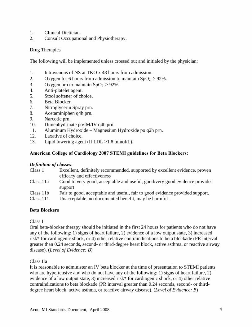
1. Clinical Dietician. 2. Consult Occupational and Physiotherapy. Drug Therapies The following will be implemented unless crossed out and initialed by the physician: 1. Intravenous of NS at TKO x 48 hours from admission. 2. Oxygen for 6 hours from admission to maintain SpO2 92%. 3. Oxygen prn to maintain SpO2 92%. 4. Anti-platelet agent. 5. Stool softener of choice. 6. Beta Blocker. 7. Nitroglycerin Spray prn. 8. Acetaminiphen q4h prn. 9. Narcotic prn. 10. Dimenhydrinate po/IM/IV q4h prn. 11. Aluminum Hydroxide – Magnesium Hydroxide po q2h prn. 12. Laxative of choice. 13. Lipid lowering agent (If LDL >1.8 mmol/L). American College of Cardiology 2007 STEMI guidelines for Beta Blockers: Definition of classes: Class 1 Excellent, definitely recommended, supported by excellent evidence, proven
efficacy and effectiveness Class 11a Good to very good, acceptable and useful, good/very good evidence provides
support Class 11b Fair to good, acceptable and useful, fair to good evidence provided support. Class 111 Unacceptable, no documented benefit, may be harmful. Beta Blockers Class I Oral beta-blocker therapy should be initiated in the first 24 hours for patients who do not have any of the following: 1) signs of heart failure, 2) evidence of a low output state, 3) increased risk* for cardiogenic shock, or 4) other relative contraindications to beta blockade (PR interval greater than 0.24 seconds, second- or third-degree heart block, active asthma, or reactive airway disease). (Level of Evidence: B) Class IIa It is reasonable to administer an IV beta blocker at the time of presentation to STEMI patients who are hypertensive and who do not have any of the following: 1) signs of heart failure, 2) evidence of a low output state, 3) increased risk* for cardiogenic shock, or 4) other relative contraindications to beta blockade (PR interval greater than 0.24 seconds, second- or third-degree heart block, active asthma, or reactive airway disease). (Level of Evidence: B)
Acute MI Standards Document, April 2008 4
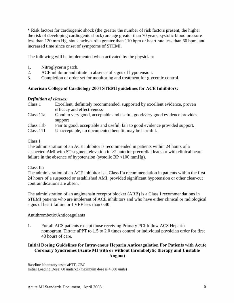
* Risk factors for cardiogenic shock (the greater the number of risk factors present, the higher the risk of developing cardiogenic shock) are age greater than 70 years, systolic blood pressure less than 120 mm Hg, sinus tachycardia greater than 110 bpm or heart rate less than 60 bpm, and increased time since onset of symptoms of STEMI. The following will be implemented when activated by the physician: 1. Nitroglycerin patch. 2. ACE inhibitor and titrate in absence of signs of hypotension. 3. Completion of order set for monitoring and treatment for glycemic control. American College of Cardiology 2004 STEMI guidelines for ACE Inhibitors: Definition of classes: Class 1 Excellent, definitely recommended, supported by excellent evidence, proven
efficacy and effectiveness Class 11a Good to very good, acceptable and useful, good/very good evidence provides
support Class 11b Fair to good, acceptable and useful, fair to good evidence provided support. Class 111 Unacceptable, no documented benefit, may be harmful. Class I The administration of an ACE inhibitor is recommended in patients within 24 hours of a suspected AMI with ST segment elevation in >2 anterior precordial leads or with clinical heart failure in the absence of hypotension (systolic BP <100 mmHg). Class IIa The administration of an ACE inhibitor is a Class IIa recommendation in patients within the first 24 hours of a suspected or established AMI, provided significant hypotension or other clear-cut contraindications are absent The administration of an angiotensin receptor blocker (ARB) is a Class I recommendations in STEMI patients who are intolerant of ACE inhibitors and who have either clinical or radiological signs of heart failure or LVEF less than 0.40. Antithrombotic/Anticoagulants 1. For all ACS patients except those receiving Primary PCI follow ACS Heparin
nomogram. Titrate aPPT to 1.5 to 2.0 times control or individual physician order for first 48 hours of care.
Initial Dosing Guidelines for Intravenous Heparin Anticoagulation For Patients with Acute Coronary Syndromes (Acute MI with or without thrombolytic therapy and Unstable
Angina)
Baseline laboratory tests: aPTT, CBC Initial Loading Dose: 60 units/kg (maximum dose is 4,000 units)
Acute MI Standards Document, April 2008 5
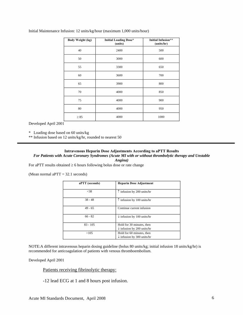
Initial Maintenance Infusion: 12 units/kg/hour (maximum 1,000 units/hour)
Body Weight (kg) Initial Loading Dose* (units)
Initial Infusion** (units/hr)
40 2400 500
50 3000 600
55 3300 650
60 3600 700
65 3900 800
70 4000 850
75 4000 900
80 4000 950
85 4000 1000
Developed April 2001 * Loading dose based on 60 units/kg ** Infusion based on 12 units/kg/hr, rounded to nearest 50
Intravenous Heparin Dose Adjustments According to aPTT Results For Patients with Acute Coronary Syndromes (Acute MI with or without thrombolytic therapy and Unstable
Angina) For aPTT results obtained 6 hours following bolus dose or rate change (Mean normal aPTT = 32.1 seconds)
aPTT (seconds) Heparin Dose Adjustment
<38 infusion by 200 units/hr
38 - 48 infusion by 100 units/hr
49 - 65 Continue current infusion
66 - 82 infusion by 100 units/hr
83 - 105 Hold for 30 minutes, then infusion by 200 units/hr
>105 Hold for 60 minutes, then infusion by 300 units/hr
NOTE:A different intravenous heparin dosing guideline (bolus 80 units/kg; initial infusion 18 units/kg/hr) is recommended for anticoagulation of patients with venous thromboembolism. Developed April 2001
Patients receiving fibrinolytic therapy:
-12 lead ECG at 1 and 8 hours post infusion.
Acute MI Standards Document, April 2008 6
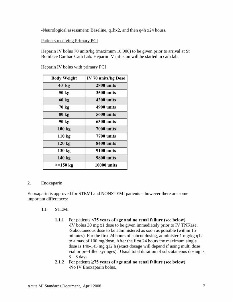
-Neurological assessment: Baseline, q1hx2, and then q4h x24 hours. Patients receiving Primary PCI Heparin IV bolus 70 units/kg (maximum 10,000) to be given prior to arrival at St Boniface Cardiac Cath Lab. Heparin IV infusion will be started in cath lab. Heparin IV bolus with primary PCI
Body Weight IV 70 units/kg Dose
40 kg 2800 units
50 kg 3500 units
60 kg 4200 units
70 kg 4900 units
80 kg 5600 units
90 kg 6300 units
100 kg 7000 units
110 kg 7700 units
120 kg 8400 units
130 kg 9100 units
140 kg 9800 units
>=150 kg 10000 units
2. Enoxaparin
Enoxaparin is approved for STEMI and NONSTEMI patients – however there are some important differences:
1.1 STEMI
1.1.1 For patients <75 years of age and no renal failure (see below) -IV bolus 30 mg x1 dose to be given immediately prior to IV TNKase. -Subcutaneous dose to be administered as soon as possible (within 15 minutes). For the first 24 hours of subcut dosing, administer 1 mg/kg q12 to a max of 100 mg/dose. After the first 24 hours the maximum single dose is 140-145 mg q12 h (exact dosage will depend if using multi dose vial or pre-filled syringes). Usual total duration of subcutaneous dosing is 3 – 8 days.
2.1.2 For patients ≥75 years of age and no renal failure (see below) -No IV Enoxaparin bolus.
Acute MI Standards Document, April 2008 7
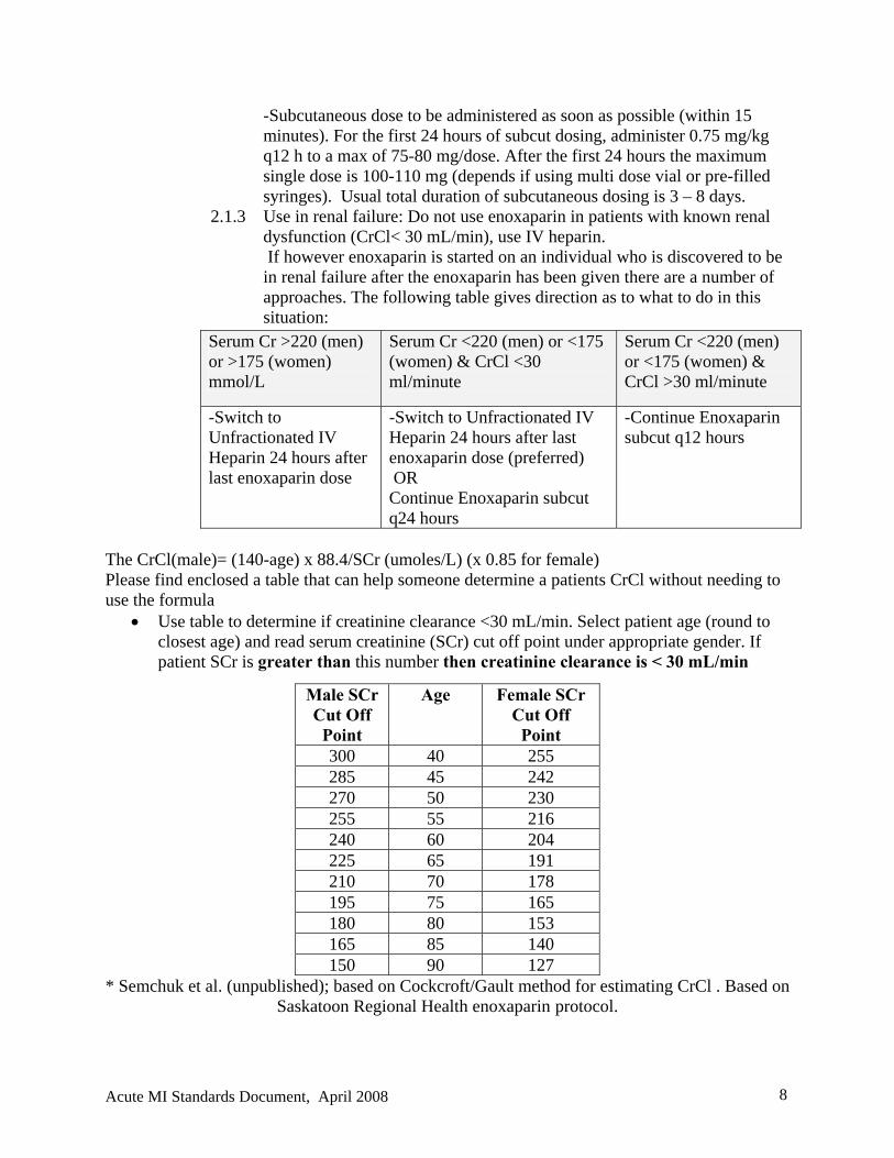
-Subcutaneous dose to be administered as soon as possible (within 15 minutes). For the first 24 hours of subcut dosing, administer 0.75 mg/kg q12 h to a max of 75-80 mg/dose. After the first 24 hours the maximum single dose is 100-110 mg (depends if using multi dose vial or pre-filled syringes). Usual total duration of subcutaneous dosing is 3 – 8 days.
2.1.3 Use in renal failure: Do not use enoxaparin in patients with known renal dysfunction (CrCl< 30 mL/min), use IV heparin. If however enoxaparin is started on an individual who is discovered to be in renal failure after the enoxaparin has been given there are a number of approaches. The following table gives direction as to what to do in this situation:
Serum Cr >220 (men) or >175 (women) mmol/L
Serum Cr <220 (men) or <175 (women) & CrCl <30 ml/minute
Serum Cr <220 (men) or <175 (women) & CrCl >30 ml/minute
-Switch to Unfractionated IV Heparin 24 hours after last enoxaparin dose
-Switch to Unfractionated IV Heparin 24 hours after last enoxaparin dose (preferred) OR Continue Enoxaparin subcut q24 hours
-Continue Enoxaparin subcut q12 hours
The CrCl(male)= (140-age) x 88.4/SCr (umoles/L) (x 0.85 for female) Please find enclosed a table that can help someone determine a patients CrCl without needing to use the formula
Use table to determine if creatinine clearance <30 mL/min. Select patient age (round to closest age) and read serum creatinine (SCr) cut off point under appropriate gender. If patient SCr is greater than this number then creatinine clearance is < 30 mL/min
* Semchuk et al. (unpublished); based on Cockcroft/Gault method for estimating CrCl . Based on Saskatoon Regional Health enoxaparin protocol.
Male SCr Cut Off
Point
Age Female SCr Cut Off
Point 300 40 255 285 45 242 270 50 230 255 55 216 240 60 204 225 65 191 210 70 178 195 75 165 180 80 153 165 85 140 150 90 127
Acute MI Standards Document, April 2008 8
2.2 NONSTEMI 2.2.1 For patients of any age:
-1 mg/kg subcutaneous q12 hours. For subcut dosing, administer 1 mg/kg q12 to a max of 140-145 mg/dose. - Usual total duration of subcutaneous dosing is 2 – 8 days. - Use in renal failure: See above chart for STEMI
Note: Enoxaparin should be injected subcut in abdominal site only - if possible avoid NSAIDs (excluding ASA) clinical evaluation of bleeding complications daily Preparation for Discharge to the Community -Patients should be risk stratified appropriately according to patient’s condition and standards of care. -It is recommended that if the patient lives in the city the patient may be discharged home if the test is booked prior to discharge and within 5 working days (if applicable). -All post discharge symptom limited stress tests to be arranged by the physician responsible for the patient’s care at 4-6 weeks post discharge. -If evidence of CHF, left ventricular assessment should be done prior to patient discharge. -The most common forms of risk stratification are treadmill EKG (both sub maximal and maximal), pharmacologic nuclear (usually with dipyridamole), exercise nuclear (sub maximal or maximal) and pharmacologic echo (usually with dobutamine).
Stressor Imager Walking on treadmill (Sub maximal) Signs, symptoms, ECG, BP Walking on treadmill (Sub maximal) Nuclear Imaging (MIBI, Thallium,
Cardiolyte) or echo Bicycle (Sub maximal) Nuclear Imaging or Echo Pharmacologic: - Adenosine (Maximal) - Dipyridamole (Maximal) - Dobutamine (Sub maximal)
Nuclear Imaging or Echo
Nursing Assessment Standards The following table defines the assessment parameters.
Standard ICU/CCU Medical Ward
ST Monitoring Ideal recommendations - Monitor Lead III, V3 and V5 for three lead systems - Monitor in lead III and V3 for two lead systems
From admission and until the physician orders the monitor to be discontinued
N/A
Acute MI Standards Document, April 2008 9
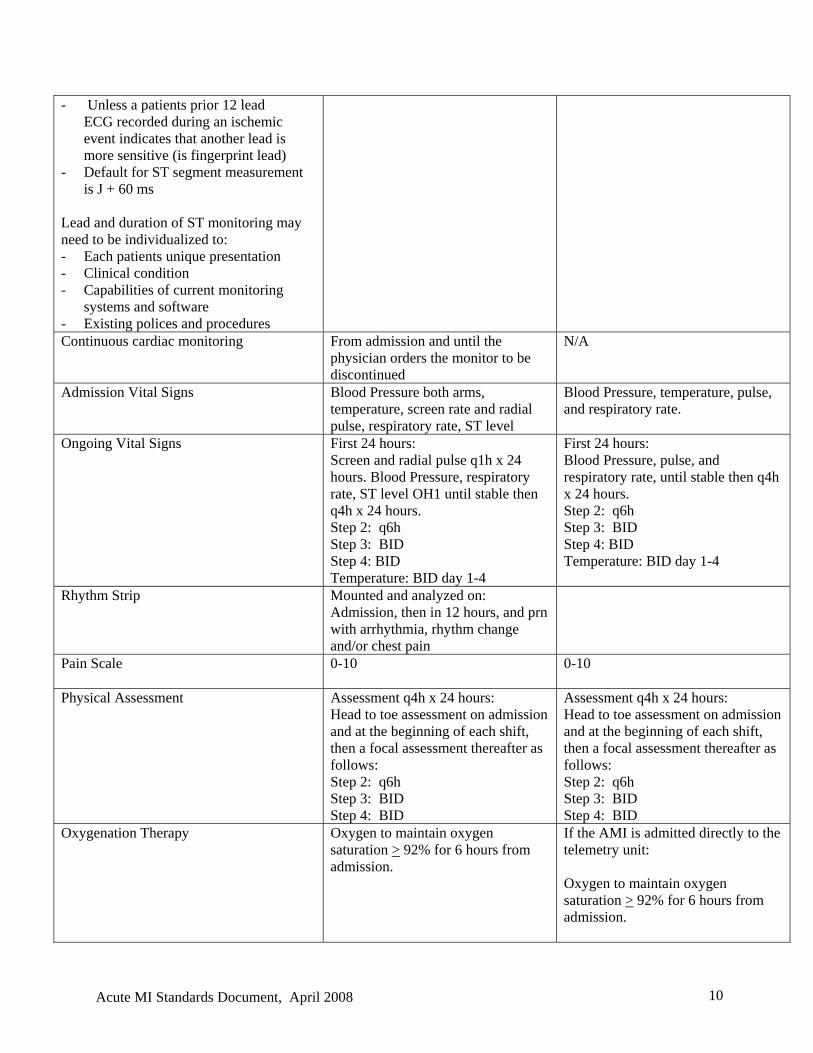
- Unless a patients prior 12 lead ECG recorded during an ischemic event indicates that another lead is more sensitive (is fingerprint lead) - Default for ST segment measurement is J + 60 ms Lead and duration of ST monitoring may need to be individualized to: - Each patients unique presentation - Clinical condition - Capabilities of current monitoring systems and software - Existing polices and procedures Continuous cardiac monitoring From admission and until the
physician orders the monitor to be discontinued
N/A
Admission Vital Signs Blood Pressure both arms, temperature, screen rate and radial pulse, respiratory rate, ST level
Blood Pressure, temperature, pulse, and respiratory rate.
Ongoing Vital Signs First 24 hours: Screen and radial pulse q1h x 24 hours. Blood Pressure, respiratory rate, ST level OH1 until stable then q4h x 24 hours. Step 2: q6h Step 3: BID Step 4: BID Temperature: BID day 1-4
First 24 hours: Blood Pressure, pulse, and respiratory rate, until stable then q4h x 24 hours. Step 2: q6h Step 3: BID Step 4: BID Temperature: BID day 1-4
Rhythm Strip Mounted and analyzed on: Admission, then in 12 hours, and prn with arrhythmia, rhythm change and/or chest pain
Pain Scale
0-10 0-10
Physical Assessment Assessment q4h x 24 hours: Head to toe assessment on admission and at the beginning of each shift, then a focal assessment thereafter as follows: Step 2: q6h Step 3: BID Step 4: BID
Assessment q4h x 24 hours: Head to toe assessment on admission and at the beginning of each shift, then a focal assessment thereafter as follows: Step 2: q6h Step 3: BID Step 4: BID
Oxygenation Therapy Oxygen to maintain oxygen saturation > 92% for 6 hours from admission.
If the AMI is admitted directly to the telemetry unit: Oxygen to maintain oxygen saturation > 92% for 6 hours from admission.
Acute MI Standards Document, April 2008 10
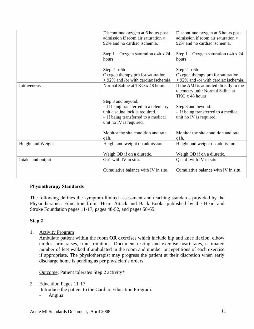
Discontinue oxygen at 6 hours post admission if room air saturation > 92% and no cardiac ischemia. Step 1 Oxygen saturation q4h x 24 hours Step 2 q6h Oxygen therapy prn for saturation < 92% and /or with cardiac ischemia.
Discontinue oxygen at 6 hours post admission if room air saturation > 92% and no cardiac ischemia. Step 1 Oxygen saturation q4h x 24 hours Step 2 q6h Oxygen therapy prn for saturation < 92% and /or with cardiac ischemia.
Intravenous Normal Saline at TKO x 48 hours Step 3 and beyond: - If being transferred to a telemetry unit a saline lock is required. - If being transferred to a medical unit no IV is required. Monitor the site condition and rate q1h.
If the AMI is admitted directly to the telemetry unit: Normal Saline at TKO x 48 hours Step 3 and beyond: - If being transferred to a medical unit no IV is required. Monitor the site condition and rate q1h.
Height and Weight Height and weight on admission. Weigh OD if on a diuretic.
Height and weight on admission. Weigh OD if on a diuretic.
Intake and output Oh1 with IV in situ. Cumulative balance with IV in situ.
Q shift with IV in situ. Cumulative balance with IV in situ.
Physiotherapy Standards The following defines the symptom-limited assessment and teaching standards provided by the Physiotherapist. Education from “Heart Attack and Back Book” published by the Heart and Stroke Foundation pages 11-17, pages 48-52, and pages 58-65. Step 2 1. Activity Program
Ambulate patient within the room OR exercises which include hip and knee flexion, elbow circles, arm raises, trunk rotations. Document resting and exercise heart rates, estimated number of feet walked if ambulated in the room and number or repetitions of each exercise if appropriate. The physiotherapist may progress the patient at their discretion when early discharge home is pending as per physician’s orders.
Outcome: Patient tolerates Step 2 activity* 2. Education Pages 11-17 Introduce the patient to the Cardiac Education Program.
- Angina
Acute MI Standards Document, April 2008 11
- Heart attack - Differences between angina and heart attack - Action plan (Nitro use and when and how to get to the hospital).
Outcome: Patient understands: -Signs and symptoms of cardiac ischemia -Difference between angina and heart attack -Knows action plan Step 3 1. Activity Program
Ambulate patient in the hall. Indicate if patient can ambulate independently or requires assistance. Document the resting and exercise heart rates.
Outcome: Patient tolerates Step 3 activity* 2. Education Pages 48-52
-Normal and abnormal response to activities -Appropriate level and progression of activities -Importance of cardiac rehabilitation Outcome: Patient understands: -Normal and abnormal responses to activity -Appropriate level and progression of activity -Importance of cardiac rehabilitation Step 4
1. Activity Program Walks up and down 10-12 steps OR modified stairs OR increased ambulation in hall. Document the resting and exercise heart rates.
Outcome: Patient tolerates Step 4 activity 2. Education Pages 51-52, 58-65 in the Heart Attack and Back Book
-Review the Home Exercise Program in Heart Attack and Back Book -Review community teaching options and benefits of cardiac rehabilitation -Review cardiac community teaching options with the patients. Ask the patient to which cardiac rehabilitation program site (Wellness Institute or Reh-Fit Centre) she/he would like the referral form faxed to
Outcome: Patient Understands: -Home exercise program -Community resources available and benefits of cardiac rehabilitation -Cardiac rehabilitation referral faxed * If on a beta-blocker exercise heart rate should not exceed an increase of 10 beats per minute
from the resting heart rate. If not on a beta blocker the heart rate should not exceed an increase of 20 beats per minute from the resting heart rate. No chest pain or excessive shortness of breath, sweating, and/or weakness. Symptom limited activity program.
Acute MI Standards Document, April 2008 12
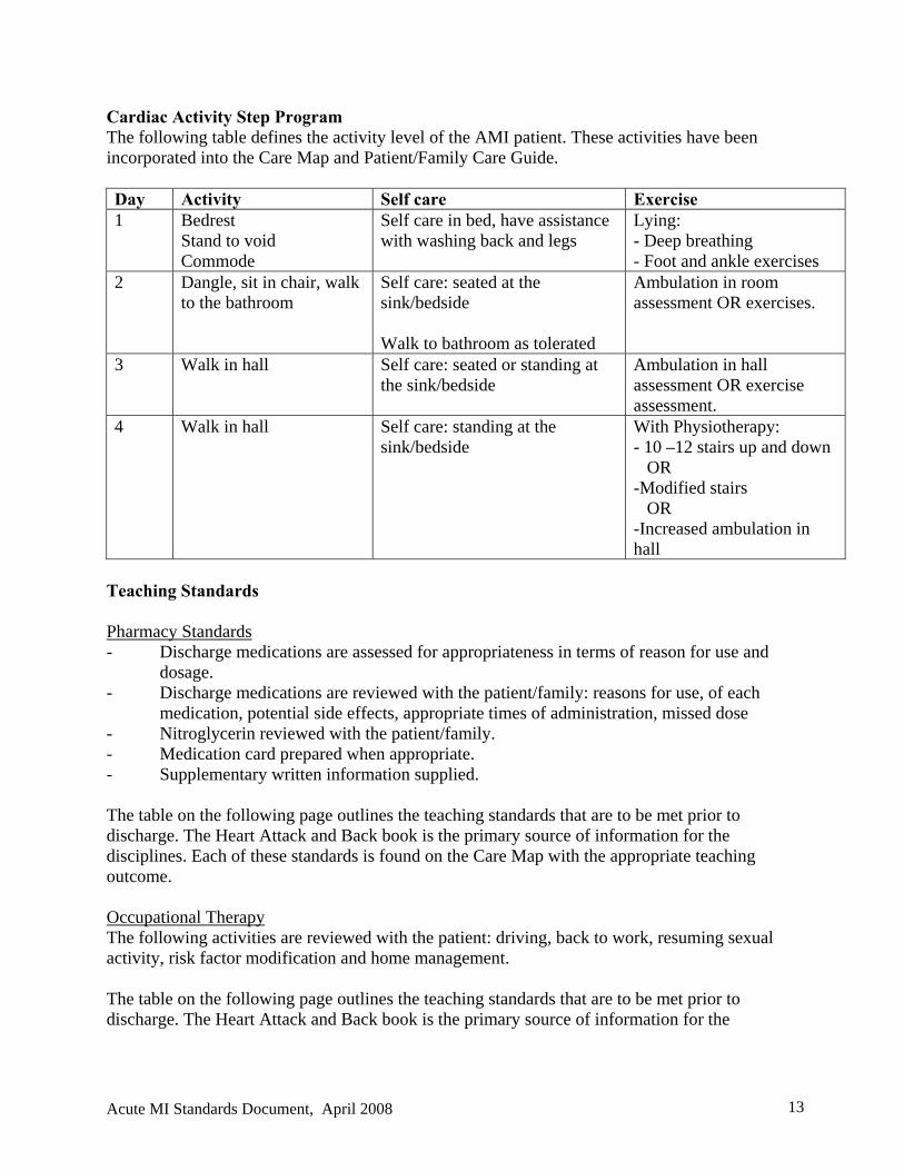
Cardiac Activity Step Program The following table defines the activity level of the AMI patient. These activities have been incorporated into the Care Map and Patient/Family Care Guide. Day Activity Self care Exercise 1 Bedrest
Stand to void Commode
Self care in bed, have assistance with washing back and legs
Lying: - Deep breathing - Foot and ankle exercises
2 Dangle, sit in chair, walk to the bathroom
Self care: seated at the sink/bedside Walk to bathroom as tolerated
Ambulation in room assessment OR exercises.
3 Walk in hall
Self care: seated or standing at the sink/bedside
Ambulation in hall assessment OR exercise assessment.
4 Walk in hall
Self care: standing at the sink/bedside
With Physiotherapy: - 10 –12 stairs up and down OR -Modified stairs OR -Increased ambulation in hall
Teaching Standards Pharmacy Standards - Discharge medications are assessed for appropriateness in terms of reason for use and
dosage. - Discharge medications are reviewed with the patient/family: reasons for use, of each
medication, potential side effects, appropriate times of administration, missed dose - Nitroglycerin reviewed with the patient/family. - Medication card prepared when appropriate. - Supplementary written information supplied. The table on the following page outlines the teaching standards that are to be met prior to discharge. The Heart Attack and Back book is the primary source of information for the disciplines. Each of these standards is found on the Care Map with the appropriate teaching outcome. Occupational Therapy The following activities are reviewed with the patient: driving, back to work, resuming sexual activity, risk factor modification and home management. The table on the following page outlines the teaching standards that are to be met prior to discharge. The Heart Attack and Back book is the primary source of information for the
Acute MI Standards Document, April 2008 13

Acute MI Standards Document, April 2008 14
disciplines. Each of these standards is found on the Care Map with the appropriate teaching outcome.
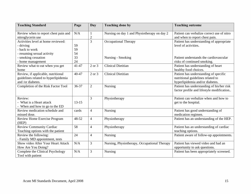
Teaching Standard Page Day Teaching done by Teaching outcome
Review when to report chest pain and nitroglycerin use
N/A 1 2
Nursing on day 1 and Physiotherapy on day 2 Patient can verbalize correct use of nitro and when to report chest pain.
Activities level at home reviewed: - driving - back to work - resuming sexual activity - smoking cessation - home management
59 59 54 33 24
3 Occupational Therapy Nursing - Smoking
Patient has understanding of appropriate level of activities. Patient understands the cardiovascular risks of continued smoking.
Review what to eat when you get home
41-47 2 or 3 Clinical Dietitian
Patient has understanding of heart healthy food choices.
Review, if applicable, nutritional guidelines related to hyperlipidemia and /or diabetes.
40-47 2 or 3 Clinical Dietitian
Patient has understanding of specific nutritional guidelines related to hyperlipidemia and/or diabetes.
Completion of the Risk Factor Tool 36-37 2 Nursing Patient has understanding of his/her risk factor profile and lifestyle modification..
Review: - What is a Heart attack - When and how to go to the ED
13-15
3 Physiotherapy Patient can verbalize when and how to get to the hospital.
Review medication schedule and missed dose.
cards 4 Nursing Patient has good understanding of medication regimen.
Review Home Exercise Program (HEP)
48-52 4 Physiotherapy Patient has an understanding of the HEP.
Review Community Cardiac Teaching options with the patient
58 4 Physiotherapy Patient has an understanding of cardiac teaching options.
Review the following: - Family MD appointment, tests
24 4 Nursing Patient aware of follow-up appointments.
Show video After Your Heart Attack How Are You Doing?
N/A 3 Nursing, Physiotherapy, Occupational Therapy
Patient has viewed video and had an opportunity to ask questions.
Complete the Clinical Psychology Tool with patient
N/A 3 Nursing Patient has been appropriately screened.
Acute MI Standards Document, April 2008 15
Community Cardiac Referral Process WRHA Sites The WRHA Cardiac Rehabilitation Program is delivered at 2 sites including the Wellness Institute at Seven Oaks General Hospital and the Reh-Fit Centre. The program is 16 weeks in length and is a comprehensive, multidisciplinary approach to the prevention, stabilization and possible reversal of cardiovascular disease. The benefits of Cardiac Rehabilitation are well established. These include a reduction in mortality of 25-40%, reduction in tobacco use, improvement in psychological well being, reduction in symptoms, reduction in recidivism, improvement in lipid profiles and improved exercise tolerance. There are few contraindications to Cardiac Rehabilitation. Essentially all patients will obtain some benefit including CHF patients and the frail elderly. These benefits range from improving the ability to perform ADL to social benefits. All AMI Care Map patients should be given the opportunity to participate in these programs. The cost to the patient at the WRHA sites is $190 +GST. Many insurance companies will cover this cost or a subsidy is available based on individual need. The gold standard is that all cardiac patients will receive cardiac rehabilitation. In the event that patients are unable to attend cardiac rehabilitation, the staff at the cardiac rehabilitation program will attempt to link the patient with appropriate cardiac resources or programs within his/her community. With this in mind, it is necessary to send referrals on all the rural patients, as the sites will follow them when they are discharged. The staff from the cardiac rehabilitation program has access to a binder that has contact numbers and locations of available rural resources for cardiac patients. For further information on the Cardiac Rehabilitation programs please call Kelly Seward at 632-3908 at the Wellness Institute or Beverly Burton-Guindon at 488-5855 at the Reh-Fit Centre. Brandon and Thunder Bay: Patients from the Brandon or Assiniboine Health Region are referred to the cardiac rehabilitation program in Brandon. For further information contact the cardiac rehab nurse at 1-204-578-4204 Patients who are residents in North Western Ontario are referred to the cardiac rehabilitation program in Thunder Bay. For further information contact the cardiac rehab nurse at 1-204-684-6001. The site Physiotherapist: 1. Reviews the benefits of cardiac rehabilitation with the patient while in hospital on Day 4
of the AMI Care Map.
Acute MI Standards Document, April 2008 16
2. Informs the patient that a staff member from the cardiac rehabilitation site of choice will contact him/her by telephone and/or letter with the available Cardiac Rehabilitation options, approximately one – two weeks post discharge.
3. Completes the referral form. 4. Faxes the form and shreds the document once the fax transmission is confirmed.
Cardiac Rehabilitation Process: The patient will be contacted either by phone and/or letter within 2 weeks of discharge.
1. He/she will be given information on the Cardiac Rehabilitation programs. Information
discussed includes the commitment required from the patient, times available for classes, benefits of cardiac rehab, cost and subsidy information, insurance information, and may assist in any other identified issues.
2. For patients who are unable to attend the 16 week Cardiac Rehabilitation program the
interviewer will provide the Heart and Stroke Foundation of Manitoba phone number for patients to call and check the availability of Heart to Heart™ programs in their community or access to other resources offered through the HSFM. For the rural patients, a cardiac resource binder may be used to direct the patient to community resources in their area. This information is also readily available at both cardiac rehabilitation sites and will be relayed to patients when appropriate.
Referral data is collected and compiled in order to evaluate these processes and facilitate solutions to problems that are identified. Status of Heart to Heart™ Sessions:
Contact the Heart and Stroke Foundation of Manitoba
Rural Manitoba Patients A binder listing cardiac resources available in rural Manitoba is available. This information is located at the Heart and Stroke Foundation of Manitoba, Health Links, the two Cardiac Rehabilitation Programs and the city facilities. Specific information includes: - Is a Heart to Heart Program available in the community? - Are there any cardiac services available? - Are nutrition services offered? - Is there a fitness/activity center? - Is there a community pharmacist? Clinical Psychology Referral Process Studies have shown that up to 25% of cardiac patients may experience depression after their AMI and a significant number have sub-clinical levels of depression that progress to a major
Acute MI Standards Document, April 2008 17
depressive episode. Similarly, up to one third of AMI patients experience severe anxiety even at six months post event. Studies have indicated that up to 25% of CAD patients suffer from Panic Disorder. Up to 50% of AMI patients complain of increased irritability even one year after their event. Moreover, these negative mood states have been shown to compromise physical as well as psychosocial recovery, and interfere with cardiac rehabilitation. A pilot project involving the psychological screening of cardiac patients at the Wellness Institute at Seven Oaks General Hospital and the Kinsmen Reh-Fit Centre has found that approximately 29% of the patients were suffering from clinically significant levels of psychological distress. The goal of the inpatient psychological screening tool is to help identify individuals who may be at risk for poor adjustment post AMI. The tool will help identify those patients who require a more formal outpatient mental status assessment and behavioral treatment planning following hospital discharge. The referral process in no way is meant to replace in-hospital assessment for acute psychological emergencies/symptoms (example suicidal ideation). Staff is to refer such patients for a psychiatric consultation prior to hospital discharge as indicated and ordered by the attending physician. Note: Clinical Health Psychology is providing an elective service for non-acutely ill post MI
patients, and psychiatrists from the Mental Health Program are the clinicians involved in the consultation process for acute psychiatric presentations
Professional staff in the hospital will review the screening tool with the patient on the third day of the AMI Care Map. The professional staff member will discuss with the patient the results of the screening tool. Patients identified at being at risk will be given the option to consent to an outpatient assessment by clinical psychology. The standard is for patients to be contacted by Clinical Health Psychology and triaged for assessment within one month of receiving the referral. Clinical psychology assessments will be conducted at St. Boniface General Hospital. There is no cost to the patient for the assessment. Treatment options available to the patient will vary. Potential referral sites include the two cardiac rehabilitation programs. The cardiac rehabilitation program offers patients a comprehensive program to facilitate behavioral modification and lifestyle changes. Other options may include: the WRHA Clinical Psychology program, the anxiety disorders clinic at St. Boniface General Hospital, Klinic counseling services, depression groups at the two tertiary sites, Interfaith Marriage and Family Institute at the University of Winnipeg, and other community resources. Patients in need of pharmacological treatment will be referred to their family physician or psychiatrist (if involved with the case). A similar referral process for psychological treatment has worked quite effectively for the patients identified as needing intervention at the two rehabilitation centres. To ensure continuity of patient care the clinical psychologist will forward assessment and follow-up treatment recommendations to the patient’s primary care provider. Charting Guidelines
Acute MI Standards Document, April 2008 18
The purpose of the Care Map is to provide a systematic means of gathering patient information, which identifies baseline data, and ongoing assessment information. Key Definitions
Interventions are patient care activities, which need to be undertaken in order to assist patients to achieve outcomes in a timely manner. These are listed on the Care Map in the appropriate category of care. The categories of care are defined as: Assessment/consults, Tests, Treatments, Meds/IV, Nutrition, Safety/Activity, Teaching, Psychosocial and Discharge Planning.
Patient Outcomes are goals to be achieved by the patient. They are to be measurable and can be defined as either intermediate or discharge outcomes. These are the shaded sections on the Care Map.
Implementation Guidelines 1. The AMI Care Map System will reside in the Emergency Department, the Intensive Care
Unit, Medical Units, and Cardiology Units. 2. After diagnosis of the AMI and prior to admission the Attending/Emergency Physician
will complete the Standard Physician Orders for the AMI Care Map. 3. The admitting unit staff will start the Care Map.
- Transcribe the orders onto the Care Map, kardex, and medication record. - Individualize the Care Map. - Give the patient the Patient/Family Care Guide and the Heart Attack and Back
Book. 4. If the patient is admitted in the early or late evening, follow these guidelines:
12-Hour Shift: complete the day column on Step 1 of the Care Map. At 1900, a same day Care Map (extension Care Map) will be required, as most of the "critical" expected outcomes will not have been met.
Process:
1. The day nurse places a second copy of the Step 1 Care Map on the patient’s chart. 2. Indicate on Care Map that a same day Care Map is being used, by circling “yes”. 3. Write in current status of orders, tests, and treatments on the same day Care Map. 4. Document the reason on the documentation section of the Care Map.
Example: May 6, 2006 19:00 Patient admitted in early evening and the length of stay too short to meet day 1 outcomes. Step 1 map repeated. Lorraine Avery, RN.
8-Hour Shift: complete the evening column on Step 1 of the Care Map. At 2300, an extension Care Map will be required, as most of the "critical” expected outcomes will not have been met.
Process:
Acute MI Standards Document, April 2008 19
1. The evening nurse places a second copy of the Step 1 Care Map “extension map” on the patient’s chart.
2. Indicate on Care Map that an extension map is being used, by circling “yes”. 3. Write in current status of orders, tests, and treatments on the same day Care Map. 4. Document the reason on the documentation section of the same day Care Map.
Orders on the Care Map 1. Standard orders are identified with a solid black box (). These are initiated on all
patients placed on the Care Map and are pre-printed on the Care Map. If an order is not appropriate the physician shall cross off and initial.
2. Individualized orders are identified with a blank box (). These require a Physician’s order to activate them. To activate the order, place a " " inside a box.
3. Additional orders are to be written on the Care Map in the appropriate category of care. Dealing with Allied Services that work Monday-Friday On Step 1 of the Care Map, all allied health personnel are notified of the admission of the AMI patient. Based on workload, the service may teach their section earlier or later if a weekend falls within the patient’s stay. Process: 1. Star (*) the Care Map in the appropriate column. 2. Document reason on the Care Map tool. Documentation Guidelines 1. Indicate the date and time of admission to the unit on Step1 of the AMI Care Map.
2. Assessment/Consults Section
Assessments Complete facilities Admission and Nursing Data Base form. Nursing assessments / assessment outcomes Nursing assessments are completed as per Nursing Assessment Protocol (Appendix A). Assessments reflect a charting by exception concept in which only abnormal assessments are identified by placing a star (*)and your initial on the Care Map under the appropriate column. Document the abnormal assessment parameter, with an action plan, on the note section of the Care Map/Same day Care Map. Thrombolytic Therapy Assessment Standards: - The use of automatic blood pressure cuffs is not recommend for 24 hours post
infusion on the limb where the thrombolytic agent has infused.
Parameter Frequency Vital Signs during Thrombolytic Therapy infusing
- Q15minutes x1 hour
Neurological Checks: - Glasgow coma scale
- Baseline in the ED, q1hx2, then q4h x24 hours.
Acute MI Standards Document, April 2008 20
- Pupil assessment - Limb assessment Use Neurological Assessment Record.
Consults: Following facility process initiate multidisciplinary consults.
3. Test Section There is space provided to transcribe tests not generic to the Care Map. - CK are drawn q8h times three. - Troponin q8h x three from admission or until first positive result obtained. - Lipid Profile consists of HDL, LDH, total cholesterol, triglycerides, and TC-HDL
ratio. After admission instruct the lab to complete the profile on the first bloodwork drawn in the Emergency Department. If the patient was admitted from another Emergency Department, draw this profile in the am.
- ALT and AST at baseline (drawn from emergency department blood work) and on Day 3.
- The WRHA ACS Heparin Nomogram may be ordered. 4. Treatment Section
- Vital signs are documented on the Care Map. The exception is the first 24-48 hours in the ICU/CCU where frequent vital signs are required. During this period use the existing facilities vitals sign form until the vitals sign frequency has decreased to the standard q4h or q6h. Outcome: Vital signs are stable.
- Oxygen Therapy to maintain O2 saturation 92% for 6 hours from admission. Discontinue O2 six hours post admission if room air saturation 92 and no cardiac ischemia. Outcome: O2 saturation > 92 % with each assessment done.
- Intake and Output q1h and cumulative balance q8h. Document on the facilities Intake and output form. Outcome: Urine output > 240 ml/8 hours.
- Cardiac monitoring including ST segment analysis: mount and analyze rhythm strip on admission, in 12 hours and prn with arrhythmia and/or rhythm change and/or chest pain.
5. Medication/Intravenous Section
- Document medication on SBGH medication kardex. - Intravenous: of NS at TKO x 48 hours from admission, check IV site and rate
q1h. Document all IV rates and solutions on the facilities Intake and Output form. Outcome: IV site patent, not reddened, and infusing at ordered rate. - Orders written by an Emergency Physician will be countersigned or re-written
within 18 hours of admission by the admitting physician. 6. Nutrition Section
- Cardiac diet is defined as modified fat, 100 mmol Na or alternative
Acute MI Standards Document, April 2008 21
Outcome: Tolerates diet. 7. Safety /Activity Section
- Activity defined in the cardiac step program on page 10. Nursing to teach deep breathing and leg/foot exercises on Step 1. These exercises are done q4h while awake until mobile.
- Orientation to unit and routine care done. - Safety: Call bell within reach and side rails up prn. Outcome: Patient safety maintained.
8. Teaching Section
Initial teaching on Step 1 is done by nursing and includes the following: Review with patient /family: diagnosis and length of stay, procedures, initial explanation of medications, next 24 hours of care, importance of reporting cardiac ischemia pain to the nurse. Outcome: Patient demonstrates understanding of importance to report chest pain. Refer to the Cardiac Teaching Section in the document for the defined teaching standards and outcomes.
9. Psychosocial Section Consult Spititual Care as per referral standards. Refer to Supporting Documentation: WRHA Referral Criteria for Spiritual Care.
Clinical Health Psychologist Outpatient Referral Process The goal of the inpatient psychological screening tool is to help identify individuals who may be at risk for poorer adjustment post AMI. The tool will help identify those patients who require a more formal outpatient psychological assessment and behavioral treatment plan following hospital discharge.
The referral process in no way is meant to replace in-hospital assessment for acute psychological emergencies/symptoms (example suicidal ideation). Staff is to refer such patients for a psychiatric consultation prior to hospital discharge as indicated and ordered by the attending physician. Note: Clinical Health Psychology is providing an elective service for non-acutely ill
post MI patients, and psychiatrists from the Mental Health Program are the clinicians involved in the consultation process for acute psychiatric presentations.
On Step 3 the Nurse in collaboration with the patient completes the Coping with a heart attack: how are you doing? Checklist. If the psychological risk factor criteria are met the nurse discusses with the patient the option of having a clinical health psychology outpatient assessment. If the patient verbally agrees, the referral is faxed. If the patient refuses the referral, document reasons on the AMI Care Map tool.
Acute MI Standards Document, April 2008 22
If patient is exhibiting acute mental distress (e.g., significant anxiety or depressive symptoms and/or suicidal ideation), medical staff to be informed in order to initiate an inpatient psychiatric consultation.
10. Discharge Planning Section
Step 1 - Assess home situation for discharge concerns and send Home Care consult if
appropriate. Refer to Supporting Documentation: WRHA Referral Criteria for Home Care for referral criteria. If patient is known to Home Care send automatic referral.
- Assess for high-risk social issues and send Social Service consult if appropriate. Refer to Supporting Documentation: WRHA Referral Criteria for Social Work.
Day 4: Discharge Medications: Prior to discharge the patient is to receive a prescription for the following medications, unless contraindicated or allergy and according to the previously written guidelines: - Nitroglycerin - Antiplatelet agents - Beta Blocker - ACE Inhibitor - Lipid -lowering agent (if LDL > 1.8 mmol/L)
If the patient does not receive the above prescriptions, document reasons on the AMI Care Map Tool. May 6/2006 18:10 Beta Blockers not ordered due to history of heart block and hypo- tension. Lorraine Avery RN
11. Plan Reviewed Section
The Registered Nurse responsible for the patient shall review the entire plan of care for his/her shift to ensure that all interventions and outcomes have been addressed. - Upon completion of the review, the “plan reviewed” section will be initialled. - Staff working part of a shift will initial only those interventions and/or outcomes
that were attended by them. The nurse assigned to the patient is responsible for signing the “plan reviewed” section to ensure its completion.
Seven Basic Documentation Rules Basic rule: Do not leave any section blank. 1. Intervention done or Outcomes met
- Each intervention or outcome in a category of care may be initialled individually OR bracketed to indicate completion.
- The bracket indicates that ALL interventions or outcomes have been addressed. If one intervention or outcome is NOT met a bracket will not be used.
Acute MI Standards Document, April 2008 23

2. Item not appropriate for your shift - Indicate N/A if “not applicable” on your shift. This will indicate that the item has
been addressed. N/A examples include: walking in the hallway on the night shift, standard diet on the night shift.
3. Item not appropriate to patient - Draw a line through it and initial the item. - A note on the AMI Care Map Tool is required to explain the reason. 4. Item ordered then discontinued - Draw a line through the item, write d/c, time, date and initial the item. 5. Item activated by a physician order
- Place a check mark in the box. - Ensure that the order has a check mark through the entire Care Map where it
appears.
6. Intervention ordered in addition to the pre-printed Standard Physician Orders. - Write it on the Care Map in the appropriate care category and time frame. 7. Intervention not done or Outcome not met - Place an * and initials in the appropriate column across from the intervention or
outcome not achieved or met. - Write a note on the AMI Care Map Tool and describe corrective action plan.
- If appropriate, write the action plan in the time frame on the Care Map in which it is to be assessed next.
Deviations from the Care Map 1. Extension Map – Not Progressing on Care Map
When an interdisciplinary team member determines that most of the "critical” (to the length of stay) expected outcomes are not met for that day (i.e. that the patient has not progressed to the next day's time frame), an extension map will be used.
Process: 1. The team determines if an extension Care Map is required. 2. The evening or night nurse places the appropriate extension day on the
patient’s chart. 3. Place the appropriate extension day over the Care Map day to be repeated.
4. Indicate on Care Map that circling “yes” is using an extension map. Write in current status of orders, tests, and treatments on the extension map.
5. Document the reason on the AMI Care Map Tool. Example: 6. Keep using the same day extension map until the patients’ condition
allows the progression of the AMI Care Map. A maximum of three (3) extension maps per “day or step” are to be used. A note on the AMI Care Map Tool is required for each time a same day Care Map is used.
Acute MI Standards Document, April 2008 24
2. Taking the Patient off the Care Map System Situations, which may require taking the patient off the Care Map System, are: 1. The patient is unstable at admission or during hospital stay. Example, ventilated
patient with cardiogenic shock. Place the AMI Care Map on hold and re-instate when stabilized.
2. The patient has significantly “deviated” from the AMI Care Map and it is not expected that the patient would be able to meet the identified outcomes in a reasonable time frame.
3. A maximum of 3 extension maps have been used for one day/step. Process: The Nurse caring for the patient shall:
1. Discuss taking the patient off the Care Map with the attending physician. 2. Document the reason on the AMI Care Map Tool. Note: “Doctor’s Order”
is not sufficient; a clinical reason shall be documented. 3. Resume site specific charting.
3. Awaiting tests/procedures and an increased length of stay (LOS) is expected Situations where an increased LOS is anticipated related to a test or procedure i.e., Coronary artery by-pass surgery coronary angiogram etc. require special consideration. The intent is NOT to discontinue the AMI Care Map documentation system but to accommodate the situation.
Process: The Nurse will:
1. Obtain order from the physician regarding the step the patient is to remain on while awaiting the test/procedure.
2. Leave the patient on this designated day. Use an extension map for this day until the test/ procedure is done. If appropriate, complete all teaching.
3. Write a note each time an extension map is used stating the reason: i.e. awaiting CABG.
Note: In this situation the limit of three same day Care Map extensions does not apply.
4. Chronological Documentation with the Care Map
Chronological documentation is only done if further explanation is required to clarify an issue. If the information is documented clearly enough on the AMI Care Map Tool do not double document on the integrated progress notes/Nurses Notes. If the staff member feels further explanation and documentation is required, state "see IPN/Nurses Notes" in the column. Follow SBGH documentation standards.
Acute MI Indicators SBGH is a participant in the Safer Health Care Now initiative and is involved in tracking and reporting AMI indicators. In addition, outcome indicators are abstracted form the AMI patient discharge information form.
Acute MI Standards Document, April 2008 25
Reference List
American College of Cardiology and American Heart Association. (2006). ACC/AHA Clinical
Performance Measures for Adults With ST-Elevation and Non–ST-Elevation Myocardial
Infarction: A Report of the American College of Cardiology/American Heart Association
Task Force on Performance Measures (Writing Committee to Develop Performance
Measures on ST-Elevation and Non–ST-Elevation Myocardial Infarction). Journal of the
American College of Cardiology, 47, 236-65.
American College of Cardiology and American Heart Association. (2004). ACC/AHA guidelines
for the management of patients with ST-elevation myocardial infarction. Retrieved
November 31, 2005, from http://www.acc.org/clinical/guidelines
American College of Cardiology and American Heart Association. (2002). Unstable Angina and
Non-ST-Segment Elevation Myocardial Infarction: ACC/AHA 2002 Guideline Update
for the Management of Patients With Unstable Angina and Non-ST-Segment Elevation
Myocardial Infarction. Journal of the American College of Cardiology, 40, 366-74.
American Association of Cardiovascular & Pulmonary Rehabilitation. (Eds.). (1999). Guidelines
for cardiac rehabilitation and secondary prevention programs (3rd ed., pp. 2-6). IL:
Human Kinetics..
American Heart Association. (2005). Cardiac rehabilitation and secondary prevention of
coronary heart disease. Circulation, 111, 369-376.
Ashton, K. (1997). Perceived Learning Needs of Men and Women after Myocardial Infarction.
Journal of Cardiovascular Nursing, 12 (1), 93-100.
Canadian Cancer Society. (2002). For smokers who don’t want to quit. Canada: Author.
Canadian Cancer Society. (2002). For smokers who want to quit. Canada: Author.
Acute MI Standards Document, April 2008 26
Candian Cardovascular Society. (2004). CCS consensus conference 2003: assessment of the
cardiac patient for fitness to drive and fly – executive summary. Canadian Journal of
Cardioloogy, 20(13), 313-325.
Gassner, L., Dunn, S., & Piller, N. (2003). Aerobic exercise and the post myocardial infarction
patient: A review of the literature. Heart and Lung, 32(4), 258-264.
Jolliffe, J. A., Ress, K., Taylor, R. S., Thompson, D., Oldridge, N., & Ebrahim, S. (2001).
Exercise-based rehabilitation for coronary heart disease. The Cochrane Database of
Systematic Reviews, Issue 1. Art. No.: CD001800. DOI: 10:1002/14651858.CD001800.
Katzmarzyk, P.T. (2004). The epidemiology of cardiovascular disease in Canada. In Canadian
guidelines for cardiac rehabilitation and cardiovascular disease prevention (2nd ed.,
pp.16-27). Winnipeg, MB: Canadian Association of Cardiac Rehabilitation.
Kinney, M.R., Molter, N., Dunbar, S.B., & Vitello-Cicciu, J. (2005). AACN’s Clinical Reference
for Critical Care Nursing (4rd ed.). Toronto: C.V. Mosby Company.
Lane, P. (1997). Creating an education model for cardiac patients. Professional Nurse, 13 (1),
5-48.
Stone, J. A., & Arthur, H. M., Austford, L., & Blair, T. (2004). Introduction to cardiac
rehabilitation. In Canadian guidelines for cardiac rehabilitation and cardiovascular
disease prevention (2nd ed., pp. 2-15). Winnipeg, MB: Canadian Association of Cardiac
Rehabilitation.
Stone, J.A., & Taylor, R.S. (2004). The science of cardiac rehabilitation In Canadian
guidelines for cardiac rehabilitation and cardiovascular disease prevention (2nd ed., pp.
192-204). Winnipeg, MB: Canadian Association of Cardiac Rehabilitation.
Taylor, R. S., Brown, A., Ebrahim, S., Jolliffe, J., Noorani, H., Rees, K., Skidmore, B., Stone, J.
Acute MI Standards Document, April 2008 27
Acute MI Standards Document, April 2008 28
A., Thompson, D. R., & Oldridge, N. (2004). Exercise-based rehabilitation for patients
with coronary heart disease: Systematic review and meta-analysis of randomized
controlled trials. The American Journal of Medicine, 116, 682-692.
Urden, L.D., Stacy, K.M. & Lough, N.E. (2002) Thelan’s Critical Care Nursing: Diagnosis and
Management (4th ed.) Toronto: Mosby
Wiens LM (1998). The effect of outpatient cardiac education on knowledge and healthy
Behaviors. Canadian Journal of Cardiovascular Nursing, 9(2), 35-42.
Witt, B., Jacobsen, S.J., Weston, S.A., Killian, J., Meverden, R. A., Allison, T.G., et al. (2004).
Cardiac rehabilitation after myocardial infarction in the community. Journal of the
American College of Cardiology, 44(5), 988-996.
Yamada, P., Holmes, V. (1998). Understanding the experience: Patients’ perception of post
myocardial infarction teaching. Progress in Cardiovascular Nursing, 13(4), 3-12, 23.
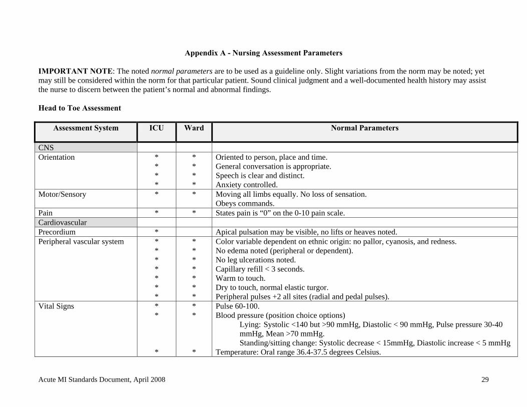
Appendix A - Nursing Assessment Parameters IMPORTANT NOTE: The noted normal parameters are to be used as a guideline only. Slight variations from the norm may be noted; yet may still be considered within the norm for that particular patient. Sound clinical judgment and a well-documented health history may assist the nurse to discern between the patient’s normal and abnormal findings. Head to Toe Assessment
Assessment System ICU Ward Normal Parameters
CNS Orientation *
* * *
* * * *
Oriented to person, place and time. General conversation is appropriate. Speech is clear and distinct. Anxiety controlled.
Motor/Sensory * * Moving all limbs equally. No loss of sensation. Obeys commands.
Pain * * States pain is “0” on the 0-10 pain scale. Cardiovascular Precordium * Apical pulsation may be visible, no lifts or heaves noted. Peripheral vascular system *
* * * * * *
* * * * * * *
Color variable dependent on ethnic origin: no pallor, cyanosis, and redness. No edema noted (peripheral or dependent). No leg ulcerations noted. Capillary refill < 3 seconds. Warm to touch. Dry to touch, normal elastic turgor. Peripheral pulses +2 all sites (radial and pedal pulses).
Vital Signs * * *
* * *
Pulse 60-100. Blood pressure (position choice options)
Lying: Systolic <140 but >90 mmHg, Diastolic < 90 mmHg, Pulse pressure 30-40 mmHg, Mean >70 mmHg.
Standing/sitting change: Systolic decrease < 15mmHg, Diastolic increase < 5 mmHg Temperature: Oral range 36.4-37.5 degrees Celsius.
Acute MI Standards Document, April 2008 29
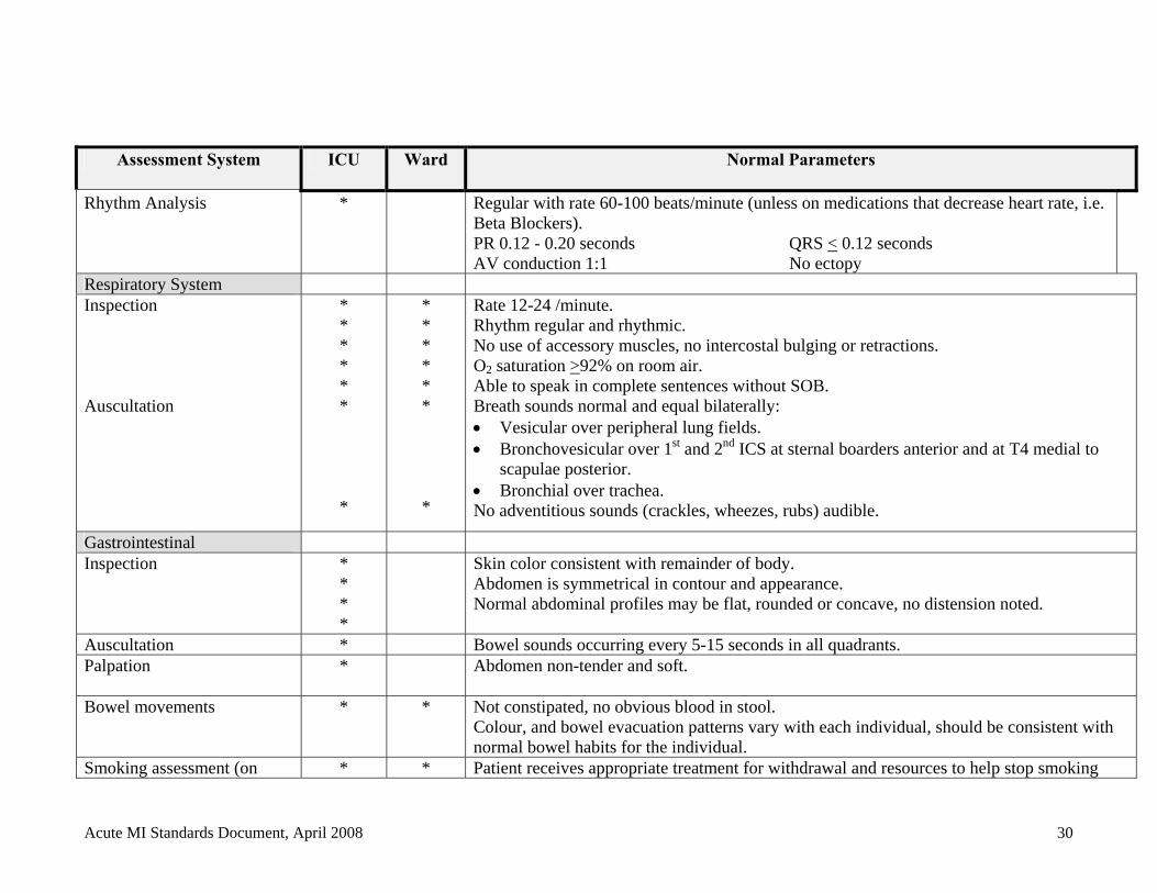
Assessment System ICU Ward Normal Parameters
Rhythm Analysis * Regular with rate 60-100 beats/minute (unless on medications that decrease heart rate, i.e. Beta Blockers). PR 0.12 - 0.20 seconds QRS < 0.12 seconds AV conduction 1:1 No ectopy
Respiratory System Inspection *
* * * *
* * * * *
Rate 12-24 /minute. Rhythm regular and rhythmic. No use of accessory muscles, no intercostal bulging or retractions. O2 saturation >92% on room air. Able to speak in complete sentences without SOB.
Auscultation * *
* *
Breath sounds normal and equal bilaterally: Vesicular over peripheral lung fields. Bronchovesicular over 1st and 2nd ICS at sternal boarders anterior and at T4 medial to
scapulae posterior. Bronchial over trachea. No adventitious sounds (crackles, wheezes, rubs) audible.
Gastrointestinal Inspection *
* * *
Skin color consistent with remainder of body. Abdomen is symmetrical in contour and appearance. Normal abdominal profiles may be flat, rounded or concave, no distension noted.
Auscultation * Bowel sounds occurring every 5-15 seconds in all quadrants. Palpation * Abdomen non-tender and soft.
Bowel movements * * Not constipated, no obvious blood in stool.
Colour, and bowel evacuation patterns vary with each individual, should be consistent with normal bowel habits for the individual.
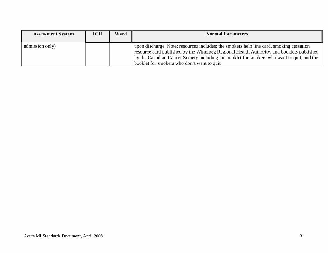
Smoking assessment (on * * Patient receives appropriate treatment for withdrawal and resources to help stop smoking
Acute MI Standards Document, April 2008 30
Assessment System ICU Ward Normal Parameters
admission only) upon discharge. Note: resources includes: the smokers help line card, smoking cessation resource card published by the Winnipeg Regional Health Authority, and booklets published by the Canadian Cancer Society including the booklet for smokers who want to quit, and the booklet for smokers who don’t want to quit.
Acute MI Standards Document, April 2008 31
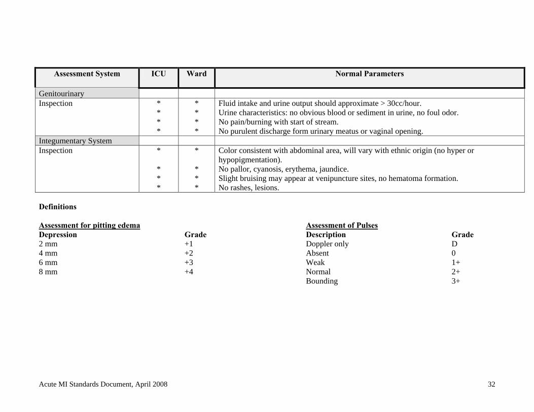
Assessment System ICU Ward Normal Parameters
Genitourinary Inspection *
* * *
* * * *
Fluid intake and urine output should approximate > 30cc/hour. Urine characteristics: no obvious blood or sediment in urine, no foul odor. No pain/burning with start of stream. No purulent discharge form urinary meatus or vaginal opening.
Integumentary System Inspection *
* * *
* * * *
Color consistent with abdominal area, will vary with ethnic origin (no hyper or hypopigmentation). No pallor, cyanosis, erythema, jaundice. Slight bruising may appear at venipuncture sites, no hematoma formation. No rashes, lesions.
Definitions Assessment for pitting edema Assessment of Pulses Depression Grade Description Grade 2 mm +1 Doppler only D 4 mm +2 Absent 0 6 mm +3 Weak 1+ 8 mm +4 Normal 2+
Bounding 3+
Acute MI Standards Document, April 2008 32
Acute MI Standards Document, April 2008 33
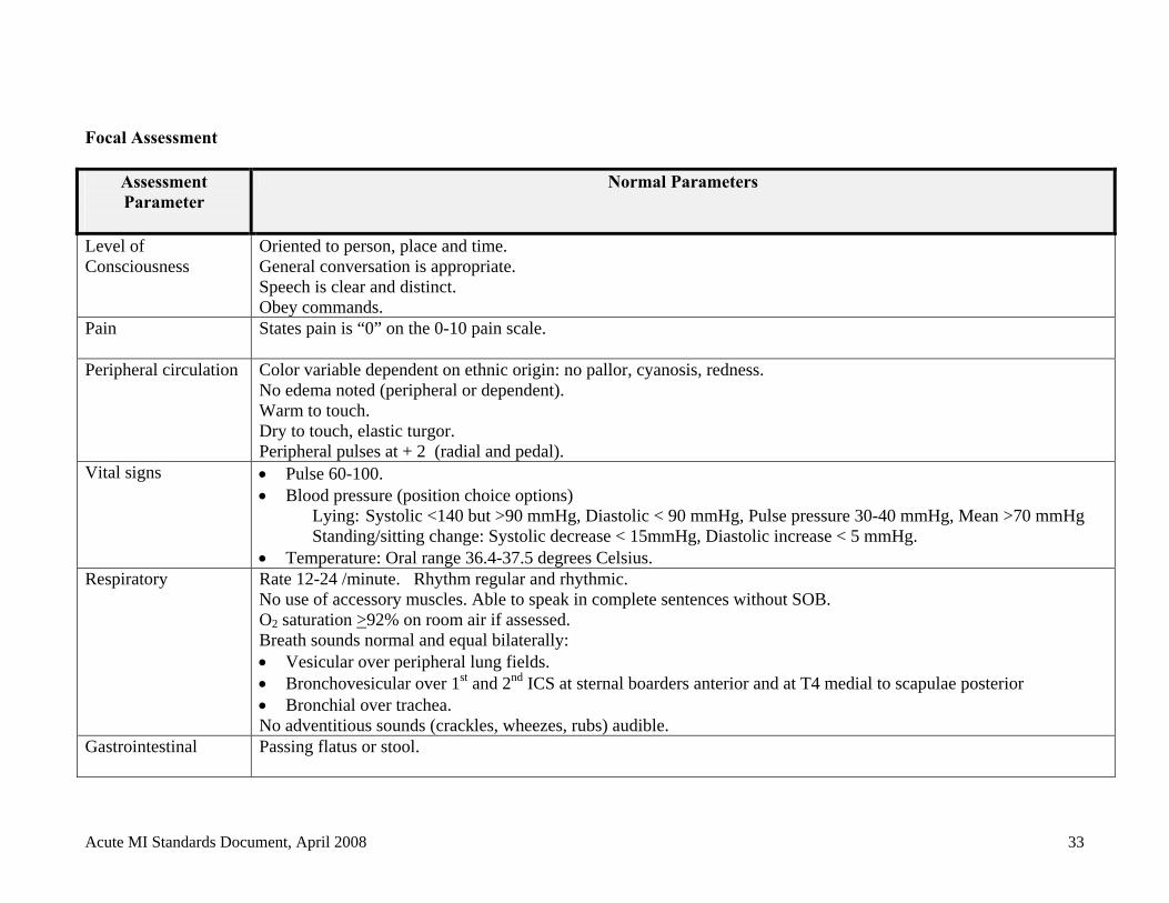
Focal Assessment
Assessment Parameter
Normal Parameters
Level of Consciousness
Oriented to person, place and time. General conversation is appropriate. Speech is clear and distinct. Obey commands.
Pain States pain is “0” on the 0-10 pain scale.
Peripheral circulation Color variable dependent on ethnic origin: no pallor, cyanosis, redness. No edema noted (peripheral or dependent). Warm to touch. Dry to touch, elastic turgor. Peripheral pulses at + 2 (radial and pedal).
Vital signs Pulse 60-100. Blood pressure (position choice options)
Lying: Systolic <140 but >90 mmHg, Diastolic < 90 mmHg, Pulse pressure 30-40 mmHg, Mean >70 mmHg Standing/sitting change: Systolic decrease < 15mmHg, Diastolic increase < 5 mmHg.
Temperature: Oral range 36.4-37.5 degrees Celsius. Respiratory Rate 12-24 /minute. Rhythm regular and rhythmic.
No use of accessory muscles. Able to speak in complete sentences without SOB. O2 saturation >92% on room air if assessed. Breath sounds normal and equal bilaterally: Vesicular over peripheral lung fields. Bronchovesicular over 1st and 2nd ICS at sternal boarders anterior and at T4 medial to scapulae posterior Bronchial over trachea. No adventitious sounds (crackles, wheezes, rubs) audible.
Gastrointestinal Passing flatus or stool.
Appendix B List of Videos
SBGH Videos 1. After your heart attack how are you doing? 2. Understanding your cardiac catheterization (Cordis 5 minutes). Information similar to
the booklet provided to patients. 3. A patient’s guide to PTCA (15 min).
These videos are available at SBGH but primarily being used with patients coming for an elective procedure. Note: A Patient education channel will soon become available at SBGH.
Manitoba Heart and Stroke Foundation Video List General
Avoiding the Surgeon's Knife VHS (60 min) - 1992 Audience - General public Profiles the participants of Dr. Dean Ornish's study on lifestyle change to reduce heart disease. Heart Safe (available in English & French) VHS (90 min) - 1993 Audience - General public Martin Sheen discusses the causes, treatment and prevention of heart disease. Useful for teaching and for support groups. The Secrets of Alive! VHS (30 min) - 1996 Audience - General public The Video guides to overall good health and wellness. It covers topics such as: nutrition; exercise; caring for your back; coping with stress; sleeping well; and dealing with anger. I Am Joe’s Heart VHS (25 min) Audience - General public, CPR courses Features dramatic vignettes from Joe’s daily life, which demonstrate the risk factors of coronary artery disease. Whimsical clay animation provides a subtle touch of humor, while graphic animation clearly and concisely details the physiology and functioning of the heart. Women and CVD Women & Heart Disease VHS (20 min) - 1991 Audience - Health professionals
Acute MI Standards Document, April 2008 34
A news report format. Highlights two women's experience with heart disease. Discusses the difficulty in diagnosing and treating women. Stresses that although diagnosis and treatment may be different the risk factors are the same. It Pays to be Heart Strong VHS (10 min) - 1998 Audience - General Public, women The video discusses the signs and symptoms of heart attack and stroke in women, including personal accounts from women affected by heart disease and candid conversations with physicians. The video also reviews the risk factors of heart disease and how to modify them. Physical Activity Fitness & Exercise VHS (30 min) Audience - General public The video highlights the physical and mental benefits of exercise; components of fitness; and guidelines for beginning and continuing a fitness program. Reviews the personal program of three individuals. Good general video, especially for those who want to start a personal program. Particularly effective opening, comparing young animals running and playing to children sitting in front of television. Flex Fit - Muscle Conditioning for Older Adults VHS (40 min) Audience - General public The video promotes exercising at your convenience, right at home using inexpensive equipment. Includes a seated version for every exercise. Fit tips and active lifestyle information for older adults are also part of this video. Smoking Butt It Out VHS (15 min) - 1984 Audience - Elementary school students, general public Directed towards 8 - 12 year olds. Uses puppets and humor to discuss the hazards of smoking, the social aspect, and role models. Diary of a Teenage Smoker VHS (30 min) - 1993 Audience - Junior High and High School students, parents and teachers. Video describing the pressures facing female adolescent and teenage smokers. Heart Health Project Workshop - Smoking VHS (28 min) - 1992 Audience - Health professionals Dr. Lertzman presents current information on smoking and heart disease including information on addiction, how to quit, and the patch. Nutrition
Acute MI Standards Document, April 2008 35
Eat Heart Smart™ VHS (30 min) - 1990 Audience - General public Reviews goals of Heart Smart™ eating such as reducing fat intake, increasing fibre, maintaining healthy body weight, reducing salt intake and limiting caffeine and alcohol intake. Shopping Heart Smart™ VHS (40 min) - 1990 Audience - General public Two reporters and a dietitian discuss the purchasing of Heart Smart™ food. The video presents general information on label reading and lowering fat through each section of the supermarket giving practical information and tips on purchasing Heart Smart™ foods. Freedom From Fat - Hidden Fat VHS (9 min) - 1987 Audience - General public, high school A humorous look at excess fat consumed in our daily diet. Discusses substitutions and more healthy choices for meals and snacks. High Blood Pressure High Blood Pressure: A Special Video VHS (30 min) Audience - General public, nursing students, health care professionals. Discusses the effects of high blood pressure on your heart and the inter-relationship with other risk factors. Includes good descriptions of high blood pressure, how it relates to heart attacks and strokes and ways to control/reduce blood pressure. Also discusses medications and lifestyle changes including relaxation techniques. Heart Attack Heart Attack: Early Warning, Early Response VHS (16 min) Audience - General public, target groups Underscores the need to seek help at the first onset of a heart attack. Shows how people tend to delay taking action by using various ploys to rationalize what is actually happening.
Acute MI Standards Document, April 2008 36

Appendix C Medication Counseling Cards
Acute MI Standards Document, April 2008 37
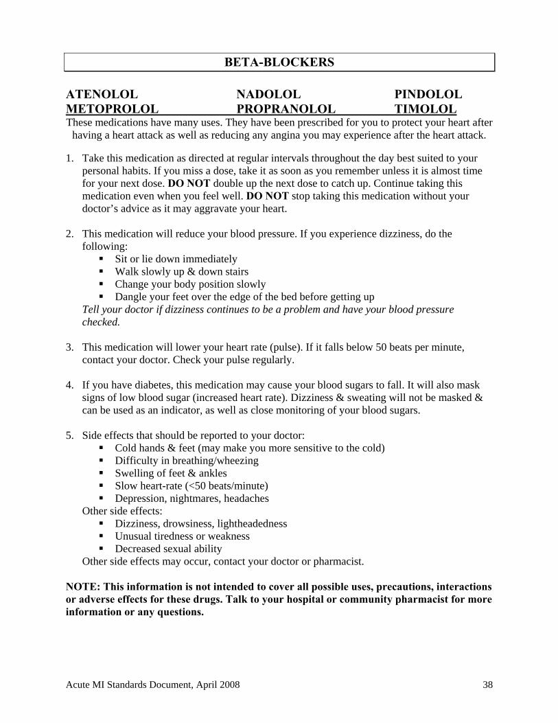
BETA-BLOCKERS ATENOLOL NADOLOL PINDOLOL METOPROLOL PROPRANOLOL TIMOLOL These medications have many uses. They have been prescribed for you to protect your heart after
having a heart attack as well as reducing any angina you may experience after the heart attack. 1. Take this medication as directed at regular intervals throughout the day best suited to your
personal habits. If you miss a dose, take it as soon as you remember unless it is almost time for your next dose. DO NOT double up the next dose to catch up. Continue taking this medication even when you feel well. DO NOT stop taking this medication without your doctor’s advice as it may aggravate your heart.
2. This medication will reduce your blood pressure. If you experience dizziness, do the
following: Sit or lie down immediately Walk slowly up & down stairs Change your body position slowly Dangle your feet over the edge of the bed before getting up
Tell your doctor if dizziness continues to be a problem and have your blood pressure checked.
3. This medication will lower your heart rate (pulse). If it falls below 50 beats per minute, contact your doctor. Check your pulse regularly.
4. If you have diabetes, this medication may cause your blood sugars to fall. It will also mask
signs of low blood sugar (increased heart rate). Dizziness & sweating will not be masked & can be used as an indicator, as well as close monitoring of your blood sugars.
5. Side effects that should be reported to your doctor:
Cold hands & feet (may make you more sensitive to the cold) Difficulty in breathing/wheezing Swelling of feet & ankles Slow heart-rate (<50 beats/minute) Depression, nightmares, headaches
Other side effects: Dizziness, drowsiness, lightheadedness Unusual tiredness or weakness Decreased sexual ability
Other side effects may occur, contact your doctor or pharmacist. NOTE: This information is not intended to cover all possible uses, precautions, interactions or adverse effects for these drugs. Talk to your hospital or community pharmacist for more information or any questions.
Acute MI Standards Document, April 2008 38
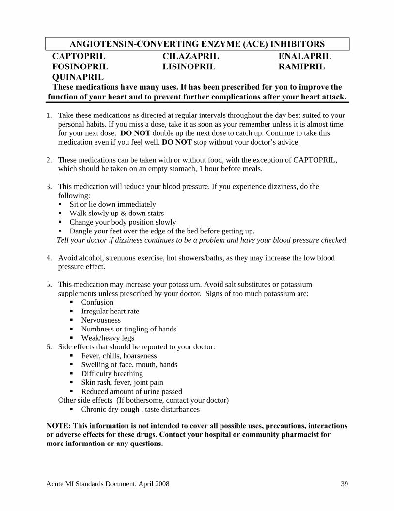
ANGIOTENSIN-CONVERTING ENZYME (ACE) INHIBITORS CAPTOPRIL CILAZAPRIL ENALAPRIL FOSINOPRIL LISINOPRIL RAMIPRIL QUINAPRIL These medications have many uses. It has been prescribed for you to improve the
function of your heart and to prevent further complications after your heart attack.
1. Take these medications as directed at regular intervals throughout the day best suited to your personal habits. If you miss a dose, take it as soon as your remember unless it is almost time for your next dose. DO NOT double up the next dose to catch up. Continue to take this medication even if you feel well. DO NOT stop without your doctor’s advice.
2. These medications can be taken with or without food, with the exception of CAPTOPRIL,
which should be taken on an empty stomach, 1 hour before meals. 3. This medication will reduce your blood pressure. If you experience dizziness, do the
following: Sit or lie down immediately Walk slowly up & down stairs Change your body position slowly Dangle your feet over the edge of the bed before getting up. Tell your doctor if dizziness continues to be a problem and have your blood pressure checked.
4. Avoid alcohol, strenuous exercise, hot showers/baths, as they may increase the low blood
pressure effect. 5. This medication may increase your potassium. Avoid salt substitutes or potassium
supplements unless prescribed by your doctor. Signs of too much potassium are: Confusion Irregular heart rate Nervousness Numbness or tingling of hands Weak/heavy legs
6. Side effects that should be reported to your doctor: Fever, chills, hoarseness Swelling of face, mouth, hands Difficulty breathing Skin rash, fever, joint pain Reduced amount of urine passed
Other side effects (If bothersome, contact your doctor) Chronic dry cough , taste disturbances
NOTE: This information is not intended to cover all possible uses, precautions, interactions or adverse effects for these drugs. Contact your hospital or community pharmacist for more information or any questions.
Acute MI Standards Document, April 2008 39
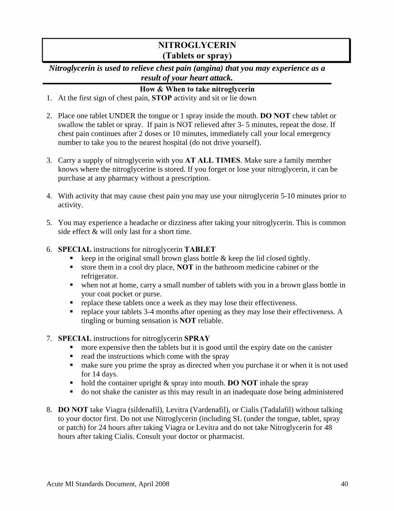
NITROGLYCERIN (Tablets or spray)
Nitroglycerin is used to relieve chest pain (angina) that you may experience as a result of your heart attack. How & When to take nitroglycerin
1. At the first sign of chest pain, STOP activity and sit or lie down
2. Place one tablet UNDER the tongue or 1 spray inside the mouth. DO NOT chew tablet or swallow the tablet or spray. If pain is NOT relieved after 3- 5 minutes, repeat the dose. If chest pain continues after 2 doses or 10 minutes, immediately call your local emergency number to take you to the nearest hospital (do not drive yourself).
3. Carry a supply of nitroglycerin with you AT ALL TIMES. Make sure a family member
knows where the nitroglycerine is stored. If you forget or lose your nitroglycerin, it can be purchase at any pharmacy without a prescription.
4. With activity that may cause chest pain you may use your nitroglycerin 5-10 minutes prior to
activity. 5. You may experience a headache or dizziness after taking your nitroglycerin. This is common
side effect & will only last for a short time. 6. SPECIAL instructions for nitroglycerin TABLET
keep in the original small brown glass bottle & keep the lid closed tightly. store them in a cool dry place, NOT in the bathroom medicine cabinet or the
refrigerator. when not at home, carry a small number of tablets with you in a brown glass bottle in
your coat pocket or purse. replace these tablets once a week as they may lose their effectiveness. replace your tablets 3-4 months after opening as they may lose their effectiveness. A
tingling or burning sensation is NOT reliable. 7. SPECIAL instructions for nitroglycerin SPRAY
more expensive then the tablets but it is good until the expiry date on the canister read the instructions which come with the spray make sure you prime the spray as directed when you purchase it or when it is not used
for 14 days. hold the container upright & spray into mouth. DO NOT inhale the spray do not shake the canister as this may result in an inadequate dose being administered
8. DO NOT take Viagra (sildenafil), Levitra (Vardenafil), or Cialis (Tadalafil) without talking
to your doctor first. Do not use Nitroglycerin (including SL (under the tongue, tablet, spray or patch) for 24 hours after taking Viagra or Levitra and do not take Nitroglycerin for 48 hours after taking Cialis. Consult your doctor or pharmacist.
Acute MI Standards Document, April 2008 40
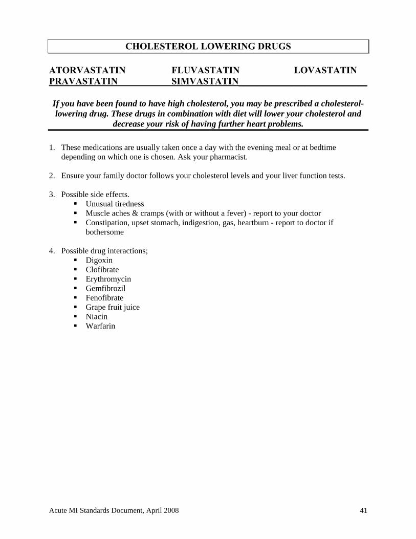
CHOLESTEROL LOWERING DRUGS ATORVASTATIN FLUVASTATIN LOVASTATIN PRAVASTATIN SIMVASTATIN___________________________
If you have been found to have high cholesterol, you may be prescribed a cholesterol-lowering drug. These drugs in combination with diet will lower your cholesterol and
decrease your risk of having further heart problems.
1. These medications are usually taken once a day with the evening meal or at bedtime
depending on which one is chosen. Ask your pharmacist. 2. Ensure your family doctor follows your cholesterol levels and your liver function tests. 3. Possible side effects.
Unusual tiredness Muscle aches & cramps (with or without a fever) - report to your doctor Constipation, upset stomach, indigestion, gas, heartburn - report to doctor if
bothersome 4. Possible drug interactions;
Digoxin Clofibrate Erythromycin Gemfibrozil Fenofibrate Grape fruit juice Niacin Warfarin
Acute MI Standards Document, April 2008 41
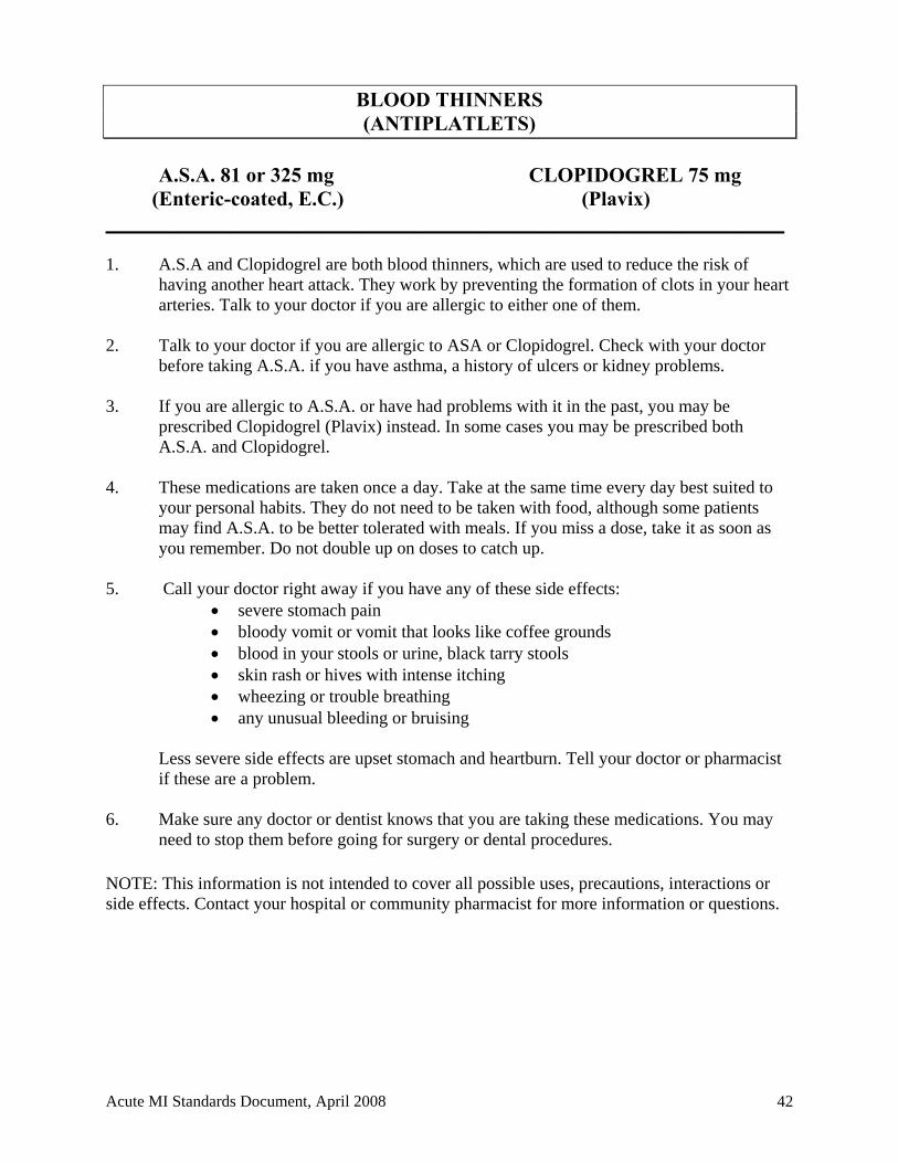
BLOOD THINNERS (ANTIPLATLETS)
A.S.A. 81 or 325 mg CLOPIDOGREL 75 mg
(Enteric-coated, E.C.) (Plavix) __________________________________________________________________ 1. A.S.A and Clopidogrel are both blood thinners, which are used to reduce the risk of
having another heart attack. They work by preventing the formation of clots in your heart arteries. Talk to your doctor if you are allergic to either one of them.
2. Talk to your doctor if you are allergic to ASA or Clopidogrel. Check with your doctor
before taking A.S.A. if you have asthma, a history of ulcers or kidney problems. 3. If you are allergic to A.S.A. or have had problems with it in the past, you may be
prescribed Clopidogrel (Plavix) instead. In some cases you may be prescribed both A.S.A. and Clopidogrel.
4. These medications are taken once a day. Take at the same time every day best suited to
your personal habits. They do not need to be taken with food, although some patients may find A.S.A. to be better tolerated with meals. If you miss a dose, take it as soon as you remember. Do not double up on doses to catch up.
5. Call your doctor right away if you have any of these side effects:
severe stomach pain bloody vomit or vomit that looks like coffee grounds blood in your stools or urine, black tarry stools skin rash or hives with intense itching wheezing or trouble breathing any unusual bleeding or bruising
Less severe side effects are upset stomach and heartburn. Tell your doctor or pharmacist if these are a problem.
6. Make sure any doctor or dentist knows that you are taking these medications. You may
need to stop them before going for surgery or dental procedures. NOTE: This information is not intended to cover all possible uses, precautions, interactions or side effects. Contact your hospital or community pharmacist for more information or questions.
Acute MI Standards Document, April 2008 42
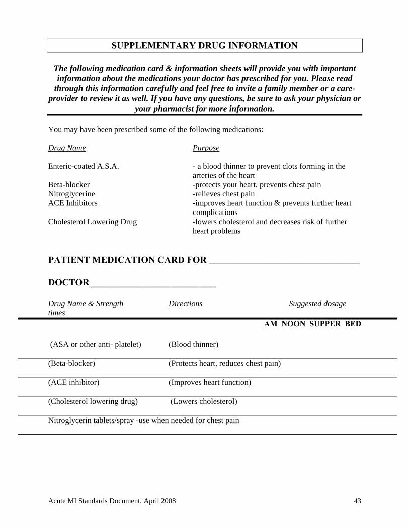
SUPPLEMENTARY DRUG INFORMATION
The following medication card & information sheets will provide you with important information about the medications your doctor has prescribed for you. Please read
through this information carefully and feel free to invite a family member or a care-provider to review it as well. If you have any questions, be sure to ask your physician or
your pharmacist for more information. You may have been prescribed some of the following medications: Drug Name Purpose Enteric-coated A.S.A. - a blood thinner to prevent clots forming in the
arteries of the heart Beta-blocker -protects your heart, prevents chest pain Nitroglycerine -relieves chest pain ACE Inhibitors -improves heart function & prevents further heart
complications Cholesterol Lowering Drug -lowers cholesterol and decreases risk of further heart problems
PATIENT MEDICATION CARD FOR ______________________________ DOCTOR___________________________ Drug Name & Strength Directions Suggested dosage imes t
AM NOON SUPPER BED
(ASA or other anti- platelet) (Blood thinner) (Beta-blocker) (Protects heart, reduces chest pain) (ACE inhibitor) (Improves heart function) (Cholesterol lowering drug) (Lowers cholesterol) Nitroglycerin tablets/spray -use when needed for chest pain
Acute MI Standards Document, April 2008 43
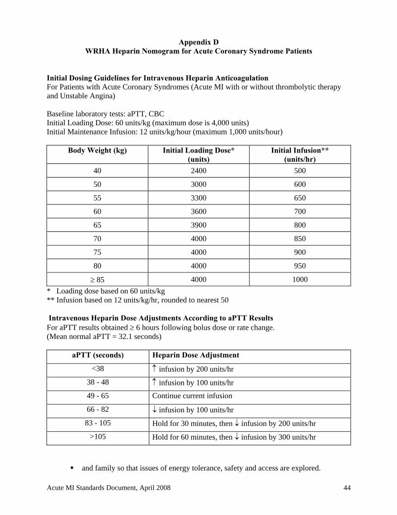
Appendix D WRHA Heparin Nomogram for Acute Coronary Syndrome Patients
Initial Dosing Guidelines for Intravenous Heparin Anticoagulation For Patients with Acute Coronary Syndromes (Acute MI with or without thrombolytic therapy and Unstable Angina) Baseline laboratory tests: aPTT, CBC Initial Loading Dose: 60 units/kg (maximum dose is 4,000 units) Initial Maintenance Infusion: 12 units/kg/hour (maximum 1,000 units/hour)
Body Weight (kg) Initial Loading Dose* (units)
Initial Infusion** (units/hr)
40 2400 500
50 3000 600
55 3300 650
60 3600 700
65 3900 800
70 4000 850
75 4000 900
80 4000 950
85 4000 1000
* Loading dose based on 60 units/kg ** Infusion based on 12 units/kg/hr, rounded to nearest 50 Intravenous Heparin Dose Adjustments According to aPTT Results For aPTT results obtained 6 hours following bolus dose or rate change. (Mean normal aPTT = 32.1 seconds)
aPTT (seconds) Heparin Dose Adjustment
<38 infusion by 200 units/hr
38 - 48 infusion by 100 units/hr
49 - 65 Continue current infusion
66 - 82 infusion by 100 units/hr
83 - 105 Hold for 30 minutes, then infusion by 200 units/hr
>105 Hold for 60 minutes, then infusion by 300 units/hr
and family so that issues of energy tolerance, safety and access are explored.
Acute MI Standards Document, April 2008 44
WRHA Cardiac Sciences Program
TITLE Criteria for Referral to Social Work
AUTHORIZED BY WRHA Cardiac Sciences Program
EFFECTIVE DATE October 2002
REVISED DATE PAGE 1 of 2
Social Work consults can be made on the social work consult form. ISSUE INDICATOR 1. DOMESTIC ABUSE OR
NEGLECT INCLUDING FINANCIAL ABUSE
- Patient/family/community makes disclosure of abuse/neglect - Patient’s bruises/injuries are consistent with abuse - Patient’s bruises/injuries are not consistent with explanation - Patient fearful of returning home; fearful of family member(s) - Patient needs place of safety - Patient has no funds for day to day needs; no knowledge of
financial situation - Patient’s basic needs have not been met
2. BARRIERS TO EFFECTIVE DISCHARGE
- Patient is unable to return to former residence due to: change in physical and/or mental functioning, residence is unsuitable to care needs, care provider is unwilling to continue in role, eviction, home requires changes before discharge (cleaning, renovations), patient is homeless, transient, patient requires institutional care
- Patient has no income or disability benefits, there is lack of income/delayed income for aids to independent living (e.g. wheelchair, transportation)
- Patient/family is not in agreement with discharge plan
3. PATIENT HAS CARE GIVING RESPONSIBILITIES
- Child under 12 is left alone - Mentally/physically incapacitated adult is left alone - Child under 12 is left alone with chronically ill adult
Acute MI Standards Document, April 2008 45
WRHA Cardiac Sciences Program
TITLE Criteria for Referral to Social Work
AUTHORIZED BY WRHA Cardiac Sciences Program
EFFECTIVE DATE October 2002
REVISED DATE PAGE 2 of 3
4. PATIENT/FAMILY EXPERIENCING COPING AND ADAPTIONAL DIFFICULTIES
- Disruption in primary role, e.g. parenting, employment - Patient/family expressing feelings of being overwhelmed - Pattern of lack of attention to ADL’s and healthy lifestyle not
related to medication/treatment - Family responding inappropriately to patient - Repetitive information seeking from a number of health care
providers - Immobilized, i.e. emotionally not ready to follow treatment
plans or make day to day decisions
5. PATIENT REQUIRES COMMUNITY RESOURCES
- Patient expresses concern re: child care, finances, employment interruptions, housing, transportation
- Patient has vocational concerns - Patient requires a change in school/work, e.g. change in hours - Patient has insufficient income to meet medical needs - Specialized transportation needed
6.STRESSED FAMILY CARE GIVERS
- Caregiver refuses help when patient requires high degree of care
- Caregiver shows exhaustion, health problems - Caregiver has emotional outbursts, e.g. crying, anger - Caregiver requests patient be hospitalized
7. PATIENTS WITH PROBLEMATIC SOCIAL SUPPORT NETWORK
- Relationship problems within network that interfere with treatment, recovery
- Patient expresses concern re: lack of supportive caring relationship
- Absence of identified next of kin - Conflict within family network that interferes with treatment - Lack of clarity/conflict within family regarding which family
member is decision-maker - Patient has problems in cognitive functioning
Acute MI Standards Document, April 2008 46
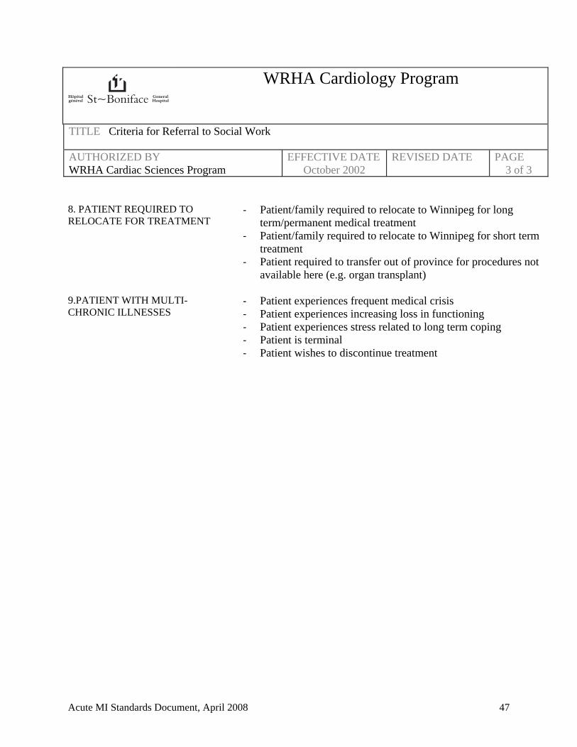
WRHA Cardiology Program
TITLE Criteria for Referral to Social Work
AUTHORIZED BY WRHA Cardiac Sciences Program
EFFECTIVE DATE October 2002
REVISED DATE PAGE 3 of 3
8. PATIENT REQUIRED TO RELOCATE FOR TREATMENT
- Patient/family required to relocate to Winnipeg for long term/permanent medical treatment
- Patient/family required to relocate to Winnipeg for short term treatment
- Patient required to transfer out of province for procedures not available here (e.g. organ transplant)
9.PATIENT WITH MULTI-CHRONIC ILLNESSES
- Patient experiences frequent medical crisis - Patient experiences increasing loss in functioning - Patient experiences stress related to long term coping - Patient is terminal - Patient wishes to discontinue treatment
Acute MI Standards Document, April 2008 47
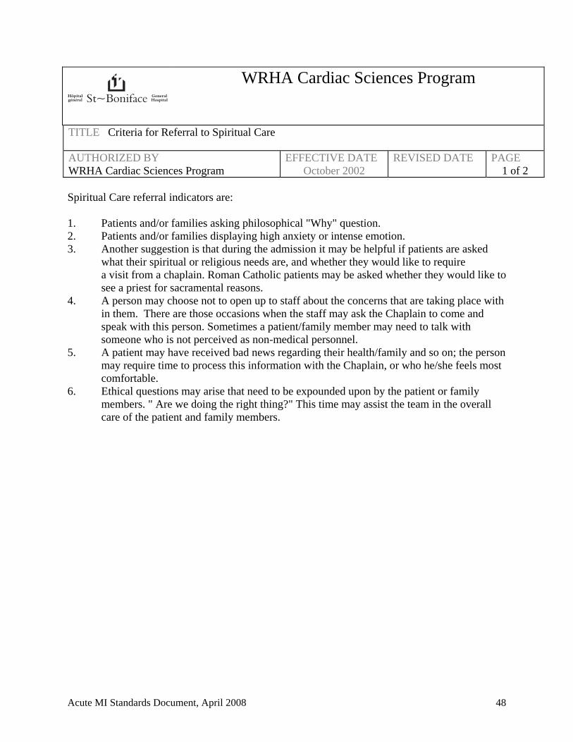
WRHA Cardiac Sciences Program
TITLE Criteria for Referral to Spiritual Care
AUTHORIZED BY WRHA Cardiac Sciences Program
EFFECTIVE DATE October 2002
REVISED DATE PAGE 1 of 2
Spiritual Care referral indicators are: 1. Patients and/or families asking philosophical "Why" question. 2. Patients and/or families displaying high anxiety or intense emotion. 3. Another suggestion is that during the admission it may be helpful if patients are asked
what their spiritual or religious needs are, and whether they would like to require a visit from a chaplain. Roman Catholic patients may be asked whether they would like to see a priest for sacramental reasons.
4. A person may choose not to open up to staff about the concerns that are taking place with in them. There are those occasions when the staff may ask the Chaplain to come and speak with this person. Sometimes a patient/family member may need to talk with someone who is not perceived as non-medical personnel.
5. A patient may have received bad news regarding their health/family and so on; the person may require time to process this information with the Chaplain, or who he/she feels most comfortable.
6. Ethical questions may arise that need to be expounded upon by the patient or family members. " Are we doing the right thing?" This time may assist the team in the overall care of the patient and family members.
Acute MI Standards Document, April 2008 48
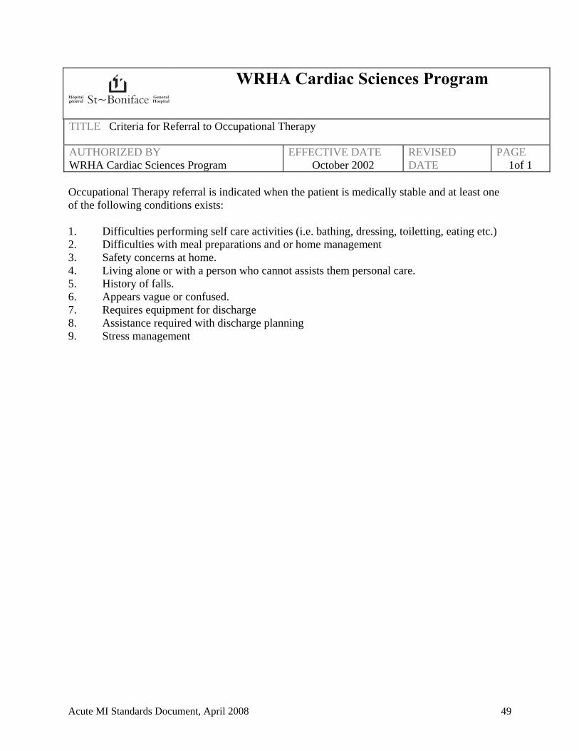
WRHA Cardiac Sciences Program
TITLE Criteria for Referral to Occupational Therapy
AUTHORIZED BY WRHA Cardiac Sciences Program
EFFECTIVE DATE October 2002
REVISED DATE
PAGE 1of 1
Occupational Therapy referral is indicated when the patient is medically stable and at least one of the following conditions exists: 1. Difficulties performing self care activities (i.e. bathing, dressing, toiletting, eating etc.) 2. Difficulties with meal preparations and or home management 3. Safety concerns at home. 4. Living alone or with a person who cannot assists them personal care. 5. History of falls. 6. Appears vague or confused. 7. Requires equipment for discharge 8. Assistance required with discharge planning 9. Stress management
Acute MI Standards Document, April 2008 49
WRHA Cardiac Sciences Program
TITLE Criteria for Referral to Clinical Dietician
AUTHORIZED BY WRHA Cardiac Sciences Program
EFFECTIVE DATE February 2001
REVISED DATE October 2002
PAGE 1 of 1
ALL Ami Care Map patients receive a referral:
Acute MI Standards Document, April 2008 50
Glycemic control and the MI Care Map
Diabetes and coronary risk The complications of diabetes can be divided between microvascular (nephropathy, retinopathy, peripheral neuropathy), and macrovascular (coronary artery disease, cerebrovascular disease).i Coronary heart disease is the highest cause of mortality among diabetic patients.ii Epidemiological studies have indicated that those who have diabetes are at same level of risk for a cardiovascular event as those who have had a previous myocardial infarction (MI). iii,iv Having the combination of diabetes and a history of MI places an individual at a very high level of cardiovascular risk. The relationship between diabetes and future coronary risk holds true whether an post-MI patient has previously known diabetes or is newly diagnosed during their MI admission. v
Long term glycemic control and coronary risk The first large study to investigate the cardiovascular effects of tighter glucose control in type I diabetes was the Diabetes Control and Complications Trial (DCCT).vi This trial compared conventional (mean Ha1c 9%) to more intensive insulin therapy (mean Ha1c 7%). After a decade of follow-up the patients in the more intensive arm had dramatic reduction in the rate of microvascular complications (retinopathy 76%; nephropathy 54%; neuropathy 60%). In terms of macrovascular events, there was a 41% reduction, which did not reach statistical significance due to the small numbers of events in this young population (median age 27 years). However in the 7 year follow-up after the trial (Epidemiology of diabetes interventions and complications: EDIC) a statistically significant 42% reduction in cardiovascular events was found.vii This result was despite the fact that during the 7 year follow up there was no significant different in the patient’s Ha1c (conventional 8.2 % vs. intensive 8%). The UKPDS was a similar study in Type II diabetes. Similarly it was found that more intensive glucose control (with sulfonyureas, insulin or metformin) resulted in clear reduction in microvascular events, but less robust reductions in macrovascular events.viii More recently the Proactive study used pioglitazone to aggressively lower glucose levels.ix The trial failed to show a significant difference in the primary composite cardiovascular endpoint, but had a significant 27% reduction in the secondary endpoint of death/MI/stroke, Acute/post MI glycemic control and coronary risk The Diabetes Mellitus Insulin Glucose Infusion Acute Myocardial Infarction (DIGAMI) trial examined tighter glucose control in the acute and post MI periodx. During the first 24-hours an insulin infusion was used to maintain a glucose level 7-10.9 mmol/L., after which multiple dose per-day insulin injections were used for at least 3 months. After the end of the year there were more people on insulin in the intervention arm 72% compared to the control 42% as well the Ha1c was lower in the intervention arm (difference 0.9). There was a 7.5% absolute reduction in mortality at the end of the year. An unresolved issue from the trial was whether the mortality benefit was purely from the long term control, or whether the early IV infusion played a role. Therefore DIGAMI-2 was devised with one arm having only an early IV infusion, one arm with IV infusion followed by intensive multi-dose therapy, and a control.xi In this trial at one year there was no difference in the Ha1c between the groups, and also no difference in mortality rates. The implication from this trial was that lowering Ha1c after an MI is important, but the method used to reach this goal (insulin, oral meds, diet) is less important. Conclusion
Acute MI Standards Document, April 2008 51
Acute MI Standards Document, April 2008 52
There is a clear relationship between diabetes and cardiovascular disease. There is an increasing body of evidence suggesting both primary and secondary prevention of cardiovascular disease with intensive glucose control. Given this evidence it is a rational step to begin to screen for patients with impair glucose control in acute MI patients. As well it is an opportune time to assess the appropriateness of the diabetes regimen for those already known to have diabetes on admission for acute MI. This is the rationale for the development of the glycemic control protocols incorporated into the AMI care map.
i Canadian Diabetes Association: 2003 Clinical Practice Guidelines for the Prevention and Management of Diabetes in Canada. Canadian Journal of Diabetes 2003;27: Suppl 2; S1-S150. ii Geiss LS, Herman WH, Smith PJ. Mortality in Non-Insulin-Dependent Diabetes. In: National Diabetes Data Group, eds. Diabetes in America. 2nd ed. Bethesda, MD: National Institutes of Health, National Institute of Diabetes and Digestive and Kidney Diseases; 1995. iii Malmberg K, Yusuf S, Gerstein HC, Brown J, Zhao F, Hunt D, Piegas L, Calvin J, Keltai M, Budaj A. Impact of diabetes on long-term prognosis in patients with unstable angina and non-Q-wave myocardial infarction: results of the OASIS (Organization to Assess Strategies for Ischemic Syndromes) Registry. Circulation. 2000;102(9):1014-1019. iv Haffner et al v Aguilar D et al. Circulation 2004; 110:1572-1578. vi Diabetes Control and Complications Trial Research Group N Eng J Med 1993;329:977-986 vii Epidemiology of diabetes interventions and complications study resreach group; N Engl J Med 2005; 353:2643-53. viii UKPDS group: Lancet 1998; 352:837-865 ix Dormandy JA et al. Lancet. 2005;366:1279-89. x DIGAMI Study Group: J Am Coll Cardiol. 1995 Jul;26(1):57-65.. xi DIGAMI-2 Study Group: Eur Heart J. 2005 Apr;26(7):650-61. Epub 2005 Feb 23