Embed Size (px)
Citation preview
Occupational Lung Disease
SS VisserInternal Medicine
UP
Classification
• Anorganic ( mineral ) dust/PneumoconiosisFibrogenic - silica, asbestos, talc, silicatesNon-fibrogenic - Fe, barium, tin• Immunologic/PharmcologicAllergic alveolitis (hypersensitivity pneumonitis)Asthma - feathers, enzymes, cotton, platinum• Irritant gases, vapours, smoke: high levels → pul-
monary oedema, low levels→industrial bronchitis• Respiratory carcinogens: asbestos, bis-chloro-
methylether
Silicosis
Professions associated with exposure:• Mining - ore of Au, Fe, Pb, Zn, Cu, rock• Production - rock & concrete works,
abrasives, pottery, isolation material• Construction - tunnels, roads and train
tracks, boiler makers, restoration of old buildings.
Prevalence and risks
• Prevalence: 22/1000 miners (1917-20) to <8/1000 miners currently
• Risk: 24 years vs 36 years exposure to:low dust levels: 5% 10%high dust levels: 15% 40%
Pathology
Macroscopic:• hard gray-black nodules upper lobes and perihilar• Massive fibrosis - large firm masses, shrunken
upper lobes, emphysematous lower lobes and subpleural blebs
• PMF (progressive massive fibrosis): upper mid and lower lobes (accelerated silicosis)
• Cavitation (ischaemic necrosis) → secondary Tb → silicotuberculosis
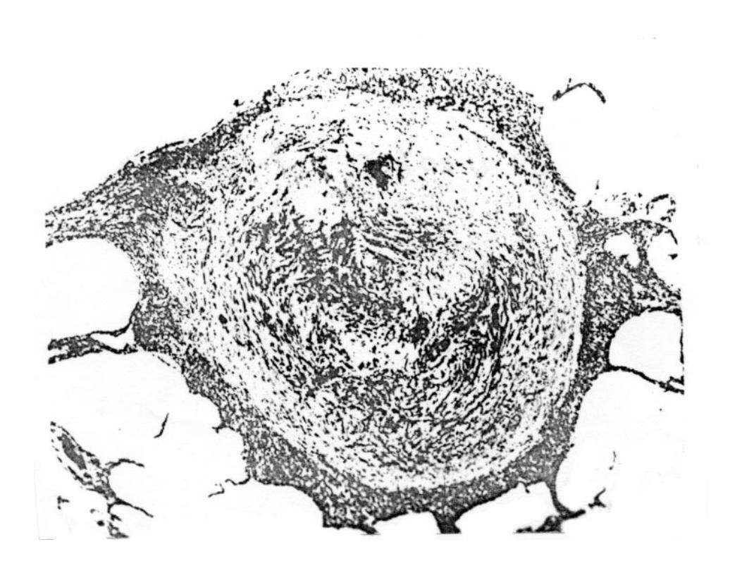
Pathology
Microscopic: silicotic nodule• Central zone: hialine connective tissue in
concentric layers - acellular, no capillaries, varying silica content, occasional ischaemia
• Middle zone: cellular connective tissue• Peripheral zone: halo of macrophages projecting
into parenchyma, high silica content• Located around respiratory bronchioli, blood
vessels, pleural surfaces, interlobular septae
Clinical manifestations
• Classic / Uncomplicated silicosis• Chronic silicosis• Accelerated silicosis• Acute silicosis• Silicoproteinosis• Silico-tuberculosis• PMF• Complicated silicosis
Diagnosis
• History of exposure• Radiology• Eliminate potentially treatable diseases
( Tb, sarcoidosis, Idiopathic pulmonary fibrosis)
• Lung biopsy
Diagnosis: Radiology
• XRChest - small nodules, 1-10 mm upper lung zones, calification
• Reticular veiling• Hilar and mediastinal lymphadenopathy (egg shell
calcification)• Cavitation• Pneumothorax• Alveolar veiling• Caplan syndrome
Diagnosis: Physiology
• Lung function: -varies from normal to obstructive or restrictive or combination
• Diffusion decreased• Hypoxaemia on exertion
Diagnosis: Serology
• Hypergammaglobulinemia• RF• ANF• S-ACE• Increased incidence of systemic sclerosis
described in SA gold miners
Treatment
• Terminate exposure to prevent PMF• Corticosteroids, pulmonary lavage, lung
transplantation• Treat complications: Tb, pneumothorax,
COPD, Cor pulmonale, collagen vascular diseases.

Asbestos
• Serpentine asbestos: Chrysotile (white asbestos) – RSA, Russia, Canada
• Amphibole asbestos: Crocidolite ( blue asbestos) – RSA Limpopo, Mpumulanga, Northern CapeAmosite ( brown asbestos) - as aboveAnthophyllite – Finland, worldwideActinolite – RSA, Taiwan
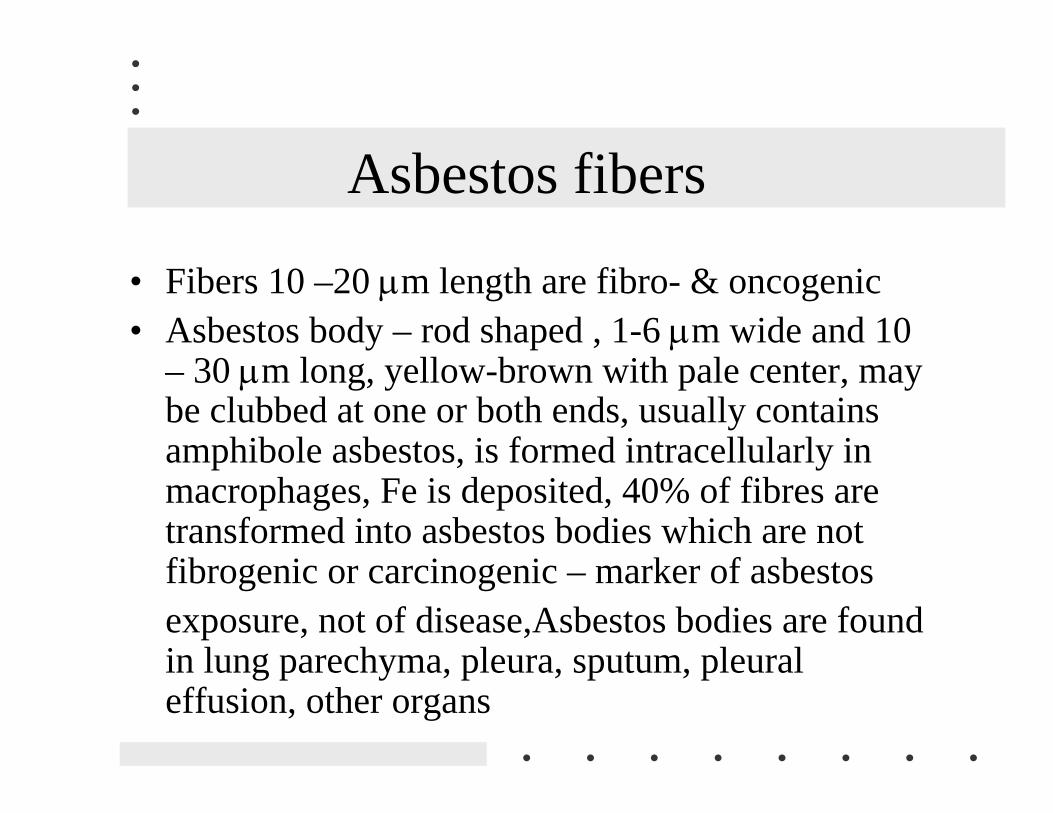
Asbestos fibers
• Fibers 10 –20 µm length are fibro- & oncogenic• Asbestos body – rod shaped , 1-6 µm wide and 10
– 30 µm long, yellow-brown with pale center, may be clubbed at one or both ends, usually contains amphibole asbestos, is formed intracellularly in macrophages, Fe is deposited, 40% of fibres are transformed into asbestos bodies which are not fibrogenic or carcinogenic – marker of asbestosexposure, not of disease,Asbestos bodies are found in lung parechyma, pleura, sputum, pleural effusion, other organs
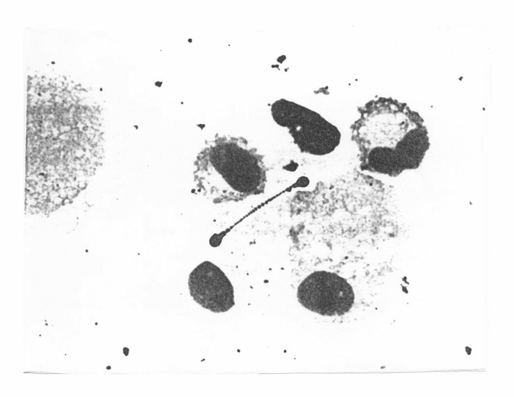
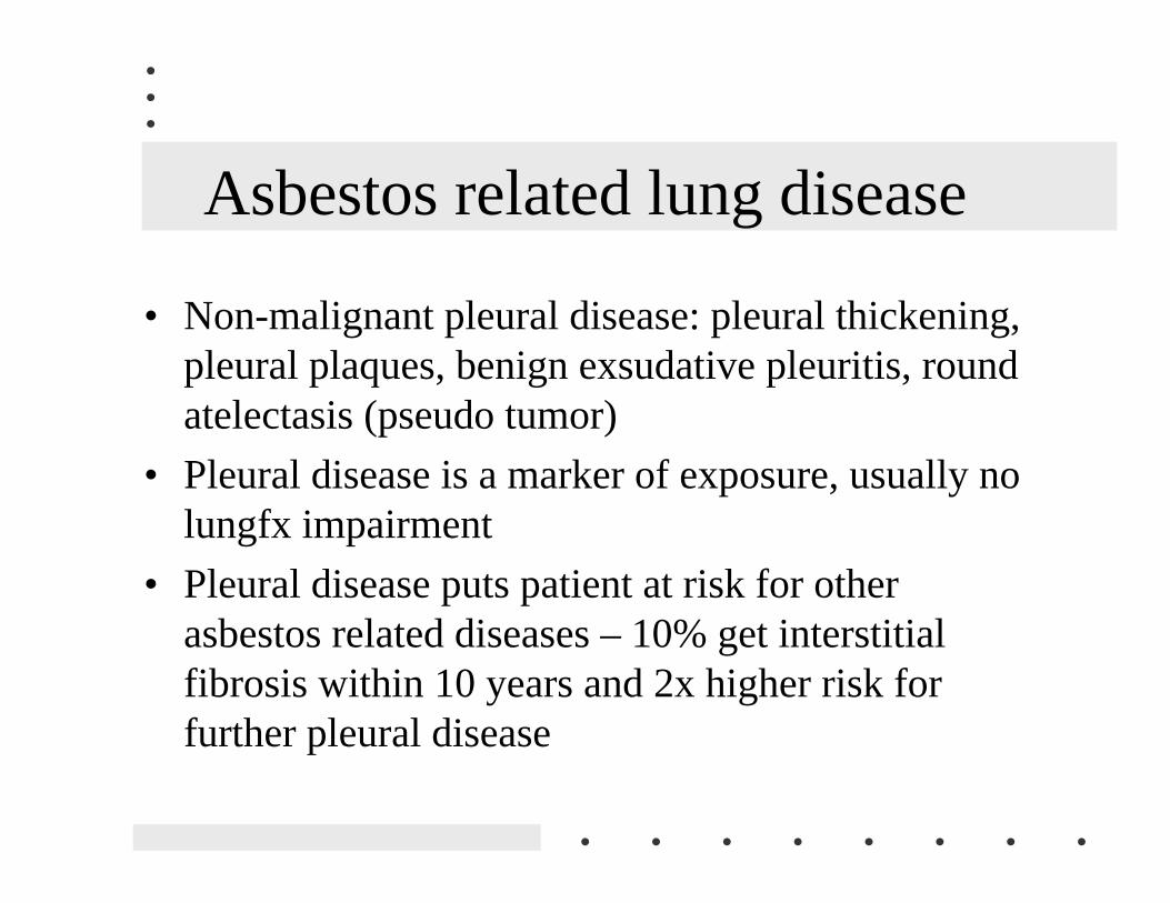
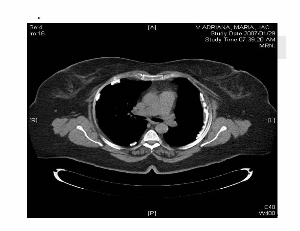
Asbestos related lung disease
• Non-malignant pleural disease: pleural thickening, pleural plaques, benign exsudative pleuritis, round atelectasis (pseudo tumor)
• Pleural disease is a marker of exposure, usually no lungfx impairment
• Pleural disease puts patient at risk for other asbestos related diseases – 10% get interstitial fibrosis within 10 years and 2x higher risk for further pleural disease
Asbestos related lung disease
• Asbestosis = parenchymal disease interstitial fibrosis
• Associated more with crocidolite• Smokers more prone to disease and XRC
interstitial infiltrates• Smokers 2,6x greater risk to die of
asbestosis
Asbestos related lung disease
• Clinical presentation: exertional dyspnea, late inspiratory creps, clubbing (60%)
• XRC: reticular veiling lower lobes, ground glass veiling, pleural changes, PMF in mixed exposure, rarely Caplan syndrome
• Lungfx: restrictive, diffusion↓, art hypoxemia, elastic recoil ↑
• Non-specific immunologic findings: ANF, RF, S-ACE elevation, ? HLAB27 association
Asbestos related lung disease
• Bronchogenic Ca: 5x higher incidence in non-smoking asbestos workers, 90x higher in smoking asbestos workers
• Prevalence for adeno ca• Chrysotile highest risk bronchus Ca• Crocidolite highest risk for mesothelioma• Other neoplasms: larynx ca, GIT ca, breast
ca, ovarian ca renal ca.
Asbestos related lung disease
• Diffuse malignant mesothelioma: 35-45 y after exposure, more with crocidolite, pleural plaques not a precursor
• 4 histologic patterns: epithelial, mesothelial, mixed, undifferentiated
• Presentation: Chest pain prominent, dyspnea, clubbing, pleural effusion
• XRC: effusion• CT: pleural based lobular mass with chest wall
and rib involvement
Coal workers pneumoconiosis( = Anthracosis)
• Simple anthracosis – asymptomatic or slight productive cough , radiologic diagnosis after 15 y exposure,
• Complicated anthracosis – chronic bronchitis, small % get PMF, functional impairment, PHT and cor pulmonale, may develop PTb, associated auto-immune reactions and ↑incidence of RA
• When exposure is terminated the simple type will not progress; PMF type will progress
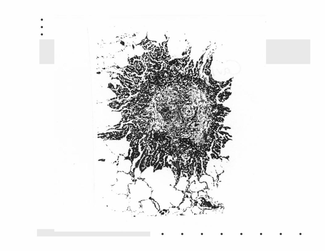
Coal workers pneumoconiosis
• Radiology: nodular veiling upper lung zones, nodules > 1 cm indicative of PMF
• Lungfx: normal – simple typerestrictive – complicated typein smokers – obstructive
• Prognosis: simple type – goodcomplicated type – cardio-respiratory failure
Metals
Metal ore – mixed dust exposure in open spaces –progressive pneumoconiosis
Welding – fumes and respirable particles ( < 1µm cs) in closed spaces – acute pulmonary disease = metal fume fever, influenza –like manifestations lasting 24 hours.
Acute Cadmium toxicity – pulmonary oedema, chemical pneumonitis, destructive emphysema with severe exposure
Metals
• Beryllium – inhaled, binds to protein and is distributed to liver, spleen and bone, may cause granulomatous ulcers of skin, perfo-ration of nasal septum, upper airway irritation, bronchiolitis, pneumonia, pulmonary oedema, chronic disease after 5-10 y – granulomatous pneumonitis (similar to hypersensitivity pneumonitis)