Embed Size (px)
Citation preview

Sputum smear microscopy in the diagnosis of pulmonary tuberculosis:
What are the options anno 2014?
Author: dr. Charlotte Trouvé Supervisor: dr. Eric Nulens
Overview • Introduction and History • CAT questions
– What are the current avaible and recommended staining methods for sputum smear microscopic examination in the diagnosis of tuberculosis?
– What are the results of our own study, where three relevant staining methods are compared to each other?
• CAT answers
• General conclusions
Tuberculosis in a nutshell
• Described by Hippocrates (5th century BC) ‘phtysis’ 24 March 1982: Tubercle bacillus (Robert Koch) • Infectious disease caused by Mycobacterium tuberculosis • Air-born transmission • Pulmonary VS extra-pulmonary • Active VS latent • Symptoms: chronic cough, fever, night sweats, weight
loss • Treatment: multiple antibiotics over a long period
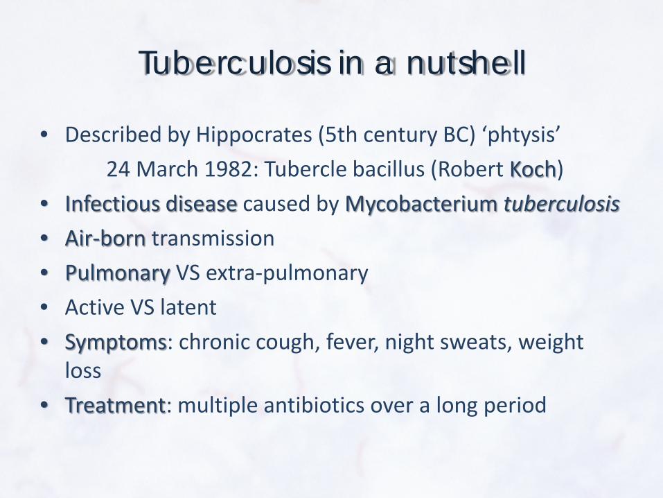
- Deadliest infectious diseases affecting humans with yearly 2 million people who die from tuberculosis
= 7% of all deaths
- Approximately 1/3 of the world population is infected with M. tuberculosis.
- 8-10 million new cases of TB per year.
- Leading cause of death among people with HIV/AIDS.
“global public health emergency”
Why Does TB Need Global Attention anno 2014?
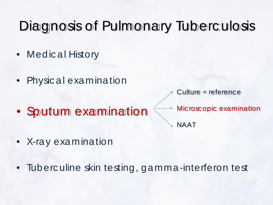
Diagnosis of Pulmonary Tuberculosis
• Medical History
• Physical examination
• Sputum examination
• X-ray examination
• Tuberculine skin testing, gamma-interferon test
Culture = reference Microscopic examination NAAT
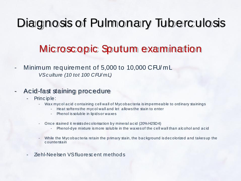
Diagnosis of Pulmonary Tuberculosis
Microscopic Sputum examination
- Minimum requirement of 5,000 to 10,000 CFU/mL VS culture (10 tot 100 CFU/mL)
- Acid-fast staining procedure
- Principle: - Wax mycol acid containing cell wall of Mycobacteria is impermeable to ordinary stainings
- Heat softens the mycol wall and let allows the stain to enter - Phenol is soluble in lipids or waxes
- Once stained it resists decolorisation by mineral acid (20% H2SO4)
- Phenol-dye mixture is more soluble in the waxes of the cell wall than alcohol and acid
- While the Mycobacteria retain the primary stain, the background is decolorized and takes up the counterstain
- Ziehl-Neelsen VS fluorescent methods
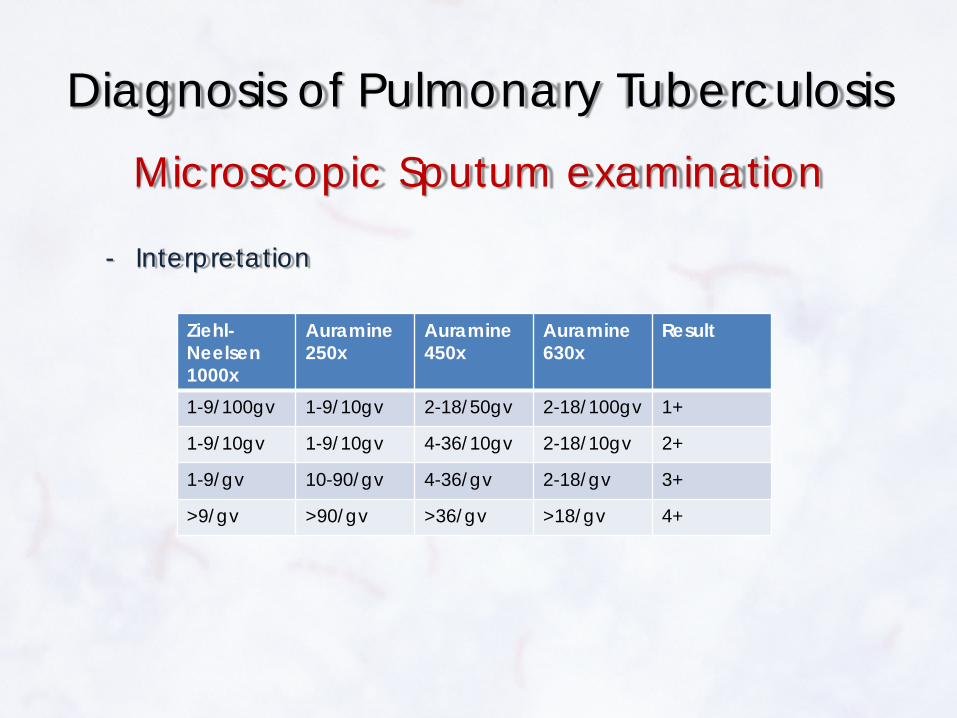
Diagnosis of Pulmonary Tuberculosis Microscopic Sputum examination
- Interpretation
Ziehl-Neelsen 1000x
Auramine 250x
Auramine 450x
Auramine 630x
Result
1-9/100gv 1-9/10gv 2-18/50gv 2-18/100gv 1+
1-9/10gv 1-9/10gv 4-36/10gv 2-18/10gv 2+
1-9/gv 10-90/gv 4-36/gv 2-18/gv 3+
>9/gv >90/gv >36/gv >18/gv 4+
PART 1:
What are the most available staining techniques for the detection of acid-fast bacilli
and what are the current recommendations?
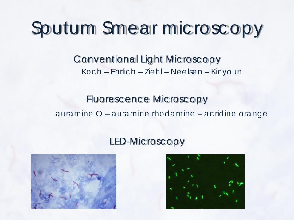
Sputum Smear microscopy Conventional Light Microscopy
Koch – Ehrlich – Ziehl – Neelsen – Kinyoun
Fluorescence Microscopy auramine O – auramine rhodamine – acridine orange
LED-Microscopy
Conventional Light Microscopy
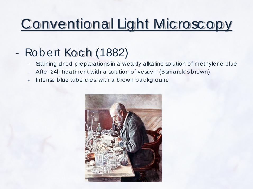
- Robert Koch (1882) - Staining dried preparations in a weakly alkaline solution of methylene blue - After 24h treatment with a solution of vesuvin (Bismarck’s brown) - Intense blue tubercles, with a brown background
Conventional Light Microscopy
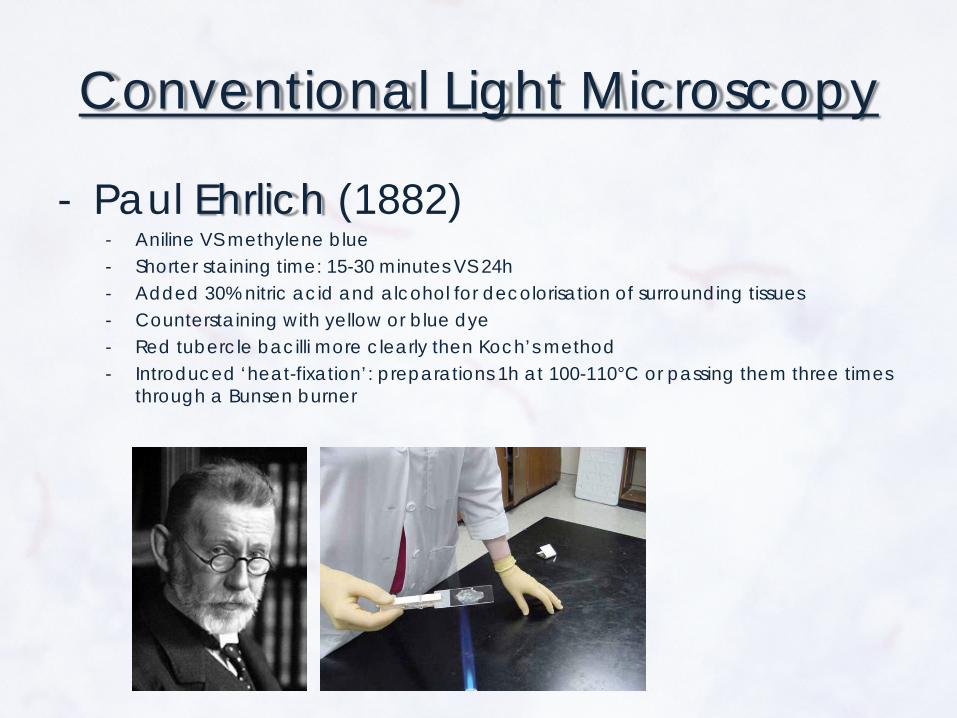
- Paul Ehrlich (1882) - Aniline VS methylene blue - Shorter staining time: 15-30 minutes VS 24h - Added 30% nitric acid and alcohol for decolorisation of surrounding tissues - Counterstaining with yellow or blue dye - Red tubercle bacilli more clearly then Koch’s method - Introduced ‘heat-fixation’: preparations 1h at 100-110°C or passing them three times
through a Bunsen burner
Conventional Light Microscopy
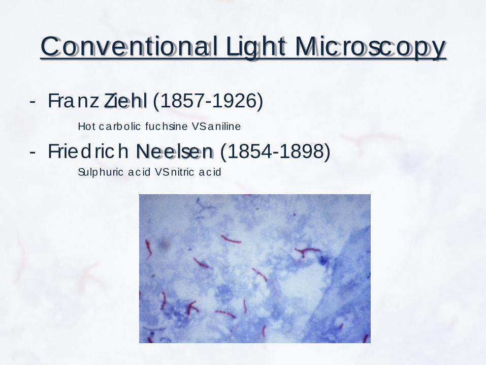
- Franz Ziehl (1857-1926) Hot carbolic fuchsine VS aniline
- Friedrich Neelsen (1854-1898) Sulphuric acid VS nitric acid
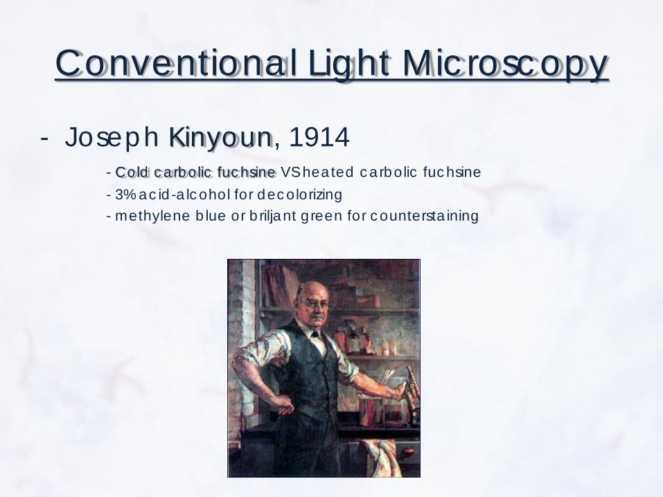
Conventional Light Microscopy
- Joseph Kinyoun, 1914 - Cold carbolic fuchsine VS heated carbolic fuchsine - 3% acid-alcohol for decolorizing - methylene blue or briljant green for counterstaining
Conventional Light Microscopy
Ziehl-Neelsen
• Most applicable and available diagnostic tool of choice for diagnosis of TB in developing countries
• Rapid, inexpensive
• Excellent reported specificities: 96% - 100% – Highly specific in areas with high incidence
Luelmo F. What is the role of sputum microscopy in patients attending health facilities? Geneva: World Health Organisation, 2004:7-13 Perkins M. New diagnostic tools for tuberculosis. Int J Tuberc Lung Dis 2010; 4:S182-88
• Variable reported sensitivities: 20% - 86%
Ramsay A. Front-loading sputum microscopy services: an opportunity to optimize smear-based case detection of tuberculosis in high prevalence countries. J Trop Med 2009; 2009:1-6 Cattamanchi A. Integrated strategies to optimize smear microscopy: a prospective observational study. Am J Respir Crit Care Med 2011; 183: 547-551 Cuevas L. A multi-country non-inferiority cluster randomized trial of frontloaded smear microscopy for the diagnosis of pulmonary tuberculosis. PLoS Med 2011; 8:e1000443 Cuevas L. LED fluorescence microscopy for the diagnosis of pulmonary tuberculosis: a multi-country cross sectional evaluation. PLoS Med 2011; 8:e1001057 Myneedu V. A pilot study of same day sputum smear examination, its feasibility and usefulness in diagnosis of pulmonary TB. Indian J Tuberc 2011; 58:160-167 Steingart K. Fluorescene versus conventional sputum smear microscopy for tuberculosis: a systematic review. Lancet Infect Dis 2006; 6: 570-581
Conventional Light Microscopy
Ziehl-Neelsen: sensitivity • Variable reported sensitivities: 20% - 86%
• Influence of other factors:
– Prevalence/severity TB – Type of specimen – Method of processing (concentrated vs direct) – Method of centrifugation – Quality examination
Iademarco et al: ZN significant more sensitive than fluorochrome staining methods if prepared and interpreted following standard recommendations!
Conventional Light Microscopy
– Ziehl-Neelsen: sensitivity • Variable reported sensitivities: 20% - 86% • Influence of other factors: Iademarco et al: ZN significant more sensitive than fluorochrome staining methods if prepared and interpreted following standard recommendations!
In reality? Sömovski et al: - large proficiency testing for ZN microscopy - 167 laboratories in the state NY, 91% used commercial
staining kits - Many unexpected errors:
- Concentration carbol fuchsine - Time of staining and counterstaining - Concentration of acid alcohol for decolorizatio - Interpretation
Conventional Light Microscopy
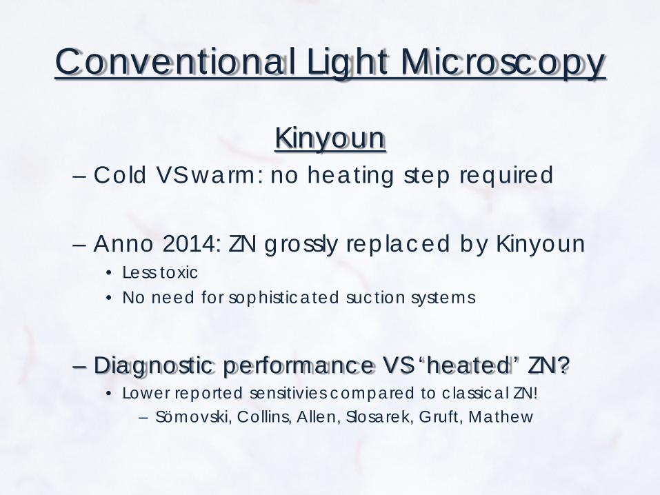
Kinyoun – Cold VS warm: no heating step required
– Anno 2014: ZN grossly replaced by Kinyoun
• Less toxic • No need for sophisticated suction systems
– Diagnostic performance VS ‘heated’ ZN?
• Lower reported sensitivies compared to classical ZN! – Sömovski, Collins, Allen, Slosarek, Gruft, Mathew
Conventional Light Microscopy
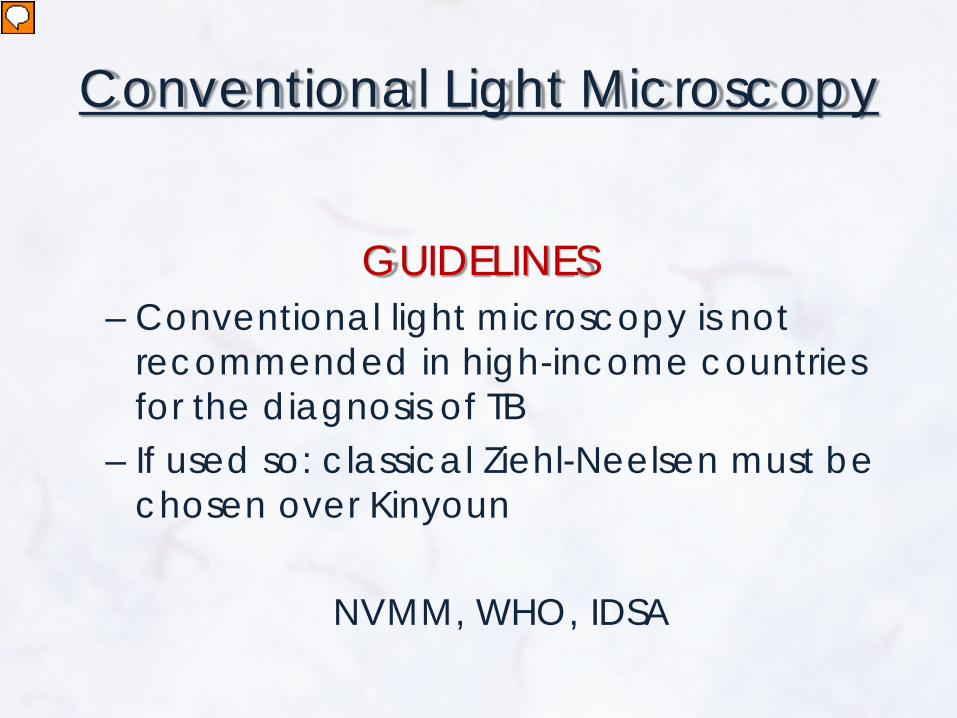
GUIDELINES
– Conventional light microscopy is not recommended in high-income countries for the diagnosis of TB
– If used so: classical Ziehl-Neelsen must be chosen over Kinyoun
NVMM, WHO, IDSA
Fluorescence Microscopy
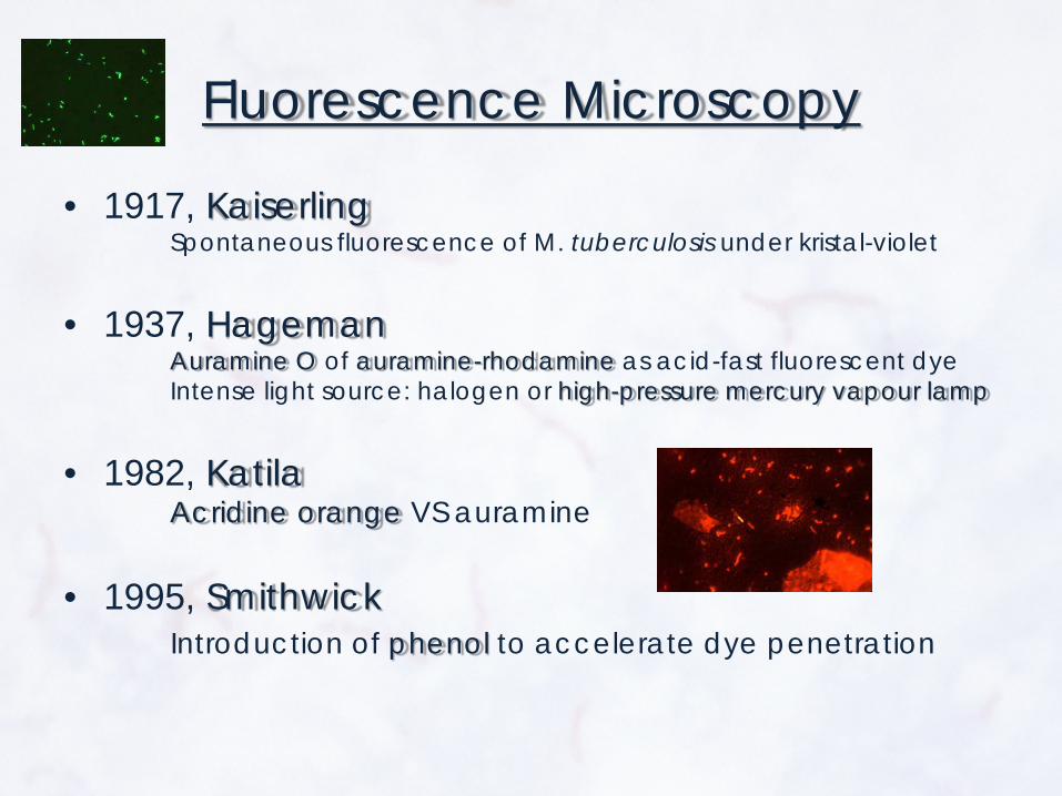
• 1917, Kaiserling Spontaneous fluorescence of M. tuberculosis under kristal-violet • 1937, Hageman Auramine O of auramine-rhodamine as acid-fast fluorescent dye Intense light source: halogen or high-pressure mercury vapour lamp
• 1982, Katila Acridine orange VS auramine
• 1995, Smithwick Introduction of phenol to accelerate dye penetration
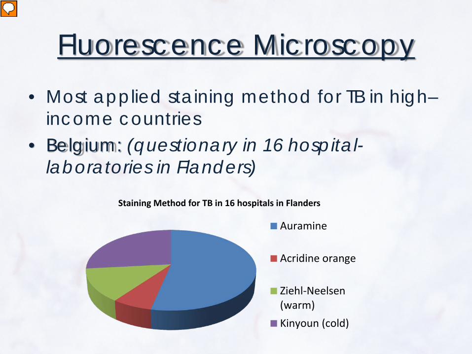
Fluorescence Microscopy • Most applied staining method for TB in high–
income countries • Belgium: (questionary in 16 hospital-
laboratories in Flanders)
Staining Method for TB in 16 hospitals in Flanders
Auramine
Acridine orange
Ziehl-Neelsen(warm)Kinyoun (cold)
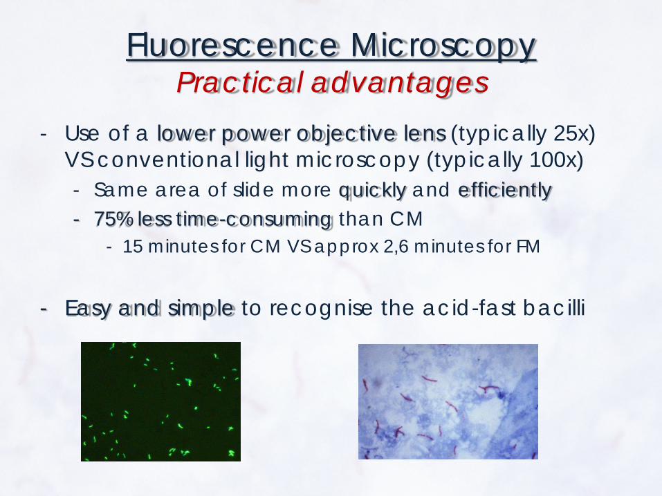
Fluorescence Microscopy Practical advantages
- Use of a lower power objective lens (typically 25x) VS conventional light microscopy (typically 100x) - Same area of slide more quickly and efficiently - 75% less time-consuming than CM
- 15 minutes for CM VS approx 2,6 minutes for FM
- Easy and simple to recognise the acid-fast bacilli
Fluorescence Microscopy Diagnostic performance
- Sensitivity - Most studies result in better sensitivities of FM compared to CM - Systematic review/thesis Henri, 2005 FM is more sensitive than CM - Systematic review Steingart, 2009
52%-97% FM 8-10% more sensitive than CM
- Specificity
- General concern related to less specific performance ⇒ Guidelines recommend to confirm acid-fact bacilli by Ziehl-Neelsen (NVMM) ⇒ BUT: - Systematic review Steingart, 2009:
- no decrease of specifity of FM compared to CM
- den Hertog et al, 2013: retrospective study of 10,276 samples - no added value of confirming auramine-positive samples with Ziehl-Neelsen - Reanalysis of these samples have no impact on patient management and thus waste of resources
Fluorescence Microscopy Practical disadvantages
- High capital cost for conventional mercury vapour lamp microscopes
BUT: Kivighja(2003), Sohn(2009): proof of cost-effectivity of FM, even in low-income countries because of the high sensitivy and greater time efficiency
- Significance maintenance of the microscopes - Limited life-span of the bulbs - Need for a dark room, away from dusty
environments - Toxic exposure when broken
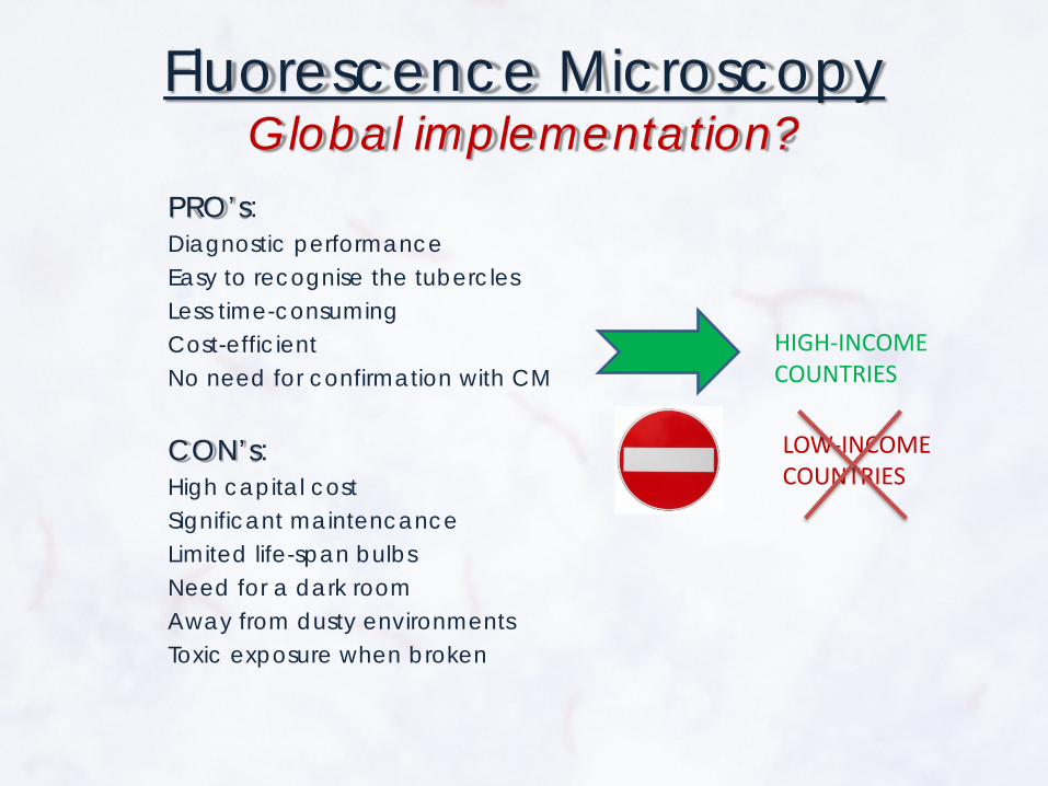
Fluorescence Microscopy Global implementation?
PRO’s: Diagnostic performance Easy to recognise the tubercles Less time-consuming Cost-efficient No need for confirmation with CM
CON’s: High capital cost Significant maintencance Limited life-span bulbs Need for a dark room Away from dusty environments Toxic exposure when broken
HIGH-INCOME COUNTRIES
LOW-INCOME COUNTRIES
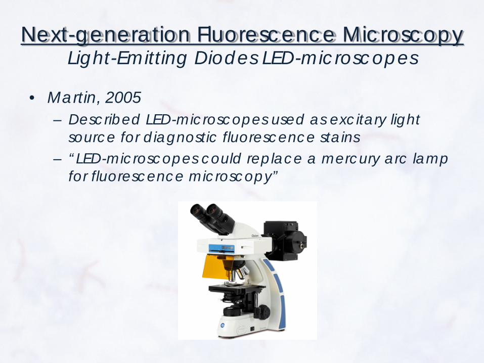
Next-generation Fluorescence Microscopy Light-Emitting Diodes LED-microscopes
• Martin, 2005 – Described LED-microscopes used as excitary light
source for diagnostic fluorescence stains – “LED-microscopes could replace a mercury arc lamp
for fluorescence microscopy”

Next-generation Fluorescence Microscopy Light-Emitting Diodes LED-microscopes
Practical advantages • No need for a dark room Improvement workflow Maximum space utilisation in the lab
• Less maintenance required than FM
• Good durability and portability
• Less capital costs than FM
• Overall better cost-efficicacy compared to CM:
– Withlaw, 2011: US$2,10CM VS US$1,63 LED – Xia, 2014: US$2,20 (+/-0,58)CM VS US$1,97 (+/-0,71)LED p<0,05
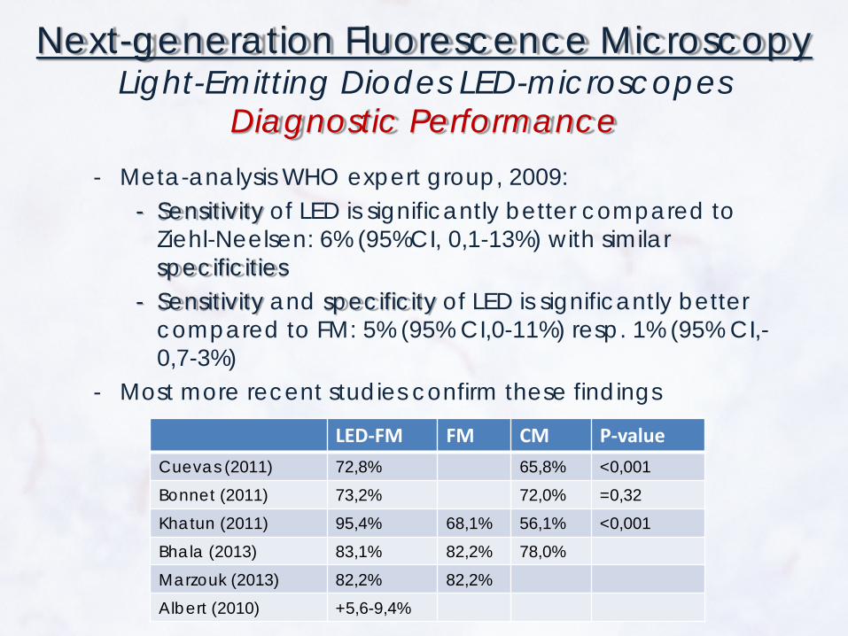
Next-generation Fluorescence Microscopy Light-Emitting Diodes LED-microscopes
Diagnostic Performance
- Meta-analysis WHO expert group, 2009: - Sensitivity of LED is significantly better compared to
Ziehl-Neelsen: 6% (95%CI, 0,1-13%) with similar specificities
- Sensitivity and specificity of LED is significantly better compared to FM: 5% (95% CI,0-11%) resp. 1% (95% CI,-0,7-3%)
- Most more recent studies confirm these findings
LED-FM FM CM P-value Cuevas (2011) 72,8% 65,8% <0,001
Bonnet (2011) 73,2% 72,0% =0,32
Khatun (2011) 95,4% 68,1% 56,1% <0,001
Bhala (2013) 83,1% 82,2% 78,0%
Marzouk (2013) 82,2% 82,2%
Albert (2010) +5,6-9,4%
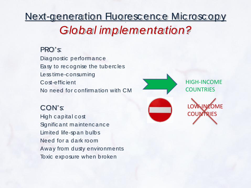
Next-generation Fluorescence Microscopy 0
Global implementation? PRO’s: Diagnostic performance Easy to recognise the tubercles Less time-consuming Cost-efficient No need for confirmation with CM
CON’s: High capital cost Significant maintencance Limited life-span bulbs Need for a dark room Away from dusty environments Toxic exposure when broken
HIGH-INCOME COUNTRIES
LOW-INCOME COUNTRIES
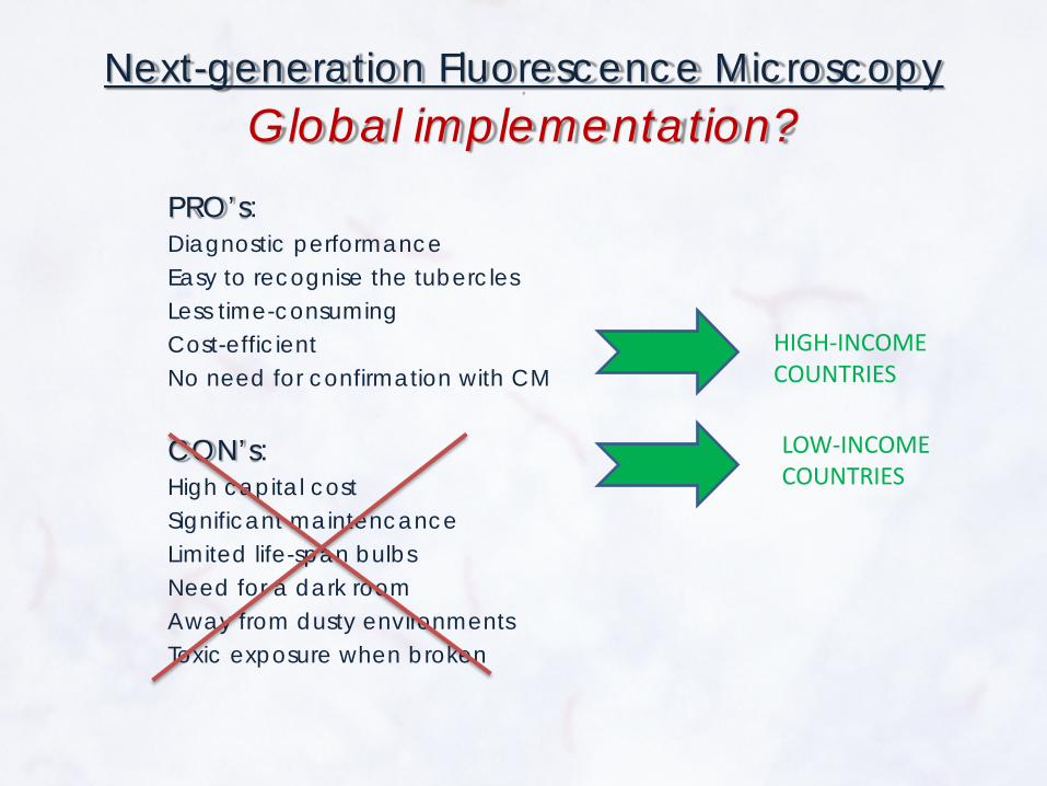
Next-generation Fluorescence Microscopy 0
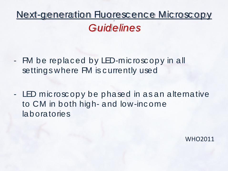
Guidelines
- FM be replaced by LED-microscopy in all
settings where FM is currently used
- LED microscopy be phased in as an alternative to CM in both high- and low-income laboratories
WHO2011
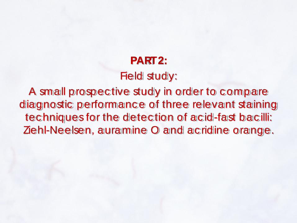
PART 2: Field study:
A small prospective study in order to compare diagnostic performance of three relevant staining techniques for the detection of acid-fast bacilli: Ziehl-Neelsen, auramine O and acridine orange.
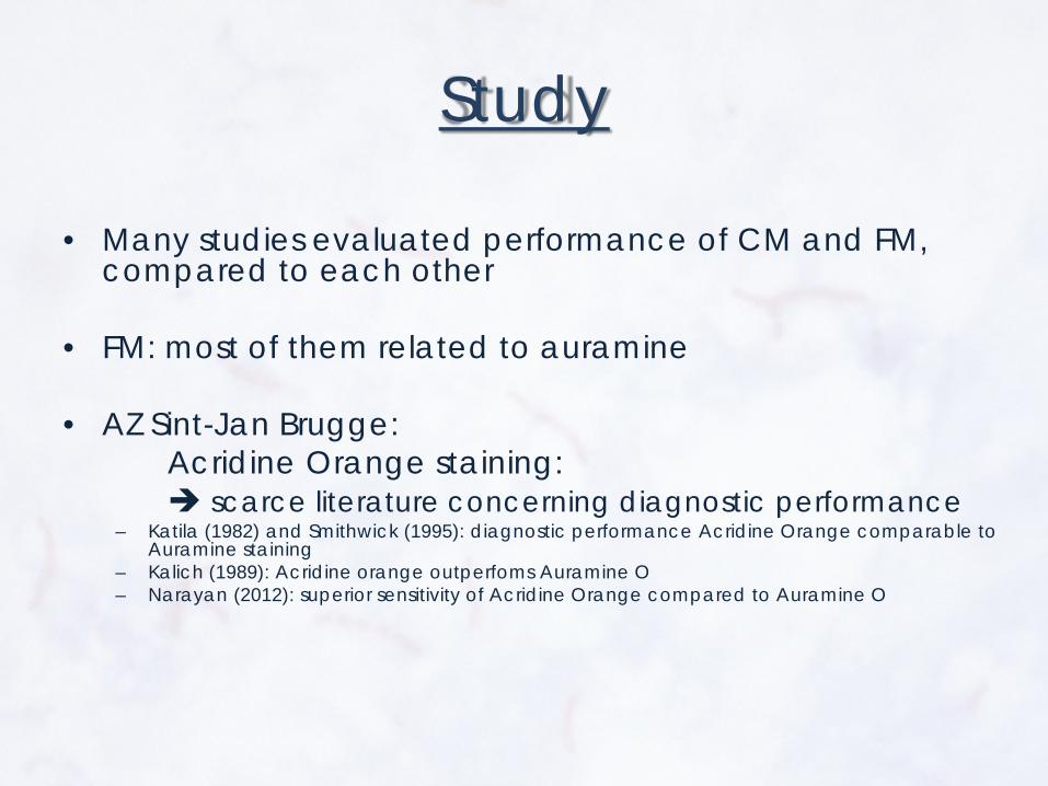
Study
• Many studies evaluated performance of CM and FM, compared to each other
• FM: most of them related to auramine
• AZ Sint-Jan Brugge: Acridine Orange staining: scarce literature concerning diagnostic performance
– Katila (1982) and Smithwick (1995): diagnostic performance Acridine Orange comparable to Auramine staining
– Kalich (1989): Acridine orange outperfoms Auramine O – Narayan (2012): superior sensitivity of Acridine Orange compared to Auramine O
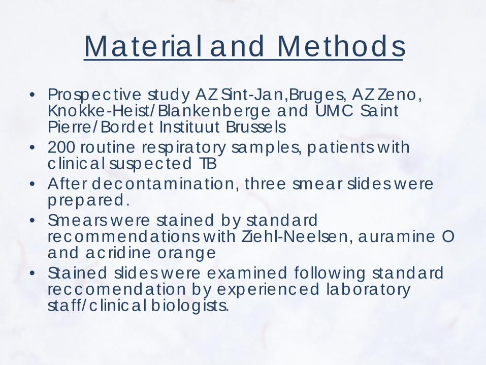
Material and Methods • Prospective study AZ Sint-Jan,Bruges, AZ Zeno,
Knokke-Heist/Blankenberge and UMC Saint Pierre/Bordet Instituut Brussels
• 200 routine respiratory samples, patients with clinical suspected TB
• After decontamination, three smear slides were prepared.
• Smears were stained by standard recommendations with Ziehl-Neelsen, auramine O and acridine orange
• Stained slides were examined following standard reccomendation by experienced laboratory staff/clinical biologists.
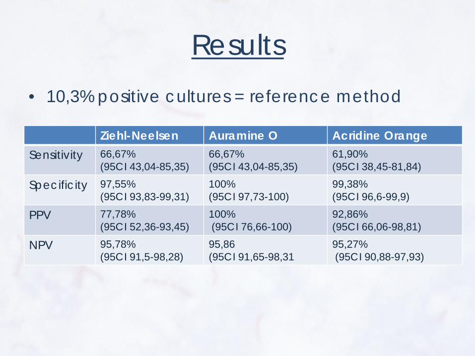
Results • 10,3% positive cultures = reference method
Ziehl-Neelsen Auramine O Acridine Orange Sensitivity 66,67%
(95CI 43,04-85,35) 66,67% (95CI 43,04-85,35)
61,90% (95CI 38,45-81,84)
Specificity 97,55% (95CI 93,83-99,31)
100% (95CI 97,73-100)
99,38% (95CI 96,6-99,9)
PPV 77,78% (95CI 52,36-93,45)
100% (95CI 76,66-100)
92,86% (95CI 66,06-98,81)
NPV 95,78% (95CI 91,5-98,28)
95,86 (95CI 91,65-98,31
95,27% (95CI 90,88-97,93)
General Answers and Conclusions • Sputum smear microscopy remains the most important diagnostic
tool for detecting acid-fast bacilli
• Guidelines and other published data: – LED fluorescence microscopy gains importance
• Ease in use/interpretation • Cost-effectivity • Diagnostic performance Reference staining method in high-income countries Not globally implemented yet
– Conventional light microscopy: Ziehl-Neelsen/Kinyoun loses importance
• More difficult to interpret • More expensive • Less performant
– However: not confirmed by our field study => if prepared and interpreted follwing standard recommendations equal sensitivities? (Iademarco and own results)
Still the most known, available and applied staining method in developing countries More and more abandoned in high income countries
Did’s and To Do’s • Field study: overall good performance of the three
staining methods. Too small for adequate conclusions concerning comparison between the methods.
• Only a limited of studies have been performed evaluating the diagnostic performance of Acridine Orange staining for the detection of acid-fast bacilli.
Because of good performance of the field study and it’s succesfull validation, the Acridine Orange staining is implemented in our lab for the detection of acid-fast bacilli.
Follow-up literature concerning diagnostic performance of Acridine Orange in the detection of acid-fact bacilli.
Literature • Daniel TM. The history of tuberculosis. Resp medicine 2006; 100:1862-1870 • Ehrlich P. A method for staining the tubercle bacillus. 1882 • Cuevas L. A multi-country non-inferiority cluster randomized trial of frontloaded smear microscopy for the diagnosis of
pulmonary tuberculosis. PLoS Med 2011; 8:e1000443 • Cuevas L. LED fluorescence microscopy for the diagnosis of pulmonary tuberculosis: a multi-country cross sectional
evaluation. PLoS Med 2011; 8:e1001057 • Steingart K. Fluorescene versus conventional sputum smear microscopy for tuberculosis: a systematic review. Lancet Infect Dis
2006; 6: 570-581 • Iademarco M. Evaluation of laboratory methods used to examine sputum specimens for Mycobacterium tuberculosis.
Abstracts of the 30th World Conference on Lung Health of the International Union Against Tuberculosis and Lung Disease; Madrid, Spain. 1999. Abstract No. 216-PD
• Somoskövi A. Lessons From a Proficiency Testing Event for Acid-Fast Microscopy. Chest 2001; 120: 250-257 • Collins F. Microscopic counts carries out on M. leprae and M. tuberculosis suspension: a comparison of three staining
procedures. Int J Lepr Other Mycobact Dis 1980; 48: 402-407 • Allen J. A modified Ziehl-Neelsen stain for mycobacteria. Med Lab Sci 1992; 49: 99-102 • Slosarek M. Comparison of microscopic positivity in smears from sputa stained according to Ziehl-Neelsen in different
modifications. J Hyg Epidemiol Micrbiol Immunol 1977; 21: 7-15 • Slosarek M. Cold staining methods for mycobacteria. J Hyg Epidemiol Microbiol Immunol 1974; 18:22-26 • Gruft H. Evaluation of Mycobacteriology Laboratories: The Acid-Fast Smear (1978) Health Lab Sci. 1978 Oct;15(4):215-20. • Mathew S. Evaluation of a cold staining method for acid-fast bacilli in sputum. Indian J Chest Dis Allied Sci. 1994 Jul-
Sep;36(3):125-31. • Katila M. Acridine orange staining of smears for demonstration of Mycobacterium tuberculosis. 1982. Eur J Clin Microbiol.
1:351-353 • Smithwick R. Phenolic Acridine Orange Fluorescent Stain for Mycobacteria. J Clin Microbiol 1995; 2763-2764 • Laifangbam S. A comparative study of fluorescent microscopy with Ziehl-Neelsen staining and culture for the diagnosis of
pulmonary tuberculosis. Kathmandu University medical journal (KUMJ) 7:27 pg 226-30 • den Hertog A. No added value of performing Ziehl-Neelsen on auramine-positive samples for tuberculosis diagnostics. Int
Tuberc Lung Dis 20103; 17:1094-1099 • World Health Organisation 2011. Fluorescent light-emitting diode (LED) microscopy for diagnosis of tuberculosis: Policy
Statement. • Marzouk M. Comparison of LED and conventional fluorescence microscopy for detection of acid-fast bacilli in an area with
high tuberculosis incidence. Diag Microbiol and Inf Dis 2013; 76: 306-308 • Xia H. Multicentre evaluation of Ziehl-Neelsen and light-emitting diode fluorescen microscopy in China. Int J Tuberc Lung Dis
2013; 17: 107-112 • Khatun Z. Usefulness of Light Emitting Diode (LED) fluorescent microscopy as a tool for rapid and effective method for the
diagnosis of pulmonary tuberculosis. Bangladesh Med Res Counc Bull 2011; 37:7-10