Embed Size (px)
Citation preview
SPOTS AND DOTS What Is That In My Lung? Elsira M. Pina, DO FCCP
University of Cincinnati Medical Center
Pulmonary-Critical Care Medicine

DISCLOSURE ANTI-TOBACCO
PRO-LUNG FORCE
TODAY’S PRESENTATION Discuss work-up of lung spot
Discuss what a lung spot could be
Discuss what a lung spot looks like
Discuss possible tests needed to evaluate the lung spot
Discuss possible biopsy approaches to determine what kind of spot is in the lung

PATIENT STORY
Mrs. Juniper sweet 65 YO woman playing with grandkids at the park Takes a daily medication for high blood pressure Has smoked 1 pack per day since age 18
Developed chest pain in the park Daughter brought her to the Emergency Department Evaluated for heart attack No heart attack Had Chest Xray done
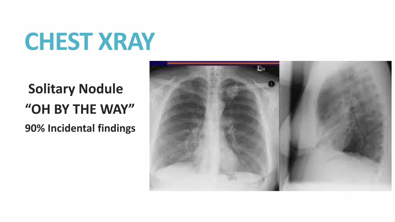
CHEST XRAY
Solitary Nodule
“OH BY THE WAY” 90% Incidental findings
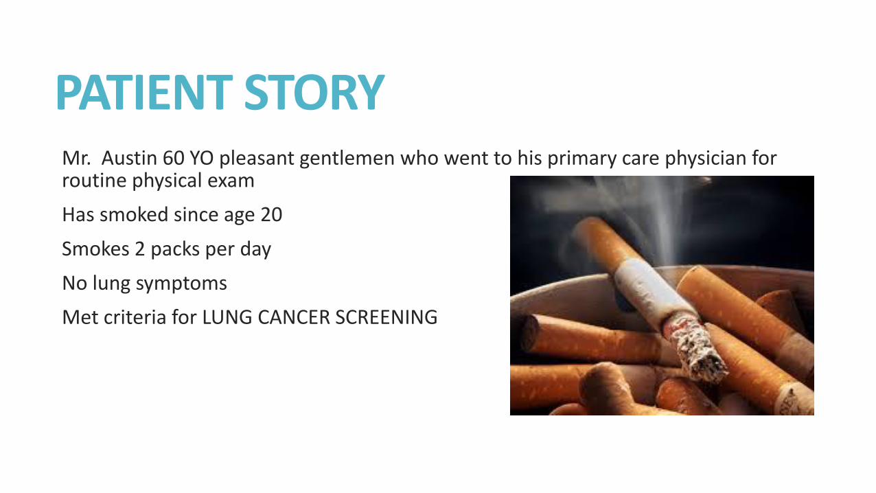
PATIENT STORY Mr. Austin 60 YO pleasant gentlemen who went to his primary care physician for routine physical exam
Has smoked since age 20
Smokes 2 packs per day
No lung symptoms
Met criteria for LUNG CANCER SCREENING
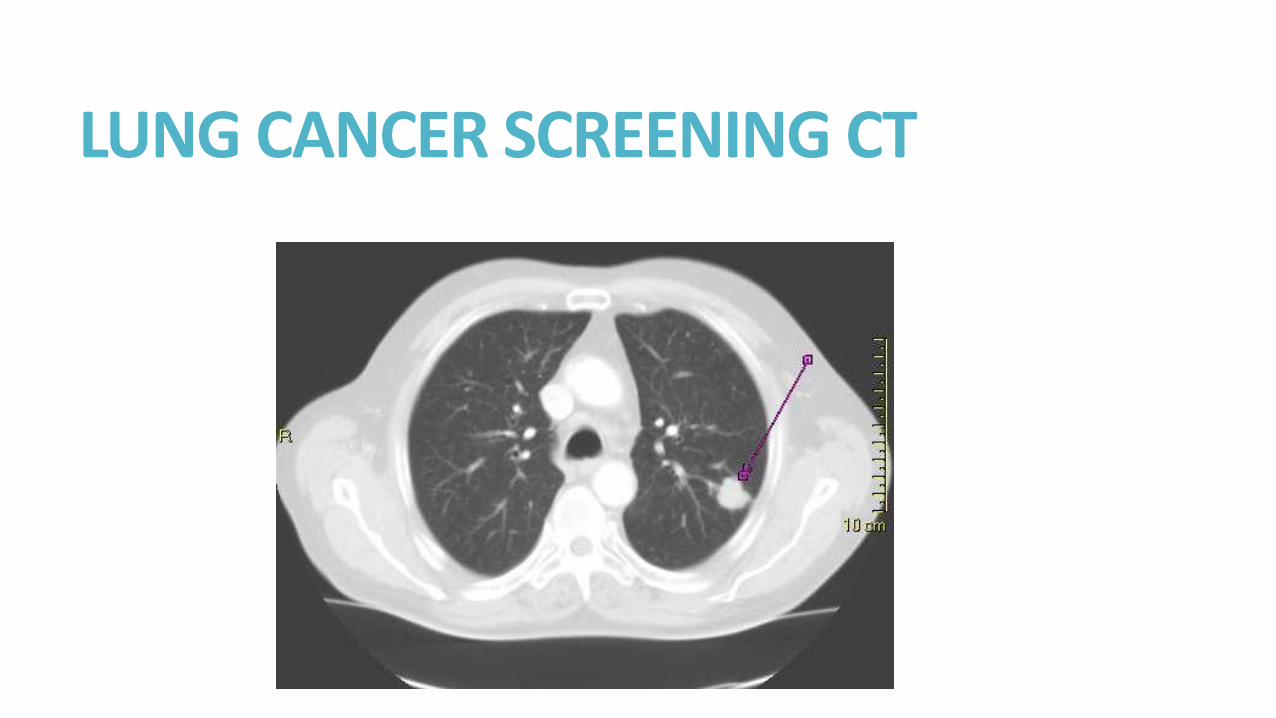
LUNG CANCER SCREENING CT
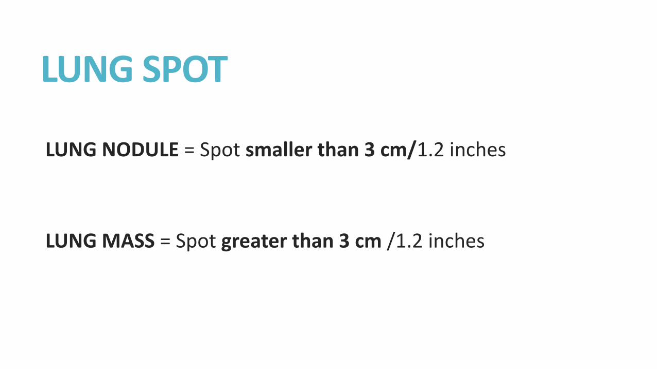
LUNG SPOT
LUNG NODULE = Spot smaller than 3 cm/1.2 inches
LUNG MASS = Spot greater than 3 cm /1.2 inches
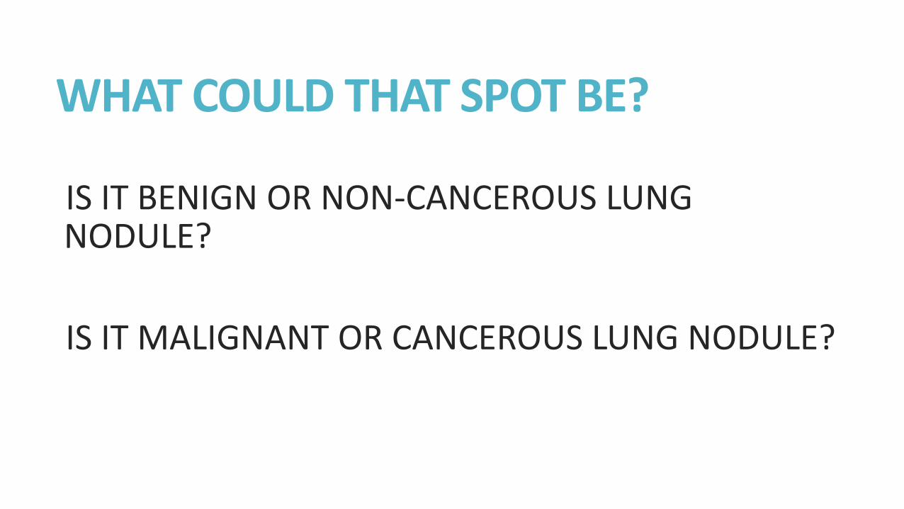
WHAT COULD THAT SPOT BE?
IS IT BENIGN OR NON-CANCEROUS LUNG NODULE?
IS IT MALIGNANT OR CANCEROUS LUNG NODULE?
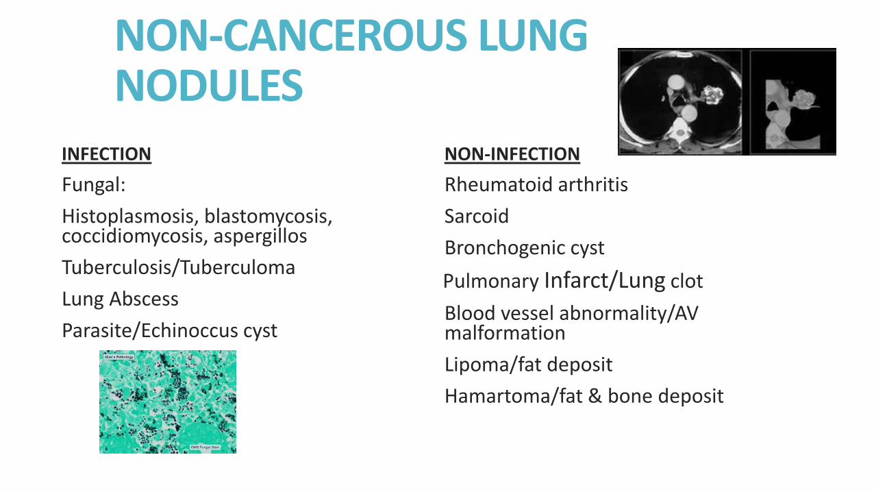
NON-CANCEROUS LUNG NODULES
INFECTION
Fungal:
Histoplasmosis, blastomycosis, coccidiomycosis, aspergillos
Tuberculosis/Tuberculoma
Lung Abscess
Parasite/Echinoccus cyst
NON-INFECTION
Rheumatoid arthritis
Sarcoid
Bronchogenic cyst
Pulmonary Infarct/Lung clot
Blood vessel abnormality/AV malformation
Lipoma/fat deposit
Hamartoma/fat & bone deposit
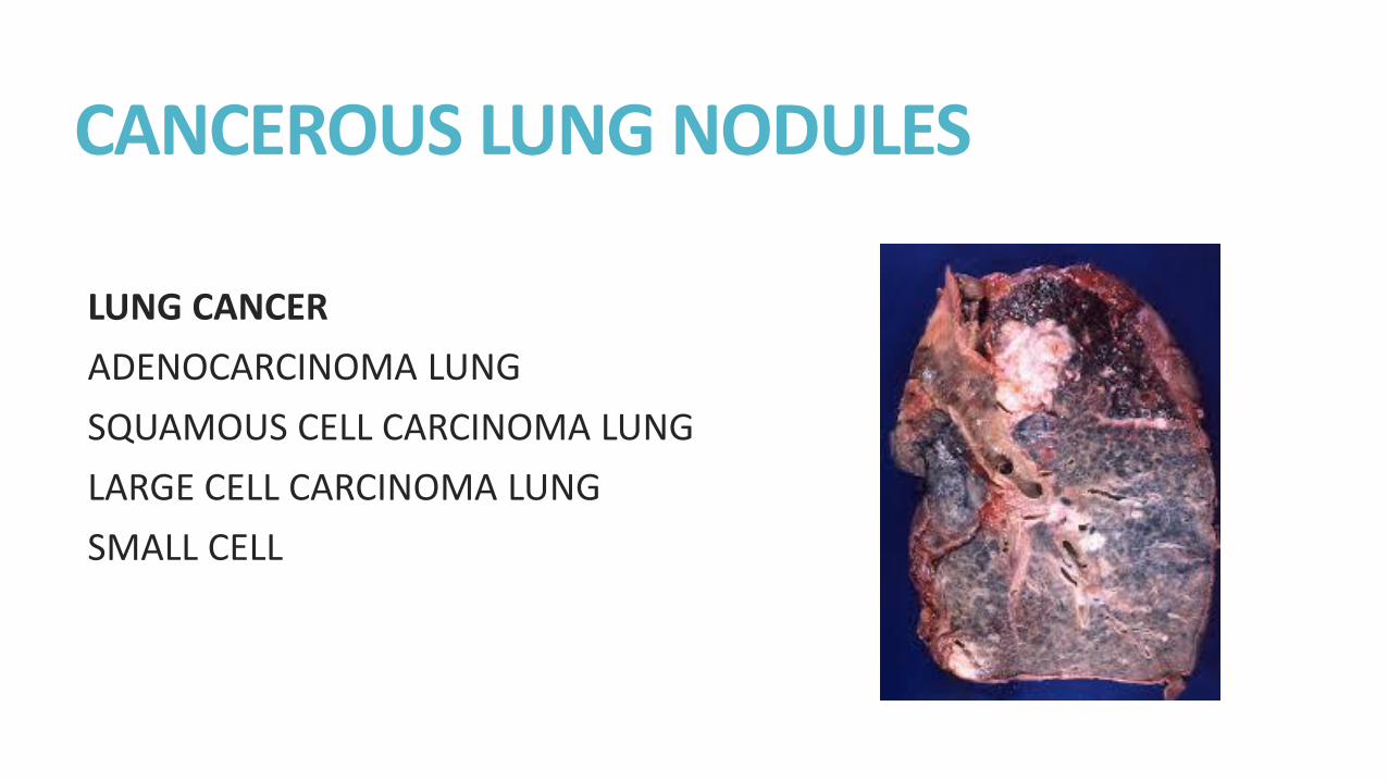
CANCEROUS LUNG NODULES
LUNG CANCER
ADENOCARCINOMA LUNG
SQUAMOUS CELL CARCINOMA LUNG
LARGE CELL CARCINOMA LUNG
SMALL CELL
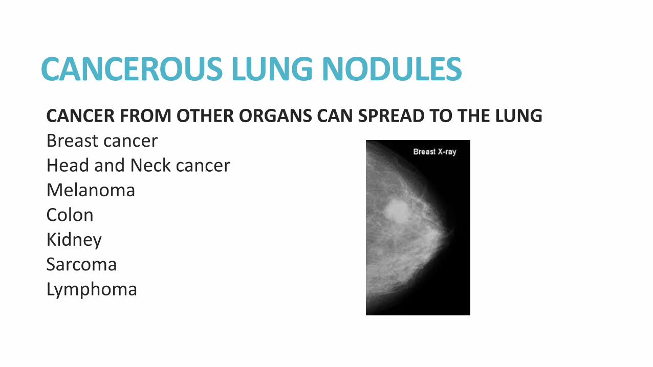
CANCEROUS LUNG NODULES CANCER FROM OTHER ORGANS CAN SPREAD TO THE LUNG Breast cancer Head and Neck cancer Melanoma Colon Kidney Sarcoma Lymphoma

CHEST XRAY vs CHEST CT
CHEST XRAYs SEE SPOTS AT LEAST 1CM OR 1/3 INCH IN SIZE
CHEST CTs CAN SEE SPOTS LESS THAN 1CM OR LESS THAN 1/3 INCH IN SIZE

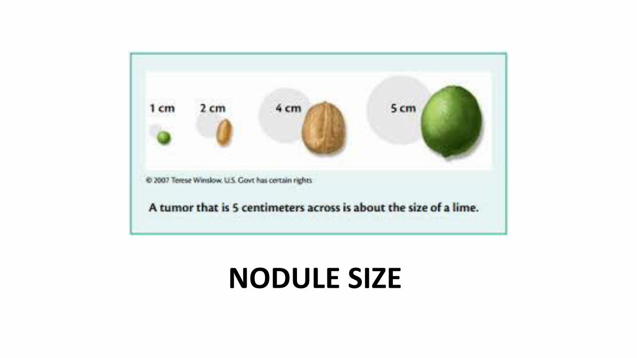
NODULE SIZE
WHAT DOES THE SPOT LOOK LIKE?
Is it completely calcified or not? ALL CALCIUM PARTIAL CALCIUM NO CALCIUM
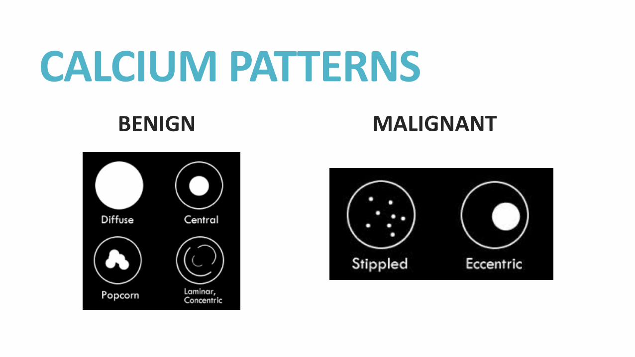
CALCIUM PATTERNS BENIGN MALIGNANT
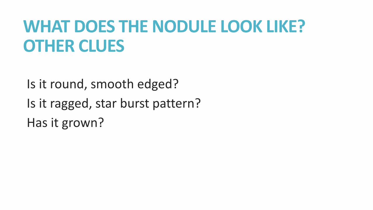
WHAT DOES THE NODULE LOOK LIKE? OTHER CLUES
Is it round, smooth edged?
Is it ragged, star burst pattern?
Has it grown?

SPICULATED SPOT
SUN BURST PATTERN MALIGNANT
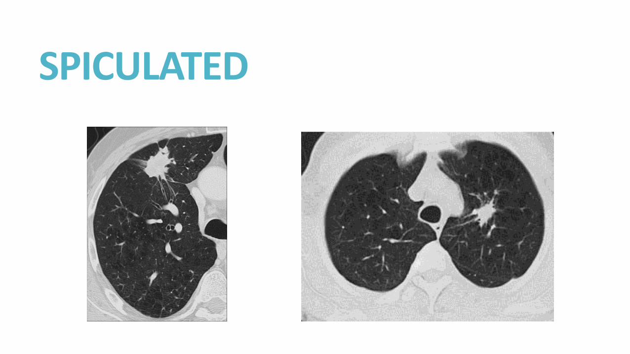
SPICULATED
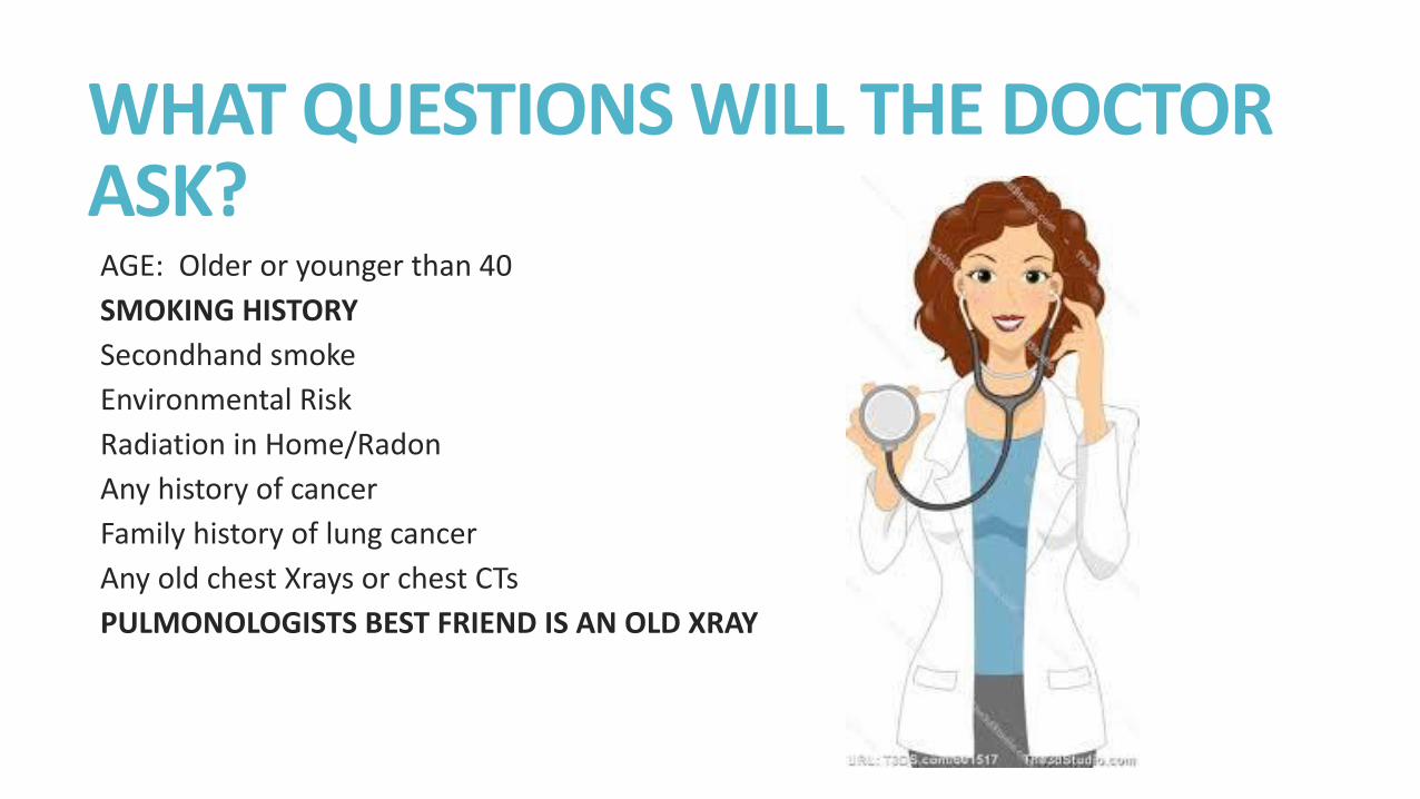
WHAT QUESTIONS WILL THE DOCTOR ASK? AGE: Older or younger than 40
SMOKING HISTORY
Secondhand smoke
Environmental Risk
Radiation in Home/Radon
Any history of cancer
Family history of lung cancer
Any old chest Xrays or chest CTs
PULMONOLOGISTS BEST FRIEND IS AN OLD XRAY
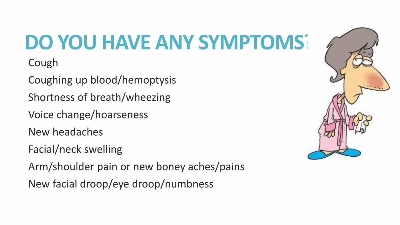
DO YOU HAVE ANY SYMPTOMS? Cough
Coughing up blood/hemoptysis
Shortness of breath/wheezing
Voice change/hoarseness
New headaches
Facial/neck swelling
Arm/shoulder pain or new boney aches/pains
New facial droop/eye droop/numbness
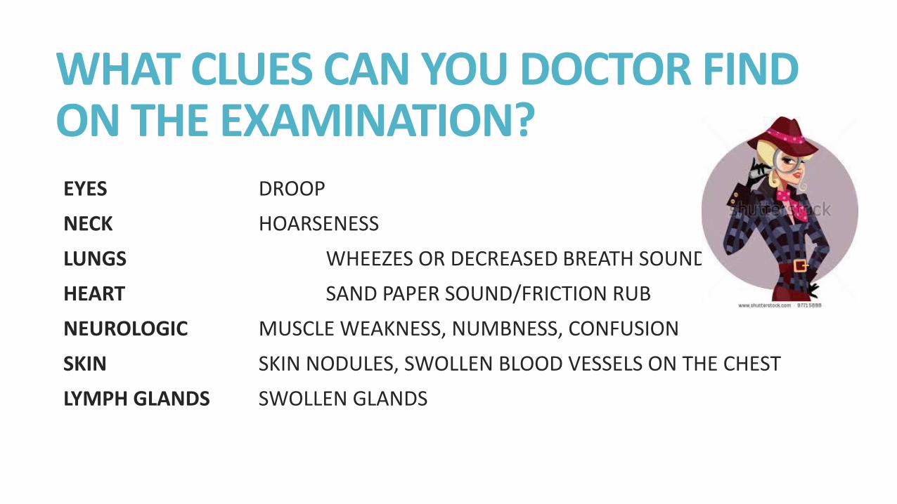
WHAT CLUES CAN YOU DOCTOR FIND ON THE EXAMINATION? EYES DROOP
NECK HOARSENESS
LUNGS WHEEZES OR DECREASED BREATH SOUNDS
HEART SAND PAPER SOUND/FRICTION RUB
NEUROLOGIC MUSCLE WEAKNESS, NUMBNESS, CONFUSION
SKIN SKIN NODULES, SWOLLEN BLOOD VESSELS ON THE CHEST
LYMPH GLANDS SWOLLEN GLANDS
WHAT INITIAL TESTING MAY I NEED?
CHEST CT
PET CT
HEAD MRI
PFTS/Pulmonary Function Testing
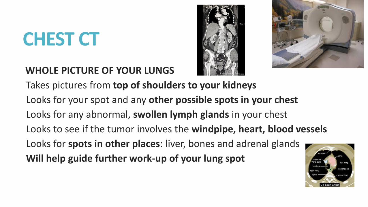
CHEST CT
WHOLE PICTURE OF YOUR LUNGS
Takes pictures from top of shoulders to your kidneys
Looks for your spot and any other possible spots in your chest
Looks for any abnormal, swollen lymph glands in your chest
Looks to see if the tumor involves the windpipe, heart, blood vessels
Looks for spots in other places: liver, bones and adrenal glands
Will help guide further work-up of your lung spot
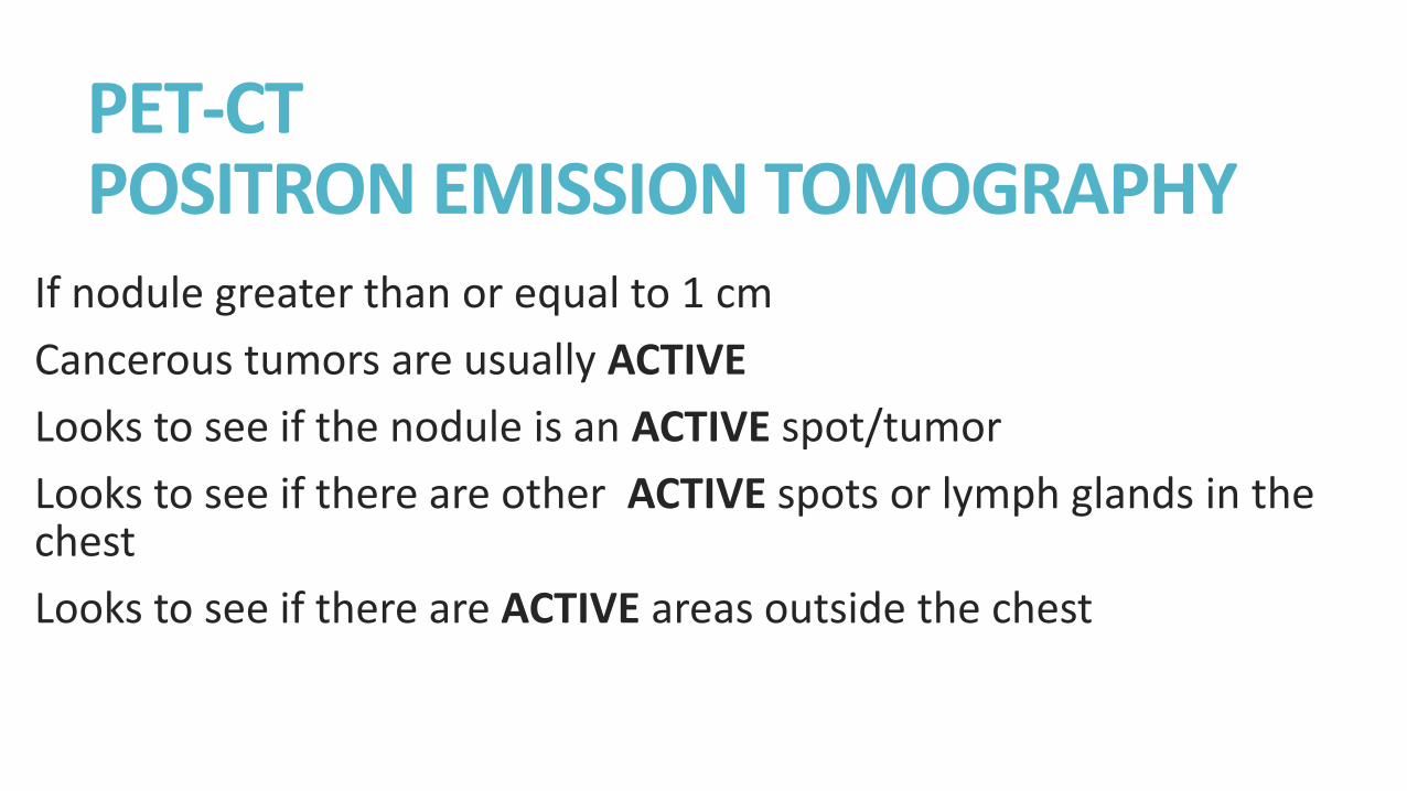
PET-CT POSITRON EMISSION TOMOGRAPHY
If nodule greater than or equal to 1 cm
Cancerous tumors are usually ACTIVE
Looks to see if the nodule is an ACTIVE spot/tumor
Looks to see if there are other ACTIVE spots or lymph glands in the chest
Looks to see if there are ACTIVE areas outside the chest
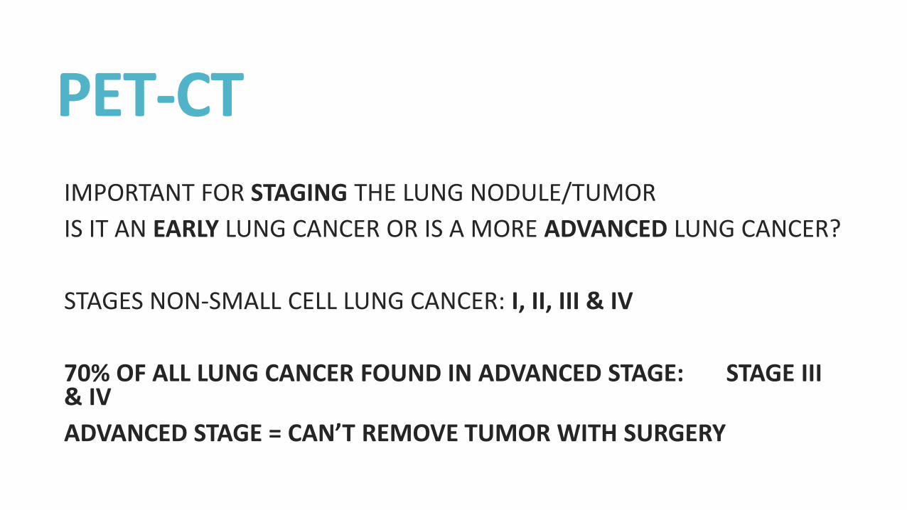
PET-CT IMPORTANT FOR STAGING THE LUNG NODULE/TUMOR
IS IT AN EARLY LUNG CANCER OR IS A MORE ADVANCED LUNG CANCER?
STAGES NON-SMALL CELL LUNG CANCER: I, II, III & IV
70% OF ALL LUNG CANCER FOUND IN ADVANCED STAGE: STAGE III & IV
ADVANCED STAGE = CAN’T REMOVE TUMOR WITH SURGERY
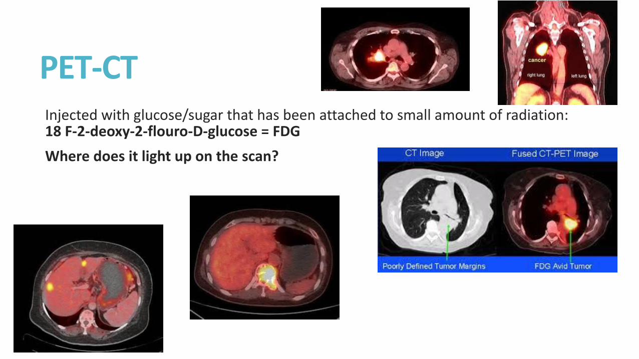
PET-CT Injected with glucose/sugar that has been attached to small amount of radiation: 18 F-2-deoxy-2-flouro-D-glucose = FDG
Where does it light up on the scan?
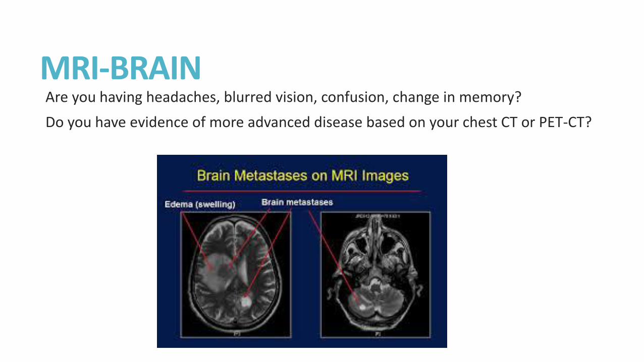
MRI-BRAIN Are you having headaches, blurred vision, confusion, change in memory?
Do you have evidence of more advanced disease based on your chest CT or PET-CT?
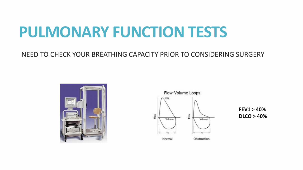
PULMONARY FUNCTION TESTS NEED TO CHECK YOUR BREATHING CAPACITY PRIOR TO CONSIDERING SURGERY
FEV1 > 40% DLCO > 40%
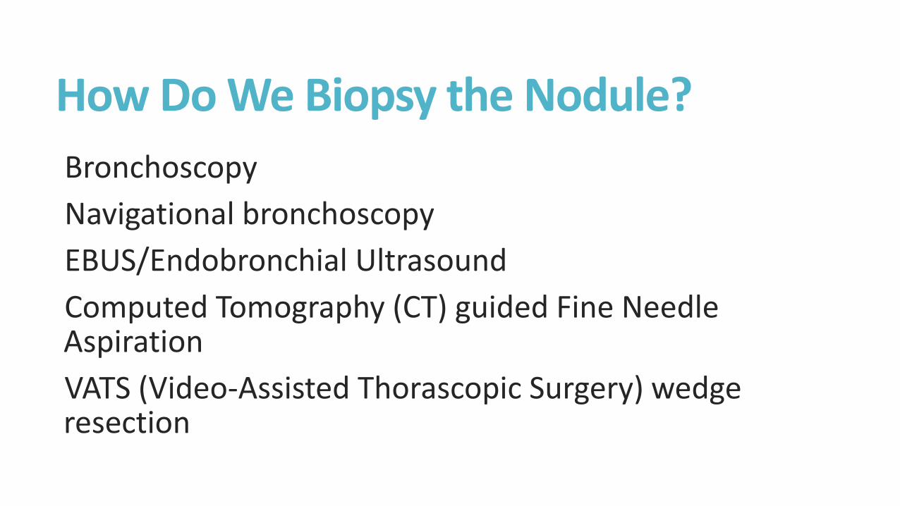
How Do We Biopsy the Nodule?
Bronchoscopy
Navigational bronchoscopy
EBUS/Endobronchial Ultrasound
Computed Tomography (CT) guided Fine Needle Aspiration
VATS (Video-Assisted Thorascopic Surgery) wedge resection
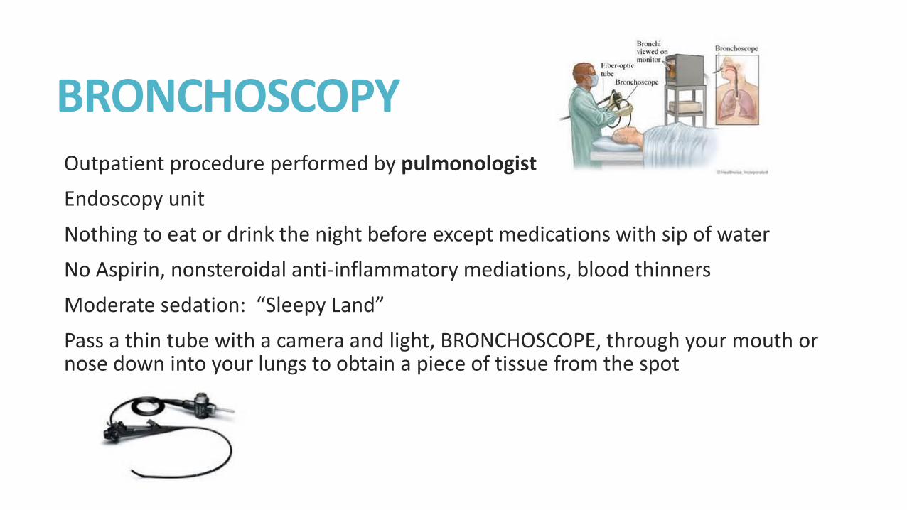
BRONCHOSCOPY Outpatient procedure performed by pulmonologist
Endoscopy unit
Nothing to eat or drink the night before except medications with sip of water
No Aspirin, nonsteroidal anti-inflammatory mediations, blood thinners
Moderate sedation: “Sleepy Land”
Pass a thin tube with a camera and light, BRONCHOSCOPE, through your mouth or nose down into your lungs to obtain a piece of tissue from the spot
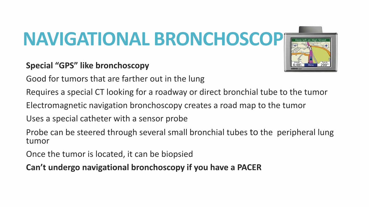
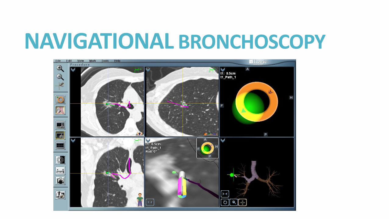
NAVIGATIONAL BRONCHOSCOPY Special “GPS” like bronchoscopy
Good for tumors that are farther out in the lung
Requires a special CT looking for a roadway or direct bronchial tube to the tumor
Electromagnetic navigation bronchoscopy creates a road map to the tumor
Uses a special catheter with a sensor probe
Probe can be steered through several small bronchial tubes to the peripheral lung tumor
Once the tumor is located, it can be biopsied
Can’t undergo navigational bronchoscopy if you have a PACER
NAVIGATIONAL BRONCHOSCOPY
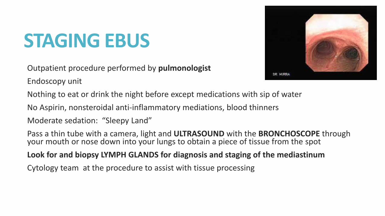
STAGING EBUS Outpatient procedure performed by pulmonologist
Endoscopy unit
Nothing to eat or drink the night before except medications with sip of water
No Aspirin, nonsteroidal anti-inflammatory mediations, blood thinners
Moderate sedation: “Sleepy Land”
Pass a thin tube with a camera, light and ULTRASOUND with the BRONCHOSCOPE through your mouth or nose down into your lungs to obtain a piece of tissue from the spot
Look for and biopsy LYMPH GLANDS for diagnosis and staging of the mediastinum
Cytology team at the procedure to assist with tissue processing
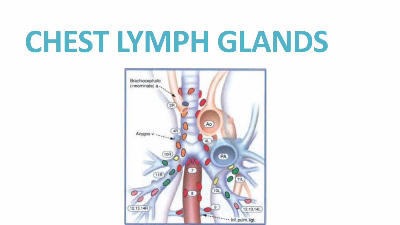
CHEST LYMPH GLANDS
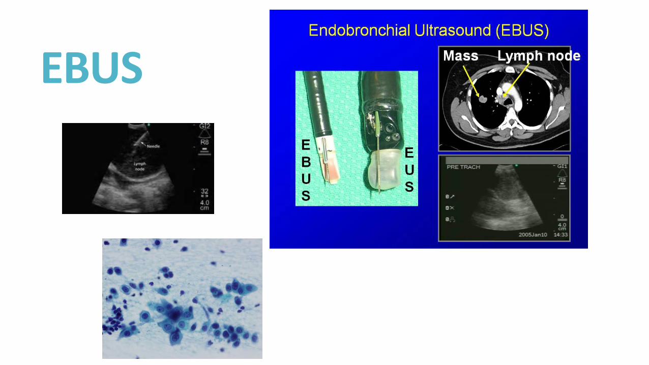
EBUS
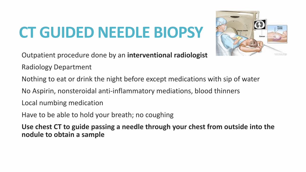
CT GUIDED NEEDLE BIOPSY Outpatient procedure done by an interventional radiologist
Radiology Department
Nothing to eat or drink the night before except medications with sip of water
No Aspirin, nonsteroidal anti-inflammatory mediations, blood thinners
Local numbing medication
Have to be able to hold your breath; no coughing
Use chest CT to guide passing a needle through your chest from outside into the nodule to obtain a sample

CT-GUIDED NEEDLE BIOPSY RIGHT LUNG MASS
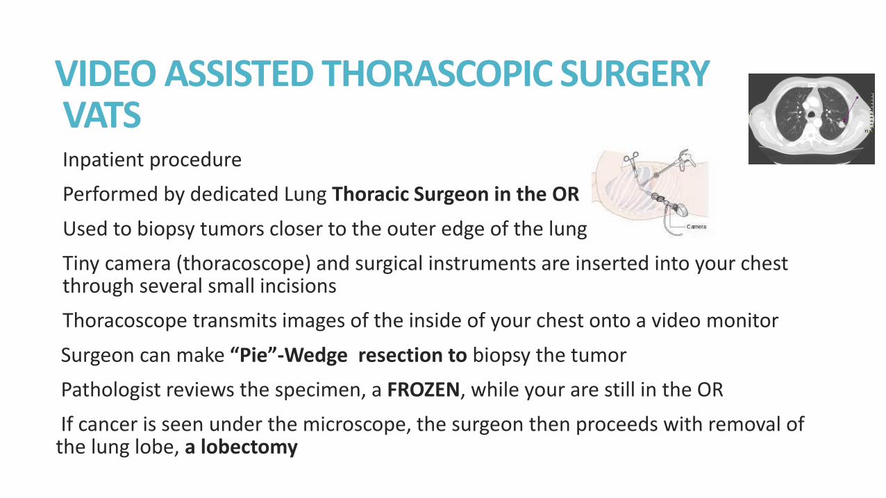
VIDEO ASSISTED THORASCOPIC SURGERY VATS Inpatient procedure
Performed by dedicated Lung Thoracic Surgeon in the OR
Used to biopsy tumors closer to the outer edge of the lung
Tiny camera (thoracoscope) and surgical instruments are inserted into your chest through several small incisions
Thoracoscope transmits images of the inside of your chest onto a video monitor
Surgeon can make “Pie”-Wedge resection to biopsy the tumor
Pathologist reviews the specimen, a FROZEN, while your are still in the OR
If cancer is seen under the microscope, the surgeon then proceeds with removal of the lung lobe, a lobectomy
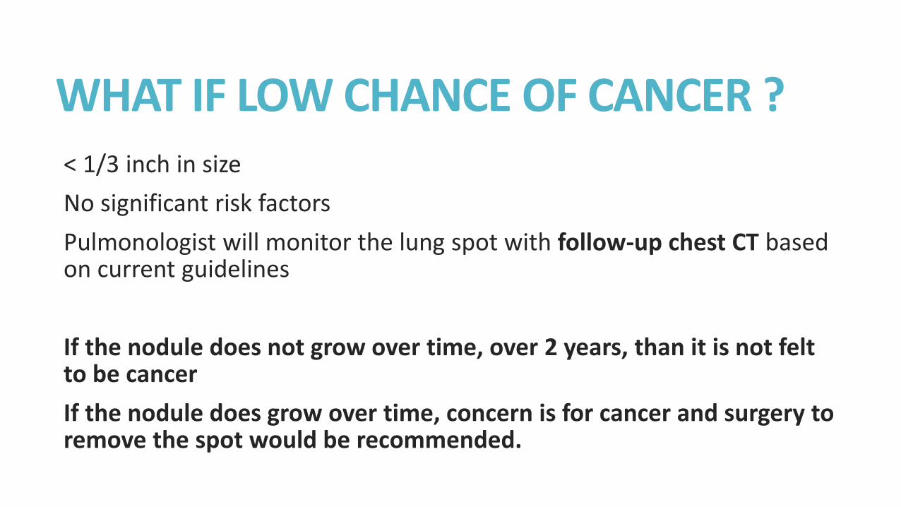
WHAT IF LOW CHANCE OF CANCER ? < 1/3 inch in size
No significant risk factors
Pulmonologist will monitor the lung spot with follow-up chest CT based on current guidelines
If the nodule does not grow over time, over 2 years, than it is not felt to be cancer
If the nodule does grow over time, concern is for cancer and surgery to remove the spot would be recommended.
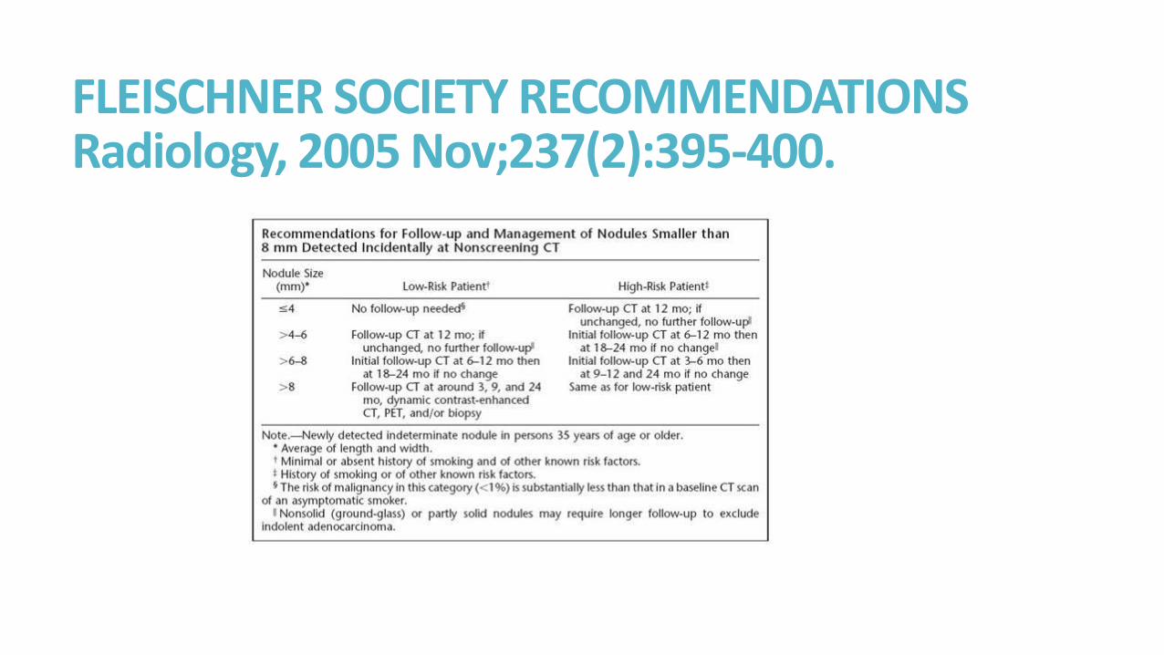
FLEISCHNER SOCIETY RECOMMENDATIONS Radiology, 2005 Nov;237(2):395-400.
WHAT IF MEDIUM CHANCE OF LUNG CANCER? Need more information about the spot If the nodule is 1/3 inch or bigger, you will need further evaluation: PET and biopsy. If the PET is ONLY ACTIVE in the spot or the biopsy show cancer cells, your doctor will recommend SURGERY If the PET is NOT ACTIVE, your doctor will discuss proceeding with biopsy versus a short term 3 month follow-up chest CT
WHAT IF HIGH RISK FOR LUNG CANCER? A SPOT FELT TO BE HIGH CHANCE OF LUNG CANCER (> 65%)
Your doctor will recommend breathing tests, PFTs, prior to surgery
Your doctor will recommend PET to check for spread outside the lungs and for any evidence of ACTIVE mediastinal lymph glands
Your doctor will recommend surgery, VATS, to remove the spot if there is no evidence of advanced disease on the PET
Your surgeon, will remove the tumor and remove the chest lymph glands at time of surgery for PATHOLOGICAL STAGING
Pathologist will look under microscope as see if there is definite cancer in the tumor and if there is any cancer in your lymph glands
SUMMARY WHAT IS THAT SPOT IN MY LUNG? Answer could be Cancer or non-cancer
Answer involves assessing risk factors for cancer
Answer involves assessing symptoms
Answer involves good physical exam
Answer involves various imaging studies
Answer may require further TISSUE BIOPSY if there is MODERATE to HIGH SUSPICION for LUNG CANCER
TISSUE IS THE ISSUE FOR DIAGNOSIS AND STAGING
YOU AND YOUR DOCTOR ARE A TEAM WORKING TO FIND AN ANSWER

THANK YOU
584-QUIT
1-800-LUNGUSA


![434805]Writi… · Web viewbig cat Only in America. Live in rainforests – swamps - grassland Fur is tan or orange with black spots with small dots in the middle. talks and ambushes](https://img.pdfslide.us/doc/110x75/5eaf0c67637c501a6267193b/434805writi-web-view-big-cat-only-in-america-live-in-rainforests-a-swamps.jpg)